UNIVERSITY OF MESSINA
Department of Chemical, Biological, Pharmaceutical
and Environmental Sciences
Doctorate School in Chemical Sciences (XXX cycle)
S.S.D. CHIM/08
Design, synthesis and biological
evaluation of novel inhibitors of
rhodesain, cysteine protease of
Trypanosoma brucei rhodesiense, as
antitrypanosomal agents
PhD student:
Santo PREVITI
Supervisor:
Prof. Silvana GRASSO
Co-Supervisor:
Prof. Roberta ETTARI
PhD Coordinator:
Prof. Sebastiano CAMPAGNA
CONTENTS
ACKNOWLEDGEMENTS. . . . . . . . . .1
LIST OF USED ACRONYMS. . . . . . . . . . . . . . . . . . 2
1. AIM OF THE RESEARCH. . . . . . . .5
2. HUMAN AFRICAN TRYPANOSOMIASIS. . . .13
2.1 Introduction. . . . . . . . . . . . . . . . . . .13
2.2 The parasites. . . . . . . . . . . .13
2.3 HAT forms: symptoms and progression of the disease. . . . . . . . . . . . . . .15
2.4 Diagnostic methods. . . . . . . . . . . . . . . . . . . 17
2.5 Pharmacological treatment. . . . . . . . 18
3. CYSTEINE PROTEASES: ALTERNATIVE AND VALID TARGETS. . . . . .25
3.1 The ideal drug . . . . . . . . . . . . . . . . .25
3.2 Potential targets. . . . . . . . . . . . . . . . . . . .26
3.3 Rhodesain: structure and functions. . . . . . . . . . . . . . . . . . . . . . .29
3.4 TbCatB: structure and functions. . . . . . . . . . . . . . . . . . 31
4. RHODESAIN INHIBITORS. . . . . . . . . . . . . . . . . . . . 34
4.1 Peptide-based inhibitors. . . . . . . . . . . . . . . . . . . . . . . . . . . . 34
4.1.1 Vinyl sulfones. . . . . . . . . . . . . . . . . . . . . . . .35
4.1.2 Azadipeptide nitriles. . . . . . . . . . . . . . . . . . . . 38
4.1.3 Aziridine-2,3-dicarboxylates. . . . . . . . . . . . . . .40
4.1.4 Peptidyl aldehydes and ketones. . . . . . . . . 41
4.2 Peptidomimetics. . . . . . . . . . . . . . . . . . . . . . . 43
4.2.1 BDZ derivatives with aldehyde warhead. . . . . . . . . . . . 44
4.2.2 BDZ derivatives bearing vinyl ester portion as warhead. . . . . . . . . 44
4.2.3 3-Bromo isoxazoline inhibitors. . . . . . . . 45
4.3 Non peptide inhibitors. . . . . . . . .47
4.3.1 Triazine nitriles. . . 47
4.3.2 Purine nitriles. . . . . . . . . . . . . . . . . . . . .51
4.3.3 Thiosemicarbazones. . . . . . . . . . . . 53
4.3.4 Other non-peptide inhibitors. . . . . . . . . . . . . . . . . . . . . 55
5. RESULTS AND DISCUSSIONS. . . . . . . . . . . . . . . . . . . 57
5.1 Series A. . . . . . . . . . . 57
5.3 Series C. . . . . . . . . . . 65 5.4 Series D. . . . . . . . . . . 68 5.5 Series E. . . . . . . . . . . 77 5.6 Series F. . . . . . . . . . . . . . . .80 6. EXPERIMENTAL SECTION. . . . . . . . . . . . . . . . . 82 6.1 Chemistry. . . . . . . . . . . 82 6.2 Biology. . . . . . . . . . . . . . . . . . . . . . . .146
6.3 Docking studies, molecular dynamics and computational chemistry. . . . 150
7. SUPPLEMENT. . . . . . . . . . . . . . . . . . . . . . . . . . .153
7.1 Introduction. . . . . . . . . . . .153
7.2 Aim of the research and results and discussions. . . . . . . . . . . . . . . .158
7.3 Experimental section. . . . . . . . . . . . . . . .163
ACKNOWLEDGEMENTS
First of all, I want to thank my supervisor Prof. Silvana Grasso, for providing me her expertise, her suggestions and all that was necessary to carry out my research project. In addition, I would like thank her for giving me the opportunity to work in Paris for three months and for the participations to the European School Medicinal Chemistry.
I would like to thank my co-supervisor, Prof. Roberta Ettari, who has always been present during these three years and always available to give me her precious opinions and advices, both professional and non-professional. I thank her for conveying to me her professionalism, work ethic and love for research, elements that has helped me to grow up both scientifically and humanly.
I want to thank Prof. Maria Zappalà, which with her experience helped me in my professional growth, encouraging me to constantly do better.
I want to thank Prof. Sandrine Ongeri, Prof. Julia Kaffy, Jean-Louis Soulier, Nicolò, Faustine and Yao-Chun for the time spent in Paris, very important for my grow up professional.
I would like to thank Prof. Tanja Schirmeister (University of Mainz) and all her research group, for the biological assays against Trypanosoma, macrophages and HeLa cells. I would like to thank Prof. Philip J. Rosenthal (University of California), for the biological evaluation of our molecules against falcipain-2 and Plasmodium falciparum.
I want to thank Dr. Sandro Cosconati (University of Campania Luigi Vanvitelli), for his important contribution by means docking and computational studies.
I want to thank Prof. Carlo De Micheli (University of Milan) and all his research group, for their contribution to obtain enantiomerically pure synthesis intermediates.
I want to thank Prof. Ettore Novellino and Prof. Luciana Marinelli (University of Naples) for the computational studies.
I would like to thank the PhDs Manuela and Milad and PhD student Santina for the time spent together in laboratories.
I want to thank my PhD colleague Fausta, always available to exchange information useful about PhD, deadlines, documents to be surrendered and reservations to use NMR instruments.
I want to thank all people who believed in me and supported me, during these three years with a message, a call, a walk in bicycle, a football match, a pizza together and in every way possible.
I would like to thank Melania, about everything that she has been for me over the last two years.
I would like to thank my sister Milena, for the continued affection and moral support, together with her boyfriend Domenico.
Lastly, I would like to thank my parents, who always supported me from the first to the last day. I owe them my good manners and without them I would not be the person I am today.
LIST OF USED ACRONYMS
Ac: Acetyl
AFC: amino-4-trifluoromethylcoumarin Ala (or A): alanine
Alloc : Allyloxycarbonyl
AMC: 7-amino-methyl-coumarin Arg (or R): arginine
Asn (or N): Asparagine Asp (or D): aspartic acid BBB: Blood-Brain Barrier BDZ: 1,4-benzodiazepine Bn: Benzyl
Boc: tert-Butyloxycarbonyl BSF: Bloodstream Form
CATT: Card Agglutination Test for Trypanosomiasis Cbz (or Z): Carbobenzyloxy
ChT-L: chymotrypsin-like C-L: caspase-like
CM: Cross-Methatesis
CNS: Central Nervous System CSF: Cerebrospinal Fluid Cys (or C): Cysteine DAG: diacylglycerol
DBF: dibromoformaldoxime DFMO: Difluoromethylornithine DIPEA: N,N-Diisopropylethylamine
DNDi: Drug for Neglected Diseases initiative EC50: Half maximal effective concentration
EDCI: 1-Ethyl-3-(3-dimethylaminopropyl)carbodiimide EDG: Electron-Donating Group
EWG: Electron-Withdrawing Group
FABS: Fluorine Atoms for Biochemical Screening FP-2: falcipain-2
FP-3: falcipan-3 Gln (or Q): Glutamine Glu (or E): Glutamic Acid Gly (or G): glycine
GPCRs: Gq Protein Coupled Receptors
GPI: glycosylphosphatidylinositol HAT: Human African Trypanosomiasis hCatB: human cathepsin B
hCatL: human cathepsin L His (or H): Histidine
HOBt: Hydroxybenzotriazole hPhe: Homophenylalanine
IC50: half maximal inhibitory concentration
IFN-γ: interferon-gamma IP3: inositol-1,4,5-triphosphate
Ki: dissociation constant
k2nd: second order rate constant
Leu (or L): Leucine
LMP: low-molecular weight protein MD: Molecular Dynamics
MECL-1: multicatalytic endopeptidase complex Mel Ox: Melarsen oxide
Mel T: Melarsen oxide-trypanothione Met (or M): Methionine
MHC: Major Histocompatibility Complex MLCK: Myosin Light Chain Kinase MW: microwave
NECT: Nifurtimox-Eflornithine Combination Therapy NTDs: Neglected Tropical Diseases
ODC: ornithine decarboxylase PARs: Protease Activated Receptors PCF: Procyclic Form
PDB: Protein Data Bank
PIP2: phosphatidylinositol-4,5-bisphosphate PLC: phospholipase C
PLP: pyridoxal 5’-phosphate Phe (or F): Phenylalanine Pro (or P): Proline
RCM: Ring-Closing Metathesis SAR: Structure-Activity Relationship SD: Standard deviation
SI: Selectivity Index
TAC: Tripartite Attachment Complex TbCatB: Trypanosoma brucei Cathepsin B TbCatL: Trypanosoma brucei Cathepsin L
TC50: toxic concentration in 50% of the cell population
TFA: Trifluoroacetic acid Thr (or T): threonine T-L: trypsin-like
TNF-α: tumour necrosis factor Trp (or W): Tryptophan Tyr (or Y): tyrosine
UPS: Ubiquitin-Proteasome System Val (or V): valine
VSGs: Variant Surface Glycoproteins WHO: World Health Organization 3FA: 3-trifluoromethyl-aniline 4FA: 4-trifluoromethyl-aniline
1. AIM OF THE RESEARCH
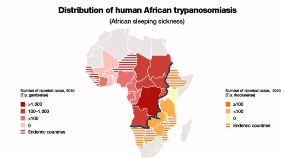
Human African Trypanosomiasis (HAT), also known as sleeping sickness, is one of the most common neglected tropical diseases (NTDs) transmitted by the bite of the tsetse fly (Glossina genus) and widespread in the sub-Saharan Africa region (Figure 1), with about 70 million people at risk of infection.1 HAT is caused by two subspecies of protozoa of
Trypanosoma genus: Trypanosoma brucei gambiense and T. b. rhodesiense,2 which are responsible for two different forms of the disease. T. b. gambiense, widespread in central and western Africa, causes the chronic form of the disease (gambiense HAT), which represents more than 95% of all reported HAT cases, characterized by a slow development. On the other hand, in the southern and eastern Africa regions, T. b. rhodesiense is responsible for the acute form of HAT (rhodesiense HAT), characterized by rapid-onset, swift progression and higher mortality rate.3 HAT is characterized by two main stages: in the early-stage, also known as stage 1 or hemolymphatic stage, the parasite invades the bloodstream and then transfers to lymph nodes, spleen and spinal fluid, causing non-specific symptoms like headache, malaise, weight loss and fatigue.
Figure 1. HAT distribution and cycle of Trypanosoma brucei.
If untreated, the stage 1 evolves to the neurological stage 2 (or late stage), during which the parasite crosses the blood-brain barrier (BBB) reaching the central nervous system (CNS), causing mental deterioration, sleep alterations, coma and lastly death.4 Unfortunately, an effective vaccine has not been developed yet, because of the high degree of antigenic
variation by variant surface glycoproteins (VSGs)5 and, consequently, chemotherapy is the sole strategy to control the infection. Only four drugs are currently available for the treatment of HAT,6 showing several problems like toxicity, parasite resistance and route of administration. Suramin and pentamidine are used only in the hemolymphatic stage, while melarsoprol and eflornithine are effective on the neurological stage, since they are able to cross the BBB. At present, eflornithine is administered in combination with nifurtimox (Nifurtimox-Eflornithine Combination Therapy: NECT), to treat melarsoprol-refractory stage 2 rhodesiense HAT and as first-line treatment for stage 2 gambiense HAT.
Within this context, several studies7 demonstrated that rhodesain, the major cysteine protease of T. b. rhodesiense, could be considered the primary target for the development of novel antitrypanosomal agents.8 Rhodesain plays key functions in the survival of the parasite and in the disease progression. In fact, it is involved in the BBB alteration9 (Figure 2A), necessary to switch from stage 1 to the neurological stage. In addition, it is also involved in the turnover of VSGs10 (Figure 2B), which allows the parasites to evade the host immune response, and in the degradation of host immunoglobulins11 (Figure 2C). Lastly, rhodesain shows an intensive lysosome proteolytic activity,12 both of parasite proteins and intracellularly transported host proteins.
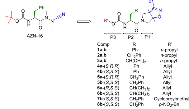
Considering the key roles played by rhodesain, during my PhD period, together with my research group, I focused my efforts on the development of novel rhodesain inhibitors as potential agents to treat HAT. We developed six different series (A-F) of molecules with the aim to obtain potent and selective rhodesain inhibitors, coupled with a good antiparasitic activity.
In series A (Figure 3), we developed novel dipeptide-like rhodesain inhibitors bearing the 3-bromoisoxazoline warhead in a constrained conformation.13 The idea to introduce 3-bromoisoxazoline nucleus takes place because 3-bromo acivicin is a potent trypanocidal agent,14 targeting the cytidine triphosphate synthetase, a glutamine amidotransferase equipped with the catalytic triad Cys/His/Glu. Starting from the potent rhodesain inhibitor AZN-16,15 we developed novel dipeptidyl analogues by replacing the azanitrile portion with a rigid bicyclic system containing the 3-bromoisoxaline nucleus, as the warhead (Figure 3).
Figure 3. Design of series A compounds starting from AZN-16 as lead compound.
At the P2 position we inserted the amino acids towards which rhodesain showed a strong preference, like Phe, hPhe and Leu, while the N-terminal portion was functionalised to give different carbamates, to evaluate the impact of various aliphatic and aromatic substituents in this position. Previously, another member of my research group synthesized compounds with Cbz, Boc and Alloc as N-protecting group, and final products were obtained as racemic mixture of diastereomers. When evaluated against rhodesain, the mixture of
diastereomeric bearing the hPhe residue at the P2 site and Alloc as N-protecting group showed the best results. Starting from these findings, we continued our investigation by replacing the allyl chain with the saturated analogue propyl group (1-3a,b), to evaluate the effect of double bond on the activity. Subsequently, we evaluated the role played by the warhead stereochemistry on the biological activity, developing the single diastereoisomers
4a-(S,R,R), 4b-(S,S,S), 5a-(S,R,R), 5b-(S,S,S), 6a-(S,R,R) and 6b-(S,S,S). Lastly, considered
that hPhe residue was well tolerated at the P2 site and S,S configured warhead showed the best inhibition results, we decided to further explore the S3 pocket by replacing N-allyl group with cyclopropylmethyl and p-NO2-Bn moieties, developing inhibitors 7b-(S,S,S)
and 8b-(S,S,S), respectively.
In series B of inhibitors, we decided to combine the 3-bromoisoxazoline warhead with a 1,4-benzodiazepine (BDZ) scaffold as specific recognition moiety, typical of various cysteine protease inhibitors.16 In particular, one of the most promising inhibitors was proven to be the Michael acceptor 9 (Figure 4), with k2nd value of 4.620.000 M-1 min-1,
coupled with a good antitrypanosomal activity (EC50 = 4.8 µM).17
Figure 4. Lead compound 9 and novel 3-bromoisoxazoline derivatives.
In this molecule, the BDZ scaffold was inserted into a peptide backbone, in which the condensed aromatic ring could mimic the Phe residue at the P2 position, while the adamantyl nucleus linked to methyl carbamoyl side chain could occupy the S3 pocket of the enzyme. Starting from this lead compound, we decided to replace the vinyl ester portion with the 3-bromoisoxazoline nucleus incorporated into a bicyclic ring, to obtain the
diastereoisomers 10a-(R,R,R) and 10b-(R,S,S).18 It is important to note that the lead compound 9 is an irreversible inhibitor, while the replacement of vinyl ester portion with the 3-bromoisoxazoline nucleus will lead to reversible inhibitors.
In series B, we discovered peptidomimetics as rhodesain inhibitors containing a 3-bromoisoxazoline nucleus, with antitrypanosomal activity similar to the lead compound 9. Inhibitors 10a-(R,R,R) and 10b-(R,S,S), showed activity against the target enzyme, in the low micromolar range, considerably lower compared to the lead compound (3 order of magnitude). We supposed that this big difference is mainly due to the warhead: probably, compounds bearing a Michael acceptor portion, with a peptidomimetic recognition moiety, are able to inhibit rhodesain to a greater extent with respect to the 3-bromoisoxazoline nucleus. The same EC50 values towards the parasite could be explained by better drug-like
properties shown by inhibitor 10b-(R,S,S). Although the latter inhibitor and the Michael acceptor 9 displayed good properties to be considered potential agents for the treatment of HAT, their high molecular weight (674.58 and 598.69, respectively), represents a great limit. Based on these considerations, we developed novel simplified BDZs 11-13a-c (Series C) bearing a Michael acceptor portion (Figure 5), with the aim to maintain/increase activity against rhodesain and parasite, and optimize the drug-like properties.19 We choose inhibitor 9 as lead compound given the deep difference between rhodesain inhibition mediated by 3-bromoisoxazoline and Michael acceptor derivatives.
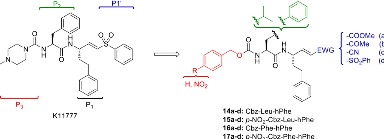
In series D of my PhD work, we focused our efforts on the development of novel peptide-based Michael acceptors 14-17a-d,20 starting from vinyl sulfone K11777 (Figure 6), a potent rhodesain inhibitor with whom the cysteine protease was co-crystallized.21
Figure 6. Vinyl sulfones K11777 and design of novel Michael acceptors.
Considered the high preference for bulky hydrophobic residues at the P2 position showed by rhodesain, we decided to maintain a Phe residue, as in K11777, or we inserted a Leu at the P2 site, while the hPhe residue was left unmodified. The N-terminal amino group was protected with Cbz or p-NO2-Cbz, the latter in agreement with the structure of inhibitor
8b-(S,S,S): the hydrophobic features of these substituents and the electron withdrawing
effect mediated by the nitro group could increase the π-π interactions at the S3 pocket. Lastly, we introduced different α,β-unsaturated portions at the P1’ site, to evaluate their different reactivity.
In light of the results obtained in series D, inhibitors bearing the vinyl ketone warhead showed the best k2nd values. Based on these data, we developed 16b derivatives, i.e. 18a-b,
19a-c, 20 and 19a-d (Figure 7), with the aim to increase selectivity and the activity against
the parasite. We decided to carry out the structure-activity relationship (SAR) study introducing novel substituents only in one position (e. g. P1’, P2 or P3), maintaining unchanged the other sites of 16b. With regard to P3 position, we inserted several substituents, linked to the amino group of Phe via urea or amide portions. In urea derivatives 18a-b, we replaced the Cbz group with N-methlypiperazine and morpholine rings, because these two portions are present in the potent rhodesain inhibitors K11777 and K11002,21,22 respectively. On the other hand, in the amide derivatives we inserted 1,3-difluorophenyl (19a) and 2,3-dihydro-1,4-benzodioxin-6-yl (19b) moieties, which are
present in potent rhodesain inhibitors reported in literature.23,15 Furthermore, we decided to introduce 5-methylbenzodioxolan-5-yl ring (19c), to evaluate the impact on the biological activity of the presence of five- or six-membered rings, both bearing two oxygen atoms.
Figure 7. Optimization of the inhibitor 16b.
At the P2 site, we replaced the Phe with the hPhe residue (20), to evaluate the impact of the chain stretch: this modification could be well tolerated, considering the size and the hydrophobic character of S2 pocket. Lastly, at the P1’ site, we decided to replace the methyl group with bulkier substituents like phenyl, cyclohexyl, ethyl and propyl groups (21a-d), to evaluate possible additional interactions with S1’ pocket.
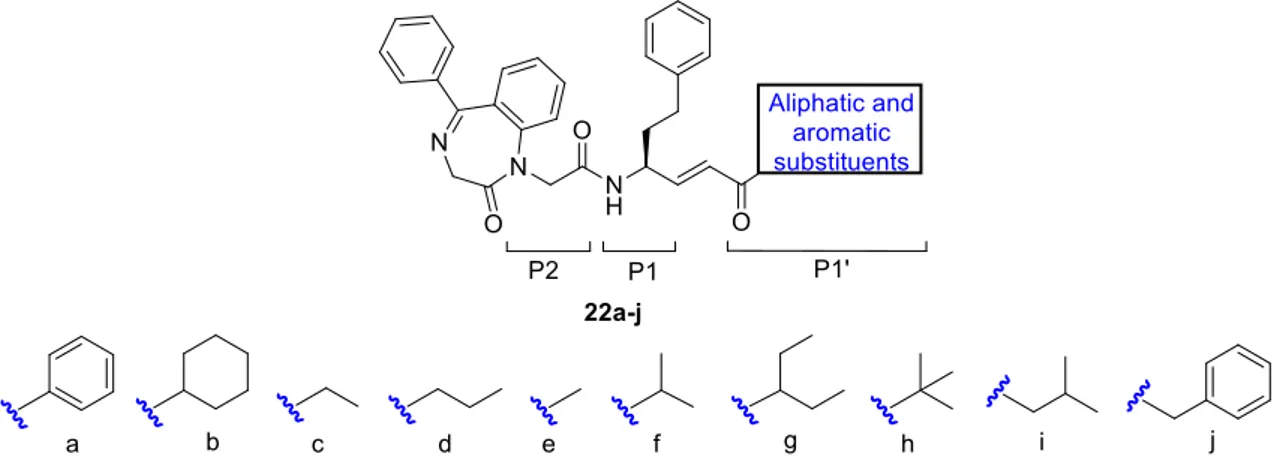
At the same time, based on the data obtained in series D, we decided to develop another series of molecules (F, Figure 8), bearing vinyl ketone warhead. Docking studies carried out on inhibitor 16b indicated the involvement of carbonyl oxygen in binding interaction by means of two H-bonds with Gln19 and Trp184 at the S1’ enzyme pocket.
Based on these data, we developed novel peptidomimetics characterized by a BDZ scaffold as recognition moiety, able to mimic Phe residue at the P2 site, hPhe at P1 position, and vinyl ketone warhead. Furthermore, we introduced several aliphatic or aromatic substituents at the carbonyl group of the warhead, with the aim to explore S1’ enzyme pocket and evaluate possible additional interactions.
The results of this wide investigation will be reported and discussed in the “Results and discussions” chapter of my PhD thesis.
CHAPTER 2. HUMAN AFRICAN TRYPANOSOMIASIS
2.1 IntroductionHuman African Trypanosomiasis (HAT), also known as sleeping sickness, is one of the most common neglected tropical diseases (NTDs), widespread in the sub-Saharan Africa and affecting 37 countries in 1.55 million km2, between latitudes 14° north and 20° south (Figure 9), with about 70 million people at risk of infection.1 It is caused by two distinct subspecies of protozoa, both belonging to Trypanosoma genus: T. brucei gambiense and T.
b. rhodesiense.2 These parasites are transmitted to human by the bite of the blood-sucking tsetse fly of Glossina genus, with a high index of infectivity and an estimated number of actual cases of 10000 per year,24 although this number is under estimated with respect to the number of real cases. As shown in figure 9, HAT is an endemic disease in the north and south of sub-Saharan region, while the highest number of HAT cases were recorded in the central area (Angola, Democratic Republic of the Congo and Tanzania).
Figure 9. The annual country reports of HAT distribution, carried out by WHO. 2.2 The parasite
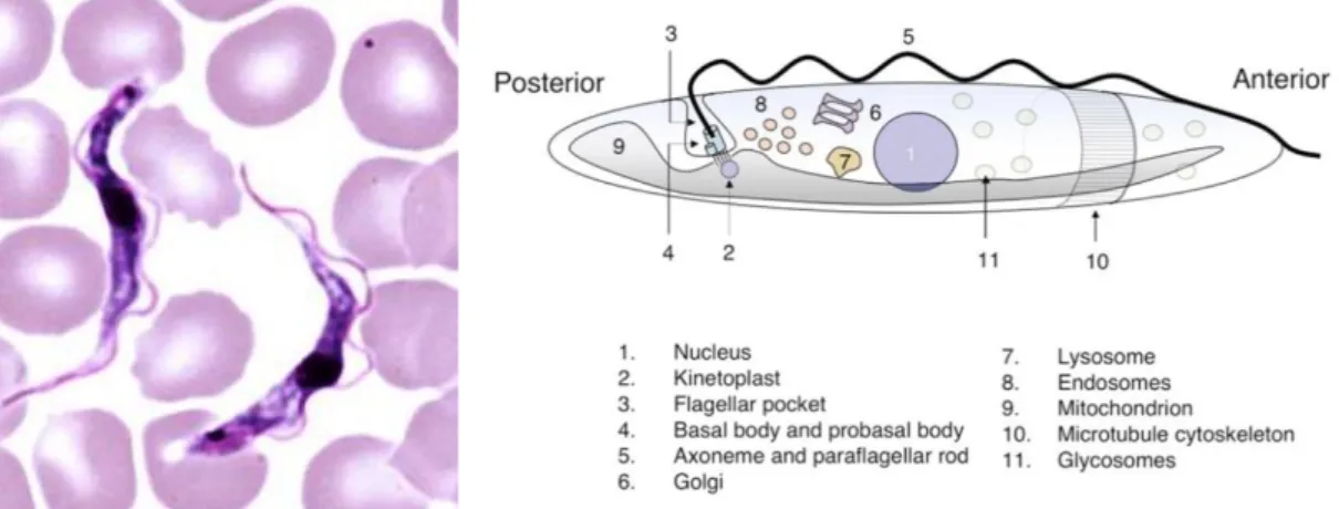
T. brucei is a typical unicellular eukaryotic cell, with a length between 8 and 50 μm; it has
a lengthened body and a streamlined and tapered shape. Its cell membrane, named pellicle, encloses the cell organelles, as the nucleus, mitochondria, endoplasmic reticulum, Golgi apparatus, ribosomes and the kinetoplast, a network of circular DNA (called kDNA) inside a large mitochondrion that contains many copies of the mitochondrial genome.25 Kinetoplast is near the basal body, but it is not distinguishable under the microscope. T.
brucei life cycle26 (Figure 10) starts with the infection of a mammalian host, when a tsetse fly bite delivers growth-arrested metacyclic trypomastigotes to the mammalian bloodstream. Then, metacyclic trypomastigotes differentiate into proliferating long forms, which maintain a bloodstream infection. Parasites eventually penetrate the blood vessel endothelium and invade extravascular tissues, including the central nervous system (CNS).
Figure 10. Life cycle of Trypanosoma brucei.
In the bloodstream, a quorum sensing-like mechanism, carries out to the differentiation of long slender forms into short stumpy forms, called also bloodstream form (BSF), which are pre-adapted for survival in the tsetse fly. When an infected host is bitten by a tsetse fly, parasites are taken up during the blood meal into the midgut, where short stumpy forms differentiate into procyclic trypomastigotes, or procyclic form (PCF), that resume cell division and establish a midgut infection. Midgut procyclic trypomastigotes initiate a migration, crossing the proventriculus, and from there onwards through the mouthparts, arrive to the salivary gland, where they attach to the salivary gland epithelium. During this migration, in the proventriculus, procyclic trypomastigotes undergo extensive restructuring, coupled to an asymmetric division, to generate one long epimastigote and one short epimastigote, which attaches to epithelial cells following arrival in the salivary gland. Attached epimastigotes replicate and ultimately complete the life cycle via an asymmetric division to generate metacyclic trypomastigotes that are free in the salivary gland lumen and are uniquely adapted to survive in the mammalian host. The T. brucei
possesses a single flagellum attached along the length of the cell (Figure 11), from which exits by a flagellar pocket, which is located at the posterior end.26 The flagellum has several functions, because is involved in cell morphogenesis and cell division, locomotion
via oscillations along the attached flagellum and cell body, establishment to the fly gut
during the procyclic phase and in in host–parasite interactions. The cell surface of the BSFs is wrapped in a dense coat of variant surface glycoproteins (VSGs), and it is replaced by an equally dense coat of procyclins when the parasite differentiates into the PCF in the tsetse fly midgut.27 The trypanosome basal bodies form the main organiser for the cytoskeleton, membranous structures and organelles.28
Figure 11. Trypanosoma b. brucei in thin blood film and simplified representation of
Trypanosoma cell architecture.
Furthermore, they serve another function to ensure inheritance of the mitochondrial genome. The kinetoplast is connected to the proximal end of both the mature basal body and pro-basal body via the tripartite attachment complex (TAC).29 The TAC represents an intricate network of filaments, connected from the proximal end of each basal body to a specialised region of the outer mitochondrial membrane, with a further set of filaments connecting the inner mitochondrial membrane and the mitochondrial DNA. This physical connection remains unchanged during cell division: the kinetoplast DNA is replicated in a periodic S phase, together with the basal body duplication. The basal body segregation ensures inheritance of the duplicated mitochondrial DNA to the two daughter cells.
2.3 HAT forms: symptoms and progression of the disease
There are two different forms of HAT (Table 1). T. b. gambiense, widespread in central and western Africa, causes the chronic gambiense form of the disease, which represents more than 95% of all reported HAT cases, characterised by a slow development
(gambiense HAT). On the other hand, in the southern and eastern Africa regions, T.
b.rhodesiense is responsible for the acute form of the HAT (rhodesiense HAT), which is
manifested with a rapid-onset, swift progression and a higher mortality rate.3
HAT is characterized by two main stages. Early-stage, also known as stage 1 or hemolymphatic stage, is characterized by non-specific symptoms.30 During this stage, the parasite invades the bloodstream causing headache, malaise, weight loss, fatigue and intermittent fever, with an onset 1–3 weeks after the tsetse fly bite. Over this time, patients might develop several problems including lymphadenopathy, enlargement of the spleen and the liver, myocarditis, pericarditis, heart attack, keratitis, conjunctivitis, endocrine dysfunction and fertility problems. Another finding that has begun to challenge the classical symptom complex is the occurrence of neurological features during early-stage disease. Some patients showed the neurological features during early-stage disease: tremors, somnolence, urinary incontinence and cranial neuropathies, symptoms that can hardly be explained by nonspecific effects at peripheral level. It is clear how the above mentioned non-specific symptoms lead to both an extension of the time to diagnose the disease and, sometimes, to an uncorrected diagnosis. This stage has a variable duration, depending on the specific subspecies: generally, 7-10 days for rhodesiense form, months for gambiense form. If untreated, the hemolymphatic stage evolves into the neurological stage (named stage 2 or late), during which the parasite invades the CNS,30 and a wide series of symptoms can take place, with almost all regions of the nervous system involved. These features include motor disturbances, as motor weakness reported, gait disturbance, tremor, abnormal movements, speech disturbances and mental alterations as the typical sleep disturbances, thus justifying the disease name, sleep abnormalities, with an inversion of the normal sleep/wake cycle, uncontrollable episodes of sleep, and an alteration of the structure of sleep itself, with the early onset of rapid eye movement sleep rather than at the end of stage 4 sleep. The behavioural disturbances like hallucinations, delirium, headache are very common. Lastly, the patients fall into a coma and, eventually, die.
Table 1. Main differences between gambiense and rhodesiense HAT forms
HAT T. b. gambiense T. b. rhodesiense
Progress Slow Rapid
Symptoms of stage I Non-specific, commons, uncorrected diagnosis CNS invasion Months (3-4) Weeks (1-2) Symptoms of stage II Motor disturbances, mental alterations, delirium
Obtaining an accurate and early diagnosis in fundamental, considered the final outcome of the disease. In the rhodesiense HAT form, trypanosomes should be identifiable on a thin layer of peripheral blood, since of the typically high level of parasitaemia.31 On the other hand, for gambiense HAT form, the parasitaemia is rarer, but the diagnosis is also possible by identifying parasites in the peripheral blood or in the lymph node aspirate. Different methods of concentration techniques might be needed to identify parasites.32
2.4 Diagnostic methods
The CATT method (Card Agglutination Test for Trypanosomiasis, figure 12) is simple and quick to effectuate, and its use is very common, in particular for population screening. Nevertheless, CATT has limitations, first and foremost the high frequency of equivocal results and limited sensitivity.33 PCR has been used to increase diagnostic accuracy,34 but such advanced equipment are not generally available in remote rural areas of Africa.
Figure 12. The example shows how interpret the CATT.
Considering the absence of reliable clinical criteria,35 a positive diagnosis by lumbar puncture, to examine the cerebrospinal fluid (CSF), is essential.36 The most widely used methods for late-stage diagnosis are the WHO criteria, meaning the presence of trypanosomes in the CSF or a white blood cell count of more than 5 cells per μL, or both. A valid method to late-stage diagnosis is measurement of CSF IgM concentrations, values that are increased when there is CNS involvement.37 Over the past few years, a great deal of efforts has gone into devising better and more reliable CNS staging markers. However, all these approaches have an intrinsic drawback: there is no standard of CNS diagnosis with which to compare any new methodology, meaning the argument is therefore somewhat circular. The problem is compounded by the dual issues of disagreement as to what criteria define CNS involvement in HAT and controversy as to which CSF
parameters should be used as an indication to use late-stage drugs.31 The sensitivity of the assay can be considerably improved by the inclusion of detergent in the reaction mix.38 For any new diagnostic test for HAT to be applicable in the affected areas in Africa, it needs to be quick, easy to undertake, inexpensive and reliable.39
2.5 Pharmacological treatment
The development of an effective vaccine is not possible due to the high degree of antigenic variation expresses from VSGs.5 The dense coat covering parasite, plays a key role against the innate immune mechanism, as the hampers the normal antibody functions of recognition of surface proteins. This shielding strategy is effective, but has one weakness: the VSG itself is recognized as an antigen and total antibody responses arise against a range of epitopes on the VSG. To permit the Trypanosoma to evade the immune response, random and background spontaneous point mutation is not enough. Trypanosoma possesses an enormous set of silent VSG genes, that differ from each other in the epitopes they encode: each silent gene has the possibility to be transcribed creating novel VSGs, which will not be recognised by immune system. In following chapter, the mechanism which allows the parasite to evade the immune system response will examined in depth. With this in mind, chemotherapy is the sole strategy to control the infection. Unfortunately, only four drugs are currently available6: suramin, pentamidine, melarsoprol and eflornithine.
The first-line treatment for rhodesiense HAT stage 1 is suramin32 (Figure 13), a polyanionic sulphated naphthylamine unable to cross the BBB, bearing an urea functional moiety and six sulfonic acids groups. It is administered as the sodium sulfonate salt by injection into a vein, because it is water-soluble but air sensible. The mechanism of action for suramin is not completely clear: it is thought that Trypanosoma is able to selectively uptake suramin by receptor-mediated endocytosis of drug that is bound to low-density lipoproteins.
Inside parasite, suramin binds to proteins, particularly to the trypanosomal glycolytic enzymes, in order to inhibit energy metabolism. Suramin was used since 1920s and, although usually effective, can result in potential complications such as renal failure, skin lesions, anaphylactic shock, bone marrow toxicity, and neurological complications such as peripheral neuropathy.
On the other hand, pentamidine(Figure 14), an aromatic diamidine, is the first-choice drug for the early-stage gambiense HAT.32 It was first used to treat HAT in 1937 and today is commercially available as white crystalline powder for reconstitution with sterile water and subsequent administration by injection or inhalation. The mechanism of action is not well understood: however, it was assumed that through cross-link between two adenines, four to five base pairs apart, pentamidine interferes with critical functions in DNA, RNA and protein synthesis. Similarly, pentamidine inhibits mitochondrial type II topoisomerase, resulting in breaks and unravelling circular mitochondrial DNA. Common side effects are burning, pain, sensation of lump in throat, chest pain, coughing, difficulty in breathing, skin rash, leukopenia, nephrotoxicity and hypotension.
Figure 14.Chemical structure of pentamidine.
The treatment of neurological stage, when the parasite crossed the BBB and invaded the CNS, provides for the use two drugs, melarsoprol and eflornithine, with serious issues, first and foremost the toxicity.
Melarsoprol (Figure 15) is a trivalent organic arsenical derivative, classified as prodrug, which is metabolized to melarsen oxide (Mel Ox) as its active form. Mel Ox is able to reacts with trypanothione,40 a spermidine-glutathione adduct that replaces glutathione in trypanosomes, forming a melarsen oxide-trypanothione adduct (Mel T, Figure 16) that competitively inhibits trypanothione reductase, killing the parasitic cell. Furthermore, it binds the vicinal sulfhydryl groups on pyruvate kinase, disrupting with energy production in the parasite. Melarsoprol was used for HAT in 1949, administered intravenously in
Figure 15. Melarsoprol and melarsen oxide after activation in vivo.
propylene glycol (extremely painful), and cannot be administered by the oral route. The inability to distinguish between host and parasites renders this drug highly toxic with many side effects. The main side effect is a post-treatment reactive encephalopathy in 10% of patients, half of whom die41: the cause of the post-treatment reactive encephalopathy is not yet known, but it is characterized by rapidly developing coma, seizures or status epilepticus and cerebral oedema. Its treatment may involve anticonvulsants, intravenous corticosteroids and acute medical supportive measures.
Figure 16. Inactivation of trypanothione by melarsen oxide.
Others common side effects are agranulocytosis, skin rashes, peripheral neuropathy, cardiac arrhythmias and a multifocal inflammatory disorder that is responsive to corticosteroids.
Eflornithine (Figure 17), also known as difluoromethylornithine (DFMO), is the first-choice drug to treat late-stage gambiense HAT and it was administered, by injection or applied to the skin, for first time in 1990.42 However, eflornithine has a principal drawback: it is ineffective against T. b. rhodesiense. It is a "suicide inhibitor," because able to bind irreversibly to the ornithine decarboxylase (ODC) and impeding the access at the
active site of the natural substrate ornithine (Figure 18). Once attached, eflornithine initially undergoes transimination which occurs with cofactor pyridoxal 5’-phosphate (PLP), followed by decarboxylation. During the decarboxylation, the fluoride atoms attached to the additional methyl group drag towards themselves the resulting negative charge from the release of carbon dioxide, with consequent release a fluoride ion.
Figure 17. Chemical structure of eflornithine.
The remaining fluoride atom, generates an electrophilic carbon, which is attacked by the nearby thiol group of Cys360, allowing eflornithine to remain permanently attached to the enzyme following the release of the second fluoride atom and subsequent transimination. The eflornithine common side effects are acne, skin reactions, itching and rash when administered topically. Otherwise, hematologic abnormalities, thrombocytopenia and reversible hearing loss are common when eflornithine is used by injection. Unfortunately, this drug with reduced toxicity is not effective in the treatment of late-stage rhodesiense HAT, due to the parasite’s low sensitivity to the molecule.
Figure 18. Mechanism of action of eflornithine.
Furthermore, several cases of drug resistance has been reported after a few years to marketing, through loss of the putative amino acid transporter TbAAT6, which has been shown in vitro.43
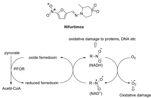
An important step forward was the development of the nifurtimox–eflornithine combination therapy (NECT)44: this combination is recommended for melarsoprol-refractory late stage rhodesiense HAT and as first-line treatment oh stage 2 gambiense HAT. Nifurtimox (Figure 19), an orally bioavailable 5-nitrofuran analogue,45 used for the treatment of Chagas disease, caused by T. cruzi: it acts through the generation of nitroanion radical causing damages to the parasite, unable to defend himself. The enzyme involved in this process is the nitro reductase, which needs oxygen for its functions. The off-label use of nifurtimox associated to eflornithine improved the late stage HAT therapy,46 above all safety and patient compliance: monotherapy with eflornithine includes infusion every 6 h for 14 days, while in combination with nifurtimox one infusion every 12 h for 7 days.
Figure 19. Chemical structure of nifurtimox and its mechanism of action.
In the last few years, the research developed two novel molecules as potential antitrypanosomal agents. The first-one is the diamine derivate pafuramidine (DB289, Figure 20),47 orally bioavailable and active against the early-stage of HAT.
This is a prodrug, which is converted in its active form furamidine (DB75): the mechanism of action is the same suggested for pentamidine. Unfortunately, in 2008 the development of this prodrug was halted in phase III clinical trials, due to its hepatic toxicity.48
At present, high expectations were addressed in the benzoxaborole derivative SCYX-7158 (Figure 21), a boron-containing molecule able to cure mice infected with T. brucei, both in hemolymphatic and neurological stage.49 Administered by oral route, in 2012, SCYX-7158 entered into phase I clinical trials,50 to evaluate its pharmacokinetic properties and toxicity for the treatment of both HAT stages: these studies confirmed that the drug penetrates the BBB and kills the parasite, coupled with acceptable values of tolerability and safety. Based on the results, in 2016, SCYX-7158 entered phase IIb/III studies in the Democratic Republic of the Congo, where numerous cases of HAT are reported.
Figure 21. Chemical structure of benzoxaborole.
Currently, the drug candidate for the treatment of advanced-stage sleeping sickness after nearly thirty years is fexinidazole (Figure 22), a 5-nitroimidazole derivative and orally bioavailable.51 The mechanism of action of fexinidazole is not clear, but it could involve bioreductive activation mediated by parasite nitroreductases. Two complementary cohort studies with fexinidazole were completed in 2016, and the results of the phase II/III study support the submission of a regulatory dossier to the European Medicines Agency, probably by the end of 2017.52
Figure 22. Fexinidaxole chemical structure.
In the present scenario, there is a clear need to find novel antitrypanosomal agents, endowed with an efficacy and safety profile, with the aim to introduce them for the
treatment of HAT. Even more interesting is the challenge to identify new trypanosomal targets on which acting for alternative therapies.
CHAPTER 3. CYSTEINE PROTEASES: ALTERNATIVE
AND VALID TARGETS
3.1 The ideal drug
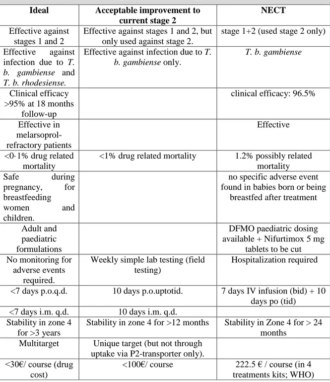
Concerning protozoal infections, the principle of magic bullet, created by Paul Ehrlich, is always the aim to be achieved: the ideal drug should be directed against a target exclusive
Table 2. Features of ideal, acceptable and NECT approach in the HAT treatment Ideal Acceptable improvement to
current stage 2
NECT
Effective against stages 1 and 2
Effective against stages 1 and 2, but only used against stage 2.
stage 1+2 (used stage 2 only) Effective against
infection due to T.
b. gambiense and T. b. rhodesiense.
Effective against infection due to T.
b. gambiense only. T. b. gambiense Clinical efficacy >95% at 18 months follow-up clinical efficacy: 96.5% Effective in melarsoprol-refractory patients Effective <0·1% drug related mortality
<1% drug related mortality 1.2% possibly related mortality Safe during pregnancy, for breastfeeding women and children.
no specific adverse event found in babies born or being
breastfed after treatment
Adult and paediatric formulations
DFMO paediatric dosing available + Nifurtimox 5 mg
tablets to be cut No monitoring for
adverse events required.
Weekly simple lab testing (field testing)
Hospitalization required
<7 days p.o.q.d. 10 days p.o.uptotid. 7 days IV infusion (bid) + 10 days po (tid)
<7 days i.m. q.d. 10 days i.m. q.d. Stability in zone 4
for >3 years
Stability in zone 4 for >12 months Stability in Zone 4 for > 24 months
Multitarget Unique target (but not through uptake via P2-transporter only). <30€/ course (drug
cost)
<100€/ course 222.5 € / course (in 4 treatments kits; WHO)
for the parasite, or otherwise significantly different in the human. Drug for Neglected Diseases initiative (DNDi) reported a list (Table 2) of requirements about ideal drug for HAT treatment.53
These include features such as the efficacy against both stages, acceptable range of toxicity, the duration of treatment, the appropriate physicochemical and pharmacokinetic properties for oral bioavailability. In the same way, an ideal target could be essential for viability of the parasite when it is inside the body both in stage 1 and stage 2, and the alteration of its functions must generate a trypanocidal effect.
3.2 Potential targets
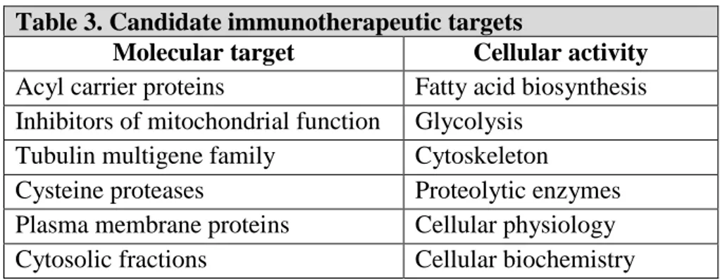
Different proteins and metabolic pathways are identified as potential target to develop novel antitrypanosomal agents: table 3 summarises the candidate molecular targets with the respective cellular activity.54 Among those, cysteine proteases play several key roles in the life cycle of parasitic organisms: their function is to catalyse the cleavage of amide linkages in macromolecular proteins and oligomeric peptides, essential function in the parasite survival into human body, tissue invasion and failure of the immune system response.55
Table 3. Candidate immunotherapeutic targets
Molecular target Cellular activity
Acyl carrier proteins Fatty acid biosynthesis Inhibitors of mitochondrial function Glycolysis
Tubulin multigene family Cytoskeleton
Cysteine proteases Proteolytic enzymes Plasma membrane proteins Cellular physiology Cytosolic fractions Cellular biochemistry
The catalytic activity can initiate both within a polypeptide chain (endoprotease activity) and from carboxy- or amino- terminal regions, (exopeptidase activity). Proteases were divided into groups on the basis of the catalytic mechanism used during the hydrolytic function: the main catalytic types are serine-, threonine-, aspartic-, metallo- and cysteine proteases.
Cysteine proteases are ubiquitously expressed both in human and in protozoans, and were successfully targeted to treat human diseases: at the same way, they play a key role in parasitic diseases and for this reason were recognized as valuable targets for the parasitic infections. Furthermore, the relative lack of redundancy in mammalian system, highlights
the importance of these enzyme families for the development of novel antiparasitic agents. The validation of this target was carried out by Scory et co-workers,56 which proved that the peptidyl inhibitor Cbz-Phe-Ala-CH2N2 23 (Figure 23) killed the bloodstream forms of
T. brucei. In addition, another study57 demonstrated that the treatment with biotinylated cysteine protease inhibitors induced T. brucei death.
Figure 23. Chemical structure of peptidyl inhibitor 23.
More in detail, cysteine proteases of parasitic organisms are divided into two main groups,55 corresponding to clans CA and CD (Figure 24). Other minor groups are clans CB and CC, identified as viral proteases. In 1879, the first cysteine protease was characterised from the fruit of Carica papaya, and was therefore called papain, and the subsequent discovered cysteine proteases, with similar chemical structure to papain, were named “papain-like”. The assignment to a family rather than to another one is a function of a number of properties, as sequence homology, possession of inserted peptide loops, and biochemical specificity to small peptide substrates.
Figure 24. Schematic graphic of the cysteine proteases of parasitic organisms.
Like all the enzymes, cysteine proteases present a substrate specificity governed by the nature of the amino acid residues in the proximity at the cleavage site. The nomenclature
used to define the binding pockets58 is relative to the cleavage site (Figure 25): Sn represents the enzyme pockets which are numbered S1-S2-Sn from cleavage site toward N-terminus (non-primed pockets), whereas are indicated as S1’-S2’-Sn’ toward C-N-terminus (primed pockets). At the same way, the number of substrate residues initiates from scissile bond and, in agreement with enzyme pocket numeration, they are numbered P1-P2-Pn, and P1’-P2’-Pn’, toward N-terminus and C-terminus, respectively.
Figure 25. Nomenclature of the substrate specificity of protease.
The majority of parasitic cysteine proteases belong to the family C1, within clan CA. In turn, family C1 consists of three subfamily: cathepsin B-like, cathepsin-L-like and bleomycin-hydrolase. The two cathepsin subfamilies are distinct55 for the presence in the cathepsin L-like subfamily of a highly conserved amino acid motif in the propeptide region of the protein, called ERFNIN motif, while cathepsin B-like proteases are characterized by the presence of an occluding loop.
The major cysteine proteases expressed by T. brucei are rhodesain, a cathepsin L-like protease also known as TbCatL, and the cathepsin B-like TbCatB: both represent promising targets for novel antitrypanosomal agents. Their importance was demonstrated in several studies7a,b: the silencing of TbCatB cured mice of trypanosomal infection, while rhodesain suppression only prevented the transition from stage 1 to stage 2 and prolonged the life span of the infected mice. Conversely, more recent studies carried out by Steverding7c and co-workers, suggested that TbCatL, rather than TbCatB, is the essential
cysteine protease of T. brucei and should be considered the primary target for HAT treatment.
3.3 Rhodesain: structure and functions
Rhodesain is the major cysteine protease of T. b. rhodesiense, consisting of a single polypeptide chain of 215 amino acids with typical papain-like folding, characterized by a (Figure 26) left (L) and a right (R) domain and with the catalytic triad (Cys25/His162/Asn182) located in a cleft between the two domains.22
Figure 26. The figure shows the secondary structure of T. brucei rhodesain, with two
domains left (green) and right (gray).
In T. b. brucei, the major cysteine protease is called brucipain, with a very high homology degree (98.4%) with rhodesain. Furthermore, rhodesain possesses a high structural similarity to cruzain, the main cysteine protease of T. cruzi, the aetiological agent of Chagas disease, and a slightly lower similarity with falcipain-2 2) and falcipain-3 (FP-3), the major cysteine proteases of the human malaria parasite Plasmodium falciparum. Like all the cysteine proteases, the catalytic mechanism starts with the conversion of the weakly nucleophilic thiol group of the Cys25 to a highly nucleophilic thiolate anion (Figure 27), and this process involves the two remaining amino acids of the catalytic triad.
The Asn182, acting as H-bond acceptor, is responsible for the stabilization of the correct tautomeric form of the His162 imidazole ring, which acts as base in the deprotonation process of thiol group. In this condition, the Cys25 thiolate anion is able, by nucleophilic attack, to cleave the amide bond. After that, other amino acid residues are concerned to permit amide bond hydrolysis and the regeneration of the free enzyme.
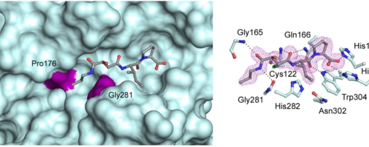
Two different crystal structures of rhodesain, in complex with vinyl sulfone inhibitors K11777 and K11002 (Figure 28), which will be described in the following chapter, were reported (PDB codes 2P7U21 and 2P86,22 respectively). Furthermore, northern blot analysis demonstrated that the amounts of rhodesain mRNA expressed, both the bloodstream and the procyclic forms of the parasite, are equal.7a
Figure 28. Crystal structure of rhodesain in complex with K11002.
Rhodesain plays key roles both in disease progression and in parasite survival, and it is considered a promising target towards which to develop novel potential antitrypanosomal agents.8 Rhodesain is necessary to cross the BBB, promoting the disease evolution from stage 1 to stage 2.9 It was hypothesized that BBB dysfunction are related to the interactions of rhodesain with Gq Protein Coupled Receptors (GPCRs), also known as Protease
Activated Receptors (PARs).59 The activation of this protein enables to cross the BBB after cytoskeletal rearrangements, inducing cell retraction and loosening of junctional complexes. Gq proteins activated by rhodesain, activate phospholipase C (PLC), in turn
generating inositol-1,4,5-triphosphate (IP3) and diacylglycerol (DAG) from phosphatidylinositol-4,5-bisphosphate (PIP2). The binding of IP3 to its receptor, placed on the endoplasmic reticulum, releases Ca2+: the resulting increase of intracellular calcium activates calmodulin of Myosin Light Chain Kinase (MLCK) and/or other effectors leading to cytoskeletal changes and barrier dysfunction. Beyond that pathway, Ca2+-independent
activation of the cytoskeleton generated by Ras-superfamily GTPases (i.e RhoA) traditionally activated by Gα12/13, could lead to alterations of the adherens junctions and
tight junctions, by means the protein p63RhoGEF.60
As mentioned above, the surface of BSF of T. brucei is covered by a dense coat, consisting of VSGs, which is attached at the C-terminus to the cytoplasmic membrane via a glycosylphosphatidylinositol (GPI) anchor.61 VSGs play a key role in elusion of the host immune response, through two main mechanisms. The first one is the duplicative transposition of a silent VSG gene into one of the telomeric VSG expression sites of the
Trypanosoma, resulting in the replacement of the previously expressed VSG gene.62 Only one of approximately one thousands of VSG genes is expressed from a VSG gene expression site: a combination of genetic recombination and VSG expression site switching is responsible for regular changes in VSG expression,63 against which the host has not yet produced specific antibodies. On the other hand, VSGs are constantly removed from the parasite’s surface by rapid internalization and recycling: it was suggested that rhodesain could act as congopain, orthologous enzyme in T. congolese, which plays a key role in the degradation of VSGs within the lysosome.10,64 This proteolytic process is fundamental to ensure the amino acids requested for synthesis of novel VSGs. The anti-VSG antibodies are internalized also, to be degraded: in particular, the peptidases destroyed the IgG, with decreasing of their amount. Pre-treatment of the Trypanosoma with cysteine protease inhibitors, K11777 and E-6465 (Figure 29), prevented IgG degradation,11 demonstrating as rhodesain contributes significantly to IgG degradation.
Figure 29. Chemical structure of potent cysteine proteases inhibitor E-64. 3.4 TbCatB: structure and functions
TbCatB is a Clan CA, family C1 (papain-like) cysteine protease belonging, differently to the rhodesain, to cathepsin B-like cysteine protease subfamily. It consists of 341 amino acids and it is composed by two domains (Figure 30), left (L) and right (R), with an
occluding loop of approximately 20 amino acids located on the surface of cathepsin B-like, and with the typical catalytic triad Cys42/His282/Asn302.22
Figure 30. Secondary structure of TbCatB. It is noted the occluding loop (red).
Furthermore, TbCatB was co-crystallized in complex with CA074 (Figure 31), a peptidyl epoxide inhibitor (PDB code 3HHI).22 By northern blot analysisit was demonstrated that TbCatB mRNA was less expressed with respect to rhodesain mRNA, both in the BSFs and PCFs.7a In addition, the mRNA for TbCatB was up-regulated in the BSF parasites: this expression difference suggests that TbCatB is regulated at the transcriptional level and carries out its activities mainly in this form. Because they lack cytochromes, the BSF of T.
brucei acquires iron from the host transferrin,7a through its internalizing receptor-mediated endocytosis.
Figure 31.Co-crystal structure TbCatB-CA074.
Then, transferrin is rapidly degraded in the “endosome/lysosome” located between the nucleus and kinetoplast of the parasite: it was demonstrated that TbCatB is the major protease involved in this fundamental process. In addition, it was observed the parasite ability to complete multiple steps of genomic replication and mitosis, but were not able to
finish the cytokinesis. It is possible that TbCatB could also be involved in a microtubule-related event: lack of cytokinesis may be an indirect consequence of iron depletion because of the incapacity of the parasite to degrade transferrin.7a
A superimposition of the rhodesain-K11002 and TbCatB-CA074 complexes brings to highlight important structural differences between the two enzymes. The S2 site of rhodesain is restricted with respect to TbCatB, due to the presence of Ala208 at the bottom of S2 site, while at the S2 pocket bears Gly328, less bulky. Furthermore, the Leu67 residue presents around the S2 site of rhodesain creates a hydrophobic environment, where the Phe residue at P2 position of K11002 well fits. On the other hand, around S2 site of TbCatB is presents Asp166, with its relative acid function. Another relevant structural feature is due to the presence in TbCatB, of the occluding loop, which creates a large prime side pocket in the catalytic site cleft.
Considering the key roles of rhodesain and TbCatB during different phases of parasite life cycle, it is presumable that the co-inhibition of both enzymes could be certainly desirable in the development of novel antitrypanosomal agents.
CHAPTER 4: RHODESAIN INHIBITORS
In this chapter, we describe the most active rhodesain inhibitors reported in the literature to date, with particular attention to their chemical composition, mechanism of action, binding mode and SAR.
4.1 Peptide-based inhibitors
Peptide molecules are recognized for their high selective, efficacy and tolerability. There is an important interest in peptides as potential therapeutic agents,66 and a growing number of peptides, approximately 140, are currently evaluated in clinical trials. Sometimes, natural peptides are not suitable for use as convenient therapeutics because of their intrinsic weaknesses, as poor chemical and physical stability, and a short circulating plasma half-life. On the contrary, peptides designed for use as drugs show various advantages, summarised in table 4 (Source: Marx, V. 2005. “Watching Peptide Drugs Grow Up.” Chemical & Engineering News).
Table 4. Features of peptide drugs
Advantages Disadvantages
High activity Low orally bioavailability High specificity Injection required
Little unspecific binding to molecular structures other than desired target
Difficult delivery and challenge to transport across membranes
Minimization to drug-drug interactions Less stable
Less accumulation in tissues Challenging and costly synthesis Lower toxicity Solubility challenges
Often very potent Risk of immunogenic effects Biological and chemical diversity Cleared of body quickly
To date, peptide inhibitors targeting rhodesain show two common structural features: a peptide fragment necessary for recognition by the enzyme and an electrophilic portion, called warhead, which undergoes the nucleophilic attack of the cysteine residue of the catalytic site.
Diazomethylketone Cbz-Phe-Ala-CH2N2 23 and E-64 (see chapter 3) are potent cathepsin
inhibitors, but they are not selective for the several type of cathepsin. To avoid indiscriminate reactivity with other thiol-containing enzymes, the intrinsic reactivity of the warhead must be sufficiently low, in order to reaction with Cys anion take places only after a specific noncovalent binding of recognition segment, in such a way that the warhead be at a distance and orientation favourable for the reaction. The important challenge is
represented on the development of cysteine protease selective inhibitors, endowed with a high selectivity degree between parasite and mammalian enzymes, by exploiting the few differences surrounding the catalytic site.
4.1.1 Vinyl sulfones
One of the most relevant class of cysteine protease inhibitors are vinyl sulfones, who act as Michael acceptors, due to their ability to covalently trap the Cys residue at the catalytic site.67 These inhibitors ensure an irreversible inhibition, because the novel covalent bond is stable and the enzyme cannot back to active form, even by increasing the concentration of natural substrate. Mechanism of action (Figure 32) was proposed: the sulfone oxygens interact by hydrogen bond with the catalytic triad residues His and Asn and these bonds would lead to polarization the vinyl group, with activating the β-carbon towards the nucleophilic attack. The negative charge resulting at the α-carbon is immediately eliminated via protonation by the histidinium residue.
Figure 32. Mechanism of action of vinyl sulfones.Many vinyl sulfones were demonstrated
to be potent rhodesain inhibitors.
As mentioned in chapter 3, given the high potency of peptide-based vinyl sulfones, two different crystal structures of rhodesain in complex with inhibitors K11777 (Figure 33) and K11002 were deposited to PDB, with codes 2P7U21 and 2P8622, respectively.
In 2001, Caffrey et al., carried out a study68 to evaluate the different reactivity of several substituents at the P1’ site, maintaining Cbz-Phe-hPhe as recognition moiety (Table 5). Data collected showed an emerging trend of reactivity: sulfonates were found to be the best inhibitors, with one order of magnitude above those displayed for aliphatic and aromatic sulfones. The introduction of bulky substituents at the P1’ site, namely CH2CH2Ph (24d)
and NHPh (24e), respectively, led to a slight decrease of potency. On the contrary, the addition of a methylene group (24b) and an oxygen atom (24a), to afford benzyl and sulfonate analogues, respectively, increased the activity against the enzyme. At the P1 position, hPhe amino acid is strongly preferred by rhodesain: this residue possesses the essential features to well fit at the S1 pocket.
Additional studies have shown that the S2 site is responsible for the specificity of rhodesain against peptidyl substrates.69 Rhodesain is able to tolerate various bulky hydrophobic residues within S2 pocket, due to the presence of the Leu67 residue which make S2 pocket mainly hydrophobic. More in detail, the highest preferences were shown towards leucine and phenylalanine, in equal measure. Moreover, Ala208 residue at the bottom of the S2 pocket makes the pocket deep, while Gln159 and Leu160 further narrow the pocket.21 However, rhodesain is 50% less reactive towards a substrate with β-branched residue at the P2 site (i.e valine) and is completely inactive in the presence of arginine (Arg) at this site.68 With regarding the S3 pocket, a comparison between the complexes rhodesain-K11777 and rhodesain-K11002, demonstrates that despite the cysteine protease is unable to establish specific polar interactions with morpholine urea portion of K11002, this group is preferred compared to N-methyl piperazine nucleus of K11777, of larger size. An comparative study21 was carried out between inhibitor K11017 (Figure 33) and K11777, which differ for amino acid residue at the P2 site. Compound K11017 showed a
k2nd value slightly higher than K11777 (k2nd =15.84 x 10⁶M-1 min-1 vs 9.00 x 10⁶M-1 min-1,
Table 5. Biological activity of different vinyl sulfonates, sulfones and sulfonamides. Comp R k2nd (M-1 min-1) 24a -OPh 3.48 x 10⁸ 24b -CH2Ph 5.35 x 10⁷ 24c -Ph 1.61 x 10⁷ 24d -CH2CH2Ph 1.09 x 10⁷ 24e -NHPh 2.24 x 10⁶
respectively): considering that both vinyl sulfones contain the amino acids preferred at the P2 site, the slight different k2nd values can be explained by additional interactions. It was
suggested that the morpholine ring at the P3 site of K11017 is able to create a network of water molecules that anchors the morpholine oxygen to the polar residues in the S3 pocket; on the contrary, K11777, containing the N-methyl-piperazine nucleus, in not able to create these polar interactions, and this might explain its reduced reactivity. When evaluated against BSFs of T. brucei, K11777 showed a good sub-micromolar activity (EC50= 0.9
μM), coupled with a poor oral bioavailability (19.9%), but better than K11002, with only 3%. In this case, the difference is primarily attributable to an increased solubility in intestinal fluids of K11777, due to N-methyl-piperazine ring.70 In addition this nucleus might vehicular the drug to concentrate within the acidic compartments,71 property known as lysosomotropism: this feature is highly desired, taking into consideration that rhodesain is mainly localized in the lysosomes. Currently, K11777 is into phase I clinical trials to evaluate tolerability, safety, and pharmacokinetics in healthy patient volunteers.
In 2013, vinyl sulfones bearing Phe or Leu at the P2 site and basic residues at the P1 site (Figure 34) were developed.72 When tested against parasite, these molecules showed an optimal antitrypanosomal activity, with EC50 values ranging from 0.07 to 26.4 µM. With a
few exception, these inhibitors displayed a more than two fold selective inhibition of parasite with respect to mammalian cells: the most promising compound of the series was proven to be inhibitor 25, with a selectivity index (SI) of 785.
Figure 34. Chemical structure of best inhibitor developed and its biological evaluation.
In the same work, were carried out computational studies on rhodesain and TbCatB. Compound 25 interacts with both enzymes by key H-bonds, in the same way of K11777. An important difference was found relatively to binding mode of naphthylsulfonylamido
moiety: in TbCatB, the Asn163 residue allows the formation of a hydrogen bond via water-mediated interaction, while in rhodesain Asn163 is replaced by Gly64, which is not able to create this interaction.
4.1.2 Azadipeptide nitriles
Another important class of peptide-based cysteine protease inhibitors is represented by azadipeptide nitriles. The mechanism of action (Figure 35) of these molecules is based on the reaction between the nitrile carbon and Cys anion, with the formation of a reversible isothiosemicarbazide adduct,73 with good stability, probably due to resonance of the nitrogen atom lone pair and the sp2-hybridized carbon atom derived from the cyano group. Moreover, the modified amide bond by N-methylation presents an improved stability towards proteolytic degradation compared to typical peptide bond.
This class of compounds shows a reversible inhibition. This type of inhibition is characterized by weak interaction between enzyme and inhibitor, and when inhibitor is removed, enzymatic activity was fully restored.
Figure 35. Mechanism of action of azadipeptide nitriles.
An equilibrium is established between the free enzyme and the non-covalent enzyme-inhibitor complex and is regulated by the dissociation constant Ki that measures the
binding affinity towards the enzyme: lower is the Ki value higher is binding affinity.In
2012, Yang et al. developed novel azadipeptide nitriles15 characterized by a common substituent at the P3 site, various hydrophobic amino acids at the P2 site (i.e Phe, Val and Leu), towards which rhodesain demonstrated a strong preference, and substituents of different sizes at the P1 site, both of aromatic and aliphatic chemical nature. As it was expected, derivatives bearing Phe (26a-e) and Leu (26f-j) residue at the P2 site showed IC50 values (Table 6) 1–2 orders of magnitude lower than the analogues bearing Val at the
same position. With regard to the P1 site, the single methyl substituent was well tolerated by rhodesain, with respect to bulkier substituents, whose presence led to reduction of