Comparison between loop-tip guidewire-assisted
and conventional endoscopic cannulation in high
risk patients
Authors Enzo Masci1, Benedetto Mangiavillano1, Carmelo Luigiano2, Alessandra Bizzotto1, Eugenio Limido3,
Paolo Cantù4, Gianpiero Manes5, Paolo Viaggi1, Giancarlo Spinzi6, Franco Radaelli6, Alberto Mariani7,
Clara Virgilio2, Angela Alibrandi8, Pier Alberto Testoni7
Institutions Institutions are listed at the end of article.
submitted 7. April 2015 accepted after revision 26. June 2015
Bibliography DOI http://dx.doi.org/ 10.1055/s-0034-1392879 Published online: 15.9.2015 Endoscopy International Open 2015; 03: E464–E470 © Georg Thieme Verlag KG Stuttgart · New York E-ISSN 2196-9736 Corresponding author Enzo Masci, MD
Gastrointestinal Endoscopy Unit San Paolo University Hospital University of Milan Via A. Di Rudinì 8 20142 Milano Italy Fax: +39-0184-536870 [email protected] License terms
Introduction
!Acute pancreatitis remains the most frequent and dreaded complication after endoscopic retro-grade cholangiopancreatography (ERCP), with rates ranging from 2 % to 40 % in published series [1–3]. Although in most cases post-ERCP pan-creatitis (PEP) manifests as a mild, uneventful dis-ease requiring 2 to 3 days of additional hospitali-zation with an expected full recovery, severe pan-creatitis is a devastating occurrence fraught with a significant mortality rate. Several risk factors for ERCP-related complications have been clearly identified, and various strategies have been pro-posed aimed at reducing the risk for PEP [4, 5] Repeated attempts at cannulation and increased pressure within the pancreatic duct (PD) due to contrast injection into the main PD result in trau-ma to the papilla and are trau-major risk factors for PEP [6]. Other factors shown to increase the risk for PEP include female sex, younger age, and sphincter of Oddi dysfunction.
Various pharmacological agents, including corti-costeroids, nonsteroidal anti-inflammatory drugs,
somatostatin analogues, and protease inhibitors, have been investigated; however, the benefit and advisability of their routine use remain uncertain [7, 8]. As well, various procedural techniques, such as the insertion of pancreatic stents or the use of pure cut electrocautery for sphincterotomy, have been suggested to minimize the development of PEP, but most studies have produced nonconfir-matory data.
Most nonrandomized and randomized studies have compared the effectiveness of guidewire-assisted cannulation (GWC) with that of con-trast-assisted cannulation (CC) for the prevention of PEP but are restricted to low risk patients, and the results so far are conflicting [9– 18]. The rationale for common bile duct (CBD) cannula-tion with the guidewire-assisted technique is that it is easier to pass a hydrophilic guidewire with a small diameter through the small opening of the CBD than a larger catheter or sphincterotome. However, this cannulation technique carries a risk for false passage, intramural dissection, perfora-tion, and PD injury [6].
Background: The guidewire biliary cannulation
(GWC) technique may increase the cannulation rate and decrease the risk for post-endoscopic retrograde cholangiopancreatography (ERCP) pancreatitis. The aim of our multicenter prospec-tive randomized controlled trial was to determine if the use of an atraumatic loop-tip guidewire re-duces the rate of post-ERCP pancreatitis (PEP) compared with the standard contrast-assisted cannulation (CC) technique.
Methods:From June 2012 to December 2013, a
total of 320 patients who had a naïve papilla and were referred for ERCP were randomly assigned to the GWC group (n = 160) or the CC group (n = 160). GWC or CC was randomly used. In cases of failed cannulation in both arms after crossover, biliary access was attempted with alternative
techniques (e. g., dual-wire technique, pancreatic duct stenting, precut).
Results:The biliary cannulation rates were 81 % in
the GWC group and 73 % in the CC group (P = n. s.). Following crossover, cannulation was successful in 8 % and 11 % of patients in the GWC and CC groups, respectively. With use of an alternative technique, the cannulation rates were 98 % in the GWC group and 96 % in the CC group, respectively. The rates of PEP were 5 % in the GWC group and 12 % in the CC group (P = 0.027). The post-inter-ventional complication rates did not differ be-tween the two groups.
Conclusion: GWC with the new wire guide is
associated with a lower rate of PEP in comparison with the CC technique.
Clinical trial reference number: NCT01771419
Recently, a new wire guide with an atraumatic loop tip has been marketed (Fusion LoopTip Wire Guide; Cook Medical, Blooming-ton, Indiana, USA). The design of the device may reduce injury to the pancreatic and bile ducts during cannulation attempts. The aim of this study was to assess if this atraumatic loop-tip guidewire reduces the occurrence of PEP (the primary end point). As a secondary end point, we evaluated whether the use of a loop-tip guidewire increases the rate of CBD cannulation in patients at higher risk for developing PEP in comparison with the CC technique.
Patients and methods
!
Between June 2012 to December 2013, a prospective study was carried out at eight Italian tertiary referral endoscopy centers. The study protocol was conducted in accordance with the princi-ples of good clinical practice and the Declaration of Helsinki (clin-ical trials reference number NCT01771419), and the institutional review boards of the hospitals approved the study protocol. All pa-tients provided written informed consent to take part in the study. Eleven endoscopists with experience of more than 150 ERCPs per year conducted the procedures (E.M., B.M., C.L., E.L., P.C., G.M., P.V., F.R., A.M., C.V., and P.A.T.). All of the endoscopists used both meth-ods (GWC and CC) to cannulate the CBD.
Fusion loop-tip guidewire
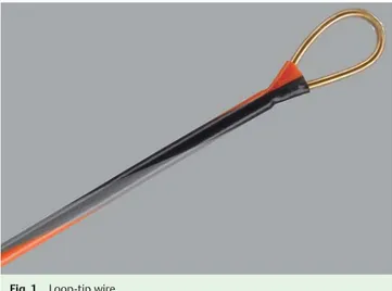
The loop-tip wire was developed to assist in cannulating the bile and pancreatic ducts and in bridging difficult strictures during ERCP. The wire has a closed distal loop that causes less trauma to the pancreaticobiliary ductal system during access. The coil is radiopaque with optimal fluoroscopic visibility. Like other types of wire, the loop-tip guidewire has spiral markings extending to the distal end, which provide an endoscopic view of its move-ment during cannulation. The core of the guidewire is made of nitinol for kink resistance and shape retention.
Eligibility criteria
All consecutive patients 18 years of age or older were considered for inclusion if they met the following criteria: were able to com-ply with the study procedures and provide written informed con-sent; had received a diagnosis of a pancreaticobiliary disease and been referred for ERCP; had an intact papilla and one or more risk factors for the development of PEP (CBD diameter < 10 mm, pre-vious episode of acute pancreatitis or recurrent acute pancreati-tis, sphincter of Oddi dysfunction type 1, female sex). The diag-nosis of a pancreaticobiliary disorder was based on clinical and laboratory data and findings on computed tomography, magnetic resonance imaging, and/or endoscopic ultrasound.
Exclusion criteria were the following: age younger than 18 years; previous sphincterotomy; previous endoscopic papillary balloon dilation; plastic or metal biliary stent placement; esophageal or gastroduodenal stenting; pancreatic or ampullary carcinoma; prior Billroth II gastrectomy; intradiverticular papilla; duodenal stenosis; contraindications to endoscopy; acute illness (hypoten-sion [blood pressure < 90 mmHg], hypoxemia [SaO2< 95 %], hemo-dynamic instability); coagulopathy (platelet count < 50 × 109/L, international normalized ratio > 1.5); inability or refusal to give written informed consent.
Randomization
Before ERCP, enrolled patients were randomly allocated to one of two groups (GWC group or CC group) on the basis of a sealed envelope method used by a physician who was not involved in performing ERCP or in the critical care of the patients. Data from consecutive patients were collected in a prospective manner through a standardized case report form and centralized at a coordinating center for analysis and interpretation.
Study protocol
ERCP procedures were conducted by experienced therapeutic endoscopists using a therapeutic video duodenoscope.
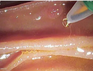
Guidewire cannulation group: In the GWC group, the sphincter-otome was preloaded with a 0.035-in loop-tip guidewire (
●
" Fig. 1). The sphincterotome was oriented from the 11- to the 12-o’clock position on the papilla and bent to align it correctly with the bile duct axis. After a minimal insertion (2– 3mm) of the sphincterotome across the ampulla, the guidewire was care-fully advanced through the CBD under fluoroscopy until it was seen entering the bile duct (●
" Fig. 2). In cases of PD cannulation, the guidewire was withdrawn, and attempts were made to redirect it toward the CBD. Such attempts were continued for no longer than 5 minutes or up to five unintentional cannulations of the PD. If biliary cannulation was achieved through guidewire in-sertion, contrast medium was injected, whereas in a case of fail-ure, the patient was crossed over to the CC technique for a time limit of 5 minutes or for a maximum of three repeated, uninten-tional contrast injections or cannulations of the main PD. Contrast-assisted cannulation group: In the CC group, the sphinc-terotome was used to cannulate the CBD directly, with its position adjusted to the correct axis for bile duct cannulation (●
" Fig. 3). If direct cannulation was not achieved, contrast opacifications were performed to visualize the CBD, and the sphincterotome was deeply reinserted under cholangiographic guidance. Such at-tempts were continued for 5 minutes or for a maximum of three unintentional PD opacifications or cannulations. If the procedure did not succeed with the CC method at the first attempt, patients were crossed over to the GWC technique with the loop-tip guide-wire, and the endoscopist attempted to cannulate for no longer than 5 minutes or five accidental main PD cannulations.The number of chances of main PD cannulation for the GWC group (five) and for the CC group (three) were chosen only for the study protocol. In cases of failed cannulation with both tech-niques, alternative techniques to access the biliary tree were
Fig. 1 Loop-tip wire.
adopted, such as dual-wire technique, PD stenting, rendez-vous, or precut, according to the endoscopist’s experience.
None of the patients received nonsteroidal anti-inflammatory drugs or a prophylactic pancreatic stent.
Definition of events
Successful deep biliary cannulation was defined as free and deep instrumentation of the biliary tree. The definition and grading of complications of ERCP and therapy were based on consensus guidelines [1].
A diagnosis of PEP was established in the presence of typical ab-dominal pain associated with serum amylase elevations more than threefold above the upper limit of normal on day 1 after the procedure. The severity of pancreatitis was graded as mild (2–3 days of hospitalization), moderate (4– 10 days of hospitali-zation), or severe (≥10 days of hospitalization or a requirement for admission to the intensive care unit and/or endoscopic/radio-logical percutaneous drainage or surgical intervention).
The severity of bleeding was defined as mild (endoscopic and clin-ical evidence of bleeding with a hemoglobin drop of < 3 g/dL and requiring no blood transfusion), moderate (requiring < 4 units of blood transfusion and no angiographic intervention/surgery), or
severe (requiring ≥4 units of blood transfusion or angiographic intervention/surgery).
The severity of perforation was defined as mild (uncertain or very slight leak of contrast and conservative medical manage-ment for≤3 days), moderate (any definite perforation requiring conservative medical treatment for 4– 10 days), or severe (any definite perforation requiring medical treatment for > 10 days or percutaneous/surgical intervention).
Outcome measures
Primary outcomes measures were the rate of successful biliary cannulation and incidence of PEP. Secondary outcome measures were the incidence of bleeding, incidence of perforation, number of attempts at biliary cannulation, and number of unintentional PD cannulations/opacifications.
Statistical analysis
Prior power calculations assumed a PEP rate of 15 % based on the CC technique. Assuming a reduction in PEP of 75 %, a sample size of 160 patients per arm was needed to achieve 80 % power with use of aχ2test of proportions at two-sided 5 % significance level. Continuous data were described as mean, standard deviation, and range according to distribution, and categorical data were measured as numbers and percentages. Comparison between groups was assessed by using the Mann–Whitney U test for quantitative variables, and by using theχ2test for qualitative vari-ables. Correlation between variables was evaluated with Spear-man’s correlation coefficient.
A logistic regression analysis was performed to identify risk factors for the development of complications. A P value below .05 was considered statistically significant. All analyses were performed with SPSS for Windows 11.0 (2001) and Methodo-logica S.R.L. (2001) software. Data were analyzed on a “per-pro-tocol” basis.
Results
!
During the study period, a total of 320 patients (142 men, 178 women, mean age 65 years) were enrolled and randomized in equal numbers to undergo ERCP with either the GWC technique (GWC group, n = 160) or the CC technique (CC group, n = 160). The patient demographics and baseline data are summarized in
●
" Table 1.There were no significant differences between the two groups in regard to age, sex, risk factors associated with the development of PEP, and serum amylase level before ERCP, and there was no sig-nificant difference between them in regard to underlying pan-creaticobiliary disorder (
●
" Table 1). The most common reason for referral in both groups was CBD stones.The study results are reported in
●
" Table 2. Primary CBD cannu-lation proved successful in 130 of 160 patients in the GWC group (81 %) and 117 of 160 patients in the CC group (73 %; P = n. s.). The mean number (± standard deviation [SD]) of attempts at biliary cannulation was 3 ± 2 in the GWC group and 4 ± 3 in the CC group (P = .002), and the mean number (± SD) of unintentional pancre-atic duct guidewire cannulations/opacifications by contrast in-jection during the attempts was 0.5 ± 1.1 in the GWC group and 0.9 ± 1.3 in the CC group (P < .001).Primary CBD cannulation within the randomly assigned tech-nique failed in 73 patients (30 in the GWC group, 43 in the CC
Fig. 2 Loop-tip wire outside the sphincterotome before cannulation of
the common bile duct.
Fig. 3 Contrast-assisted cannulation without use of a guidewire.
group) in the allotted time. CBD cannulation with the CC tech-nique after crossover was realized in 13 of the 30 patients (43 %) in whom primary cannulation with the GWC technique had been unsuccessful. After crossover, CBD cannulation was achieved with the GWC technique in 18 of the 43 patients (42 %) in the CC group in whom primary cannulation had failed.
At the crossover step, the mean number (± SD) of attempts to achieve cannulation (GWC group 1 ± 2 vs. CC group 1 ± 3) and the mean number (±SD) of unintentional pancreatic duct opacifica-tions/cannulations (GWC group 0.2 ± 0.7 vs. CC group 0.2 ± 7) were similar in the two groups.
When an alternative rescue cannulation technique was used, the overall successful cannulation rate was 98 % in the GWC group and 96 % in the CC group.
Rates of overall procedure-related complications (PEP, hyper-amylasemia, bleeding) did not significantly differ between the two groups. Neither death nor perforation occurred in either group.
PEP occurred in 27 of the 320 patients (8.4 %): 8 patients in the GWC group (5 %; 4 mild, 3 moderate, 1 severe) and 19 patients in the CC group (11.9 %; 14 mild, 4 moderate, 1 severe) (P = .027). In the GWC group, PEP was significantly associated with the fol-lowing: number of pancreatic cannulations during primary CBD cannulation attempts (P = .001); number of attempts to achieve primary biliary cannulation (P = .006); number of pancreatic opacifications at crossover (P < .001); and number of attempts to achieve CBD cannulation at crossover (P = .001), precut (P < .001), and pancreatic sphincterotomy (P < .001).
Table 1 Demographic and clinical data of the patients enrolled in a study comparing guidewire-assisted and contrast injection-assisted bile duct cannulation.
All patients (n = 320) Guidewire group (n = 160) Contrast group (n = 160) P value Age, mean ± SD, y 65 ± 16 64 ± 15 65 ± 17 n.s. Sex, male/female, n 142/178 70/90 72/88 n. s.
Previous acute or recurrent pancreatitis, n (%) 71 (22) 37 (23) 34 (21) n. s.
Age≤ 40 y, n (%) 29 (9) 13 (8) 16 (10) n. s.
Common bile duct≤ 1 cm, n (%) 268 (84) 131 (82) 137 (85) n.s.
Indication
Common bile duct stones, n (%) 277 (87) 141 (88) 136 (85) n. s.
Intrahepatic lithiasis, n (%) 3 (0.9) 1 (0.6) 2 (1.3) n. s.
Sphincter of Oddi dysfunction, n (%) 22 (7) 10 (6) 12 (7.5) n. s.
Cholangitis, n (%) 34 (11) 16 (10) 18 (11) n. s.
Bile leak, n (%) 10 (3) 5 (3) 5 (3) n. s.
Preprocedural serum amylase level, mean ± SD, U/L 186 ± 475 248 ± 608 124 ± 278 n .s.
SD, standard deviation; n. s., not significant.
Table 2 Results of the comparison of the two techniques.
Guidewire group (n = 160)
Contrast group (n = 160)
P value
Successful primary common bile duct cannulation, n (%) 130 (81) 117 (73) n. s.
Attempts at bile duct cannulation, mean ± SD 3 ± 2 4 ± 3 .002
Pancreatic opacifications/cannulations, mean ± SD 0.5 ± 1.1 0.9 ± 1.3 < .001
Number of crossovers (%) 30 (19) 43 (27) n. s.
Number of attempts at secondary bile duct cannulation, mean ± SD 1 ± 2 1 ± 3 n. s.
Number of pancreatic opacifications/cannulations, mean ± SD 0.2 ± 0.7 0.2 ± 07 n. s.
Number of common bile duct cannulations successful at crossovers (%) 13 (8) 18 (11) n. s.
Alternative cannulation technique, n (%) 15 (9.3) 25 (15.6) n. s.
Precut 8 12 n. s.
Rendez-vous – 1 n. s.
Double-guidewire 7 12 n. s.
Successful final bile duct cannulation, n (%) 157 (98) 153 (96) n. s.
Biliary sphincterotomy, n (%) 154 (96) 152 (95) n. s.
Pancreatic sphincterotomy, n (%) 2 (1.3) 3 (1.9) n. s.
Common bile duct stones extraction, n (%) 134 (84) 120 (75) n. s.
Intrahepatic stones extraction, n (%) 1 (0.6) 3 (1.9) n. s.
Plastic biliary stenting, n (%) 14 (9) 14 (9) n. s.
Metal biliary stenting, n (%) 1 (0.6) 1 (0.6) n. s.
Nasobiliary drainage, n (%) 14 (9) 13 (8) n. s.
Serum amylase level at 24 h, mean ± SD, U/L 283 ± 420 404 ± 749 n. s.
Complications, n (%) 11 (6.9) 22 (13.7) n. s.
Pancreatitis 8 (5) 19 (11.9) .027
Bleeding 2 3 n. s.
Others 1 – n. s.
n. s., not significant; SD, standard deviation.
On univariate analysis, risk factors for PEP in the GWC group were the following: number of pancreatic cannulations at the primary attempt (P < .001), number of CBD cannulation attempts (P = .02), number of pancreatic opacifications at crossover (P = .001), and number of attempts to achieve CBD cannulation at crossover (P = .005) and precut (P = .001). Multivariate analysis identified as an independent risk factor for PEP only the number of pan-creatic cannulations at the primary attempt (P = .003).
In the CC group, PEP was related to pancreatic sphincterotomy (P = .036) and endoscopic nasobiliary drainage (P = .005). On uni-variate analysis, risk factors for PEP in the CC group were pancre-atic sphincterotomy (P = .021) and nasobiliary drainage (P = .002). On multivariate analysis, pancreatic sphincterotomy and the placement of a nasobiliary drain were confirmed as independent risk factors for PEP (P = .015 and P = .002, respectively).
Post-ERCP hyperamylasemia, assessed at 24 hours after the pro-cedure, occurred in 46.9 % (75/160) of patients in the GWC group and 56.2 % (90/160) of patients in the CC group (P = n. s.). Post-ERCP hyperamylasemia in the GWC group showed an asso-ciation with previous acute pancreatitis or recurrent episodes of acute pancreatitis (P = .031), cholangitis (P = .001), number of un-intentional PD cannulations at primary attempts (P = .007), num-ber of attempts to achieve CBD cannulation (P < .001), numnum-ber of pancreatic opacifications at crossover (P = .04), number of at-tempts to achieve CBD cannulation at crossover (P = .019) and precut (P = .027), and nasobiliary drainage (P = .004).
Hyperamylasemia in the CC group was related to previous acute or recurrent pancreatitis (P = .002), a diameter of the CBD of 10 mm or less (P = .021), number of pancreatic ductal opacifications at primary attempts (P = .02), and number of at-tempts to achieve primary CBD cannulation (P = .002).
Post-ERCP bleeding occurred in 1.6 % (5/320) of patients (2 pa-tients in the GWC group and 3 papa-tients in the CC group; P = n. s.). Hemostasis was ensured with either endoscopic or conservative management. Bleeding in the GWC group was related to previous acute or recurrent pancreatitis (P = .009), number of attempts to achieve primary CBD cannulation (P = .022), and number of biliary cannulation attempts at crossover (P = .002), the last being the only risk factor for bleeding on univariate analysis (P = .027). Bleeding in the CC group was significantly associated with chol-angitis (P = .002). On univariate analysis, cholchol-angitis appeared to be the only risk factor for bleeding (P = .022).
Mallory–Weiss syndrome developed in one patient in the GWC group after ERCP that required endoscopic hemostasis with clips.
Discussion
!
To the best of our knowledge, this is the first prospective study assessing the use of the Fusion LoopTip Wire Guide with atrau-matic tip for CBD cannulation and the first randomized con-trolled trial comparing the GWC technique and the CC technique for the prevention of PEP in the setting of high risk patients. Patient-related and procedure-specific factors known to increase the risk for PEP include female gender, young age (< 40 years), suspected sphincter of Oddi dysfunction, difficult cannulation with prolonged cannulation attempts, and contrast injection of the PD [4]. Moreover, Nakai et al. recently showed that guidewire insertion into the PD is a risk factor for PEP [7]. Numerous efforts have been made to identify pharmacological agents with the potential to prevent PEP; however, the evidence has been incon-clusive [8, 9].
Various procedural techniques intended to minimize the risk for the development of PEP have been assessed, but only GWC and prophylactic PD stenting have proved somewhat effective [10]. Omuta et al. found that a J-shaped guidewire facilitated selective biliary cannulation [11], but Tsuchiya et al. demonstrated, in a prospective multicenter controlled study, that the J-tip guidewire was not significantly different from other guidewires in regard to rates of CBD cannulation [12]. Most published studies comparing the effectiveness of GWC and CC have shown divergent results for the two techniques in both preventing PEP and increasing the biliary cannulation rate [13].
Vandervoort et al. [14] reported a higher rate of PEP, up to 10.2 %, associated with the GWC technique (overall PEP complication rate of 7.2 %); however, in this study, the patients undergoing GWC were probably a subgroup who had failed CC and were therefore at higher risk for the development of pancreatitis. On the other hand, in the study by Artifon et al. [15], GWC was asso-ciated with a significantly lower likelihood of PEP (adjusted odds ratio [OR] 0.43, 95 %CI 0.21–0.89, P=.02); however, about 11 % of patients had suspected sphincter of Oddi dysfunction, which in-dependently increases the risk for PEP.
Recently, Sasahira et al. showed that during wire-guided cannu-lation, conversion to a double-guidewire technique when the wire cannulated the PD at first attempt neither facilitated selec-tive CBD cannulation nor decreased PEP incidence compared with repeated use of a single-wire technique [16]. This finding is consistent with the study by Lee et al. [17], in which GWC was associated with a lower rate of PEP than CC, but multivariate analysis showed that GWC does not prevent PEP in patients with suspected sphincter of Oddi dysfunction and unintentional PD guidewire cannulation.
In a cohort of 400 patients observed by Lella et al. [18], equally randomized to bile duct cannulation with a 0.035-in soft-tipped Teflon guidewire or to traditional methods of cannulation, none of the patients in the GWC group developed pancreatitis, whereas the incidence of PEP in the CC group was 4 %.
Nambu et al. [19] found a lower incidence of PEP with the GWC method than with the contrast injection technique, but the rate of successful CBD cannulation was comparable between the two groups.
In the study by Katsinelos et al. [20], a total of 332 patients were randomly assigned to cannulation with a standard catheter (n = 165) or a hydrophilic guidewire (n = 167); the primary success rate of selective CBD cannulation was higher in the GWC group (81.4 %) than in the standard catheter group (53.9 %; P < .001), but the frequencies of post-interventional pancreatitis and he-morrhage did not differ.
These results are also in accordance with the study of Bailey et al. [21], who demonstrated that the GWC technique improved the primary success rate for CBD cannulation, although it did not re-duce the incidence of PEP compared with the CC technique. Finally, in the latest prospective multicenter randomized con-trolled crossover trial to address this issue, Kobayashi et al. [22] showed that the GWC technique does not reduce the risk for PEP and does not improve the success rate of selective bile duct can-nulation.
In our study, as in previous studies [15–19], the incidence of PEP was significantly lower with the GWC technique than with the CC technique. The overall rate of PEP was 8.4 % (27/320 patients), slightly higher than that in previously published data (mean rate of 5.5 %) [15–22]. A possible explanation is that our study includ-ed only patients at high risk for PEP. We allowinclud-ed five chances of
main PD cannulation for the GWC group and three chances for the CC group only for the study protocol. However, despite our having selected only high risk patients, the rate of PEP in the GWC group was 5 %, similar to that previously reported in the lit-erature (mean rate of 4.7 %) [15–22].
These results suggest that the design of this new guidewire with an atraumatic loop tip may reduce injury to the pancreatic and biliary ducts during cannulation attempts. The loop-shaped tip may facilitate passage through the epithelial folds of the intra-duodenal biliary segments, reducing possible trauma, whereas standard guidewire tips may become stuck in the epithelial folds or cause flexion of the intraduodenal biliary segments, increasing the risk for papillary trauma during attempts at CBD cannulation. Trauma to the papilla resulting from repeated attempts at cannu-lation and/or increased pressure within the PD due to contrast in-jection are major risk factors for PEP; however, other factors have also been shown to increase the risk for PEP, include female sex, younger age, sphincter of Oddi dysfunction, and pancreatic inter-ventions, such as pancreatic sphincterotomy [6, 23].
Several mechanisms have been postulated to explain how PEP is prevented with the GWC technique. They include facilitated selective biliary cannulation, limited papillary trauma, and mini-mized inadvertent injection of contrast into the PD or the papilla itself, which reduce potential mechanical, chemical, and hydro-static injury to the pancreas in comparison with the CC tech-nique. However, inadvertent guidewire insertion into the PD may result in injury to the papilla or the PD, increasing the like-lihood of PEP.
This finding was evident in our study; however, multivariate analysis identified only the number of pancreatic cannulations at the primary attempt as an independent risk factor for PEP in the GWC group. Parameters inducing trauma to the papilla, such as the number of attempts at CBD cannulation, number of attempts at biliary cannulation at crossover and precut, and num-ber of pancreatic opacifications with use of the CC technique at crossover, were identified as risk factors for PEP only on univari-ate analysis in the GWC group.
In the CC group, in accordance with the findings of previous stud-ies [6, 23], on multivariate analysis only pancreatic sphincterot-omy and the placement of a nasobiliary drain were found to be independent risks factors for PEP.
As in the study of Nambu et al. [19], in our series the GWC tech-nique showed no significant advantage over the CC techtech-nique with regard to the success rate of biliary cannulation. This finding might be related to the fact that only experienced endoscopists with similar skill levels for both GWC and CC were involved in the study. Risks associated with the GWC technique include per-foration of the bile duct, PD, and duodenum by the guidewire. The studies comparing CC with standard GWC of the common bile duct are summarized in
●
" Table 3.In our study, complications consisted of 5 cases of post-sphinc-terotomy bleeding (1.6 %) and 1 case of Mallory–Weiss syndrome. No perforation or mortality occurred, and as in previous reports [14, 15, 20, 23], the overall complication rates did not differ signif-icantly between the two groups. A future randomized controlled trial comparing the loop-tip wire and a standard wire is need. In conclusion, in our study of a high risk cohort, the use of a new guidewire with an atraumatic loop tip for biliary cannulation resulted in a significantly lower incidence of PEP than did the CC method. However, the rate of successful selective biliary cannula-tion and the incidence of complicacannula-tions were comparable be-tween the two groups.
Competing interests: There are no financial arrangements or commercial associations (e. g., equity ownership or interest, con-sultancy, patent and licensing agreement, or institutional and corporate associations) that might be a conflict of interest in rela-tion to the manuscript submitted.
Institutions
1Gastrointestinal Endoscopy Unit, San Paolo University Hospital,
University of Milan, Milan, Italy
2Unit of Gastroenterology and Digestive Endoscopy, ARNAS Garibaldi,
Catania, Italy
3Digestive Endoscopy Unit, Hospital of Busto Arsizio, Busto Arsizio,
Varese, Italy
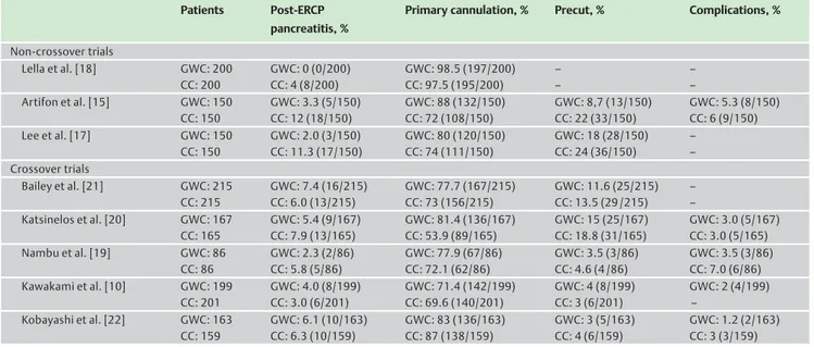
Table 3 Summary of studies comparing contrast cannulation (CC) with standard guidewire cannulation (GWC) of the common bile duct, reported in the
discussion.
Patients Post-ERCP
pancreatitis, %
Primary cannulation, % Precut, % Complications, %
Non-crossover trials Lella et al. [18] GWC: 200 CC: 200 GWC: 0 (0/200) CC: 4 (8/200) GWC: 98.5 (197/200) CC: 97.5 (195/200) – – – – Artifon et al. [15] GWC: 150 CC: 150 GWC: 3.3 (5/150) CC: 12 (18/150) GWC: 88 (132/150) CC: 72 (108/150) GWC: 8,7 (13/150) CC: 22 (33/150) GWC: 5.3 (8/150) CC: 6 (9/150) Lee et al. [17] GWC: 150 CC: 150 GWC: 2.0 (3/150) CC: 11.3 (17/150) GWC: 80 (120/150) CC: 74 (111/150) GWC: 18 (28/150) CC: 24 (36/150) – – Crossover trials Bailey et al. [21] GWC: 215 CC: 215 GWC: 7.4 (16/215) CC: 6.0 (13/215) GWC: 77.7 (167/215) CC: 73 (156/215) GWC: 11.6 (25/215) CC: 13.5 (29 /215) – – Katsinelos et al. [20] GWC: 167 CC: 165 GWC: 5.4 (9/167) CC: 7.9 (13/165) GWC: 81.4 (136/167) CC: 53.9 (89/165) GWC: 15 (25/167) CC: 18.8 (31/165) GWC: 3.0 (5/167) CC: 3.0 (5/165) Nambu et al. [19] GWC: 86 CC: 86 GWC: 2.3 (2/86) CC: 5.8 (5/86) GWC: 77.9 (67/86) CC: 72.1 (62/86) GWC: 3.5 (3/86) CC: 4.6 (4 /86) GWC: 3.5 (3/86) CC: 7.0 (6/86) Kawakami et al. [10] GWC: 199 CC: 201 GWC: 4.0 (8/199) CC: 3.0 (6/201) GWC: 71.4 (142/199) CC: 69.6 (140/201) GWC: 4 (8/199) CC: 3 (6/201) GWC: 2 (4/199) – Kobayashi et al. [22] GWC: 163 CC: 159 GWC: 6.1 (10/163) CC: 6.3 (10/159) GWC: 83 (136/163) CC: 87 (138/159) GWC: 3 (5/163) CC: 4 (6/159) GWC: 1.2 (2/163) CC: 3 (3/159)
ERCP, endoscopic retrograde cholangiopancreatography
4Gastroenterology Department, University of Milan, IRCCS Fondazione Policlinico, Milan, Italy
5Unit of Digestive Endoscopy, University Hospital L. Sacco, Milan, Italy
6Gastroenterology Unit, Valduce Hospital, Como, Italy
7Division of Gastroenterology and Gastrointestinal Endoscopy, Vita-Salute
San Raffaele University, Scientific Institute San Raffaele, Milan, Italy
8Department of Statistics, University of Messina, Messina, Italy
References
1 Cotton PB, Lehman G, Vennes J et al. Endoscopic sphincterotomy com-plications and their management: an attempt at consensus. Gastro-intest Endosc 1991; 37: 383–393
2 Masci E, Toti G, Mariani A et al. Complications of diagnostic and thera-peutic ERCP: a prospective multicenter study. Am J Gastroenterol 2001; 96: 417–423
3 Freeman ML, Nelson DB, Sherman S et al. Complications of endoscopic biliary sphincterotomy. N Engl J Med 1996; 335: 909–918
4 Masci E, Mariani A, Curioni S et al. Risk factors for pancreatitis after endoscopic retrograde cholangiopancreatography: a meta-analysis. Endoscopy 2003; 35: 830–834
5 Freeman ML, Guda NM. ERCP cannulation: a review of reported tech-niques. Gastrointest Endosc 2005; 61: 112–125
6 Cotton PB, Garrow DA, Gallagher J et al. Risk factors for complications after ERCP: a multivariate analysis of 11,497 procedures over 12 years. Gastrointest Endosc 2009; 70: 80–88
7 Nakai Y, Isayama H, Sasahira N et al. Risk factors for post-ERCP pan-creatitis in wire-guided cannulation for therapeutic biliary ERCP. Gas-trointest Endosc 2015; 81: 119–126
8 Woods KE, Willingham FF. Endoscopic retrograde cholangiopancrea-tography associated pancreatitis: a 15 year review. World J Gastro-intest Endosc 2010; 2: 165–178
9 Enns R, Lee S. Preventing post-ERCP pancreatitis. Gastrointest Endosc
2013; 78: 851–853
10 Kawakami H, Maguchi H, Mukai T et al. A multicentric prospective, ran-domized study of selective bile duct cannulation performed by multi-ple endoscopists: the BIDMEN study. Gastrointest Endosc 2012; 75:
362–372
11 Omuta S, Maetani I, Shigoka H et al. Newly designed J-shaped tip guide-wire: a preliminary feasibility study in wire-guided cannulation.
World J Gastroenterol 2013; 19: 4531–4536
12 Tsuchiya T, Itoi T, Maetani I et al. Effectiveness of the J-tip guidewire for selective biliary cannulation compared to conventional guidewires (the JANGLE study). Dig Dis Sci Apr 23. [Epub ahead of print] 13 Mariani A, Giussani A, Di Leo M et al. Guidewire biliary cannulation
does not reduce post-ERCP pancreatitis, compared with the contrast injection technique in low-risk and high-risk patients. Gastrointest Endosc 2012; 75: 339–346
14 Vandervoort J, Soetikno RM, Tham TC et al. Risk factors for complica-tions after performance of ERCP. Gastrointest Endosc 2002; 56: 652– 656
15 Artifon EL, Sakai P, Cunha JE et al. Guidewire cannulation reduces risk of post-ERCP pancreatitis and facilitates bile duct cannulation. Am J Gastroenterol 2007; 102: 2147–2153
16 Sasahira N, Kawakami H, Isayama H et al. Early use of double-guide-wire technique to facilitate selective bile duct cannulation: the multi-center randomized controlled EDUCATION trial. Endoscopy 2015; 47: 421–429
17 Lee TH, Park do H, Park JY et al. Can wire-guided cannulation prevent post-ERCP pancreatitis? A prospective randomized trial. Gastrointest Endosc 2009; 60: 444–449
18 Lella F, Bagnolo F, Colombo E et al. A simple way of avoiding post-ERCP
pancreatitis. Gastrointest Endosc 2004; 59: 830–834
19 Nambu T, Ukita T, Shigoka H et al. Wire-guided selective cannulation of the bile duct with a sphincterotome: a prospective randomized com-parative study with the standard method. Scand J Gastroenterol
2011; 46: 109–115
20 Katsinelos P, Paroutoglou G, Kountouras J et al. A comparative study of standard ERCP catheter and hydrophilic guide wire in the selective
cannulation of the common bile duct. Endoscopy 2008; 40: 302–307
21 Bailey AA, Bourke MJ, Williams SJ et al. A prospective randomized trial of cannulation techniques in ERCP: effect on technical success and
post-ERCP pancreatitis. Endoscopy 2008; 40: 296–301
22 Kobayashi G, Fujita N, Imaizumi K et al. Wire-guided biliary cannula-tion technique does not reduce the risk of post-ERCP pancreatitis: multicenter randomized controlled trial. Dig Endosc 2013; 25: 295– 302
23 Freeman ML, Di Sario JA, Nelson DB et al. Risk factors for post-ERCP pan-creatitis: a prospective, multicenter study. Gastrointest Endosc 2001;
54: 425–434