DOI: 10.1002/ejoc.201101273
Looking for a Robust, Synthetic, Fully-Extended (2.0
5
-Helical) Peptide
Structure – Effect of Terminal Groups
Fernando Formaggio,*
[a]Marco Crisma,
[a]Cristina Peggion,
[a]Alessandro Moretto,
[a]Mariano Venanzi,
[b]and Claudio Toniolo*
[a]Keywords: Amino acids / Peptides / Solution peptide synthesis / Conformation analysis
The incorporation ofα-amino acids with a quaternary α-car-bon atom into a peptide provides a tool to effectively restrict the available range of its backbone conformations. Specifi-cally, under favorable conditions, Cα,α-diethylglycine (Deg)
homopeptides are known to preferentially adopt the fully-extended (2.05-helical) structure, which is characterized by
Introduction
The fully-extended (2.0
5-helical) or planar sheet peptide
structure represents an extremely appealing molecular
spacer in long-range donor–acceptor studies as it is
en-dowed with the longest distance between two consecutive
α-amino acid α-carbon atoms.
[1]This conformation, although
proposed at an early stage in 3D-structural studies of
pro-teins,
[2]is extremely rare. Indeed, as predicted,
[2]it has so
far only been authenticated in the –(Gly)
4– sequence of
His-tRNA-synthetase.
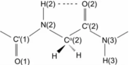
[3]The twofold (2.0) repeating motif of
this peptide conformation is based on the 2
씮2
intramolec-ularly H-bonded form depicted in Figure 1. The relative
dis-position of the two dipoles, N(2)–H(2) and C
⬘(2)=O(2), is
such that there is obviously some interaction between
them.
[4,5]As these four atoms, along with the α-carbon
atom of the second residue, are involved in a cyclic
Figure 1. The 2씮2 intramolecularly H-bonded (C5) peptide
con-formation for a Gly residue.
[a] ICB, Padova Unit, CNR, Department of Chemical Sciences, University of Padova,
35131 Padova, Italy Fax: +39-049-8275829
E-mail: [email protected] [email protected]
[b] Department of Chemical Sciences and Technologies, University of Rome “Tor Vergata”,
00133 Rome, Italy
the longest possible separation between two adjacent α-amino acid Cαatoms. We have investigated the influence of
the nature of the N- and/or C-terminal protecting (or block-ing) groups on the relative stabilities of the fully-extended conformation vs. the competing, shorter 310-helical structure
in a synthetic Deg homopeptide series.
(pentagonal) structure, this conformation is termed the C
5structure.
Apart from protein residues, we and others have shown
theoretically and experimentally that specific
backbone-modified
α-amino
acids,
e.g.
C
α,β-didehydroalanine
(ΔAla)
[6](Scheme 1) and, in particular, the subclass of
(achiral and chiral) C
α,α-dialkylated glycines with both side
chains longer than methyl but not interconnected in a cyclic
system,
[1,7–22]typically favor a monomeric (not
self-associ-ated) fully-extended structure. The simplest member of this
subclass is the achiral C
α,α-diethylglycine (Deg, Scheme 1).
On the other hand, C
α-methylated, C
α-alkylated (methyl or
longer alkyl group) α-amino acids, the prototype of which
is α-aminoisobutyric acid (Aib),
[7,10–13,16–19,23–26]and C
α,α-cyclized Gly residues (1-aminocycloalkane-1-carboxylic
ac-ids, Ac
nc)
[10,16]are known to strongly prefer the much more
compact 3
10-
[27]or α-helical structures (
φ = ⫾60° ⫾20°; ψ
=
⫾30° ⫾15°). However, the fully-extended conformation
seems to be rather fragile as even a modest sequence
modifi-Scheme 1. Structures of the α-amino acids, Pyr and –NH–TEMPO terminal moieties discussed.
cation, e.g. incorporation of a single α-aminobutyric acid
residue (with only one ethyl side chain) into a host (Deg)
npeptide may force it into the competing 3
10helix.
[21,28]In search of a robust fully-extended peptide structure, we
have studied the conformational effects induced by the type
of N- and/or C-protecting (or blocking) groups on the
rela-tive propensities of well-characterized Deg
homopept-ides
[29,30](to the tetramer level) to adopt either the
fully-extended or the 3
10-helical conformation. To this end, we
utilized FTIR, 1D and 2D NMR spectroscopy, and
steady-state fluorescence techniques. The results were compared
with those obtained for Aib homopeptides (with the same
protecting or blocking groups) used as 3
10-helical
stan-dards.
Results and Discussion
Synthesis
For the synthesis of the Pyr–(Deg)
n–NH–TEMPO
homopeptide series (Pyr = 1-pyrenylcarbonyl; n = 1–4;
NH–TEMPO =
4-amino-1-oxyl-2,2,6,6-tetramethylpiperid-inyl, Scheme 1), we first prepared the corresponding Tfa–
(Deg)
n–OtBu (Tfa = trifluoroacetyl) peptides.
[29,30]The
ini-tial procedure involves the syntheses of Tfa–Deg–OH and
H–Deg–OtBu (H–Deg–OtBu was prepared by catalytic
hy-drogenation of Z–Deg–OtBu,
[31]Z = benzyloxycarbonyl).
Throughout the series, peptide bond formation was
achieved from Tfa–Deg–OH and H–(Deg)
n–OtBu (n = 1–
3) in the presence of
O-(7-azabenzotriazol-1-yl)-1,1,3,3-tet-ramethyluronium (HATU) hexafluorophosphate
[32,33]and
N
,N-diisopropylethylamine (DIEA) in anhydrous
acetoni-trile. Selective, reductive Tfa removal was achieved by
treat-ment of the N-protected peptides with NaBH
4in
ethan-ol.
[14,30]The N-terminal Pyr moiety was introduced in the
Tfa-deprotected H–(Deg)
n–OtBu (n = 1–4) peptides by
1-pyren-ylcarboxylic acid in the presence of HATU/DIEA in
anhy-drous dichloromethane. The Pyr-protected peptide free
ac-ids were synthesized from the corresponding tert-butyl
es-ters in trifluoroacetic acid (TFA)/dichloromethane (1:1).
Fi-nally, the target Pyr–(Deg)
n–NH–TEMPO (n = 1–4)
homo-peptides were obtained from the corresponding free acids
and NH
2-TEMPO as described above for the
correspond-ing Pyr-protected peptide tert-butyl esters.
Syntheses of the known Z–(Aib)
n–OtBu (n = 1–5)
homo-peptides
[34,35]were performed either by the
1-(3-dimeth-ylamino)propyl-3-ethylcarbodiimide
(EDC)/7-aza-1-hy-droxy-1,2,3-benzotriazole (HOAt) method to activate Z–
Aib–OH in anhydrous dichloromethane with
N-methyl-morpholine (for the dimer and trimer) or via the
intermedi-ate 5(4H)-oxazolone from Z–(Aib)
2–OH
[31,34–37]in
anhy-drous CH
3CN under reflux (for the tetramer and
penta-mer). This oxazolone was, in turn, prepared by treatment
of the N
α-protected dipeptide free acid with EDC in
anhy-drous CH
3CN. The Pyr–(Aib)
n–OtBu and Pyr–(Aib)
n–NH–
TEMPO (n = 1–5) series were synthesized from the
corre-sponding Z–(Aib)
n–OtBu and Pyr–(Aib)
n–OH oligomers,
respectively, as described for the (Deg)
nseries.
Solution Conformational Analysis
The preferred solution conformations of the Pyr–
(Deg)
n–NH–TEMPO (OtBu) oligopeptides were
investi-gated and compared with those of the corresponding
(Aib)
ncompounds by FTIR and NMR spectroscopy.
The FTIR spectra in the conformationally informative
N–H stretching region (3500–3200 cm
–1) of the Pyr-blocked
(Deg)
nand (Aib)
nhomooligopeptide series to the tetramer
and pentamer levels, respectively, are shown in Figure 2.
Above 3400 cm
–1, the two Aib series (Figure 2, A and B)
exhibit one (or two) bands associated with the free
(sol-vated) N–H vibrations.
[4,38,39]Below 3400 cm
–1, a strong
band is seen at 3368 cm
–1in the trimer ester, and at
3346 cm
–1in the dimer amide, which are associated with
H-bonded N–H vibrations.
[4,38,39]Both bands significantly
shift to lower wavenumbers (to 3348 and 3328 cm
–1,
respec-tively) and their relative intensities markedly and linearly
increase upon main-chain elongation. The spectra do not
change in the concentration range investigated (10.0–
0.1 m
m, not shown), which strongly supports the view that
the observed H-bonding is intramolecular. These results,
which match those reported for the Z-protected (Aib)
nseries,
[40]are typical of a 3
10
helix, which is cooperatively
stabilized with increasing backbone length.
The FTIR spectra of the Pyr–(Deg)
n–OtBu oligomers
(Figure 2, C) are significantly different from those of the
(Aib)
nseries discussed above. In particular, an intense
ab-sorption at 3395 cm
–1occurs at the monomer level. This
band is assigned to the C-terminal conformer II
(Scheme 2), where the H-bonding acceptor of the C
5struc-ture is the ester carbonyl oxygen atom.
[41]The amount of
free (solvated) N–H stretching vibrations (a weak band at
about 3440 cm
–1) is very low. Upon main-chain elongation
to dimer, trimer, and tetramer, an absorption appears at the
lower frequency of 3364 cm
–1. The position of this band
does not change from dimer to tetramer. Again, a variation
of the peptide concentration modifies the spectra only
slightly. This general behavior, which is in good agreement
with that of the Tfa-protected Deg,
[9]Beg (C
α-n-butyl, C
α-ethylglycine),
[42]and Epg (C
α-ethyl, C
α-n-pentylglycine)
[41]series, is attributed to an ever increasing contribution of the
C
5internal conformer I (Scheme 2).
The FTIR spectra of the Pyr–(Deg)
n–NH–TEMPO
series (Figure 2, D) provide evidence for somewhat
interme-diate behavior between those of the (Aib)
nand (Deg)
nester
series. Based on a comparison between the spectra of Pyr–
NH–TEMPO and Pyr–(Deg)
2–OtBu, the two bands in the
spectrum of the monopeptide Pyr–Deg–NH–TEMPO at
3438 and 3374 cm
–1are assigned to the free NH group of
the –NH–TEMPO moiety and the C
5H-bonded NH group
of Deg, respectively. In the spectrum of the Deg dipeptide
amide, an additional shoulder is seen near 3345 cm
–1. This
band, more clearly observed in the spectrum of the
corre-sponding Aib dipeptide amide (Figure 2, B), is assigned to
an intramolecularly H-bonded, folded C
10(β-turn)
con-former, which is the basic unit of a 3
10helix.
[27]Elongation
of the peptide backbone in this Deg series generated spectra
Figure 2. FTIR spectra (3550–3200 cm–1) of (A) Pyr–(Aib)n–OtBu
(n = 1–5), (B) Pyr–(Aib)n–NH–TEMPO (n = 1–5), (C) Pyr– (Deg)n–OtBu (n = 1–4), and (D) Pyr–(Deg)n–NH–TEMPO (n = 0– 4) in CDCl3(peptide concentration: 1 mm).
Scheme 2. The C5internal (I) and C-terminal (II) conformers of
the Deg homopeptides.
that tend towards those of the corresponding 3
10-helical
Aib peptide series with a predominant band near 3340 cm
–1and an increasingly less intense shoulder near 3370 cm
–1.
Peptide concentration does not significantly affect the
spec-tra.
Looking at all the FTIR data, we conclude that a
C-terminal secondary amide offers the Deg peptide series a
chance to fold in a 3
10helix where the starting point is
pro-vided by the H-bond donor –NH–TEMPO group. The
po-sition of this conformational equilibrium is shifted from the
C
5conformation to the 3
10helix as the peptide backbone
is elongated to the tetramer level. Contrary to the behavior
of the Pyr-blocked Deg peptide esters, this phenomenon
makes the preferred conformation of the longer Deg
pept-ide ampept-ides more similar to that of the corresponding Aib
amide peptides.
[20]More detailed information on the secondary structural
propensities of the Pyr-blocked Deg homooligopeptide
es-ters was extracted from 1D and 2D NMR spectra. The
NH
proton
resonances
were
assigned
from
the
NH(i)
씮 NH(i+1) space connectivities obtained from 2D
ROESY experiments
[43,44]and a comparison with those of
the corresponding Tfa-protected analogs
[9]in the same
sol-vent. A section of the ROESY spectrum of the Deg trimer
is illustrated in part B of Figure 3. The NH(1) chemical
shifts of the Pyr-blocked and Tfa-protected homotrimers
reveal a slight conformational change at the N-terminus.
Indeed, this backbone region is more rigid in the
Tfa-pro-tected compound by virtue of three-center F···H···O
H-bonding (missing in the Pyr peptide), which is reflected in
a shift to lower field (ca. 8.0 ppm) of the Tfa-NH proton
[9]compared to about 7.6 ppm for the Pyr-NH proton. This
investigation was conducted in parallel with a study of the
related (Aib)
noligomers (the ROESY spectrum of the
pentapeptide is shown in part A of Figure 3).
First, we performed a solvent titration of the NH proton
chemical shifts; as an example, those of the –(Deg)
4– ester
are reported in Figure 4 in comparison with those of the
–(Aib)
4– ester. The polar solvent dimethyl sulfoxide
(DMSO)
[45]added to the CDCl
3
solution is expected to
in-teract strongly with the exposed (not intramolecularly
H-bonded) peptide/amide NH protons through N–H···O=S
H-bond formation, which would induce a downfield shift
in their resonances.
[46]Interestingly, only one class of NH
protons is observed in each of the four Deg homooligomers,
and all the NH proton chemical shifts are only slightly
sen-sitive to the presence of the perturbing solvent. The lack of
solvent accessibility is compatible with the occurrence of
the fully-extended conformation in CDCl
3solution for
these compounds. These results are at variance with those
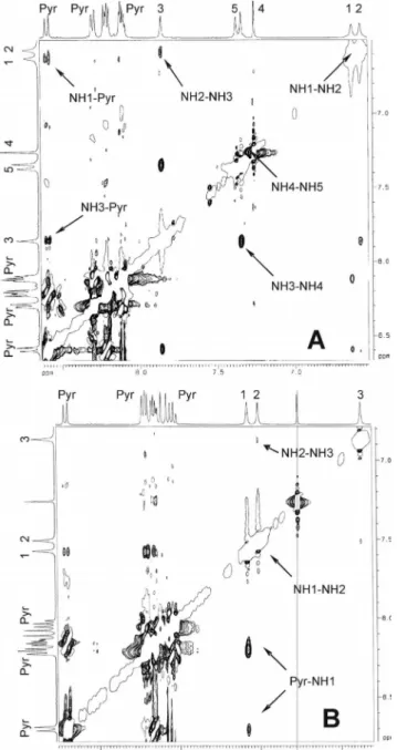
Figure 3. Sections of the ROESY spectra of (A) Pyr–(Aib)5–OtBu
and (B) Pyr–(Deg)3–OtBu in CDCl3solution (peptide
concentra-tion: 10 mm). The NH(i)씮NH(i+1) and Pyr씮NH1 cross-peaks are indicated.
of the (Aib)
n(n = 1–5) oligomers, where two classes of NH
proton chemical shifts were observed. The first class
(en-compassing the first and second NH groups, numbering the
residues from the N-terminus) includes NH protons that
are very sensitive to DMSO, whereas the second class (all
other NHs) includes NH protons that are marginally
sensi-tive to DMSO. Therefore, we propose that the (Aib)
noligo-mers adopt the 3
10helix where the H-bonding donor NH
groups are those of the third, fourth, and fifth residues.
The ROESY spectra of the (Deg)
nand (Aib)
noligopept-ides (Figure 3) differ in several aspects. In the spectrum of
the –(Aib)
5– oligomer, all NH(i)
씮 NH(i+1) cross-peaks
are clearly observed. This finding strongly supports the
Figure 4. Plots of the NH proton chemical shifts in the1H NMR
spectra of (A) Pyr–(Aib)4–OtBu and (B) Pyr–(Deg)4–OtBu as a
function of the increasing amount of DMSO added to the CDCl3
solution (v/v) (peptide concentration: 1 mm).
view that this peptide is highly folded. Two other
3D-struc-turally informative cross-peaks involve a Pyr aromatic
pro-ton and either the Aib
1or the Aib
3NH proton. This
con-nectivity indicates the onset of a helical structure.
Analo-gous data were found for the shorter (Aib)
noligomers. In
contrast, in the spectrum of the –(Deg)
3–oligomer, all
NH(i)
씮NH(i+ 1) cross-peaks are extremely weak (similar
findings were obtained for the Tfa-protected Deg
homo-oligomers). In addition, a Pyr aromatic proton interacts
with the Deg
1NH proton but not with the other two NH
protons in this compound. A different ROESY section of
the Pyr-blocked –(Deg)
3– spectrum (not shown) emphasizes
the occurrence of cross-peaks between the NH(i) protons
and the C
βH
2
(and C
γH
3) protons of the preceding (i – 1)
residue. Again, this spatial vicinity is compatible with the
presence of a fully-extended conformation. In summary,
our FTIR and NMR spectroscopic studies show that the
preferred conformation of the Pyr-blocked (Deg)
nand
(Aib)
noligomer esters is remarkably different; the (Aib)
npeptides fold in a 3
10helix,
[20]whereas the (Deg)
npeptides
prefer the fully-extended structure.
Molecular Spacers
The fully-extended peptide conformation is extremely
promising as a molecular spacer in spectroscopic analysis.
We utilized this secondary structure in a steady-state
fluo-rescence study. To this end, we incorporated the Pyr
photo-sensitizer group
[47,48]at the N-terminus of the backbone
and the paramagnetic, free radical quencher –NH–
TEMPO
[49,50]moiety at the C-terminus. The quenching
phenomenon is explained in terms of an intramolecular
ef-fect, as no intermolecular quenching is observed in the
highly diluted peptide solution (10
–7m in MeOH).
More-over, negligible quenching was seen in a 1:1 mixture of Pyr–
Deg–OtBu and CH
3–CONH–TEMPO at the same
concen-tration. The Pyr–NH···CONH–TEMPO donor···acceptor
distance is believed to play a major role in these
experi-ments, and the pyrenyl···nitroxide relative orientation may
exert some effect as well.
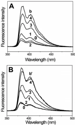
[48]Figure 5 (A and B) shows the steady-state fluorescence
spectra of the Pyr–(Aib)
n–NH–TEMPO (n = 0–4) and the
Pyr–(Deg)
n–NH–TEMPO (n = 0–4) series, respectively.
Spectra of the reference compounds Pyr–(Aib)
4–OtBu and
Pyr–(Deg)
4–OtBu are also shown. Figure 5 and Table 1,
which lists the percentages of fluorescence quenching in
peptides of different lengths, indicate that the trend is
sim-ilar for both series.
Figure 5. Steady-state fluorescence spectra of (A) Pyr–(Aib)4–OtBu
(blank b) and Pyr–(Aib)n–NH–TEMPO (n = 0–4), and (B) Pyr– (Deg)4–OtBu (blank b⬘) and Pyr–(Deg)n–NH–TEMPO (n = 0–4)
in MeOH (peptide concentration: 10–7m; λ
exc= 340 nm).
Table 1. Extent of fluorescence quenching observed for Pyr–NH– TEMPO (n = 0, reference), Pyr–(Aib)n–NH–TEMPO (n = 1–4) and Pyr–(Deg)n–NH–TEMPO (n = 1–4). n Pyr–(Aib)n–NH–TEMPO Pyr–(Deg)n–NH–TEMPO [%] [%] 0 80 80 1 68 51 2 84 79 3 42 40 4 20 12
Specifically, the extent of quenching decreases with the
increasing number of spacer units with the remarkable
ex-ception of the two Pyr–(X)
2–NH–TEMPO (X = Aib, Deg)
peptides, which exhibit a quenching efficiency as high as
that of the quencher derivative (n = 0). This finding
sup-ports the view that both peptide series are highly folded in
the 3
10-helical conformation because the fluorophore and
quencher groups are expected to be spatially close in the
n
= 2 compounds in this threefold 3D-structure. This
con-clusion is in agreement with the FTIR spectroscopic results
discussed above for the same two peptide series. For both
series the amount of fluorescence surviving in the
tetra-peptide amide is significantly high (80–90 %). The extent of
quenching is less significant for the Deg series relative to
its Aib counterpart. This is consistent with a mixture of
predominantly 3
10-helices with a high amount of the
fully-extended conformation in the Deg peptides, whereas the
fully-extended conformer is absent in the Aib peptides (the
fluorophore···quencher separation is longer in the
fully-extended conformation).
Conclusions
Over the last 25 years, the highly crystalline nature of
peptides rich in C
α-tetrasubstituted residues has been
ex-ploited to characterize the fully-extended (C
5)
conforma-tion and the related 2.0
5helix by X-ray diffraction. In
par-ticular, multiple, consecutive C
5conformations have been
observed in homopeptides with two side chains longer than
methyl,
[10,11,14,16,18,19,41,42]which is the case for achiral Deg.
Further evidence for this conclusion has come from
spec-troscopic studies in solution and conformational energy
computations. Interestingly, the axial translation per residue
in the 2.0
5helix is about 3.80 Å, the longest possible for a
single amino acid, which makes this conformation
ex-tremely attractive to use as a spacer or bridge. However,
we
[21,28]and Tanaka et al.
[14]have found that this type of
helical structure is not very robust, as subtle perturbations
in the chemical structure and environment can induce a
dramatic conformational switch to the 40 % shorter 3
10he-lix.
In this work, with the aim of detecting the most
appro-priate chemical structures for the stabilization of the 2.0
5helix, we synthesized and characterized several Deg
homo-peptides (to the tetramer level), which differ in the nature
of the N- and/or C-terminal protecting (or blocking)
groups, and compared them to the corresponding Aib
homopeptides, which represent classical model 3
10helices.
Our present and published findings, combined with those
of Tanaka et al.,
[14]led us to conclude that:
(i) The N-terminal group most suitable for stabilizing the
2.0
5helix is Tfa, thanks to its ability to generate an
ad-ditional intramolecular F···H–N H-bond at the N-terminus.
However, other amides (including Pyr) or urethane
N-ter-minal groups may accomplish a similar role, at least in a
low polarity solvent (CHCl
3).
(ii) Any ester (but especially a tert-butyl ester) group at
the C-terminus is compatible with the 2.0
5helix.
Con-versely, a secondary amide induces the formation of a 3
10helix, particularly in homologs to the tetramer level,
be-cause of its extra H-bonding donor NH group, which is
unsatisfied in the 2.0
5helix.
(iii) The absence or nature of the solvent is crucial in
governing the Deg homopeptide conformation. In
particu-lar, crystal-packing forces and the type of crystallization
solvent may induce a 3
10-helical structure in a peptide that
is fully extended in CDCl
3. However, in general, only
lim-ited and scattered information is available on the role of the
solvent on this conformational equilibrium. It is evident
that this phenomenon deserves a more detailed
investiga-tion, which is currently in progress in our laboratories.
Experimental Section
Synthesis and Characterization: Materials and reagents were of the
highest commercially available grade and used without further pu-rification. Melting points were determined in open capillaries with a Leitz Laborlux 12 apparatus. Solid-state IR spectra (KBr disk) were recorded with a Perkin–Elmer model 1720X FTIR spectro-photometer. TLC was performed with Merck Kieselgel 60F254
pre-coated plates using the following solvent systems: (1) chloroform/ ethanol, 9:1; (2) 1-butanol/acetic acid/water, 3:1:1; and (3) toluene/ ethanol, 7:1. The chromatograms were visualized by UV fluores-cence or developed by chlorine/starch/potassium iodide or nin-hydrin chromatic reaction as appropriate. All compounds were obtained in a chromatographically homogeneous state. Flash chromatography was carried out with Merck silica gel 60 (40– 63 μm mesh). MS (ESI mode) were measured with a Perseptive Biosystems (Mariner model) ESI-TOF spectrometer.
General Procedure for the Coupling Reaction between Pyr–(X)n–OH (C component) and H–(X)n–OtBu (X = Deg or Aib) or NH2 -TEMPO (N component): To a solution of the C component
(0.80 mmol) in anhydrous CH2Cl2 at 0 °C were added HATU
(0.83 mmol) and DIEA (0.83 mmol). After 10 min, the N-compo-nent (0.80 mmol) was added. The resulting solution was heated to reflux for 4–7 d with stirring. The solvent was evaporated under reduced pressure and EtOAc was added. The organic phase was washed with 10 % KHSO4, H2O, 5 % NaHCO3, and water and
dried with Na2SO4. The solution was filtered, and the solvent was
evaporated under reduced pressure. The product was purified using flash chromatography and an appropriate CH2Cl2/ethanol mixture
as eluant.
General Procedure for the Removal of the tert-Butyl Ester C Protec-tion: The C-protected peptide was dissolved in TFA/anhydrous
CH2Cl2(1:1) and the solution was stirred at room temperature for
30 min. The solvent mixture was evaporated under reduced pres-sure and the product was repeatedly triturated with diethyl ether to remove the remaining TFA. The product was collected by fil-tration and dried with KOH in a dessiccator under vacuum.
Pyr–Deg–OtBu: Yield 75 %; m.p. 121–122 °C [from
EtOAc/petro-leum ether (PE)]. Rf1 = 0.95; Rf2 = 0.95; Rf3 = 0.80. 1H NMR
(400 MHz, CDCl3): δ = 8.66 (d, 1 H, Pyr CH), 8.24–8.03 (m, 8 H,
Pyr 8⫻CH), 7.13 (s, 1 H, NH), 2.83 (qd, 2 H, 1 β-CH2), 2.00 (qd,
2 H, 1 β-CH2), 1.48 (s, 9 H, OtBu 3⫻CH3), 0.99 (t, 6 H, 2 γ-CH3)
ppm. IR (KBr): ν˜ = 3387, 1723, 1652 cm–1.
Pyr–(Deg)2–OtBu: Yield 49 %; m.p. 117–118 °C (from EtOAc/PE).
Rf1 = 0.95; Rf2 = 0.95; Rf3 = 0.75.1H NMR (400 MHz, CDCl3): δ = 8.69 (d, 1 H, Pyr CH), 8.23–8.04 (m, 8 H, Pyr 8⫻CH), 7.56 (s, 1 H, NH1), 7.00 (s, 1 H, NH2), 3.02 (qd, 2 H, 1 β-CH 21), 2.49 (qd, 2 H, 1 β-CH22),1.88 (qd, 2 H, 1 β-CH21), 1.78 (qd, 2 H, 1 β-CH22), 1.53 (s, 9 H, OtBu 3⫻CH3), 1.06 (t, 6 H, 2 γ-CH31), 0.81 (t, 6 H, 2 γ-CH32) ppm. IR (KBr): ν˜ = 3394, 3360, 3311, 1734, 1650 cm–1.
Pyr–(Deg)3–OtBu: Yield 58 %; m.p. 200–201 °C (from CH2Cl2/PE).
Rf1 = 0.95; Rf2 = 0.95; Rf3 = 0.70.1H NMR (400 MHz, CDCl3): δ = 8.70 (d, 1 H, Pyr CH), 8.23–8.03 (m, 8 H, Pyr 8⫻CH), 7.58 (s, 1 H, NH1), 7.51 (s, 1 H, NH2), 6.87 (s, 1 H, NH3), 2.94 (qd, 2 H, 1 β-CH21), 2.68 (qd, 2 H, 1 β-CH22), 2.47 (qd, 2 H, 1 β-CH23), 1.93 (qd, 2 H, 1 β-CH21), 1.80 (qd, 2 H, 1 β-CH23), 1.68 (qd, 2 H, 1 β-CH22), 1.50 (s, 9 H, OtBu 3⫻CH3), 1.06 (t, 6 H, 2 γ-CH31), 0.88 (t, 6 H, 2 γ-CH32), 0.81 (t, 6 H, 2 γ-CH33) ppm. IR (KBr): ν˜ = 3394, 3377, 3353, 1721, 1676, 1649 cm–1.
Pyr–(Deg)4–OtBu: Yield 47 %; m.p. 205–206 °C (from EtOAc/PE).
Rf1 = 0.95; Rf2 = 0.95; Rf3 = 0.60.1H NMR (400 MHz, CDCl3): δ = 8.70 (d, 1 H, Pyr CH), 8.23–8.03 (m, 8 H, Pyr 8⫻CH), 7.59 (s, 1 H, NH1), 7.52 (s, 1 H, NH2), 7.40 (s, 1 H, NH3), 6.86 (s, 1 H, NH4), 3.0 (qd, 2 H, 1 β-CH 21), 2.66 (m, 4 H, 1 β-CH22, 1 β-CH23), 2.44 (qd, 2 H, 1 β-CH24), 1.94 (qd, 2 H, 1 β-CH21), 1.79–1.71 (m, 6 H, 1 β-CH221 β-CH231 β-CH24), 1.50 (s, 9 H, OtBu 3⫻CH3), 1.06 (t, 6 H, 2 γ-CH31), 0.87 (m, 12 H, 2 γ-CH32and 2 γ-CH33), 0.78 (t, 6 H, 2 γ-CH34) ppm. IR (KBr): ν˜ = 3401, 3358, 1720, 1679, 1653 cm–1. Pyr–Deg–OH: Yield 85 %; m.p. 244–245 °C. Rf1 = 0.95; Rf2 = 0.95; Rf3 = 0.15.1H NMR (400 MHz, DMSO): δ = 8.37–8.12 (m, 9 H, Pyr CH), 2.04 (m, 4 H, 2 β-CH2), 0.91 (t, 6 H, 2 γ-CH3) ppm. IR (KBr): ν˜ = 3379, 1717, 1615 cm–1.
Pyr–(Deg)2–OH: Yield 98 %; m.p. 234–235 °C. Rf1 = 0.95; Rf2 =
0.95; Rf3 = 0.15.1H NMR (400 MHz, DMSO): δ = 8.17–8.09 (m,
9 H, Pyr CH), 7.57 (s, 1 H, NH), 7.01 (s, 1 H, NH), 2.29 (m, 6 H, 2 β-CH21and 1 β-CH22),1.87 (qd, 2 H, 1 β-CH22), 1.07 (t, 6 H, 2
γ-CH31), 0.82 (t, 6 H, 2 γ-CH32) ppm. IR (KBr): ν˜ = 3400, 3347,
3291, 1712, 1658, 1632 cm–1.
Pyr–(Deg)3–OH: Yield 86 %; m.p. 224–225 °C. Rf1 = 0.95; Rf2 =
0.95; Rf3 = 0.10. IR (KBr): ν˜ = 3383, 3341, 1724, 1676, 1659 cm–1.
Pyr–(Deg)4–OH: Yield 91 %; m.p. 213–214 °C. Rf1 = 0.95; Rf2 =
0.95; Rf3 = 0.10.1H NMR (400 MHz, DMSO): δ = 8.59 (d, 1 H,
Pyr CH), 8.37–8.12 (m, 8 H, Pyr 8⫻CH), 8.53, 7.96, 7.58, 7.47 (4s, 4 H, 4 NH), 1.98 (qd, 2 H, 1 β-CH2), 1.95–1.84 (m, 14 H, 7
β-CH2), 1.17–0.71 (m, 24 H, 8 γ-CH3) ppm. IR (KBr): ν˜ = 3421,
1738, 1653, 1639 cm–1.
Pyr–NH–TEMPO: Yield 51 %; m.p. 202–203 °C (from EtOAc/PE).
Rf1 = 0.90; Rf2 = 0.95; Rf3 = 0.35. IR (KBr): ν˜ = 3470, 3269, 1737,
1635 cm–1. MS (ESI-TOF): m/z = 401.23 [M + H]+.
Pyr–Deg–NH–TEMPO: Yield 63 %; m.p. 246–247 °C (from
CH2Cl2/PE). Rf1 = 0.95; Rf2 = 0.95; Rf3 = 0.35. MS (ESI-TOF):
m/z = 513.32 [M + H]+.
Pyr–(Deg)2–NH–TEMPO: Yield 81 %; m.p. 242–243 °C (from
CH2Cl2/PE). Rf1 = 0.70; Rf2 = 0.95; Rf3 = 0.25. IR (KBr): ν˜ =
3430, 3346, 3318, 1672, 1653, 1633 cm–1. MS (ESI-TOF): m/z =
626.41 [M + H]+.
Pyr–(Deg)3–NH–TEMPO: Yield 58 %; m.p. 247–248 °C (from
CH2Cl2/PE). Rf1 = 0.70; Rf2 = 0.95; Rf3 = 0.20. IR (KBr): ν˜ =
3330, 1631 cm–1. MS (ESI-TOF): m/z = 739.35 [M + H]+.
Pyr–(Deg)4–NH–TEMPO: Yield 80 %; m.p. 251–252 °C (from
CH2Cl2/PE). Rf1 = 0.70; Rf2 = 0.95; Rf3 = 0.15. IR (KBr): ν˜ =
3429, 3327, 1652 cm–1. MS (ESI-TOF): m/z = 853.32 [M + H]+.
Pyr–Aib–OtBu: Yield 86 %; m.p. 117–118 °C (from EtOAc/PE). Rf1
= 0.90; Rf2 = 0.90; Rf3 = 0.65.1H NMR (400 MHz, CDCl3): δ =
8.65 (d, 1 H, Pyr CH), 8.23 (d, 2 H, Pyr 2⫻CH), 8.14 (m, 3 H, Pyr 3⫻CH), 8.05 (m, 3 H, Pyr 3 ⫻CH), 6.72 (s, 1 H, NH), 1.78 (s, 6 H, 2 β-CH3), 1.56 (s, 9 H, OtBu 3⫻CH3) ppm. IR (KBr): ν˜
= 3323, 1725, 1624 cm–1.
Pyr–(Aib)2–OtBu: Yield 95 %; m.p. 210–211 °C (from EtOAc/PE).
δ = 8.59 (d, 1 H, Pyr 1 CH), 8.21 (d, 2 H, Pyr 2⫻CH), 8.11 (m, 3 H, Pyr 3⫻CH), 8.03 (m, 3 H, Pyr 3⫻CH), 7.33 (s, 1 H, NH), 6.88 (s, 1 H, NH), 1.84 (s, 6 H, 2 β-CH3), 1.63 (s, 6 H, 2 β-CH3), 1.48
(s, 9 H, OtBu 3⫻ CH3) ppm. IR (KBr): ν˜ = 3373, 3358, 1712, 1666,
1649 cm–1.
Pyr–(Aib)3–OtBu: Yield 85 %; m.p. 201–202 °C (from CH2Cl2/PE).
Rf1 = 0.60; Rf2 = 0.95; Rf3 = 0.30.1H NMR (400 MHz, CDCl3):
δ = (d, 1 H, Pyr CH), 8.22 (d, 2 H, Pyr 2⫻CH), 8.13 (m, 3 H, Pyr 3⫻CH), 8.08 (m, 3 H, Pyr 3⫻CH), 7.42 (s, 1 H, NH3), 6.87 (s, 1
H, NH2), 6.83 (s, 1 H, NH1), 1.75 (s, 6 H, 2 β-CH
3), 1.59 (s, 6 H,
2 β-CH3), 1.51 (s, 6 H, 2 β-CH3) ppm. IR (KBr): ν˜ = 3412, 3335,
1735, 1668, 1650 cm–1.
Pyr–(Aib)4–OtBu: Yield 52 %; m.p. 236–237 °C. Rf1 = 0.50; Rf2 =
0.95; Rf3 = 0.15.1H NMR (400 MHz, CDCl3): δ = 8.59–8.62 (d, 1 H, Pyr CH), 8.28–8.30 (d, 2 H, Pyr 2⫻CH), 8.18–8.23 (m, 3 H, Pyr 3⫻CH), 8.09–8.12 (m, 3 H, Pyr 3 ⫻CH), 7.56 (s, 1 H, NH3), 7.33 (s, 1 H, NH4), 6.63 (s, 1 H, NH2), 6.59 (s, 1 H, NH1), 1.75 (s, 6 H, 2 β-CH31), 1.55 (s, 12 H, 2 β-CH32and 2 β-CH33), 1.52 (s, 6 H, 2 β-CH34), 1.39 (s, 9 H, OtBu 3⫻CH3) ppm. IR (KBr): ν˜ = 3340, 1730, 1661, 1641 cm–1.
Pyr–(Aib)5–OtBu: Yield 40 %; m.p. 253–254 °C. Rf1 = 0.45; Rf2 =
0.95; Rf3 = 0.15.1H NMR (400 MHz, CDCl3): δ = 8.58–8.60 (d, 1 H, Pyr CH), 8.28–8.33 (d, 2 H, Pyr 2⫻CH), 8.19–8.23 (m, 3 H, Pyr 3⫻CH), 8.09–8.13 (m, 3 H, Pyr 3⫻CH), 7.86 (s, 1 H, NH3), 7.37 (s, 1 H, NH5), 7.34 (s, 1 H, NH4), 6.63 (s, 1 H, NH1), 6.58 (s, 1 H, NH2), 1.74 (s, 6 H, 2 β-CH 31), 1.56 (s, 12 H, 2 β-CH32and 2 β-CH34), 1.50 (s, 12 H, 2 β-CH33, 2 β-CH35), 1.44 (s, 9 H, OtBu 3⫻ CH3) ppm. IR (KBr): ν˜ = 3331, 1734, 1662 cm–1. Pyr–Aib–OH: Yield 87 %; m.p. 243–244 °C. Rf1 = 0.25; Rf2 = 0.95; Rf3 = 0.05.1H NMR (400 MHz, DMSO): δ = 8.94 (s, 1 H, NH), 8.52 (d, 1 H, Pyr CH), 8.37 (d, 2 H, Pyr 2⫻CH), 8.35–8.06 (m, 6 H, Pyr 6⫻CH), 1.55 (s, 6 H, 2 β-CH3) ppm. IR (KBr): ν˜ = 3329, 1705, 1632 cm–1.
Pyr–(Aib)2–OH: Yield 98 %; m.p. 160–161 °C. Rf1 = 0.20; Rf2 =
0.90; Rf3 = 0.10.1H NMR (400 MHz, DMSO): δ = 8.49 (d, 1 H,
Pyr CH), 8.17 (d, 2 H, Pyr 2⫻CH), 8.06 (m, 3 H, Pyr 3⫻CH), 7.97 (m, 3 H, Pyr 3⫻CH), 7.72 (s, 1 H, NH), 6.79 (s, 1 H, NH), 1.78 (s, 6 H, 2 β-CH3), 1.64 (s, 6 H, 2 β-CH3) ppm. IR (KBr): ν˜ =
3357, 3299, 1739, 1663, 1629 cm–1.
Pyr–(Aib)3–OH: Yield 71 %; m.p. 225–226 °C. Rf1 = 0.10; Rf2 =
0.85; Rf3 = 0.10.1H NMR (400 MHz, DMSO): δ = 8.90 (s, 1 H,
NH), 8.60–8.51 (d, 1 H, Pyr CH), 8.41–8.08 (m, 8 H, Pyr 8⫻CH), 7.85 (s, 1 H, NH), 7.73 (s, 1 H, NH), 1.54 (s, 6 H, 2 β-CH3), 1.42
(s, 6 H, 2 β-CH3), 1.36 (s, 6 H, 2 β-CH3) ppm. IR (KBr): ν˜ = 3410,
3292, 1726, 1657, 1636 cm–1.
Pyr–(Aib)4–OH: Yield 98 %; m.p. 232–233 °C. Rf1 = 0.10; Rf2 =
0.85; Rf3 = 0.10.1H NMR (400 MHz, DMSO): δ = 9.09 (s, 1 H, NH), 8.51–8.55 (d, 1 H, Pyr CH), 8.34–8.38 (d, 2 H, Pyr 2⫻CH), 8.31 (s, 1 H, NH), 8.25–8.26 (m, 3 H, Pyr 3⫻CH), 8.08–8.16 (m, 3 H, Pyr 3⫻CH), 7.76 (s, 1 H, NH), 7.58 (s, 1 H, NH), 1.56 (s, 6 H, 2 β-CH3), 1.38 (s, 12 H, 4 β-CH3), 1.34 (s, 6 H, 2 β-CH3) ppm. IR (KBr): ν˜ = 3284, 1746, 1658, 1635 cm–1.
Pyr–(Aib)5–OH: Yield 87 %; m.p. 240–241 °C. Rf1 = 0.10; Rf2 =
0.85; Rf3 = 0.10.1H NMR (400 MHz, DMSO): δ = 9.15 (s, 1 H, NH), 8.51–8.56 (d, 1 H, Pyr CH), 8.47 (s, 1 H, NH), 8.35–8.38 (d, 2 H, Pyr 2⫻ CH), 8.32 (s, 1 H, NH), 8.26–8.27 (m, 3 H, Pyr 3⫻CH), 8.07–8.25 (m, 3 H, Pyr 3⫻CH), 7.61 (s, 1 H, NH), 7.35 (s, 1 H, NH),1.57 (s, 6 H, 2 β-CH3), 1.42 (s, 6 H, 2 β-CH3), 1.38 (s, 6 H, 2 β-CH3), 1.36 (s, 6 H, 2 β-CH3), 1.34 (s, 6 H, 2 β-CH3) ppm. IR (KBr): ν˜ = 3431, 3312, 1738, 1661 cm–1.
Pyr–Aib–NH–TEMPO: Yield 79 %; m.p. 228–229 °C (from EtOAc/
PE). Rf1 = 0.75; Rf2 = 0.95; Rf3 = 0.20. IR (KBr): ν˜ = 3373, 1649
cm–1. MS (ESI-TOF): m/z = 486.32 [M + H]+.
Pyr–(Aib)2–NH–TEMPO: Yield 51 %; m.p. 232–233 °C (from
EtOAc/PE). Rf1 = 0.55; Rf2 = 0.90; Rf3 = 0.10. IR (KBr): ν˜ =
3392, 3271, 1687, 1637 cm–1. MS (ESI-TOF): m/z = 570.35 [M +
H]+.
Pyr–(Aib)3–NH–TEMPO: Yield 57 %; m.p. 252–253 °C (from
EtOAc/PE). Rf1 = 0.55; Rf2 = 0.90; Rf3 = 0.10. IR (KBr): ν˜ =
3431, 3341, 3281, 1658, 1650 cm–1. MS (ESI-TOF): m/z = 655.42
[M + H]+.
Pyr–(Aib)4–NH–TEMPO: Yield 72 %; m.p. 275–276 °C (from
CH2Cl2/PE). Rf1 = 0.50; Rf2 = 0.95; Rf3 = 0.10. IR (KBr): ν˜ =
3433, 3327, 1662 cm–1. MS (ESI-TOF): m/z = 740.49 [M + H]+.
Pyr–(Aib)5–NH–TEMPO: Yield 71 %; m.p. 266–267 °C (from
EtOAc/PE). Rf1 = 0.50; Rf2 = 0.95; Rf3 = 0.10. IR (KBr): ν˜ =
3433, 3326, 1660 cm–1. MS (ESI-TOF): m/z = 825.57 [M + H]+.
FTIR Absorption: The solution FTIR absorption spectra were
re-corded with a Perkin–Elmer 1720X spectrophotometer, nitrogen flushed, equipped with a sample-shuttle device, at 2 cm–1nominal
resolution, averaging 100 scans. Solvent (base-line) spectra were re-corded under the same conditions. Cells with path lengths of 0.1, 1.0, and 10 mm (with CaF2 windows) were used. Spectrograde
CDCl3(99.8 % D) was purchased from Fluka.
1H NMR Spectroscopy: The1H NMR spectra were recorded with
a Bruker AM 400 spectrometer. Measurements were carried out in CDCl3(99.96 % D; Aldrich) and DMSO (99.96 % D6, Acros
Or-ganics) with tetramethylsilane as the internal standard. Splitting patterns are abbreviated as follows: (s) singlet, (d) doublet, (t) trip-let, (q) quartet; (qd) quartet of doublets, (m) multiplet. Compounds containing the –NH–TEMPO moiety could not be characterized by this technique due to the dramatic line broadening of all signals because of the presence of the paramagnetic nitroxyl group. The 2D ROESY[43,44] experiments were performed with a Bruker
AVANCE DRX-400 spectrometer, operating at 400 MHz, equipped with a 5 mm probe BBI-Z grad. Processing of the experimental data was carried out using TOPSPIN 1.3.
Fluorescence: Steady-state fluorescence spectra of the
Pyr-contain-ing compounds were carried out with a Perkin–Elmer LS 50B spec-trofluorimeter. Cells with path lengths of 1 cm were used. Spectro-grade MeOH was purchased from Fluka.
Acknowledgments
F. F. and A. M. are grateful to the University of Padova for finan-cial support (PRAT 2007).
[1] C. Toniolo, E. Benedetti, in: Molecular Conformation and Bio-logical Interactions(Eds.: P. Balaram, S. Ramaseshan), Indian Academy of Sciences, Bangalore, India, 1991, pp. 511–521. [2] L. Pauling, R. B. Corey, Proc. Natl. Acad. Sci. USA 1951, 37,
729–740.
[3] A. Åberg, A. Yaremchuk, M. Tukalo, B. Rasmussen, S. Cus-ack, Biochemistry 1999, 36, 3084–3094.
[4] M. T. Cung, M. Marraud, J. Néel, Ann. Chim. 1972, 7, 183– 209.
[5] C. Toniolo, C. R. C. Crit. Rev. Biochem. 1980, 9, 1–44. [6] M. Crisma, F. Formaggio, C. Toniolo, T. Yoshikawa, T.
Waka-miya, J. Am. Chem. Soc. 1999, 121, 3272–3278.
[7] V. Barone, F. Lelj, A. Bavoso, B. Di Blasio, P. Grimaldi, V. Pavone, C. Pedone, Biopolymers 1985, 24, 1759–1767.
[8] E. Benedetti, V. Barone, A. Bavoso, B. Di Blasio, F. Lelj, V. Pavone, C. Pedone, G. M. Bonora, C. Toniolo, M. T. Leplawy, K. Kaczmarek, A. Redlinski, Biopolymers 1988, 27, 357–371. [9] C. Toniolo, G. M. Bonora, A. Bavoso, E. Benedetti, B. Di
Bla-sio, V. Pavone, C. Pedone, V. Barone, F. Lelj, M. T. Leplawy, K. Kaczmarek, A. Redlinski, Biopolymers 1988, 27, 373–379. [10] C. Toniolo, E. Benedetti, Macromolecules 1991, 24, 4004–4009. [11] E. Benedetti, B. Di Blasio, V. Pavone, C. Pedone, C. Toniolo,
M. Crisma, Biopolymers 1992, 32, 453–456.
[12] K. Ramnarayan, M. F. Chan, V. N. Balaji, S. Profeta Jr., S. N. Rao, Int. J. Pept. Protein Res. 1995, 45, 366–376.
[13] M. Cirilli, V. M. Coiro, A. Di Nola, F. Mazza, Biopolymers
1998, 46, 239–244.
[14] M. Tanaka, N. Imawaka, M. Kurihara, H. Suemune, Helv. Chim. Acta1999, 82, 494–510.
[15] M. Kurihara, M. Tanaka, M. Oba, H. Suemune, N. Miyata, in: Peptides 2000(Eds.: J. Martinez, J.-A. Fehrentz), EDK, Paris, France, 2001, pp. 427–428.
[16] C. Toniolo, M. Crisma, F. Formaggio, C. Peggion, Biopolymers
2001, 60, 396–419.
[17] M. A. C. Preto, A. Melo, S. P. G. Costa, H. L. S. Maia, M. J. Ramos, J. Phys. Chem. B 2003, 107, 14556–14562.
[18] C. Toniolo, M. Crisma, F. Formaggio, C. Peggion, Q. B. Broxt-erman, B. Kaptein, Biopolymers 2004, 76, 162–176.
[19] M. Crisma, F. Formaggio, A. Moretto, C. Toniolo, Biopolymers
2006, 84, 3–12.
[20] M. Crisma, E. Andreetto, M. De Zotti, A. Moretto, C. Peg-gion, F. Formaggio, C. Toniolo, J. Pept. Sci. 2007, 13, 190–205. [21] J. Torras, D. Zanuy, M. Crisma, C. Toniolo, O. Betran, C.
Ale-man, Biopolymers 2008, 90, 695–706.
[22] A. Moretto, M. De Zotti, M. Crisma, F. Formaggio, C. Toni-olo, Int. J. Pept. Res. Ther. 2008, 14, 307–314.
[23] G. R. Marshall, in: Intra-Science Chemistry Report, vol. 5 (Ed.: N. Kharasch), Gordon and Breach, New York, 1971, pp. 305– 316.
[24] N. Shamala, R. Nagaraj, P. Balaram, J. Chem. Soc., Chem. Commun.1978, 996–997.
[25] I. L. Karle, P. Balaram, Biochemistry 1990, 29, 6747–6756. [26] G. Jung, H. Brückner, H. Schmitt, in: Structure and Activity
of Natural Products(Eds.: G. Voelter, G. Weitzel), de Gruyter, Berlin, 1981, pp. 75–114.
[27] C. Toniolo, E. Benedetti, Trends Biochem. Sci. 1991, 16, 350– 353.
[28] E. Benedetti, C. Pedone, V. Pavone, B. Di Blasio, M. Saviano, R. Fattorusso, M. Crisma, F. Formaggio, G. M. Bonora, C. Toniolo, K. Kaczmarek, A. Redlinski, M. T. Leplawy, Biopoly-mers1994, 34, 1409–1418.
[29] Z. Kaminski, M. T. Leplawy, A. Olma, A. Redlinski, in: Pept-ides 1980(Ed.: K. Brunfeldt), Scriptor, Copenhagen, Denmark,
1981, pp. 201–206.
[30] M. T. Leplawy, K. Kaczmarek, A. Redlinski, in: Peptides: Chemistry and Biology(Ed.: G. R. Marshall), ESCOM, Leiden, The Netherlands, 1988, pp. 239–241.
[31] W. J. McGahren, M. Goodman, Tetrahedron 1967, 23, 2017– 2030.
[32] L. A. Carpino, J. Am. Chem. Soc. 1993, 115, 4397–4398. [33] L. A. Carpino, A. El-Faham, C. A. Minor, F. Albericio, J.
Chem. Soc., Chem. Commun.1994, 201–203.
[34] D. S. Jones, G. W. Kenner, J. Preston, R. C. Sheppard, J. Chem. Soc.1965, 6227–6239.
[35] E. Benedetti, A. Bavoso, B. Di Blasio, V. Pavone, C. Pedone, M. Crisma, G. M. Bonora, C. Toniolo, J. Am. Chem. Soc. 1982, 104, 2437–2444.
[36] C. Toniolo, G. M. Bonora, M. Crisma, E. Benedetti, A. Ba-voso, B. Di Blasio, V. Pavone, C. Pedone, Int. J. Pept. Protein Res.1983, 22, 603–610.
[37] M. T. Leplawy, D. S. Jones, G. W. Kenner, R. C. Sheppard, Tet-rahedron1960, 11, 39–51.
[38] S. Mizushima, T. Shimanouchi, M. Tsuboi, P. Souda, J. Am. Chem. Soc.1952, 74, 270–271.
[39] E. S. Pysh, C. Toniolo, J. Am. Chem. Soc. 1977, 99, 6211–6219. [40] C. Toniolo, G. M. Bonora, V. Barone, A. Bavoso, E. Benedetti, B. Di Blasio, P. Grimaldi, F. Lelj, V. Pavone, C. Pedone, Macro-molecules1985, 18, 895–902.
[41] M. Crisma, A. Moretto, C. Peggion, L. Panella, B. Kaptein, Q. B. Broxterman, F. Formaggio, C. Toniolo, Amino Acids
2011, 41, 629–641.
[42] N. Imawaka, M. Tanaka, H. Suemune, Helv. Chim. Acta 2000, 83, 2823–2835.
[43] A. A. Bothner-By, R. L. Stephens, J. Lee, C. D. Warren, R. W. Jeanloz, J. Am. Chem. Soc. 1984, 106, 811–813.
[44] A. Bax, D. Davis, J. Magn. Reson. 1985, 63, 207–213. [45] D. Martin, H. G. Hauthal, in: Dimethyl Sulphoxide, van
Ros-trand-Reinhold, Wokingham, UK, 1975.
[46] K. D. Kopple, M. Ohnishi, A. Go, J. Am. Chem. Soc. 1969, 91, 4264–4272.
[47] E. Prasad, K. R. Gopidas, J. Am. Chem. Soc. 2000, 122, 3191– 3196.
[48] M. Sisido, S. Hoshino, H. Kusano, M. Kuragaki, M. Makino, H. Sasaki, T. A. Smith, J. P. Ghiggino, J. Phys. Chem. B 2001, 105, 10407–10415.
[49] C. Toniolo, M. Crisma, F. Formaggio, Biopolymers 1998, 47, 153–158.
[50] B. Pispisa, C. Mazzuca, A. Palleschi, L. Stella, M. Venanzi, M. Wakselman, J.-P. Mazaleyrat, M. Rainaldi, F. Formaggio, C. Toniolo, Chem. Eur. J. 2003, 9, 4084–4093.
Received: August 31, 2011 Published Online: November 16, 2011