PhD Course in Biochemistry
XXXI Cycle (Academic Years 2015-‐2018)
Molecular mechanisms of folding of Intrinsically
Disordered Proteins
PhD student
Francesca Troilo
Tutors
PhD coordinator
Prof. Stefano Gianni Prof. Stefano Gianni
Dott.ssa Sonia Longhi
PhD Course in Biochemistry
XXXI Cycle (Academic Years 2015-‐2018)
Molecular mechanisms of folding of Intrinsically
Disordered Proteins
PhD student
Francesca Troilo
Tutors
PhD coordinator
Prof. Stefano Gianni Prof. Stefano Gianni
Dott.ssa Sonia Longhi
SCIENTIFIC COMMUNICATIONS
Publications:
1. Troilo F, Bonetti D, Longhi S, Gianni S. The fuzzy appendage of measles virus NTAIL hampers interaction with XD through a combination
of entropy and enthalpy. IN PREPARATION.
2. Troilo F, Bignon C, Gianni S, Fuxreiter M, Longhi S. Experimental characterization of fuzzy protein assemblies: interactions of paramyxoviral NTAIL domains with their functional partners. Methos
in Enzymology, (2018) in press.
3. Troilo F, Bonetti D, Camilloni C, Toto A, Longhi S, Brunori M,
Gianni S. Folding Mechanism of the SH3 Domain from Grb2. J Phys Chem B. (2018). doi: 10.1021/acs.jpcb.8b06320. PMID: 30091591
4. Bonetti D, Troilo F, Toto A, Travaglini-‐Allocatelli C, Brunori M, Gianni S. Mechanism of Folding and Binding of the N-‐Terminal SH2
Domain from SHP2. J Phys Chem B. (2018). doi: 10.1021/acs.jpcb.8b05651. PMID: 30047735
5. Bonetti D, Troilo F, Brunori M, Longhi S, Gianni S. How Robust Is
the Mechanism of Folding-‐Upon-‐Binding for an Intrinsically Disordered Protein? Biophys J. (2018) 114(8):1889-‐1894. doi:
6. Bignon C, Troilo F, Gianni S, Longhi S. Partner-‐Mediated
Polymorphism of an Intrinsically Disordered Protein. J Mol Biol.
(2018) 430(16):2493-‐2507. doi: 10.1016/j.jmb.2017.11.012. PMID: 29197511
7. Bonetti D, Troilo F, Toto A, Brunori M, Longhi S, Gianni S.
Analyzing the Folding and Binding Steps of an Intrinsically Disordered Protein by Protein Engineering. Biochemistry. (2017)
56(29):3780-‐3786. doi: 10.1021/acs.biochem.7b00350. PMID: 28661120
8. Troilo F, Bonetti D, Toto A, Visconti L, Brunori M, Longhi S, Gianni
S. The Folding Pathway of the KIX Domain. ACS Chem Biol. (2017) 12(6):1683-‐1690. doi: 10.1021/acschembio.7b00289. PMID: 28459531
Selected communications by the candidate at scientific conferences:
1. Francesca Troilo, Christophe BIGNON, Daniela BONETTI, Stefano
GIANNI and Sonia LONGHI. “Towards a better understanding of
the molecular mechanisms by which fuzzy regions affect the folding rate of adjacent molecular recognition elements” 2nd
NGP-‐Net Symposium Non-‐globular Proteins in Molecular Physiopathology (2016) – Belgrade, Serbia (poster presentation).
2. Francesca Troilo, “Molecular mechanisms of folding of an intrinsically disordered protein” workshop “Physics of
Biomolecules: Structure, Dynamics and Function”(2018)-‐ Bressanone, Italy (short talk presentation)
3. Francesca TROILO, Christophe BIGNON, Daniela BONETTI, Stefano
GIANNI and Sonia LONGHI. “The fuzzy appendage of measles
virus NTAIL hampers interaction with XD through a combination
of entropy and enthalpy”. BeMM Symposium (2018) – Rome,
Italy (poster presentation)
INDEX
INTRODUCTION 1
1. Intrinsically Disordered Proteins 3 2. Folding of IDPs: kinetic studies 5 2.1. Induced fit and conformational selection mechanisms 8
2.2. Φ-value analysis 11
2.2.1. Φ-value analysis: IDPs interactions 14 2.2.2. Linear Free Energy Relationship 16
3. Templated folding 17
4. Fuzziness in IDP complexes 18
5. Frustration 19
6. Experimental system: Measles Virus NTAIL 21
6.1. MeV NTAIL-XD interaction: kinetic studies 25 6.2. MeV NTAIL-hsp70 interaction 28 6.3. Fuzziness in MeV NTAIL 28
OBJECTIVES 33
MATERIALS AND METHODS 37
1. Constructs generation 38
1.1. NTAIL site-directed variants 38
1.3. Artificial NTAIL 39 1.4. X Domain: Y480W and I504A variants 39
2. Protein expression and purification 40 2.1. NTAIL site-directed variants 40
2.2. NTAIL truncation variants and artNTAIL 41
2.3. XD variants 41
2.4. α-MoREs peptides 42
3. Modeling 42
4. Circular dichroism measurements 43 5. Temperature-jump measurements 43
6. Data analysis 46
RESULTS AND DISCUSSION 47
Paper 1: “Analyzing the Folding and Binding Steps of an
Intrinsically Disordered Protein by Protein Engineering” 48
Paper 2: “Partner-Mediated Polymorphism of an Intrinsically
Disordered Protein” 55 62
Paper 3: “How Robust Is the Mechanism of Folding-
Upon-Binding for an Intrinsically Disordered Protein?” 70
Paper 4: “The fuzzy appendage of measles virus NTAIL hampers interaction with XD through a combination of entropy
CONCLUSIONS AND PROSPECTS 93 Conclusion 1. 94 Conclusion 2. 95 Conclusion 3. 96 Conclusion 4. 96 Future Prospects. 98 BIBLIOGRAPHY 101 ATTACHMENTS 111
Paper 1: “The folding pathway of the KIX domain” 112
Paper 2: “The folding mechanism of the SH3 domain from Grb2” 121
Paper 3: “The Mechanism of Folding and Binding of the
N-Terminal SH2 Domain from SHP2” 129 135
Review: “Experimental characterization of fuzzy protein
assemblies: interactions of Paramyxoviral NTAIL domains with
1. Intrinsically Disordered Proteins
During the last decades, the well-known and accepted dogma that posits that a protein needs to have a well-defined three-dimensional structure to carry out its function was broken by the discovery of intrinsically disordered proteins.
Intrinsically disordered proteins (IDPs) are ubiquitous proteins lacking a well-defined three-dimensional structure and existing as an ensemble of conformations under physiological conditions of pH and salinity. Despite (or thanks to) the lack of structure, these proteins are fully functional and are involved in many biological functions. The intrinsic disorder may concern the whole protein or just some regions in the protein, named intrinsically disordered regions (IDRs) (Uversky, 2000)(Dunker et al., 2001)(Uversky, 2002) (Tompa, 2011)(Uversky & Dunker, 2010)(Wright & Dyson, 1999)(Dunker et al., 2013)(Habchi, Tompa, Longhi, & Uversky, 2014)(Dyson, 2011)(Tompa, 2012)(Uversky, 2013). IDPs/IDRs interact with their physiological partners, via particular regions called Molecular Recognition Elements (MoREs). MoREs are regions with an inherent propensity to fold that undergo a disorder-to-order transition upon binding (Dyson & Wright, 2005)(Dyson & Wright, 2002) and that can fold into α-helices (α-MoREs), β-strands (β-MoREs), or can adopt an irregular structure (i-MoREs) or a combination of different secondary structures (complex-MoREs).
Protein disorder is very common in nature: the presence of IDPs or IDRs is reported in all living (bacteria, archaea, eukaryota) and semi-living (i.e. viruses) organisms.
IDPs/IDRs presents some features that make them different with respect to globular proteins:
• Low resistance to proteolysis
• Low content of hydrophobic (Isoleucine, Leucine, Valine) and aromatic (Tryptophan, Tyrosine, Phenylalanine) residues, normally involved in the formation of the hydrophobic core of globular proteins, as well as of Cysteine residues, that are involved in protein conformation stability via disulphide bonds formation and coordination of prosthetic groups
• High content of Alanine and Glycine residues, of polar residues (Arginine, Glycine, Glutamine, Serine, Glutamate, Lysine) and of Proline that, despite being hydrophobic, is a structure-breaking amino acid.
• High Stokes radius
• Unusual mobility in SDS-PAGE
These peculiar features have allowed the development of bioinformatics tools for the predictions of intrinsic disorder. Bioinformatics analyses indicate that more than 50% of eukaryotic proteins are predicted to contain long (more than 30 contiguous amino acids) disordered regions. Besides the function as linkers or spacers, disorder confers many advantages to proteins: it confers flexibility, plasticity and capability to interact with multiple partners (proteins, small ligands, DNA/RNA). The generally high entropic penalty associated with the disorder-to-order transition lead to low-affinity, though specific, interactions that are ideally suited in regulatory pathways. Thanks also to the ability to undergo disorder-to-order transitions upon binding, IDPs/IDRs are indeed involved in many biological functions. Significant role of IDPs/IDRs is reported in regulation, recognition, signaling and control pathways (i.e. hub and scaffolding proteins use the disorder to bind multiple partners to activate different pathways). IDPs/IDRs may act as inhibitors or
activators, regulating the function of the partner. Besides, given the open and extended structure, IDPs/IDRs are also able to interact with small ligands (e.g. metals or ions) neutralizing or storing them and releasing them when needed by the cell. The high flexibility and exposure to the solvent make disordered regions good substrates for regulatory, proteolytic attack and post-translational modifications (acetylation, hydroxylation, ubiquitination, methylation, phosphorylation). Furthermore, disorder is often observed in proteins constituting channels, that take advantage of the flexibility and capability to recognize specifically different partners and undergoes conformational changes upon the interaction with ligands. IDPs/IDRs are also involved in regulation of transcription and translation and cell cycle control. Therefore, these proteins are fundamental for many biological functions and the defect in their functions and/or in their abundance may lead to diseases.
2. Folding of IDPs: kinetic studies
Following interaction with physiological partners, IDPs/IDRs undergo a (partial) disorder-to-order transition that takes place in MoREs. Therefore, studying the folding of IDPs/IDRs implies studying their interaction with their partner(s). The folding-upon-binding reaction of IDPs/IDRs involves at least two steps: the formation of the interacting complex and the conformational change of the IDP/IDR (Kiefhaber, Bachmann, & Jensen, 2012).
A powerful experimental technique to obtain information about the induced folding of IDPs/IDRs is the study of the kinetics of the interaction with their partners. A common way to study the kinetics is to induce the folding of the IDP/IDR by mixing the disordered protein with its ligand and
in these mixing techniques are stopped-flow (milliseconds time scale) or continuous flow (microseconds time scale). These techniques, despite powerful to follow the kinetics of reactions, suffer from the limitation of the so-called “dead time” of the instrument. The latter is defined as the time elapsing between the end of the mixing of the solutions and the beginning of the kinetic measurement. For the stopped-flow the dead time is about 2-3 milliseconds (ms), while for the continuous flow it is about 50 microseconds (µs). Therefore, the mixing techniques cannot be used to study ultra-fast reactions that are lost in the instrument dead time.
To study ultra-fast reactions relaxation techniques are used. In these methods a system at equilibrium (in this case the IDP bound to the partner and hence folded) is perturbed by a rapid change in the experimental conditions, such as a rapid increase in temperature (temperature-jump) or pressure (pressure-jump). The return to the new equilibrium is then measured through spectroscopic techniques. In this way, reactions occurring in nano- or microseconds time scale may be studied too. The relaxation process is usually estimated following the variation of an optical probe, such as the fluorescence of an aromatic residue (tryptophan, tyrosine), as a function of the acquisition time and the fit of the resulting curve is then used to calculate the observed rate constant of the reaction (kobs)(Figure 1).
Figure 1. Example of a single exponential variation of tryptophan
fluorescence as a function of the acquisition time in a temperature-jump binding experiment. The single exponential trace is fitted by a single exponential equation that allows calculating the kobs of the reaction. ∆𝐹 is the
amplitude of the trace, t is the time of acquisition and 𝐹! is the final value of the fluorescence.
As already mentioned, the reaction between IDPs/IDRs and their partners involves at least two steps: formation of the complex (regulated by the association and dissociation rate constants, kon and koff) and the induced
folding of the IDP/IDR (regulated by the folding and unfolding rate constants, kf and ku), which tend to be cooperative. Therefore, in most of the
cases, it is hard distinguishing between the two steps since the reaction occurs in an all-or-none way and only one rate constant, representing the
52 53 54 55 56 57 0 0,005 0,01 0,015 F lu ore sce nce (R el . U ni ts) Time (s) y = m1*exp(-m2*M0)+m3 Error Value 0,21991 2,0634 m1 221,87 1665,2 m2 0,015389 54,036 m3 NA 47,794 Chisq NA 0,56661 R Fl uo re sc en ce ( R el at iv e U ni ts ) Time (s)
𝐹
(!)= ∆𝐹 ∙ 𝑒
(!!!"#∙! )+ 𝐹
!IDPs/IDRs) the more performing a quantitative analysis becomes hard. For short IDPs/IDRs, usually, merely two states are experimentally visible: the unbound state, with the isolate free species, and the complex, formed by the folded IDP/IDR bound to the partner. In this case a possible accumulation of intermediate(s) specie(s) is not visible. When a reaction occurs following a two-state mechanism, it is represented by a linear dependence of the observed rate constant (kobs) as a function of protein concentrations. In many cases, the
linear dependence of the kobs with protein concentration represents an
apparent two-state mechanism “hiding” intermediate(s) step(s), which are not experimentally detectable. The presence of intermediate(s) steps occurring in the reaction is usually manifested by a hyperbolic dependence of the kobs as a
function of protein concentration.
2.1. Induced fit and conformational selection mechanisms
Two extreme mechanisms are used to describe the folding upon binding process of IDPs/IDRs: conformational selection (Monod, Wyman, & Changeux, 1965) and induced-fit (Kiefhaber et al., 2012). In the conformational selection mechanism, the disordered protein folds before the binding with the partner, while, in the induced fit, folding occurs after binding. However, it was observed that in many cases the interaction is described by a combination of the two pathways. In some cases indeed it is reported that part of the protein folds before the interaction (conformational selection) and the rest after the binding (induced fit) (Csermely, Palotai, & Nussinov 2010), while in other cases a flux through the two pathways occurring simultaneously is detected (Hammes, Chang, & Oas, 2009).
To simplify the kinetic studies of these complicated reactions, a common approach is performing the experiments in pseudo-first order conditions. Pseudo first-order conditions are reached when one of the
reactants is used in high excess with respect to the other one, such that the reaction rate will depend just on the concentration of the reactant present at low concentration. Under these conditions, the system approaches a first-order scenario as showed in the following scheme:
𝐴 + 𝐵 ! ! ! ! 𝐴𝐵 𝐴 ≫ 𝐵 𝐵 ! ! ! ! ! 𝐴𝐵
Kinetic binding experiments, performed under pseudo-first order conditions, may be used to distinguish between an induced fit mechanism and a conformational selection mechanism (Gianni, Dogan, & Jemth, 2014a). In the induced fit mechanism, folding occurs after binding. If this mechanism is working, kinetic experiments, conducted under pseudo-first order conditions with respect to each of the two reactants, will return the same hyperbolic behavior of kobs as a function of the concentration of the protein in
excess irrespective of which of the two interacting species is used as the excess ligand (Figure 2, panel A). When the conformational selection mechanism is working, folding occurs before binding and the kobs will display
a hyperbolic dependence as a function of protein concentration only when the specie at low, constant concentration is the unfolded protein, while, when it is the IDP/IDR that is in excess the kobs will return a linear behavior (Figure 2,
Figure 2. Schematic representation of induced fit (panel A) and
conformational selection (panel B) mechanism under pseudo-first order conditions. IDPU is the intrinsically disordered protein unfolded, IDPF is the intrinsically disordered protein folded, L is the partner; kf and ku are the
! ! ≫ !"# ! ! !"!!!! !!!!!!!!!""!!! !!!!!" ! !!!! !!!"#!−!!!!! !!!!!!!!!!!!!!! !!!!!!!!!!!!!!! !"#!− !!! ! ! 0 500 1000 1500 2000 2500 3000 0 10 20 30 40 50 kobs (s -1 ) [IDP] M 0 500 1000 1500 2000 2500 3000 0 10 20 30 40 50 k obs (s -1) [L] M !"#!+ !!!! !!"" !!!!!" !!!"#!−!!!!! !!!!!!!!! !!!!!!!!! !"#!− !!! ! ! !"# ≫ ! ! ! !!! !!!!!!!!!""!!!! !!!!!" ! !!!!! !!!"#!−!!!!! !!!!!!!!!!!!!!!! !!!!!!!!!!!!!!!! !"#!− !!! ! [IDP]&μM& [L]&μM& A. Induced fit ! ! ! ! ≫ !"# ! ! !"#!!! !!!!!!!!!!!!!!!!!!! !!!!!!!!!!!!!!!!!!!! !!!"#!!!! !!!!!!!!!""!!!!!!! !!!!!!!"! ! !!!! !"#!− !!! ! 0 500 1000 1500 2000 2500 3000 0 10 20 30 40 50 kobs (s -1 ) [IDP] M 0 500 1000 1500 2000 2500 3000 0 10 20 30 40 50 k obs (s -1) [L] M !"#!+ !!!! !!!!!!!!!!! !!!!!!!!!!!! !!!"#!+!!!!! !!!!""!!! !!!!!!"!!! !"#!− !!! ! !"# ≫ ! → !"#! ≫ ! ! ! !!!! !!!!!!!!!!!!!""!""!!!!!!!! !!!!!!"!"" !"# !!!!!! !"#!− !!! ! ! [IDP]&μM& [L]&μM& B. Conformational selection
folding and unfolding rate constant, kon and koff are the association and
dissociation rate constant.
2.2. Φ-value analysis
Kinetic experiments are also used to obtain structural information about the binding and folding steps of IDPs/IDRs upon interaction with their partner(s), through the study of the transition state of the reaction.
According to the “transition state theory”, introduced by Eyring in 1935, the transition state is the high free energy barrier through which the reactants are converted in products in a chemical reaction. The transition state may be described as an ensemble of conformations having the same probability to go back to the reactants or to move on to the products. By definition, the transition states never accumulate, so they can just be studied indirectly (Eyring, 1935). A powerful technique to obtain information about transition states of reactions is the Φ-value analysis. The Φ-value analysis, developed by Alan Fersht and co-workers in the late 1980s, is a method that, combining protein engineering and kinetic studies, allows inferring the structure of the transition state(s) and intermediate(s) in reactions such as protein folding, binding or catalysis, at the level of single residues (Fersht, 1995)(Fersht, Leatherbarrow & Wells, 1986)(Fersht, Leatherbarrow, & Wells, 1987)(Matouschek et al, 1989). This technique, which was initially introduced to describe the folding of barnase (Fersht, Matouchek & Serrano, 1992), is now widely used to study the folding mechanisms of small proteins. In particular, this strategy consists in the introduction of single amino acidic substitutions in the protein of interest and in the evaluation of the effect of the variation on the thermodynamics and kinetics of the system allowing interaction patterns in the transition state to be mapped out. The Φ-value is
between the denatured (D) (e.g. unfolded) state and the transition state (TS) and that between the unfolded state and the native state (N) upon mutation (see Figure 3):
Φ =∆∆𝐺!!!" ∆∆𝐺!!!
Where the change in free energy variation between D and TS (∆∆𝐺!!!") is defined as:
∆∆𝐺!!!"= ∆𝐺!!!"!" − ∆𝐺 !!!"!"#
∆∆𝐺!!!" = −𝑅𝑇𝑙𝑛 𝑘!!" 𝑘!!"#
and the change in equilibrium free energy variation (∆∆𝐺!!!), is defined as
follow: ∆∆𝐺!!! = ∆𝐺!!!!" − ∆𝐺 !!!!"# ∆∆𝐺!!! = 𝑅𝑇𝑙𝑛𝐾!"!"#− 𝑅𝑇𝑙𝑛𝐾 !"!" ∆∆𝐺!!! = 𝑅𝑇𝑙𝑛𝑘!!"# 𝑘!!"# 𝑘!!" 𝑘!!"
Keq is the equilibrium constant defined as the ratio between kF and kU, kF and
kU are respectively the rate constant of folding and unfolding, experimentally
The Φ-values range from 0 to 1: a Φ-value equal to 0 means that the variation perturbs the transition state in the same way as the denatured state, while a Φ-value equal to 1 indicates that the effect of the substitution is the same in the transition state and in the native state. In other words, the Φ-value is a measure of the degree of nativeness of the TS. If the introduced substitution concerns a residue that is involved in the TS (i.e. a residue in a region that starts to fold in the TS), this will give rise to a Φ-value close to 1. Conversely, if the substitution pertains a residue that does not participate to folding (i.e. a residue that is located far from the region that starts to fold in the TS) then this will lead to a Φ-value close to 0 (see also below).
Usually conservative substitutions are introduced in order to induce small perturbations: buried hydrophobic side chains are truncated in order to avoid altering the stereochemistry (i.e., Ile/Val; Ala/Gly; Leu/Ala; Thr/Ser; Phe/Ala) (Fersht & Sato, 2004) and to rule out the possibility that the observed effect is imputable to the variation per se.
In the case of folding, where the reactant is the denatured state (D) and the product is the native state (N), the transition state (TS) represents an ensemble of conformations with the same probability to fold or unfold (Fersht, 2000) and the Φ-value indicates whether a residue is structured in the transition state in the same way as in the native state (Φ-value equal or close to 1) or whether it is as unstructured as in the denatured state (Φ-value equal or close to 0) (Figure 3).
Figure 3. Schematic representation of the Φ-value analysis of a folding
reaction. D and D’ are the denatured state of the wt and the variant; N and N’ are the native state of the wt and the variant; TS and TS’ are the transition state of the wt and the variant.
Calculating the Φ-values for each variant allows drawing the structure of the transition state, mapping the residues with high, low or intermediate Φ-values on the structure of the protein to assess the degree of similarity with the native state and hypothesize the mechanism through which folding occurs.
The experimentally measured Φ-values can be also used as restrains to perform molecular dynamics simulations.
2.2.1. Φ-value analysis: IDPs interactions
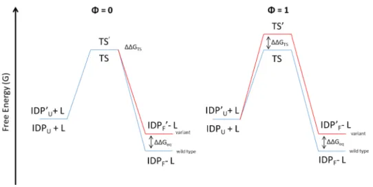
The Φ-value analysis is also used to characterize the folding upon binding of IDPs/IDRs (Figure 4).
Figure 4. Schematic (simplified) representation of the Φ-value analysis of a
folding upon binding reaction. IDPU and IDPF are the disordered protein unfolded and folded respectively. IDP’U is the IDP, bearing the amminoacidic substitution, unfolded; TS’ is the TS of the variant; IDP’F is the IDP, bearing the amminoacidic substitution, folded.
In the case of IDPs/IDRs, the reactants are the disordered protein and its partner and the product is the complex with the folded IDP/IDR. The scheme reported in Figure 4 is an extreme simplification. In fact, in this case, the analysis is harder because the significance of the Φ-values changes depending on which step (i.e. the folding or the binding), is the rate-limiting step (representing the highest free energy barrier of the reaction that defines the transition state) of the reaction. To increase the complexity, most of the times is not possible distinguishing, experimentally, which is the limiting step of the reaction since, usually, a simple (apparent) two-state mechanism
performing a Φ-value analysis, assessing whether the mechanism is induced fit or conformational selection. In the conformational selection mechanism, the IDP/IDR folds before the binding with the partner so, if the folding is the rate-limiting step, the Φ-value analysis acquires the same significance of a classical folding analysis and does not give information about the stability of the complex. When the rate-limiting step is the binding, high Φ-values (equal or close to 1) indicate that the residue is folded in the transition state and fractional Φ-values (comprised between 0 and 1) indicate that the residue, in the transition state, is involved in the interaction with the partner.
In the induced fit mechanism, the folding occurs after the binding. In this case, when the binding is the rate-limiting step, in the transition state the IDP/IDR will be mostly unfolded, because the folding can’t occur before the limiting step, and it will be represented by low Φ-values (equal or close to 0). When the folding is the rate-limiting step, the Φ-value has the same meaning of a classical folding analysis (Gianni, Dogan, & Jemth, 2014b).
Therefore, the Φ-value analysis allows obtaining information on the folding upon binding mechanisms of IDPs/IDRs since it consents hypothesizing the molecular mechanism that govern the process and allows identifying, for example, the residues that are important for the initial interaction or that fold before others.
2.2.2. Linear Free Energy Relationship
An additional classical procedure to assess the structural features of the transition state, based on the comparison of kinetic data obtained from the wt protein and its variants, consists in performing the Linear Free Energy Relationship analysis (LFER plot). The LFER plot is obtained by plotting the change in free energy in the transition state as a function of the change in free energy in the equilibrium state. LFERs were originally introduced to assess
the position of the transition state along the reaction coordinate during the formation of a covalent bond (Leffler, 1953). Upon altering the structure and thus the reactivity of a substrate, the dependence of activation free energy on ground-state free energy generally yields a linear profile. The slope of the observed correlation, called α, reflects the position of the transition state along the reaction coordinate. A linear LFER plot is a typical index that the folding occurs following the so-called nucleation-condensation mechanism whereby the whole protein self-assembles around a weakly formed nucleus and the transition state resembles a distorted version of the native state. The α value reflects the degree of similarity between the native and transition state. A high α value indicates that the transition state resemble a similar, although distorted, version of the native state (in the case of folding upon binding reaction the native state is the bound, folded state) while, a low α value indicates that the transition state is closer to the denatured state (in the case of a folding upon binding reaction the denatured state is the unbound, unfolded state).
3. Templated folding
A recent finding is that IDPs/IDRs could adapt their folding process depending on the structure of the partner. Φ-value analysis and molecular dynamic simulations studies of the folding after binding interaction between the intrinsically disordered c-Myb and the KIX domain revealed a structural malleability of the transition state dictated by the topology of KIX (Toto et al., 2016). This behavior suggested that IDPs/IDRs could fold via a templated mechanism involving a heterogeneous nucleation process induced by the structure of the partner.
This mechanism is in contrast with the so-called homogeneous nucleation typically observed for globular proteins, where the transition state is similar to the native state presenting an elevated fraction of native-like contacts.
This property increases the specificity of the molecular recognition process by which IDPs/IDRs interact with their partners avoiding miss-interactions.
4. Fuzziness in IDP complexes
The regions in IDPs/IDRs that undergo a disorder-to-order transition upon binding to a partner (i.e. MoREs) are usually short regions, while a considerable part of the sequence remains disordered also in the bound complex. These regions are called “fuzzy” and this type of complexes are called “fuzzy complexes”. Fuzzy complexes can be classified in four structural categories: in the polymorphic complexes the IDP/IDR assumes different conformations on the partner surface (Renault et al., 2008) in the flanking complexes fuzzy regions close to the bound motifs provide additional contacts to the interaction increasing the binding affinity (Billeter et al. 1993); in the clamps complexes the fuzzy regions don’t bind the partner and connect globular binding domains or motifs interacting with the partners (Clerici et al., 2009). Lastly, in the random complexes, the IDP/IDR remains completely disordered also upon the binding (Pometun, Chekmenev, & Wittebort, 2004). These four general categories can also be present in a same protein in different combinations.
Fuzzy regions also influence the interaction between IDP/IDR and partners enhancing or decreasing the binding through various mechanisms. In the conformational selection, fuzzy regions modulate the conformational equilibrium, stabilizing interaction-prone structural elements and moving the conformational equilibrium prior to binding (Adams, McBryant, Wade,
Woodcock, & Hansen, 2007)(Ghosh et al., 2010). In the flexibility/entropy modulation, fuzzy regions influence the flexibility and energy landscape of the binding interface performing transient contacts leading either to a decrease of the entropic cost of the binding and to an increase in the affinity (Pufall et al., 2005)(Lee et al., 2008). In the competitive binding, fuzzy regions compete with the binding partner, blocking the access to the binding surface, through electrostatic interactions or steric hindrance, resulting in a sort of auto-inhibition of the interaction (Stott, Watson, Howe, Grossmann, & Thomas, 2010). Finally, in the tethering mechanism, fuzzy regions enhance the binding affinity increasing the local concentration of globular, low-affinity binding domains (Daughdrill, Narayanaswami, Gilmore, Belczyk, & Brown, 2007)(Vise, Baral, Latos, & Daughdrill, 2005)(Borcherds, Becker, Chen, Chen, & Daughdrill, 2017). Therefore, the residual structural disorder in the bound form plays a role in the biological functionality of IDPs allowing a fine regulation of their activity and of that of the bound partner.
5. Frustration
Frustration in a system arises from the impossibility to achieve a specific goal. In folding, a system is considered frustrated when energetic contributions cannot be simultaneously minimized by a single conformation (Oliveberg & Wolynes, 2005). In general, proteins have the ability to find spontaneously a specific conformation, the native state, which is at the bottom of the funnel, according to the energy landscape theory. The energy landscape is the mathematical function that describes the intramolecular and solvation free energy of a given protein as a function of the microscopic degrees of freedom (Dill, Ozkan, Shell, & Weikl, 2008). The energy landscape theory was developed using tools from the statistical mechanics of
simplified this theory leading to the conclusion that proteins have funnel-shaped energy landscapes since they present many high-energy states and few low-energy states. Energy landscapes are multidimensional surfaces where the vertical axis of the funnel represents the free energy of a given chain configuration, the many lateral axis represent the conformational coordinates and each conformation is represented by a point on the energy surface (Dill & Chan, 1997). According to this model, the native state represents the bottom of the funnel (i.e. the free energy minimum) and the denatured state is a set of different configurations represented by all the points on the landscape surface (Figure 5). This funnel structure is only possible for those selected sequences that are chosen so that energetic conflicts are for the most part avoided and the native structure is more stable than expected for random associating residues: this hypothesis is known as the principle of minimal frustration (Ferreiro et al., 2010). Thus, proteins are usually minimally frustrated and this feature makes protein structure robust to mutations. However, sometimes, local frustration could be a functionally useful adaptation arising from random evolution (Ferreiro, Hegler, Komives, & Wolynes, 2007). Local frustration could be useful because it may sculpt protein dynamics for specific functions, for example a specific site frustrated in a monomeric protein may become less frustrated after association in a larger assembly, driving specific association (Wang & Verkhivker, 2003)(Papoian, Ulander, & Wolynes, 2003). Also enzymes show a certain degree of frustration in their catalytic sites (Ellerby et al., 1990)(Meiering, Serrano & Fersht, 1992); therefore, looking at frustrated sites can give a hint about functional constraints on the evolution of protein energy landscapes. Considering that proteins have a certain degree of frustration, their free energy landscape will be characterized by some local minima, which are likely to contain misfolded elements. It is possible to indicate frustrated sites,
experimentally, performing, for example the value analysis: negative Φ-values or Φ-Φ-values greater than 1 are usually a signature of frustrated sites.
Figure 5. Schematic tridimensional representation of the energy landscape.
The bottom of the funnel represents the low-energy native state and the other points of the surface are high-energy unfolded structures. Folding can occur via alternative microscopic trajectories (adapted from Dill & MacCallum, 2012).
6. Experimental system: Measles Virus NTAIL
Protein disorder is highly abundant in the proteome of viruses. In addition to the biological functions carried out in eukaryotic organisms (e.g. protein-protein recognition and interaction, regulation and signal transduction, regulation of transcription and translation) the presence of disorder in viral proteins allows viruses to increase their adaptability to hostile environment and, give capability of IDPs/IDRs to bind to many different partners, confers them the ability to interact with multiple cellular factors and to highjack cellular processes at their own advantage (Tokuriki, Oldfield, Uversky, Berezovsky, & Tawfik, 2009). Moreover it was suggested
that protein disorder in viruses could be connected to the elevated mutational frequency typically occurring in viral genomes: spontaneous mutations are indeed expected to have a lower effect in a disordered protein than in a well-structured protein (Xue et al., 2010)(Uversky & Longhi, 2011).
An interesting and widely studied disordered domain is the C-terminal domain of the Nucleoprotein (NTAIL) of Measles Virus. Measles Virus (MeV), a member of the Paramyxoviridae family, is a severe human pathogen responsible for the measles disease. The genome of MeV consists of a negative, single-stranded RNA, which is enveloped by the nucleoprotein (N) in a helical nucleocapsid. The nucleocapsid, and not naked RNA, is the substrate used by the RNA-dependent RNA polymerase (RdRp) for transcription of viral genes and replication of the genome. The RdRp is composed of the large (L) protein and the phosphoprotein (P). The P protein possesses a long N-terminal, intrinsically disordered domain (PNT amino acids 1-230) (Karlin, Longhi, & Canard, 2002a), and a C-terminal region (PCT, amino acids 231-507) organized in alternating structured and disordered modules (Karlin, Canard, & Longhi, 2003). The N protein consists of a structured N-terminal domain (NCORE, amino acids 1-400) that is responsible for binding to RNA and self-assembly of N to yield the nucleocapsid (Karlin, Longhi, & Canard, 2002b)(Gutsche, Desfosses, Effantin, & Ling, 2015), and a C-terminal disordered domain (NTAIL, amino acids 451-525) (Longhi et al., 2003) exposed at nucleocapsid surface. NTAIL contains three regions conserved in Morbillivirus members named Box 1 (amino acids 401-420), Box 2 (amino acids 489-506), and Box 3 (amino acids 517-525) (Figure 6). These regions present a higher content in hydrophobic residues, which facilitates protein-protein interactions. Being exposed at the surface of the nucleocapsid, NTAIL is able to interact with different partners: it binds the P viral protein but also some cellular factors
including the major inducible 70 kDa heat shock protein (hsp70) (Zhang et al., 2005), the interferon regulatory factor 3 (IRF3) (Colombo et al., 2009), peroxiredoxin 1 (Watanabe et al., 2011), the cell protein responsible for the nuclear export of N (Sato et al., 2006), casein kinase II, components of the cell cytoskeleton and also immune system factors.
During my PhD I have studied in particular the interaction between MeV NTAIL and the P protein. NTAIL interacts with the C-terminal X domain (XD, amino acids 459-507) of the P protein. This interaction is vital for the virus, leading to the recruitment of the polymerase complex onto the nucleocapsid template to allow transcription and replication to take place (Figure 7).
Figure 6. Schematic representation of the Nucleoprotein (A) and
Phosphoprotein (B) of MeV. Figure adapted from (Longhi et al., 2017)
B
A
Figure 7. Schematic representation of the MeV helical nucleocapsid and of
its interaction with the RNA-dependent RNA polymerase via the NTAIL-XD interaction. Figure adapted from Habchi & Longhi 2012.
The NTAIL domain interacts with XD via a short, order-prone molecular recognition element (α-MoRE, amino acids 486-502), located within Box 2 that, upon binding, undergoes an α-helical folding. The structure of the complex between MeV XD and the α-MoRE of NTAIL is available (Protein Data Bank: 1T6O). XD is composed of three α helices; after the binding (and folding) of the α-MoRE, a four-helix complex is formed (Figure 8).
Figure 8. Representation of the three-dimensional structure of the four-helix
complex between XD (in blue) and the α-MoRE (in green) of NTAIL (aa 486-504). Protein Data Bank code: 1T6O
6.1. MeV NTAIL-XD interaction: kinetic studies
Kinetic binding experiments were performed to study the interaction between MeV NTAIL and XD. Since the reaction is very fast (milliseconds time scale), to avoid losing the reaction in the stopped-flow dead time, the temperature-jump apparatus was used. As mentioned before, and better explained in the Materials and Methods section, the temperature-jump (t-jump) is a relaxation technique that allows measuring ultra fast reactions. The t-jump, via a rapid increase in temperature, perturbs an equilibrium system (in this case the complex between two interacting proteins), and then, taking advantage of the change in the tryptophan fluorescence upon (un)binding, it
measures the relaxation to the new equilibrium, allowing the observed rate constant of the binding reaction to be calculated.
The kinetics of interaction between MeV NTAIL and XD, performed under pseudo-first order conditions, showed a hyperbolic dependence of the kobs irrespective of whether NTAIL or XD was used as the excess ligand, a
behavior that constitutes a signature of folding after binding (Figure 9) (Dosnon et al., 2015).
Figure 9. Top: schematic representation of the folding after binding process
of the XD-NTAIL interaction. kon and koff are the association and dissociation
rate constants and kf and ku are the folding and unfolding rate constants.
Bottom: hyperbolic dependence of the kobs as a function of XD (open circles)
The hyperbolic dependence of kobs as a function of NTAIL concentration
allowed the binding, bimolecular step to be separated from the folding, monomolecular step (Dosnon et al., 2015).
An experiment performed in the presence of TFE (2,2,2-trifluoroethanol), a secondary structure stabilizer able to induce folding in peptides and proteins (Jasanoff & Fersht, 1994)(Myers, Pace, & Scholtz, 1998), showed that at high protein concentrations the reaction is limited by the folding rate of NTAIL. In fact, upon increasing the TFE concentration (i.e. upon increasing the stabilization of the α-MoRE helix) a transition from a hyperbolic to a linear dependence of the kobs was observed (Figure 10)
Figure 10. Kinetic binding experiment between NTAIL and XD, under pseudo-first order conditions, in the presence of increasing concentrations of TFE (Figure from: Dosnon et al. 2015).
6.2. MeV NTAIL-hsp70 interaction
MeV NTAIL also interacts with the 70 kDa heat shock protein (hsp70) of the host cells. Hsp70 is an ubiquitously expressed protein involved in protein folding (acting as chaperone) and in the protection of the cell under stress conditions. Hsp70 is a large protein composed of three major functional domain: the N-terminal ATPase domain responsible for the interaction and hydrolysis of ATP; the substrate binding domain composed of a 15 kDa β-sheet subdomain and a 10 kDa helical subdomain; and a C-terminal domain rich in α-helices acting as a 'lid' for the substrate binding domain.
NTAIL interacts with hsp70 through Box 2 and also, although with lower affinity, through Box 3 (Zhang et al., 2005)(Carsillo, Traylor, Choi, Niewiesk, & Oglesbee, 2006). Despite the affinity of NTAIL towards hsp70 is lower (70 µM) (Couturier et al., 2010) than that towards XD (3 µM) (Dosnon et al., 2015), hsp70 competes with XD for the binding with NTAIL thus reducing the stability of the NTAIL-XD complex (Zhang et al., 2005).
6.3. Fuzziness in MeV NTAIL complexes
After binding to XD, the NTAIL regions close to the α-MoRE remain conspicuously disordered (Longhi, 2012). In a work published in 2016, Gruet and co-workers showed that gradual removal of the long N-terminal fuzzy appendage leads to an increase in the binding affinity between NTAIL and XD (Gruet et al., 2016). In those studies, the authors made use of a protein
complementation assay based on split-GFP re-assembly (Magliery & Regan, 2006)(Wilson, Magliery & Regan, 2004). In this assay, XD, fused to the C-terminal domain of the GFP (Green Fluorescent Protein), is co-expressed in E. coli with NTAIL, fused to the N-terminal domain of GFP. Upon interaction between NTAIL and XD, and only in this case, the two GFP fragments can assemble in an irreversible manner, leading to reconstitution of the fluorophore and hence to a fluorescence signal that can be measured. The fluorescence is proportional to the binding strength between the two interacting proteins. Those studies demonstrated that the fuzzy region of NTAIL decreases the binding strength between NTAIL and XD.
The same experiments were also performed with hsp70: despite the lower affinity, and the likely different mechanisms of interaction, the truncation of the fuzzy region was found to lead to an increase in the binding strength with Hsp70 too.
The same results were also obtained in split luciferase reassembly assays. In this case, the reporter is luciferase, the assay is performed in eukaryotic cells, and the reassembly of the two halves of luciferase, leading to luminescence, is reversible. In addition, pull-down experiments that made use of XD fused to GFP and of purified, histidine-tagged NTAIL either in its full-length or in its truncated form, confirmed that truncated NTAIL has a higher affinity towards XD.
Thus, the fuzzy region in NTAIL seems to compete with the partner, through mechanisms that are still to be unveiled, decreasing the binding affinity.
In the same work the authors showed that the residual disorder in NTAIL also affects the folding rate of the α-MoRE. They performed kinetic binding experiments (under pseudo-first order conditions) between XD and either NTAIL full-length or a synthetic peptide mimicking just the α-MoRE region of NTAIL. In the case of full-length NTAIL, a hyperbolic dependence of kobs on ligand concentration was observed (the folding of NTAIL becoming
rate-limiting at high reactant concentrations). Conversely, when the MoRE peptide was used, a linear dependence was observed (Figure 11), meaning that the folding rate of the α-MoRE is increased, and the overall reaction can be described as schematized in Figure 12. This behavior suggests that the fuzzy appendage of NTAIL influences the folding of the MoRE, lowering its rate constant.
Figure 11. Kinetic binding experiments between XD and full-length NTAIL (empty circles) and the α-MoRE peptide (full circles). Figure modified from Gruet et al. 2016.
Figure 12. Schematic representation of the binding reaction between the
α-MoRE and XD. The overall reaction is described by a simple two-state mechanism with an apparent association rate constant (𝑘!"!"") and an apparent dissociation rate constant (𝑘!""!"").
The mechanisms regulating the interaction of intrinsically disordered proteins with their partners and the resulting disorder-to-order transitions are still far to be clearly understood.
In this thesis, using the C-terminal intrinsically disordered domain of the Nucleoprotein of Measles virus (NTAIL) and its interaction with its partners as a model system, I will attempt at better understanding the mechanisms regulating the interaction between IDPs and partners, the folding pathway of Molecular Recognition Elements and the role of the regions retaining intrinsic disorder also upon binding.
The first goal of my thesis is the study of the molecular mechanism of the interaction between MeV NTAIL and its partner XD (the C-terminal domain of the MeV phosphoprotein) at single residues level performing the Φ-value analysis. To this end, I will first design and produce a series of single site variants of MeV NTAIL in the α-MoRE region (amino acids 488-505) and then will perform kinetics binding experiments between each variant of NTAIL and its partner XD.
A work published in 2016 by Bonetti and co-workers showed that XD, in native conditions, populates an alternative state similar to an on-pathway intermediate retaining a native-like secondary structure but displaying some differences in the overall topological structure. It was observed that the substitution of the Isoleucine with Alanine in position 504 of XD leads to a variant that populates only the native state, i.e. devoid of the intermediate (Bonetti et al. 2016). Therefore, I will perform kinetic binding experiments using the XD I504A variant and the same single site NTAIL variants used for the Φ-value analysis with wt XD, so as to assess whether the folding after binding mechanism of NTAIL depends or not on the structure of the partner.
Subsequently, I will also produce a series of truncation variants of NTAIL, through gradual removal, by blocks of ten amino acids, of the region of NTAIL that remains disordered in the complex with XD, so as to study the effect of the residual disorder on the rate of folding of the α-MoRE. Concomitantly, I will also assess which is the minimal region responsible for the shift from a hyperbolic to a linear dependence of the kobs as a function of
NTAIL concentrations (see Figure 11). Once established whether and which part of the fuzzy region plays a role in the regulation of the folding rate of the α-MoRE, I will perform single site variation in this region and t-jump experiment with XD to assess the nature of the inhibitory effect of the fuzzy appendage. In particular, I will ascertain if the fuzzy region inhibits the folding of the MoRE through an entropic effect, merely due to the length of the sequence, or whether on the contrary it exerts an enthalpic effect due to specific aminoacidic interactions. Moreover, to assess the possible enthalpic effect of the fuzzy region on the rate of folding of the α-MoRE, I will perform kinetic binding experiment using an artificial variant of NTAIL, described in Gruet et al., 2016, where all the amino acids in the region comprised between residues 401 and 480 are replaced with “disordered prone” residues, in order to obtain a different (i.e. highly dissimilar) and more disordered variant of NTAIL.
Finally, I will also study two variants of the isolate α-MoRE region: AlaMoRE, a variant obtained by substituting with Alanine all the amino acids not essential for the interaction with XD, expected to be more ordered; and GlyMoRE, a variant obtained by substituting with Glycine all the amino acids not essential for the interaction with XD in order to obtain a more disordered sequence. Using far-UV CD spectroscopy I will assess whether the content in secondary structure in AlaMoRE and in GlyMoRE is increased
perform kinetics binding experiments with wt XD to assess whether a different content in (dis)order of the MoRE region affects its folding rate.
1. Constructs generation
1.1. NTAIL site-directed variants
The constructs encoding the site directed variants of NTAIL were obtained using the gene encoding wild-type (wt) NTAIL, inserted in the pET28 expression vector, as template to perform site-directed mutagenesis using the QuickChange Lightning Site-Directed Mutagenesis kit (Agilent technologies) according to the manufacturer’s instructions. All substitutions were conservative and were confirmed by DNA sequencing.
1.2. NTAIL truncation variants
The constructs encoding N-terminally heaxahistidine tagged NTAIL truncation variants (from 401 to 481) with a tobacco etch virus (TEV) protease cleavage site (amino acidic sequence: ENLYFQGS) immediately upstream of the codons encoding amino acids 401, 411, 421, 431, 441, 451, 461, 471 and 481 of NTAIL were already available (Gruet et al. 2016).
The NTAIL truncation variants were also cloned into the pETG20A expression vector that drives the expression of the protein of interest fused to a Thioredoxin (Trx) solubility tag. To this end, the already available pDEST14 constructs encoding N-terminally hexahistidine tagged NTAIL, with a TEV protease cleavage site at different NTAIL positions, were used as a templates in a first PCR amplification that used attB2 as reverse primer and as forward primer an oligonucleotide encompassing the attB1 site followed by the sequence encoding the TEV protease cleavage site, and by a sequence annealing with the 17 NTAIL nucleotides downstream of the TEV cleavage site. After digestion with DpnI (1 hour at 37°C), 1 µl of the first PCR reaction was used as template for a second PCR amplification performed using primers attL1a and attL2a. These primers encode approximately half of the attL1 and attL2 recombination sites, respectively, and have been shown
to be good substrates for LR clonase (Fu et al., 2008). As the attL1a and attL2a sequences encompass the attB1 and attB2 sequences, respectively, amplicons flanked by attB sequences as obtained from the first PCR product provide the necessary homology for attL1a and attL2a primer hybridization, respectively. 1.5 µl of the second PCR product were used in a 5 µl LR reaction to insert the truncation variant sequences into the pETG20A plasmid by Gateway cloning. These steps were performed to produce all the truncation variant constructs bearing the Trx tag.
1.3. Artificial NTAIL
An already available construct encoding the artificial NTAIL sequence cloned into the pNGG plasmid (Gruet et al. 2016) was used as a template in a first PCR amplification aimed at inserting the TEV protease cleavage site upstream residue 401. The forward primer contained the attB1 site, followed by the sequence encoding for the TEV protease cleavage site and by 24 nucleotides annealing with the 5’ region of artNTAIL. The reverse primer was attB2. After digestion with DpnI, the artificial amplicon bearing the TEV cleavage site was used as a template for a second PCR amplification with primers attL1a and attL2a. The resulting amplicon was then inserted in the pETG20A plasmid through an LR reaction, as described for the truncation variants.
1.4. X Domain: Y480W and I504A variants
The construct encoding XD with a C-terminal hexahistidine tag and bearing the Y480W substitution (referred to as pseudo-wt), has been already described (Dosnon et al. 2015), as was the construct encoding pseudo-wt bearing the additional I504A substitution (Bonetti et al. 2016).
2. Protein expression and purification 2.1. NTAIL site-directed variants
All variants were expressed in the Escherichia coli Rosetta T7 pLysS (Novagen) strain. Cultures were grown in Luria Bertani (LB) medium containing 100 µg/ml ampicillin and 34 µg/ml chloramphenicol at 37°C until the OD600 reached 0.6-0.8 and then protein expression was induced with 1 mM IPTG (isopropyl-β-D-thiogalactopyranoside). After induction, cells were grown at 25°C over night and then collected by centrifugation. Cells were resuspended in 5ml/gr of pellet of buffer A (50 mM sodium phosphate pH 7.2, 300 mM NaCl) supplemented with 0.1 mg/mL lysozyme, 10 µg/mL DNase I and protease inhibitor cocktail (Sigma) and disrupted by sonication. The lysate was clarified by centrifugation at 12,000 rpm for 40 minutes at 4 °C. The soluble fraction bacterial lysate was loaded onto a 5 ml His trap FF column (GE, Healthcare) preloaded with Ni2+ ions, previously equilibrated in buffer A. After a washing step with buffer A supplemented with 1 M NaCl, the proteins were eluted with buffer A containing 250 mM imidazole. The proteins were then concentrated until 1 – 2 ml and loaded onto a Superdex 75 16/60 column (GE, Healthcare). Isocratic elution was carried out in SEC buffer (10 mM sodium phosphate pH 7.0, 150 mM NaCl). The purity of the proteins was confirmed by SDS-PAGE and the concentration was calculated measuring the absorbance at 280 nm, using the theoretical extinction coefficient, as provided from ExPASy (https://www.expasy.org/), and applying the Lambert-Beer equation.
2.2. NTAIL truncation variants and artNTAIL
The NTAIL truncation variants and artNTAIL were expressed in Terrific Broth (TB) medium containing 100 µg/ml ampicillin and 34 µg/ml chloramphenicol. The E. coli strain Rosetta T7 pLysS (Novagen) was used. Cells were grown in the same conditions described for the single-site NTAIL variants except that cells were collected by centrifugation after 4 hours at 37°C.
Cell lysis was carried out as described above and the soluble fraction obtained after centrifugation was loaded onto a 5 ml His trap FF column (GE, Healthcare) as described for the single site variants. In this case, the buffer used to equilibrate the column was 50 mM Tris/HCl pH 8.0, 300 mM NaCl, 10 mM imidazole, 1 mM phenyl-methyl-sulphonyl-fluoride (PMSF). After sample injection, the column was washed with 50 mM Tris/HCl pH 8.0, 1 M NaCl, 20 mM Imidazole. Elution was carried out with 50 mM Tris/HCl pH 8.0, 300 mM NaCl, 250 mM Imidazole. Eluents from IMAC were desalted using a desalting column (GE, Healthcare) previously equilibrated with desalting buffer (20 mM Tris/HCl pH 8.0, 300 mM NaCl). Cleavage with TEV protease was carried out overnight at 4°C in desalting buffer using 1 mg of TEV protease per 20 mg of target protein. Samples were then loaded onto 2.5 ml Ni2+ IDA Agarose resin, previously equilibrated in desalting buffer, and incubated 15 minutes at 4°C. The non-retained fraction was recovered, concentrated up to 1 – 2 ml, and then loaded onto a Superdex 75 16/60 column (GE Healthcare) and eluted in SEC buffer. The purity of the protein was confirmed by SDS-PAGE.
2.3. XD variants
(LB) medium containing 100 µg/ml ampicillin at 37°C until the OD600 reached 0.6 and then protein expression was induced with 1 mM IPTG. After induction, cells were grown at 25°C for 2 days and then collected by centrifugation. Cells were resuspended in buffer A (50 mM sodium phosphate, 300 mM NaCl) supplemented with protease inhibitor cocktail (Sigma). The lysate was clarified by centrifugation at 12,000 rpm for 40 minutes at 4 °C.
The soluble fraction of the bacterial lysate was loaded onto a 5 ml His trap FF column (GE, Healthcare). After a washing step with buffer A supplemented with 1 M NaCl, the proteins were eluted with buffer A containing 1 M imidazole. The eluted protein was then desalted with a desalting column (GE, Healthcare) using buffer A. The purity of the protein was verified by SDS-PAGE.
2.4. α-MoREs peptides
Peptides mimicking the wt MoRE and its variants enriched in Alanine (AlaMoRE, supposed to be more ordered) and in glycine (GlyMoRE, supposed to be more disordered) were purchased from JPT (Berlin, Germany). The MoRE peptides were designed to encompass residues 486-504 of NTAIL, and possessed an additional non-native tyrosine at the C-terminus to allow their concentration to be inferred from the absorbance at 280 nm using the Lambert-Beer equation.
3. Modeling
The structural models of the free form of the three MoRE peptides were obtained using the PEP-FOLD3 server (http://mobyle.rpbs.univ-paris-diderot.fr/cgi-bin/portal.py#forms::PEP-FOLD3, Lamiable et al. 2016).
4. Circular dichroism measurements
The far-UV CD spectra of the MoRE peptides were measured using a Jasco 810 dichrograph, flushed with N2 and equipped with a Peltier thermoregulation system. Peptides were dissolved in 10 mM sodium phosphate pH 7.0 at a concentration of 0.1 mg/mL in a final volume of 300 µl. One-mm thick quartz cuvettes were used. Spectra were measured between 190 and 260 nm at 20°C. The scanning speed was 50 nm/min, with data pitch of 0.2 nm. Each spectrum is the average of three acquisitions. Spectra were smoothed using the ‘‘means-movement’’ smoothing procedure implemented in the Spectra Manager package. The BESTSEL website (http://bestsel.elte.hu/, Micsonai A, Wien F, Kernya L, Lee YH, Goto Y, Refregiers M, et al. Accurate secondary structure prediction and fold recognition for circular dichroism spectroscopy. Proc Natl Acad Sci U S A. 2015;112:E3095-103) was used to analyze the experimental data in the 190– 250 nm range.
5. Temperature-jump measurements
The interaction between MeV NTAIL and XD is an extremely rapid reaction. Thus, the classical stopped-flow mixing methods cannot be use because of the dead-time of about 1-2 ms occurring in this technique. Therefore, to study the kinetics of interaction between NTAIL and XD, the temperature jump (t-jump) method, a kinetic technique that allows measuring ultra rapid reactions, was used. The t-jump methodology is a relaxation method whereby the solution containing a mixture of the interacting proteins at equilibrium is perturbed by a rapid increase in temperature. After trigger,
the relaxation to the new equilibrium is observed. The rapid heating is due to