REVIEW
Imaging of the scrotum: beyond sonography
Gian Carlo Parenti1&Francesco Feletti1&Aldo Carnevale2&Licia Uccelli3&Melchiore Giganti2
Received: 28 August 2017 / Revised: 18 December 2017 / Accepted: 27 December 2017 / Published online: 15 February 2018 # The Author(s) 2018. This article is an open access publication
Abstract
The aim of this article is to describe the role of second-level imaging techniques after an initial ultrasonography evaluation in the assessment of scrotal diseases. While ultrasonography remains central as the primary imaging modality for the evaluation of pathologic conditions of the scrotum, the role of magnetic resonance imaging continues to evolve: it can actually be valuable as a problem-solving tool when sonographic findings are equivocal or inconclusive. Magnetic resonance imaging of the scrotum may provide accurate detection and characterization of scrotal diseases, well depicting the precise location of scrotal masses (intratesticular or extratesticular) and reliably characterizing benign conditions simulating neoplastic processes, thus preventing unnecessary radical surgery. Advanced magnetic resonance techniques, most of all diffusion weighted imaging and magnetic resonance spectroscopy, play in the meanwhile a more significant role in evaluating scrotal diseases.
Teaching points
• Multiparametric ultrasonography usually represents the initial imaging modality for approaching scrotal diseases. • MRI is well established as a problem-solving tool for inconclusive sonographic findings.
• Advanced MRI techniques can be successfully applied in scrotal pathology assessment. • MRI is valuable in differentiating benign conditions from neoplastic processes. • CT plays a role in trauma assessment and cancer staging alongside PET/CT.
Keywords Magnetic resonance imaging . Scrotum . Testis . Ultrasonography . Male urogenital diseases
Introduction
Multiparametric ultrasonography (US), encompassing in this definition conventional Brightness-Mode US, Eco-colour Doppler analysis, contrast-enhanced ultrasonography (CEUS) and elastosonography, usually represents the initial imaging modality for approaching scrotal diseases, thanks to
a proven diagnostic accuracy, cost-effectiveness and large availability of this technique [1–3].
Nevertheless, US correlates to some important limitations: the operator-dependent nature of the modality, low panoramicity due to small field of view and poor tissue char-acterization [1,4].
Therefore, other imaging modalities could integrate ultra-sound examination, in order to clarify any inconclusive or equivocal finding, or with the purpose of longitudinal moni-toring scrotal disease.
In the literature, the role of MRI is well established as a second level imaging modality useful for an increasing num-ber of indications [5–8].
Advanced techniques, namely diffusion weighted imaging (DWI), proton magnetic resonance spectroscopy (H1MRS) and magnetization transfer ratio (MTR), applied successfully in the evaluation of other anatomical districts, could be also used as an adjunctive imaging tool to assess testicular pathol-ogy [9–15].
The aim of this article is to describe the role of second-level imaging modalities, after ultrasonography examination, in the assessment of scrotal diseases.
* Aldo Carnevale [email protected]
1
Department of Diagnostic Imaging of Romagna, Section of Radiology, Ospedale Civile Santa Maria delle Croci, 48100 Ravenna, Italy
2
Department of Morphology, Surgery and Experimental Medicine, Section of Radiology, University of Ferrara, Via Ludovico Ariosto 35, 44121 Ferrara, Italy
3 Department of Morphology, Surgery and Experimental Medicine,
Section of Nuclear Medicine, University of Ferrara, Via Ludovico Ariosto 35, 44121 Ferrara, Italy
Materials and methods
An extended systematic search in Medline database (via PubMed), including articles related to human medicine pub-lished in the last 10 years to January 1, 2017, was performed. Keywords were chosen according to Medical Subject Heading (MeSH) terms: (“Diagnostic Imaging”[Mesh]) AND (“Testis”[Mesh]) OR (“Scrotum”[Mesh]) NOT “Ultrasonography”[Mesh].
The bibliographic search produced 443 results; two independent reviewers (G.P. and F.F. with more than 25 and 11 years of experience in urogenital radiology, respectively) screened the initial search results on the basis of titles and abstracts. Both reviewers had experi-ence in data extraction for retrospective and prospective studies.
Articles addressing an issue pertinent to magnetic res-onance imaging (MRI), computed tomography (CT) or positron emission tomography (PET)-CT examination of scrotal diseases were considered potentially eligible for inclusion. Studies only focused on scrotal ultrasound were excluded. Because of the variety of the issues dealt with by this work and due to limited data avail-able from the literature, the reviewers included case re-ports considered to contribute relevant information about imaging techniques application or for future research. All the papers not included in the first literature selec-tion of each of the two reviewers, were jointly re-analysed and discussed to obtain an agreement.
The full articles of selected studies were retrieved and ad-ditional searches of their reference lists performed to identify other potentially eligible articles. All non-English written studies were evaluated only by the information taken from the abstract.
In order to aid the selection procedure, a score was assigned to each article that met the inclusion criteria according to the 2011 Oxford Center of Evidence-Based Medicine (CEBM) levels of evidence [16], as follows:
1. systematic review of cross sectional studies with consis-tently applied reference standard and blinding;
2. individual cross sectional studies with consistently ap-plied reference standard and blinding;
3. non-consecutive studies, or studies without consistently applied reference standards;
4. case-control studies, or poor or non-independent refer-ence standard;
5. mechanism-based reasoning.
Finally, 58 articles were considered relevant for the scopes of the literature review and thus included in the present work.
Results
MRI, thanks to high spatial resolution and a large field of view, can be considered an ideal second-level technique [4], useful as a problem-solving tool when sonographic findings are equivocal [6].
We already found in a previously published study [7] that MRI proved to be necessary in 5.74% of patients (tot. n = 801) suspected to have scrotal disease after sonographic evaluation. In 47.8% of the cases, MRI managed to achieve a correct diagnosis, while in 37% of the cases contributed to exclude focal lesions. As Muglia et al. [8] reported in their work, also in our experience MRI was necessary in 5.02% of patients (tot. n = 622) undergone US, and resulted to add specific value in 82% of the cases.
In addition to MRI, Computed Tomography (CT) and pos-itron emission tomography-CT (PET-CT) have a second-level role approaching neoplastic processes, as shown below.
This review is organized on a pathology basis, as follows: trauma, inflammatory pathology, tumours, infarct, varicocele and infertility functional assessment, congenital abnormalities and scrotal hernia.
Trauma
Kim et al. [17] proved a 100% diagnostic accuracy for MRI in blunt scrotal trauma (7/7 cases). More in detail, authors report-ed two doubtful cases on sonographic evaluation for which MRI managed to achieve a diagnosis of epididymal hematoma.
In all cases of trauma (4) reported by Muglia et al., MRI identified a testicular hematoma or hematocele, assessing its extension and eventually inguinal canal involvement [8].
In our experience, in three patients with suspected neoplas-tic lesion according to US findings, MRI was useful in prov-ing a testicular hematoma leadprov-ing to a conservative approach. Furthermore, in a case it revealed an intrascrotal tear of tunica albuginea surrounding corpus cavernosum [7,18] not noted on US examination.
CT provides excellent depiction of bone fragments, defin-ing their presence and location, and eventual testicular dislo-cation resulting from a blunt pelvic injury [19–21].
Inflammatory processes
In our series, we described a case of epididymo-orchitis evolved into abscess formation, complicated by an intra-scrotal fistula poorly characterized by sonography; in that oc-casion, particularly thanks to T2-weighted sequences, MRI allowed identifying fistulous track and its relationship to close structures [7]. We also reported three cases of severely inho-mogeneous testicular echogenicity, in which MRI allowed excluding focal signal intensity abnormalities or focal areas
of pathologic contrast-enhancement (CE), differentiating be-tween a chronic orchitis from a neoplastic process, thus preventing unnecessary surgery [7].
In the literature, nevertheless we found cases of chronic granulomatous orchitis confused with tumour and undergone orchiectomy on the basis of MRI findings of testicular en-largement, hypointensity on T2-weighted images and in-creased signal on DW images [22].
Finally, MRI utility in early diagnosis of Fournier gangrene and in pre-operative planning is illustrated in a few works, accurately depicting fascial spread of the inflammatory pro-cess and necrotic component [23].
Tumours
In a study conducted on 165 testicle-containing scrotal com-partments (84 patients), MRI proved 100% sensitivity, speci-ficity, positive predictive value and negative predictive value for precise localization of scrotal focal lesions, excellently differentiating testicular from extra-testicular ones [24].
Muglia et al. found that MRI allowed reliable characteriza-tion of testicular tumours and pseudotumors in 88% of the cases (n = 15); findings not recognized at US that were essen-tial to define the neoplastic nature of the process were the presence of hemorrhagic intralesional components and exten-sion to tunica albuginea or epididymis [8]. Nevertheless, in the same series, two lesions suspected for tumours at MRI re-vealed chronic orchitis at pathological exam after surgery.
Some reports [25,26] described hypointensity signal on T2-weighted images and lack of restricted diffusion on DWI as useful in discriminate between paratesticular fibrous pseudotumor and malignancies, clearly affecting patient man-agement avoiding radical surgical intervention.
In our experience, MRI had an essential role in 15 cases of testicular neoplasms: in 11 cases of malignant germ cell tu-mours and 4 cases of benign lesions, of which two were Sertoli cell tumours and two were adenomatoid tumours. In all these cases, MRI was useful for preoperative planning, providing excellent depiction of eventual infiltration of tunica albuginea, tunica vaginalis, epididymis and paraepididymal soft tissues [7].
In a study conducted on 33 cases of testicular masses de-tected on US, Tsili et al. suggested that sensitivity and speci-ficity of MRI in differentiating benign from malignant lesions was 100% (95% CI, 87.9–100%) and 87.5% (95% CI, 52.9– 97.7%), respectively, with an overall accuracy of 96.4% and a satisfactory rate of correspondence between MRI findings and histopathologic diagnosis in regard to local extension of tes-ticular neoplasms (92.8%) [27]. All malignant tumours exhib-ited mainly hypointense signal compared to contralateral nor-mal testis on T1-weighted images, whereas signal intensity on T2-weighted images was low in seminomas, showing intralesional septal contrast enhancement after gadolinium
administration. Non-seminomatous tumours were heteroge-neous on both T1 and T2-weighted images, containing cystic or necrotic components, with heterogeneous enhancement [27]. In the same study, investigators also described a hypointense testicular focal lesion with contrast enhancement, falsely suspected for malignancy but revealed to be granulo-matous orchitis after surgical biopsy.
Manganaro et al., in their work concerning contrast en-hanced MRI on 44 patients with nonpalpable testicular le-sions, found in benign lesions a more rapid and intense wash-in (18/21, 85.7%, vs. 5/23, 21.7%) and a prolonged wash-out (15/21, 71.4% vs. 0/23), whereas malignant tumours showed a weak and progressive wash-in (18/23, 78.3% vs. 3/21, 14,3%) and an absent wash-out (21/23, 91.3% vs. 6/21, 28.6%) [28].
The role of DW imaging has been evaluated in a study conducted on 50 patients with monolateral testicular disease. A cut-off ADC value of≤0.99 × 10⁻³mm2/s had a sensitivity of 93.3%, specificity of 90%, positive predictive value of 87.5% and negative predictive value of 94.7% in the charac-terization of intratesticular masses [29]. Tsilli et al., in a series of 26 men, suggested that ADC values could be used to dif-ferentiate seminomas from non-seminomatous germ cells tu-mours before surgical intervention [30]. Moreover, a retro-spective evaluation of 56 cases advocated that ADC values do not provide detection of Testicular Intraepithelial Neoplasia (TIN) and could not be considered as a non-invasive screening tool for patients requiring surgical biopsy [31].
In a meta-analysis on 130 patients, fluorine-18-fluorodeoxyglucose (FDG)-PET-CT proved to be the best im-aging technique capable to detect residual disease in metasta-tic seminomas after chemotherapy, correctly directing thera-peutic management [32]; FDG-PET demonstrated higher specificity (92% vs. 59%) and sensitivity (72% vs. 63%), as well as a higher positive (70% vs. 28%) and negative predic-tive value (93% vs. 86%) than the solely size-based CT as-sessment of residual tumours.
Testicular infarction
In a previously published study [33], in 14 patients suspected for Segmental Testicular Infarction (STI) at US examination MRI allowed a correct diagnosis. In more detail, in 92% of the cases (n = 13) diagnosis was confirmed by imaging findings, i.e. hypointense signal on both T-1 and T2-weighted images, and lack of internal contrast enhancement of the infarcted tissue. In a single case, STI was excluded for the presence of a mild intralesional contrast enhancement and lack of change at close imaging follow-up; the patient underwent orchiecto-my with a histopathology report of B-cells lymphoma.
Urgent MRI, including dynamic contrast enhanced (DCE)-MRI, has a 100% sensitivity in the diagnosis of testicular
torsion, whereas T2- and T2*-weighted images have a 100% accuracy in detection of testicular necrosis [34].
Varicocele and infertility functional assessment
After a sonographic diagnosis and evaluation of varicocele, magnetic resonance angiography (MRA) allows depiction of eventual associated conditions, e.g. nutcracker syndrome [35]. Similarly, antegrade venography may identify the level of a
possible parallel duplication of internal spermatic vein, allowing an optimal treatment [36].
Karakas et al. [37] investigated the role of ADC values in determining testicular fibrosis; the authors found that ADC values were significantly lower among 25 patients presenting with varicocele compared to those of healthy volunteers; ADC values were also negatively correlated with venous diameter. Also, in hydrocele evaluation, in a series of 49 patients, altered ADC values were reported consequentially to a me-chanical effect of the pressure of the fluid, with possible dys-function of testicular tissue and a negative effect on fertility [38].
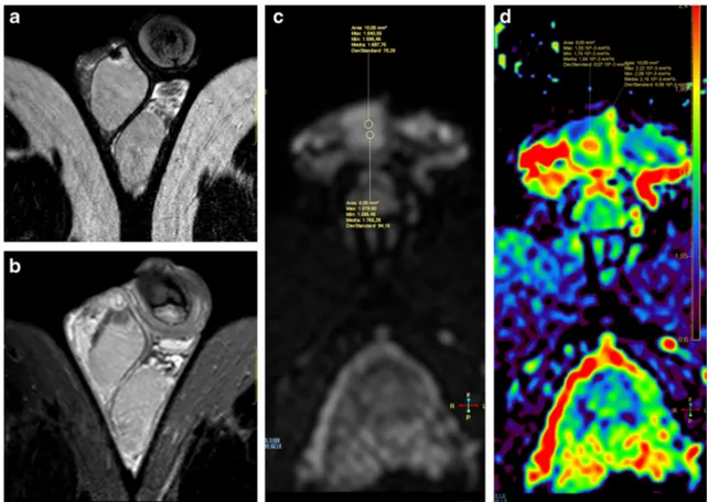
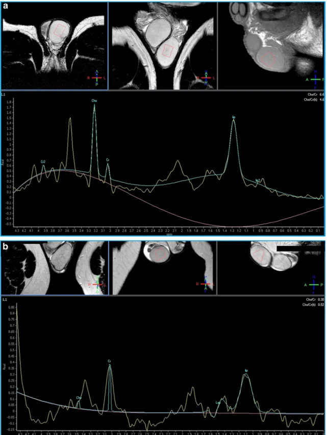
To assess the utility of MR spectroscopy in the evaluation of male infertility, we have studied 14 patients affected with documented fertility dysfunction (five patients presenting
Fig. 1 a-c Left testicle rupture. MRI shows a tunica albuginea tear and testicle rupture. a T1-weighted SPIR TSE sequence, axial view, b T2-weighted FFE sequence, axial view; c T2-T2-weighted TSE sequence, coronal view
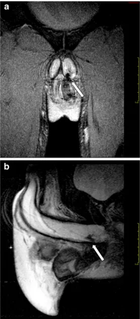
Fig. 2 a-b Intrascrotal rupture of the corpus cavernosum (arrow). a T2-weighted FFE sequence, coronal view; b T2-T2-weighted SPIR TSE sequence, sagittal view
with oligospermia, three with asthenospermia, six with oligo-asthenospermia). The choline principal peak was significantly lower in the altered semen analysis group than in normal con-trols (0.69 vs 1.34, 95% CI: 0.52–0.85; p < 0.001). Since patients proved to have semen alterations did not show any significant morphological finding in testicle structure both at US and MRI, excepted for varicocele in seven cases, we con-cluded that 1HMRS is capable to reveal spermatogenesis dis-orders in patients with normal testis at US and MRI examina-tion [39].
Congenital abnormalities and scrotal hernia
In a meta-analysis (8 studies) on 171 patients with 193 non-palpable testicles [40], investigators proved that conventional MRI has a low sensitivity (estimated median sensitivity of 62%), but high specificity (100%) in the identification of
nonpalpable cryptorchid testes, and has limited value in locat-ing intra-abdominal viable undescended testicles, whereas it fails in determining the presence of most atrophic ones. However, T2-weighted sequences with fat-suppression tech-niques, DWI [41] or MRA may significantly improve accura-cy of MR in cryptorchidism assessment [40].
MRI could allow or confirm the diagnosis of trans-verse testicular ectopia and eventually show any other associated variants or abnormalities [42, 43]. In a case report of an accessory scrotum, Ziegelmann et al. found that MRI was capable to define the complex relation-ships between the accessory scrotum and ano-sphincteric complex, as well as identify associated peritoneal lipo-ma [44].
In our series, MRI confirmed a sonographic diagnosis of inguinal-scrotal hernia in three patients, demonstrating the presence of intestinal loops in hernia sac [7].
Fig. 3 a-b Intrascrotal abscess. a T2-weighted TSE sequence, axial view, and b T2-weighted TSE sequence, sagittal view, showing fistulous track (arrow)
Fig. 4 a-e MR characterization of post-traumatic fibrosis. a T2-weighted TSE sequence, axial view and b post-contrast T1-weighted SPIR TSE sequence, axial view, depicting an ill-defined hypointense on T2-weighted image (T2-wi) area, with mild contrast enhancement (arrow), consistent with fibrosis; c DWI, b = 800; d-e ADC map
Discussion
Trauma
MRI in scrotal trauma assessment provides excellent depic-tion of hematomas based on the signal characteristics of blood
products, i.e. hyperintense signal on T1-weighted images re-lated to methaemoglobin, associated to hypointense on T2-weighted images perilesional hemosiderin rind.
On MRI, considering that testicular tumours are incidental-ly identified in 15% of patients after a traumatic episode, a confident differentiation between a hematoma and a Fig. 5 a-c MR study in a case of
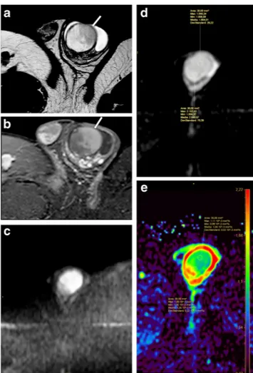
benign Leydig cells tumour. a T2-weighted TSE sequence, sagittal view and b post-contrast T1-weighted SPIR TSE sequence, axial view, depicting a round hypointense on T2-wi testicular focal lesion (arrow), with homogeneous
hyperenhancement. c ADC map
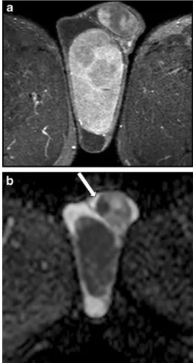
Fig. 6 a-d MR study of a multifocal mixed embryonal carcinoma. a T2-weighted TSE sequence, axial view; b postcontrast T1-weighted SPIR TSE sequence.; c-d ADC
neoplastic mass can be easily achieved in most cases, despite confusing US appearance of an inhomogeneous focal lesion, with intralesional vascular signals occasionally observed at colour Doppler analysis [45].
Moreover, due to a large field of view and optimal soft tissue characterization, MRI not only defines the extension of traumatic testicular injuries previously identified on US (Fig. 1), but also demonstrates an as-sociated tunica albuginea tear in corpora cavernosa, pro-viding information in regard to the vascularization, fun-damental for an optimal surgery planning (Fig. 2). On this regard, we consider MRI usage for monitoring re-sults of conservative therapies when a tunica albuginea tear is recognized, but a normal capsular blood flow of the tunica vasculosa is preserved [7].
CT is crucial in the evaluation of complex pelvic traumas involving the scrotum, revealing the presence and precise lo-cation of bone fragments, as well as testicular dislolo-cation [19–21].
Inflammatory processes
MR imaging may prove useful in inflammatory scrotal condi-tions as an adjunct to US in defining the extent of inflamma-tion, thereby aiding in the differential diagnosis from neo-plasms. Actually, based only on imaging findings, inflamma-tion if localized can mimic a neoplastic process [46]; in this setting, clinical correlation is usually decisive for orienting the differential diagnosis [27].
Acute orchitis usually manifests as an area of heteroge-neous signal intensity, reduced on T1-weighted images and increased on T2, in association to epididymis swelling and scrotal layers thickening [47].
In granulomatous orchitis, the absence of areas of altered signal intensity and contrast enhancement can orientate the diagnosis [7], as well as a concomitant epididymal involvement [27]; nevertheless, a chronic orchitis can simulate a neoplasm despite using more advanced MR imaging techniques [6, 22, 27]. In these cases, biopsy [6] or MRI follow-up after medical treat-ment may prevent unnecessary orchiectomy.
Moreover, MRI can confirm the sonographic detection of a complicated testicular cyst, depicted as slightly hypoechoic on US and presenting as a well-marginated area of typical high signal intensity on T2-weighted images with also high signal intensity on T1-weighted images due to inflammatory content [7].
MRI is also helpful for the exact depiction of more con-spicuous inflammatory processes, demonstrating extrascrotal tissues involvement of great intrascrotal abscesses [7]. T2-weighted sequences enable excellent depiction of fistulous tracks (Fig.3).
Tumours
MRI is highly valuable in the characterization of testicular masses detected at US examination [7], notably of nonpalpable testicular lesions [28]. This technique actually enables an accurate differentiation of tumours from pseudotumors, namely chronic orchitis, post-traumatic fibro-sis (Fig. 4) and rete testis ectasia [8], and, among tumours, allows identifying morphological features of malignancy (Fig. 5), for instance extratesticular structures involvement [8], or the presence of multifocal disease (Figs. 6 and7), proving high rate of correspondence between imaging find-ings and histopathological diagnosis in regard to local extent [27].
Scrotal MRI performs well with respect to the differentia-tion of extratesticular from intra-testicular disease, and can aid in narrowing the differential diagnosis in cases of paratesticular masses [48]. Paratesticular sarcomas, that may involve the testicle itself [49], can mimic an inguinal hernia at
Fig. 7 a-b MRI in a case of bilateral seminoma. a Post-contrast T1-weighted SPIR TSE sequence, axial view and b ADC map, demonstrating a large right testicular mass and a smaller left intratesticular one (arrow), with heterogeneous contrast enhancement and restricted diffusion
clinical and sonographic evaluation; CT and MRI both confi-dently demonstrate the presence of fat component, calcifica-tions, sepiments or intralesional contrast enhancement, help-ing discernhelp-ing these tumours from hernias and spermatic cord lipomas [47,49–52]. Moreover, as assumed in a recent retro-spective analysis based on 77 cases of paratesticular sarcomas, CT and MRI are able to supply prognostically important data for local recurrence concerning tumour size and boundaries; however, the differential diagnosis on the sole basis of imag-ing can be difficult, with well-differentiated liposarcomas be-ing the most relevant diagnostic challenge [50].
Most paratesticular masses are benign, therefore radical orchiectomy may be avoided: a multiparametric MR protocol including DWI, MTI and DCE could provide important addi-tional information in the assessment of benign nature of paratesticular masses, including cellular angiofibroma the tu-nica vaginalis, thus preventing unnecessary radical surgery [53].
According to ESUR guidelines, follow-up in testicular microlithiasis is advised if any risk factors are known [54]; in our center, we sometimes use MRI to rule out parenchymal focal abnormalities in patients with grade III microlithiasis [7]. Nevertheless, MRI is unable to depict testicular microlithiasis, but this may facilitate the detection of parenchymal areas with pathological signal or contrast enhancement in affected
patients, since the parenchymal echostructural inhomogeneity associated with this condition could not allow malignant le-sions to be confidently excluded at US examination.
MRI proved a useful tool for local staging of testicular neoplasms [29], for testis-sparing surgery planning, if indicat-ed [48], and for the differentiation between fibrosis and local recurrence after nodulectomy (Fig.8).
In testicular cancer staging after therapy, FDG-PET/CT allows evaluating the presence and vitality of residual disease; nevertheless, false positive results may be reported in seminomas [55], and false negatives in cystic mature terato-mas [56].
FDG-PET/CT could be considered a useful adjunct to other imaging modalities in the diagnosis of testicular lymphoma [57] and could depict scrotal extension of peritoneal mesothe-lioma [58].
Infarction
Sonographic diagnosis of segmental testicular infarction (STI) is challenging; it’s a rare entity and US appearance is often nonspecific. Anamnestic and clinical information aid orienting the differential diagnosis, but contrast-enhanced MRI is supportive, excluding neoplastic nature of a focal pa-renchymal lesion [7].
Fig. 8 a-d Hypoechogenic lesion at the site of previous tumour enucleation (Leydig cells tumour). MRI allowed the identification of an area of fibrosis. a T2-weighted TSE sequence, coronal view; b postcontrast T1-weighted SPIR TSE sequence; c-d. ADC map
In STI, MRI findings are the following: low signal intensity and a typical rim enhancement after gadolinium administra-tion, the latter related to granulation tissue during the subacute phase [33].
DWI and ADC map of a focal lesion are valuable to ex-clude or confirm the diagnosis, also through monitoring any change during follow-up MR studies.
In patients with suspected torsion, because the best chance of testicular preservation occurs with expeditious
management, it’s recommended an immediate surgical intervention, also without any imaging confirm [59]. Nevertheless, our experience suggests that MRI is useful in selected complex cases, characterized by chronic or intermittent clinical manifestations, including those cases appearing at birth and intrauterine torsion [7]. DWI analysis with ADC values measurement allows identify-ing testicular torsion without usidentify-ing contrast material [60].
Varicocele and infertility functional assessment
The role of CT and MR in the evaluation of varicocele is primarily related to the capability to accurately delineate the retroperitoneal anatomy and eventual associated or predispos-ing conditions [61], notably in those cases of isolated right sided varicocele or monolateral acute varicocele in elderly patients [62].
MRA and retrograde venography depict of a detailed map of the venous spermatic vascularity, useful in se-lected cases to detect and describe specific vascular abnormalities.
Since varicocele is a frequent condition, often accidentally encountered during US examination, it’s indispensable to ob-tain functional data to direct exactly therapeutic management. For instance, digital infrared thermography is an imaging mo-dality assessing increased superficial temperature related to varicocele [63].
In a study conducted on 20 cases involving FDG-PET/CT usage for non-neoplastic andrological scopes, investigators found that testicular mean standardized uptake value (SUVmean) showed an inverse correlation with sperm con-centration; significant differences were noted between normospermic and oligospermic men, suggesting possible fu-ture applications in assisted reproductive technologies and in other andrological or urological fields [64].
DWI with ADC values measurement could be used as a testicular fibrosis index [37].
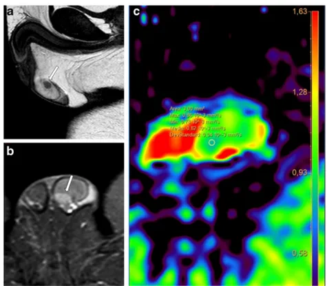
Our experience with 1HMRS in the evaluation of infertility proved that alterations in choline, creatine, lipids and lactate concentrations in testicular tissue correlate to the level of sper-matogenesis (Fig.9). These values could represent a sign of testicular dysfunction in patients with no evidence of morpho-logical abnormalities [39]. Therefore, 1HMRS may be useful in men presenting with varicocele or other known risk factors for infertility, with no evidence of morphological testicular alterations, to indicate the most appropriate management. This diagnostic approach could be particularly suitable for paediatric patients affected with varicocele, for monitoring testicular function after treatment, or in those cases in which spermiogram cannot be obtained for psychological or reli-gious reasons.
Congenital abnormalities and scrotal hernia
Accurate diagnosis and appropriate treatment of cryptor-chidism lead to the highest chance of proper testicular function in the reproductive capacity terms, and facilitate early detection of malignant degeneration. MRI plays a role in cryptorchid testis investigation: the overall accuracy rate in testes detection is superior for MRI than US, with the first performing much better at locating intra-abdominal testicles [40], also considering its larger field of view. Moreover, undescended testis alterations in echostructure are frequently noted, and MRI allows confi-dently ruling out superimposed malignancies.
In newborns and paediatric patients with urological or anorectal abnormalities, MRI provides valuable information for preoperative planning, accurately defining even complex anatomic relationships [44].
Finally, MRI is not routinely performed in investigat-ing investigat-inguinal-scrotal herniae, but is capable of con-firming the diagnosis after clinical and US examination and to reliably depict intestinal loops within the hernia sac [7].
Conclusions
MRI provides excellent characterization of scrotal focal le-sions and valuable depiction of local extent of pathological processes, emerging as the dominant problem-solving imag-ing modality after a first inconclusive sonographic exam (Table1).
CT finds a role in traumatic injuries assessment and in cancer staging together with PET/CT.
Advanced MR techniques, most of all DWI and 1HMRS, are playing a more significant role in scrotal diseases evalua-tion: they could improve tissue characterization and even pro-vide functional information, for instance allowing early non-invasive identification of patients with spermatogenesis dysfunction.
Table 1 Summary of the principal indications for scrotal MRI, as derived from literature analysis
• Correct definition of scrotal masses localisation (testicular vs. extratesticular compartment)
• Characterization of testicular focal lesions after inconclusive findings at ultrasonography
• Local staging of testicular cancer
• Investigation of cryptorchidism (especially for intraabdominal undescended testes)
Open AccessThis article is distributed under the terms of the Creative C o m m o n s A t t r i b u t i o n 4 . 0 I n t e r n a t i o n a l L i c e n s e ( h t t p : / / creativecommons.org/licenses/by/4.0/), which permits unrestricted use, distribution, and reproduction in any medium, provided you give appro-priate credit to the original author(s) and the source, provide a link to the Creative Commons license, and indicate if changes were made.
References
1. Kühn AL, Scortegagna E, Nowitzki KM, Kim YH (2016) Ultrasonography of the scrotum in adults. Ultrasonography 35: 180–197
2. Crawford P, Crop JA (2014) Evaluation of scrotal masses. Am Fam Physician 89:723–727
3. Dieckmann K-P, Frey U, Lock G (2013) Contemporary diagnostic work-up of testicular germ cell tumours. Nat Rev Urol 10:703–712 4. Studniarek M, Skrobisz-Balandowska K, Modzelewska E (2015)
Scrotal imaging. J Ultrasound 15:245–258
5. Remer EM, Casalino DD, Arellano RS et al (2012) ACR appropri-ateness criteria ® acute onset of scrotal pain–without trauma, with-out antecedent mass. Ultrasound Q 28:47–51
6. Parker RA, Menias CO, Quazi R et al (2015) MR imaging of the penis and scrotum. Radiographics 35:1033–1050
7. Parenti GC, Feletti F, Brandini F et al (2009) Imaging of the scro-tum: role of MRI. Radiol Med 114:414–424
8. Muglia V, Tucci S, Elias J et al (2002) Magnetic resonance imaging of scrotal diseases: when it makes the difference. Urology 59:419– 423
9. Baleato-González S, García-Figueiras R, Santiago-Pérez MI et al (2015) Usefulness of 1H magnetic resonance spectroscopy in hu-man testes: preliminary study. Clin Radiol 70:1026–1031 10. Yamaguchi M, Mitsumori F, Watanabe H et al (2006) In vivo
lo-calized 1H MR spectroscopy of rat testes: stimulated echo acquisi-tion mode (STEAM) combined with short TI inversion recovery (STIR) improves the detection of metabolite signals. Magn Reson Med 55:749–754
11. Aaronson DS, Iman R, Walsh TJ et al (2010) A novel application of 1H magnetic resonance spectroscopy: non-invasive identification of spermatogenesis in men with non-obstructive azoospermia. Hum Reprod 25:847–852
12. Firat Uǧraş M, Karakaş HM, Erdem G, Kurus M, Ugras M, Celik T, Kahraman B, Doǧanay SAK (2008) 1H magnetic resonance spec-troscopy of the normal testis: preliminary findings. Magn Reson Imaging 26:215–220
13. Tsili AC, Giannakis D, Sylakos A et al (2014) Apparent diffusion coefficient values of normal testis and variations with age. Asian J Androl 16:493
14. Tsili AC, Ntorkou A, Baltogiannis D et al (2016) Magnetization transfer imaging of normal and abnormal testis: preliminary results. Eur Radiol 26:613–621
15. Tsili AC, Argyropoulou MI, Giannakis D et al (2012) Diffusion-weighted MR imaging of normal and abnormal scrotum: prelimi-nary results. Asian J Androl 14:649–654
16. Howick J, Chalmers I, Glasziou P, et al (2011) The Oxford 2011 levels of evidence. Oxford Centre Evidence-Based Medicine 17. Kim SH, Park S, Choi SH et al (2009) The efficacy of magnetic
resonance imaging for the diagnosis of testicular rupture: a prospec-tive preliminary study. J Trauma 66:239–242
18. Antonini G, Vicini P, Sansalone S et al (2014) Penile fracture: Penoscrotal approach with degloving of penis after magnetic reso-nance imaging (MRI). Arch Ital Urol Androl 86:39–40
19. Schicho A, Riepl C (2015) Femoral-head dislocation to the scro-tum. N Engl J Med 372:863–863
20. Boudissa M, Ruatti S, Maisse N et al (2013) Bilateral testicular dislocation with pelvic ring fracture: a case report and literature review. Orthop Traumatol Surg Res 99:485–487
21. Rushambuza RPM (2013) The role of diagnostic CT imaging in the acute assessment of battlefield external genital injuries. J R Army Med Corps 159:i21–i25
22. Imamura T, Horiuchi E (2016) A case of granulomatous Orchitis. Hinyokika Kiyo 62:45–47
23. Yoneda A, Fujita F, Tokai H et al (2010) MRI can determine the adequate area for debridement in the case of Fournier’s gangrene. Int Surg 95:76–79
24. Mohrs OK, Thoms H, Egner T et al (2012) MRI of patients with suspected scrotal or testicular lesions: diagnostic value in daily practice. Am J Roentgenol 199:609–615
25. Bulakci M, Tefik T, Kartal MG et al (2016) Imaging appearances of Paratesticular fibrous Pseudotumor. Polish. J Radiol 81:10–14 26. Kim KH, Sung DJ, Han NY et al (2015) Immunoglobulin
G4-related paratesticular fibrous pseudotumor and retroperitoneal fi-brosis: a case report. Urol Int 94:369–372
27. Tsili AC, Argyropoulou MI, Giannakis D et al (2010) MRI in the characterization and local staging of testicular neoplasms. Am J Roentgenol 194:682–689
28. Manganaro L, Vinci V, Pozza C et al (2015) A prospective study on contrast-enhanced magnetic resonance imaging of testicular le-sions: distinctive features of Leydig cell tumours. Eur Radiol 25: 3586–3595
29. Algebally AM, Tantawy HI, Yousef RRH et al (2015) Advantage of adding diffusion weighted imaging to routine MRI Examinations in the Diagnostics of scrotal lesions. Pol J Radiol 80:442–449 30. Tsili AC, Sylakos A, Ntorkou A et al (2015) Apparent diffusion
coefficient values and dynamic contrast enhancement patterns in differentiating seminomas from nonseminomatous testicular neo-plasms. Eur J Radiol 84:1219–1226
31. Tsili AC, Ntorkou A, Baltogiannis D et al (2015) The role of ap-parent diffusion coefficient values in detecting testicular intraepithelial neoplasia: preliminary results. Eur J Radiol 84:828– 833
32. Muller J, Schrader AJ, Jentzmik F, Schrader M (2011) Assessment of residual tumours after systemic treatment of metastatic seminoma: (1)(8)F-2-fluoro-2-deoxy-D-glucose positron emission tomography - meta-analysis of diagnostic value. Urologe A 50: 322–327
33. Parenti GC, Sartoni M, Gaddoni E et al (2012) Imaging of segmen-tal testicular infarction: our experience and literature review. Radiol Med 117:1161–1175
34. Watanabe Y, Nagayama M, Okumura A et al (2007) MR imaging of testicular torsion: features of testicular hemorrhagic necrosis and clinical outcomes. J Magn Reson Imaging 26:100–108
35. Gulleroglu K (2014) Nutcracker syndrome. World J Nephrol 3:277 36. Nagappan P, Keene D, Ferrara F et al (2015) Antegrade venography identifies parallel venous duplications in the majority of adolescents with varicocele. J Urol 193:286–290
37. Karakas E, Karakas O, Cullu N et al (2014) Diffusion-weighted MRI of the testes in patients with varicocele: a preliminary study. Am J Roentgenol 202:324–328
38. Gulum M, Cece H, Yeni E et al (2012) Diffusion-weighted MRI of the testis in hydrocele: a pilot study. Urol Int 89:191–195 39. Parenti GC, Albarello F, Campioni P (2016) Role of MR
spectros-copy (H1-MRS) of the testis in men with semen analysis altered. Reprod Syst Sex Disord 5: doi:https://doi.org/10.4172/2161-038X. 1000182
40. Krishnaswami S, Fonnesbeck C, Penson D, McPheeters ML (2013) Magnetic resonance imaging for locating nonpalpable undescended testicles: a meta-analysis. Pediatrics 131:e1908–e1916
41. Kantarci M, Doganay S, Yalcin A, et al (2010) Diagnostic perfor-mance of diffusion-weighted mri in the detection of nonpalpable
undescended testes: comparison with conventional MRI and surgi-cal findings. Am J Roentgenol 195: doi:https://doi.org/10.2214/ AJR.10.4221
42. Yamada K, Takahata A, Ichijo Y et al (2014) A case of testicular seminoma in persistent Mullerian duct syndrome with transverse testicular ectopia. Abdom Imaging 40:475–479
43. Yildiz A, Yiǧiter M, Oral A, Bakan V (2014) Transverse testicular ectopia. Pediatr Int 56:102–105
44. Ziegelmann MJ, Viers BR, Cockerill PA et al (2017) The utility of magnetic resonance imaging in the evaluation of accessory scrotum in the newborn. Urology 99:228–230
45. Wittenberg AF, Tobias T, Rzeszotarski M, Minotti AJ (2006) Sonography of the acute scrotum: the four T’s of testicular imaging. Curr Probl Diagn Radiol 35:12–21
46. Karam JA, Baker LA (2007) Focal orchitis presenting as bilateral testicular masses. J Pediatr Urol 3:337–339
47. Cassidy FH, Ishioka KM, McMahon CJ et al (2010) MR imaging of scrotal tumors and pseudotumors. Radiographics 30:665–683 48. Tsili AC, Giannakis D, Sylakos A et al (2014) MR imaging of
scrotum. Magn Reson Imaging Clin N Am 22:217–238
49. Wolfman DJ, Marko J, Gould CF et al (2015) Mesenchymal Extratesticular tumors and Tumorlike conditions: from the radio-logic pathology archives. Radiographics 35:1943–1954
50. Ap Dafydd D, Messiou C, Thway K et al (2017) Paratesticular sarcoma: typical presentation, imaging features, and clinical chal-lenges. Urology 100:163–168
51. Woodward PJ, Schwab CM, Sesterhenn IA (2003) From the ar-chives of the AFIP: extratesticular scrotal masses: radiologic-pathologic correlation. Radiographics 23:215–240
52. Shah T, Abu-Sanad O, Marsh H (2016) Role of magnetic resonance imaging in the early diagnosis of paratesticular rhabdomyosarcoma. Ann R Coll Surg Engl 98:e74–e76
53. Ntorkou AA, Tsili AC, Giannakis D et al (2016) Magnetic reso-nance imaging findings of cellular angiofibroma of the tunica vaginalis of the testis: a case report. J Med Case Rep 10:71 54. Richenberg J, Belfield J, Ramchandani P et al (2014) Testicular
microlithiasis imaging and follow-up: guidelines of the ESUR scro-tal imaging subcommittee. Eur Radiol 25:323–330
55. Bilen MA, Hariri H, Leon C, et al (2014) Positive FDG-PET/CT scans of a residual seminoma after chemotherapy and radiotherapy: Case report and review of the literature. Clin Genitourin Cancer 12: e147–e150
56. Becherer A (2011) PET in testicular cancer. Humana Press, pp 225– 241
57. Sidhu P, Lin P, Son H et al (2014) Testicular fluorine-18 fludeoxyglucose uptake on positron emission tomography CT in patients with lymphoma: clinical significance and management im-pact. Br J Radiol 87:20140472
58. Ozguven S, Aras M, Dede F et al (2014) Scrotal peritoneal meso-thelioma on PET/CT. Clin Nucl Med 39:1045–1046
59. Bowlin PR, Gatti JM, Murphy JP (2017) Pediatric testicular torsion. Surg Clin North Am 97:161–172
60. Maki D, Watanabe Y, Nagayama M et al (2011) Diffusion-weighted magnetic resonance imaging in the detection of testicular torsion: feasibility study. J Magn Reson imaging JMRI 34:1137–1142 61. Belay R, Huang G, Shen J-C, Ko EK (2016) Diagnosis of clinical
and subclinical varicocele: how has it evolved? Asian J Androl 18: 182–185
62. Nana GR, Basra M, Maudgil DD, Rao AR (2014) Left renal vein thrombosis: a rare cause of acute scrotal pain. BMJ Case Rep 2014: bcr2013202237-bcr2013202237. doi: https://doi.org/10.1136/bcr-2013-202237
63. Kulis T, Knezevic M, Karlovic K et al (2013) Infrared digital ther-mography of scrotum in early selection of progressive varicocele. Med Hypotheses 81:544–546
64. Dierickx LO, Huyghe E, Nogueira D et al (2012) Functional testic-ular evaluation using PET/CT with 18F-fluorodeoxyglucose. Eur J Nucl Med Mol Imaging 39:129–137
Publisher’s Note
Springer Nature remains neutral with regard to jurisdictional claims in published maps and institutional affiliations.