UNIVERSIT `A DEGLI STUDI DI CATANIA DOTTORATO DI RICERCA IN FISICA
Stefania Tropea
EXPERIMENTAL STUDY ON
CARBON FRAGMENTATION FOR HADRONTHERAPY
Ph.D. Thesis Tutor: Prof. F. Cappuzzello Co-Tutors: Dr. C. Agodi Dr. M. De Napoli Ph.D. Coordinator: Prof. F. Riggi XXVI CICLO 2010/2013
ii
“Nothing in life is to be feared, it is only to be understood. Now is the time to understand more, so that we may fear less.” Marie Curie
Contents
Introduction v
1 Hadrontherapy 1
1.1 Historical background and evolution of hadrontherapy . . . . 3
1.2 Physical aspects of radiotherapy with ion beams . . . 10
1.2.1 Dose and therapeutic ratio . . . 10
1.2.2 Inverse depth-dose profile: stopping of ions in matter . 13 1.2.3 Range straggling and lateral scattering . . . 18
1.3 The problem of nuclear fragmentation . . . 22
1.4 Radiobiological rationale of ion beam therapy . . . 29
1.4.1 Radiation damage by photons and heavy ions . . . 29
1.4.2 Relative Biological Effectiveness (RBE) . . . 33
1.5 Carbon ion therapy highlights . . . 40
1.6 Treatment planning for ion beam therapy: the INFN TPS project . . . 41
1.7 Hadrontherapy in the world . . . 45
2 The Experiments 49 2.1 Fragmentation at Fermi energies on a thin target . . . 49
2.1.1 Experimental apparatus . . . 50
2.1.2 Trigger of the experiment . . . 55
2.2 Fragmentation at Fermi energies on thick targets . . . 56
2.3 The experiment at relativistic energies . . . 59
2.3.1 The Interaction Region . . . 61
2.3.2 The TP-MUSIC IV time projection chamber . . . 67
2.3.3 The ToF-Wall detector . . . 68
2.3.4 The LAND detector . . . 73
2.3.5 The Veto Counter . . . 75
iv CONTENTS
3 Carbon fragmentation at Fermi energies 77
3.1 Thin target measurements . . . 77
3.1.1 Calibration procedures . . . 78
3.1.2 Cross section angular distributions . . . 82
3.1.3 Double-differential cross sections: comparisons with GEANT4 models . . . 84
3.2 Thick targets measurements . . . 101
3.2.1 Detector calibration . . . 102
3.2.2 Fragments energy spectra . . . 104
3.2.3 GEANT4 BIC model validation . . . 107
3.2.4 Fragments build-up curves . . . 118
4 Carbon fragmentation at relativistic energies 121 4.1 The global reconstruction algorithm . . . 121
4.2 Mass distributions . . . 124
4.3 Preliminary cross-section angular distributions . . . 126
4.4 Preliminary double-differential cross sections . . . 130
4.5 The Monte Carlo simulation . . . 132
4.6 Cross sections comparisons between data and FLUKA MC code134 Conclusions and outlook 145 Appendix A GEANT4 ion interaction models for hadrontherapy applica-tions 149 4.7 The Binary Light Ion Reaction model . . . 150
4.8 The Quantum Molecular Dynamics model . . . 153
4.9 Pre-equilibrium decay and de-excitation models . . . 154
Appendix B Nucleus-nucleus models in the FLUKA Monte Carlo code 157 4.10 The rQMD model in FLUKA . . . 158
4.11 The BME model in FLUKA . . . 159
Acknowledgements 161
Introduction
Nowadays, one of the most advanced methods for solid tumors treatment is represented by hadrontherapy, a radiotherapy technique which applies col-limated beams of protons or heavier ions for the sterilization of tumor cells. Particularly, hadrontherapy is a high precision technique of external radio-therapy which yields a better perspective for defeating radioresistant tumors and it has given a boost to the use of radiation in the fight against cancer.
Due to their very favourable profile of the released dose in tissue, the charged hadron beams can be very effective in destroying the tumor and sparing the adjacent healthy tissue in comparison to the standard X-ray based treatments. On the other hand, the use of accelerated particles re-quires appropriate methods for accurately evaluate the dose distribution inside and outside the planned target volume during the irradiation treat-ment.
The most important difference between protons and heavier ions is the increased biological effectiveness of the latter, i.e. a lower physical dose is needed with ions to obtain a given biological effect. For carbon ions, which are considered the optimal choice, the effect of the favourable absorbed dose distribution, which is highly localized, is enhanced by the large relative bi-ological effectiveness (RBE) towards the end of the particle range, offering an additional advantage for slow growing radioresistant tumors.
Nevertheless, the carbon ions based therapies are still not completely under control as great uncertainties affect the dose distributions inside the patient due to the nuclear interactions between carbon ions and the tra-versed human body. Indeed, when the carbon beam penetrates matter, the primary ions can be fragmented as a result of the collisions with the tissue atomic nuclei. The collisions along the carbon path lead to the attenuation of the primary beam intensity and the production of secondary fragments.
Neutrons and ions lighter than carbon are produced as hydrogen, helium, lithium, beryllium and boron isotopes. These lighter fragments have longer
vi
ranges and wider energy distributions with respect to the primary particles and give rise to a characteristic dose tail behind the Bragg peak. As far as the biological effect of ion radiation is dependent on the particle field composition, a detailed knowledge of the fragmentation process is essential in order to guarantee the appropriate treatment accuracy.
Currently, the Monte Carlo codes are the most powerful tools able to precisely compute the biological dose to be delivered within a modern treat-ment planning system. However, the accuracy of a Monte Carlo simulation is associated to the reliability of the physical processes implemented in the code. In particular, for a realistic estimation of fragmentation products, nucleus-nucleus models inside the code have to be validated versus experi-mental data, which are still a small amount in the literature.
As a consequence, the main goal of the present work consist in estimating the double-differential fragmentation cross sections with respect to energy and angle in the energy range of interest for hadrontherapy.
After an overview of the historical development of radiotherapy and, more specifically, of hadrontherapy, the physical and biological rationale of carbon ions application in tumor treatments are dealt with in Chapter 1, focusing particularly on the fragmentation issue. In order to extract fragmentation cross sections in different experimental conditions, two mea-surements were performed at intermediate energies on both a thin carbon target and different tissue-equivalent targets, and a third one were done in the relativistic energy domain on a thicker carbon target. In Chapter 2 the experimental devices used in order to perform the three experiments have been described.
The first two experiments were carried out at the Laboratori Nazionali del Sud (LNS-INFN) in Catania with a beam of carbon ions at 62 AMeV. The cross sections angular and energy distributions were obtained for the thin carbon target analysis and also a comparison with those extracted by means of the GEANT4 Monte Carlo code were performed. The results are shown and discussed in Chapter 3 together with those associated to the sec-ond experiment done at the same energy but on thick tissue-equivalent tar-gets. In Chapter 4 the preliminary results of the measurement performed at the GSI laboratory (Darmstadt, Germany) with a 400 AMeV carbon beam and the comparison with those obtained with the FLUKA Monte Carlo code are presented and discussed. In the end, the conclusions of the whole work done are drawn and the future perspectives are outlined.
Chapter 1
Hadrontherapy
Cancer is a group of diseases characterized by uncontrolled growth and spread of abnormal cells. If the spread is not restrained, it can result in death. Cancer is caused by both external factors (tobacco, chemicals, radi-ation, and infectious organisms) and internal factors (inherited mutations, hormones, immune conditions, and mutations that occur from metabolism). These causal factors may act together or in sequence to initiate or promote carcinogenesis.
Malignant neoplasms are the main cause of death among persons aged 45 to 64, for both men and women. Overall, an estimated 12.7 million new cancer cases and 7.6 million cancer deaths occurred in 2008 worldwide, with 56% of the new cases and 63% of the deaths occurred in the less developed regions of the world. As shown in Figure 1.0.1, the most commonly diag-nosed cancers worldwide are lung (12.7%), breast (10.9%) and colorectal cancers (9.7%). The most common causes of cancer death are lung (18.2%), stomach (9.7 %) and liver cancers (9.2%).
Faced with increasing numbers of cancer cases, a major public health issue, researchers and practitioners are mobilising the world over. Indeed, this pathology is rising sharply in Europe: between 2000 and 2020 the number of new cases is expected to rise by 50% [1].
The goal of any cancer therapy is to destroy the malignant cells in the body while doing minimal damage to the healthy tissue. Modern cancer ther-apies, no matter if they are chemotherapy, targeted medications, surgery, X-ray therapy, or particle beam therapy, are all about collateral damage: destroy the cancer but safeguarding the patient life [2]. However, radiation therapy can be considered the most important, effective and cost effective treatment modality for all types of solid malignancies.
2
Figure 1.0.1: Incidence and mortality data for all ages and both sexes [2].
It has been estimated that about 45% of all cancer patients can be cured (excluding those suffering from non-melanoma skin cancers). Ra-diation therapy contributes to the cure of approximately 23% of all cancer patients, when used alone (12%) or in combination with surgery (6%) or chemotherapy-immunotherapy (5%). Thus, about half of the cancer patients who are cured benefit from radiation therapy. This proportion illustrates the important role of radiation therapy in cancer management [3]. Up to 2008, in industrialized countries, about 70% of cancer patients have been re-ferred to a radiation therapy department for at least part of the treatment. The majority has been treated with ”conventional” photon beam therapy, which for that reason remains the reference radiation treatment modality.
The impressive development and progress in conformal therapy with pho-tons and, more recently, with propho-tons, raises a difficult issue: the extent to which photon or proton beam therapy has reached a plateau in development (at least as far as physical selectivity is concerned). A search for improve-ment is actually directed to alternative radiation modalities, such as ion beam therapy.
1.1 Historical background and evolution of hadrontherapy 3
1.1
Historical background and evolution of
hadron-therapy
The era of radiation treatment began in the closing years of the nine-teenth century, chiefly with the X-rays discovery, done by German physicist Wilhelm Conrad R¨ontgen on November 8, 1895 [4], and reported to the world shortly after the first of the year 1896 [5]. R¨ontgen’s discovery was a scientific bombshell, and was received with extraordinary interest by both scientists and laymen. The discovery of a new form of energy that could penetrate solid objects and record their structure excited R¨ontgen’s scien-tific contemporaries who recognized instantly that this finding could change medical practice forever. A century later, the vastly more sophisticated arts of medical imaging are still based upon the recognition that body parts ab-sorb a beam of X-rays according to their density, producing an image which allows identification of body structures, as well as the recognition of abnor-malities, reflective of injury and disease conditions.
By the first few months of 1896, X-rays were being used to treat skin lesions prior to any understanding of the beams physical or biological char-acteristics. The driving force was, of course, patients overwhelming need of treatment for uncontrollable and debilitating diseases. Particularly, Leopold Freund, a dermatologist and professor of radiology at the Medical Univer-sity of Vienna, was the first physician to use ionizing radiation for therapeu-tic purposes. In 1896, a year after discovery of X-rays and the same year that Antoine Henri Becquerel discovered radioactivity, Freund successfully treated a five-year-old patient in Vienna suffering from hairy moles covering her whole back. The case was published in 1901 and in 1903 Freund pub-lished the first textbook on radiation therapy [6].
Shortly thereafter, in 1898, Marie and Pierre Curie discovery of radium and polonium stimulated speculation that radioactivity also could be used to treat disease. Indeed, reports of the use of radium (curietherapy1) oc-curred throughout the first decade of the twentieth century.
In retrospect, it is clear that lack of knowledge of the biological effects and mechanisms of actions of the new rays led to much morbidity and poor
1Curietherapy (nowadays referred to as brachytherapy) is a term used to describe the
short distance treatment of cancer with radiation from small, encapsulated radionuclide sources. This type of treatment is given by placing sources directly into or near the volume to be treated. The dose is then delivered continuously, either over a short period of time or over the lifetime of the source to a complete decay. Most common brachytherapy sources emit photons. However, in a few specialized situations, β or neutron emitting sources are used.
4 1.1 Historical background and evolution of hadrontherapy
cancer control. Particularly, it took until 1904, when Edison’s assistant Clarence Dally died following injuries to his hands and arms, that physicians and physicists took the possibly fatal power of the X-rays into account. Dur-ing this discovery era, which lasted until the early 1920’s, radiation therapy remained a more or less empirical science, as far as the major progress orig-inated from clinical application. However, two general tendencies started to be visible: the clinical results were improved by a greater conformity of the applied radiation to the target volume and by an increased biological effectiveness of the radiation.
The next major era in radiation therapy began during the 1920’s, pri-marily as the result of two major contributions. The first was the invention by W.D.Coolidge [7] of a sealed-off vacuum X-ray tube, equipped with an hot tungsten cathode, which could be operated at the unprecedented en-ergies of 180.000 to 200.000 volts, thus introducing the kilovoltage era in radiotherapy. Unfortunately, X-rays generated by these tubes were fairly soft and, from the medical point of view, the depth-dose curves were par-ticularly disadvantageous since the maximum dose would be delivered at the skin surface and then would rapidly fall off with the depth in the tissue. Secondly, that era witnessed an important advance for all uses of X-rays and γ-rays, the adoption of the r¨ontgen R as the internationally accepted unit of radiation exposure by the International Congress of Radiology in 1928 [8], later succeeded by the absorbed dose unit, called rad2.
Although the kilovoltage era was one of great achievement, radiothera-pists were severely prevented by the physical limitations of dose distribution since, with energies between 50 kV and 200 kV, it was very difficult to deliver sufficient doses into deep-seated tumours, primarily because of the associ-ated unavoidable skin toxicity. It was obvious that beams of higher energy were needed, and by the early 1950’s, several groups of physicists had be-gun to come up with new ideas for devices of much higher energy such as the cobalt teletherapy machines and the megavoltage linear electron accel-erators, giving rise to the the megavoltage era. Following the discovery of artificial radioactivity in 1934 done by Ir`ene and Fr´ed´eric Joliot-Curie,60Co was adopted as an alternative source of high-energy γ-rays for
teleradiother-2More precisely, exposure is given by X=dQ/dm, where dQ is the absolute value of the
total charge of the ions of one sign produced in air when all the electrons and positrons, liberated or created by photons in mass dm of air, are completely stopped in air. The unit used for exposure is the r¨ontgen R, where 1 R = 2.58 × 10⁻⁴C/kg. In the SI system of units, r¨ontgen is no longer used and the unit of exposure is simply 2.58 × 10⁻⁴ C/kg of air [9]. The old unit of the absorbed dose rad (see Section 1.2.1) is defined so that 1 rad = 100 erg/g.
1.1 Historical background and evolution of hadrontherapy 5
apy, with a higher dose rate than could be achieved with radium. Moreover, the 1.33 MeV maximum energy of the emitted gamma made it possible to obtain far better depth-dose curves, showing a maximum at about 5 mm below the skin surface and markedly decreasing the dose to the superficial tissues, as shown in Figure 1.1.1.
Figure 1.1.1: Percentage Depth-Dose (PDD) curves in water for a 10 × 10 cm2field at a Source to Surface Distance (SSD) of 100 cm for various megavoltage photon beams ranging from60Co γ-rays to 25 MV X-rays [9].
On the other hand, the most important developments in radiotherapy arose when a high voltage accelerator was developed in 1932 by R. Van de Graaff [10] and, five years later, the first hospital-based accelerator of this type, a 1 MeV air-insulated machine, was installed in Boston. A further im-provement in treatment delivery techniques came with the development of the betatron in 1943 by D.W. Kerst [11], through which high energy X-rays and electron beam therapy became feasible. As a result, the first patient was irradiated in 1949 with X-rays generated by 20 MeV electrons from a Kerst betatron installed in Urbana (USA). Betatrons were widely circulated but, since the mid 1970’s, their application showed a gradual decline because of some disadvatages such as the relatively low intensity of the X-ray beams produced, the small treatment field area together with the relevant weight, which made these machines unhandy. In the mean time, the advances made during the World War II made it possible to use microwave generators for electron acceleration, leading to the born of the first radiofrequency linear
6 1.1 Historical background and evolution of hadrontherapy
accelerators designed by C.W. Miller [12], which were soon to take up a dominant place on the world market of medical accelerators [13] replacing betatrons.
Nowadays, as sources of radiation for modern radiotherapy with colli-mated beams, the electron linear accelerators (linacs) are still used. Such accelerators are capable of producing both electrons and photons beams with energies varying between 3 and 25 MeV. While electron beams are suitable for the treatment of superficial or semi-deep tumors, the photon beams from a linear accelerator can be applied for a very efficient treatment of tumors situated at a depth of many centimeters inside the body with respect to the skin surface. In order to irradiate selectively such targets, thus achieving a better conformation of dose to the tumor, sophisticated irradiation tech-niques have been developed which involve the use of multiple beam entry ports onto a point, usually conciding with the geometrical center of the tar-get (cross-fire technique). These irradiation techniques are applied by having the structure containing the linac rotate around a horizontal axis (gantry). The most recent Intensity Modulated Radio-Therapy (IMRT) makes use of 6-10 entrance ports. The beams may be non-coplanar and their intensity is varied across the irradiation field by means of variable collimators (multi-leaf collimators) that are computer controlled. Nowadays, in the developed countries, every years about 20000 patients out of 10 million inhabitants, are treated with high-energy photons and about 8000 linacs are used worldwide for cancer treatment.
Despite the remarkable development of conventional radiotherapy, it was found that some tumors, called radioresistant tumors, respond poorly to the photon treatments and sometimes even non-radioresistant tumors, located near critical body parts, can not be given a tumorocidal dose because of unavoidable dose to the surrounding normal tissues. In this context, the tendency which drove the historical development of radiotherapy was the searching of an increased biological effectiveness of radiation. In order to overcome both the physical and the biological limitations of conventional radiotherapy the use of neutrons, protons and heavier charged particles was proposed, which led to the born of the “Hadrontherapy”.
“Hadrontherapy” is a collective word covering all forms of radiation ther-apy which use beams of particles made up of quarks: neutrons, protons, pions, antiprotons, helium (i.e. alpha particles), lithium, boron, carbon and oxygen ions. As in the case of photons, the use of hadrons for medical ap-plications is sensibly influenced by the scientific progress and it is strictly related to the historical development of the accelerators technology [14].
1.1 Historical background and evolution of hadrontherapy 7
Fast neutrons (i.e. neutrons having kinetic energies between a few MeV and a few tens of MeV) were the first hadrons used in radiotherapy soon after the invention of the cyclotron by Ernest Lawrence and Stanley Livingston in 1930 [15]. Soon after, in 1935, John Lawrence, who was a medical doctor in Yale, joined his brother Ernest in Berkeley for appliying the new powerful accelerator for medical purposes [16]. The two main applications were the production of radioisotopes and, later, the therapeutical use of fast neutron beams. Neutrons act via their scattering and recoil ions which are, in bio-logical tissues, mostly low energy protons and, as a consequence, produce a greater biological damage with respect to photons. At the end of 1938, the first patients were treated with neutrons but the technique was primitive and the doses given to healthy tissues too high. Indeed, even if a better tu-mour control was achieved, thanks to the increased biological effectiveness of neutrons, the poor depth dose profile unfortunately compensated this ad-vantage with severe late effects in normal tissues. For this reason, some years later, in 1948, after the effects evaluated on 226 patients, Dr. Robert Stone concluded that neutron therapy had not to be continued [17]. It has to be noted that today neutron therapy is mostly restricted, in some laboratories, for the treatment of radio resistant tumours of the salivary glands, while in most countries this technique has been terminated.
The application of high-energy beams of heavy charged particles to radio-therapy was first considered by Robert R. Wilson, who was one of Lawrence’s students. In 1945 he designed a new 160 MeV cyclotron and, one year later, proposed the use of proton beams in radiation oncology [18]. In fact he had measured depth dose profiles at the Berkeley cyclotron with a signifi-cant increase in dose at the end of particle range, the so called Bragg peak, which had been observed fifty years before in the tracks of alpha particles by William Henry Bragg [19]. As soon as Wilson analized the stopping process of protons in matter, he understood that, due to the Bragg peak, the dose can be concentrated on the tumour target sparing healthy tissues better than what can be done with X-rays and wrote the famous seminal paper [18], which is considered the first work on hadrontherapy (see Figure 1.1.2). Two years later the 184-inch synchrocyclotron at Lawrence Berkeley Laboratory (LBL) became available for experiments and the physical and radiobiological properties of proton beams were thoroughly investigated by Cornelius Tobias [20], thus confirming the predictions made by Wilson. Pa-tient treatments started in 1954 at LBL, first with protons and later, in 1957, with helium beams. Radiotherapy with heavier ions started in 1975 at the Bevalac facility of the LBL mostly appliying beams of 20Ne.
8 1.1 Historical background and evolution of hadrontherapy
In the first trials at Berkeley, the beam was distributed to the target volume adapting methods from conventional photon therapy, in which the photon beam is passively shaped by collimators and absorbers. Thus, the energy modulation of charged particle beams was first performed with mod-ified collimator and absorber techniques [21].
Figure 1.1.2: The original picture from Robert R. Wilson’s paper on protonther-apy [18].
In other words, ions were treated as photons without making use of their most important characteristic, i.e. the electric charge, which makes their beams easy to detect and to control by means of magnetic fields. This was also because of the fact that, in those early times, the computer power available was too poor for a control system of an active beam scanning, as described in the next sections.
It has to be remarked that the first hadrontherapy treatments were per-formed by means of particle accelerators that had originally been built for nuclear physics experiments and were then adapted to tumour therapy. This was the case in Berkeley as well as at Harvard Cyclotron Facility, which made the largest impact on the development of protontherapy and where, up to now, the highest number of patients have been successfully treated. Some years later, other nuclear physics laboratory in USSR and Japan set up proton beams for therapy and in 1984 the Paul Scherrer Institute (Switzer-land) did the same. The clinical proton beam currently used in the facility
1.1 Historical background and evolution of hadrontherapy 9
CATANA3 for eye melanoma treatments at Laboratori Nazionali del Sud (LNS, Catania, Italy) is also an example of beams produced by a supercon-ducting cyclotron (SC) originally designed for nuclear physics experiments and adapted for therapy [22].
As far as all the treatment facilities were located in physics laboratories, the irradiation condition was far from ideal and, although many times it was felt and said that hadrontherapy field could not develop without dedicated equipment, this step took almost 20 years. Indeed, the first hospital-based centre was built at the Loma Linda University Center (California), which signed an agreement with Fermilab (founded and directed for many years by Robert Wilson) and treated the first patient in 1990. Afterwards, a smooth conversion from a physics laboratory to a hospital facility took place in Japan, where from 1983 to 2000 about 700 patients were treated at the Pro-ton Medical Research Center (PMRC, University of Tsukuba). Moreover, USA proton therapy was further expanded during the 1990s. Over 50000 patients have worldwide been treated with proton beams by now and other facilities are under construction or in planning stages [23].
Between 1954 and 1974 at Berkeley, under the leadership of C. Tobias, about 1000 pituitary tumors were treated with protons. A few years later, heavier ions, helium in 1957 and argon in 1975, came into use at the LBL. As a consequence, 2800 patients received treatments to the pituitary glands with helium beams, thus achieving a better dose conformation to the tu-mor with respect to protons, and moving the first step towards the light ion radiosurgery. About 20 years later, argon beams were tried in order to increase the effectiveness against radioresistant tumors, but problems arose owing to non-tolerable side effects in the normal tissue. After a few irradia-tions, Tobias and collaborators used lighter ions, first silicon and then neon ions for 433 overall patients, until Bevalac stopped operation in 1993. Only towards the end of the program it was found that the neon charge (Z=10) is too large and undesiderable effects were produced in the traversed and downstream healthy tissues [24].
Further experimental studies were needed, but only in the early 1990’s carbon ions were recognized as the optimal ion choice. In fact their effects in the tissue entrance are similar to those of X-rays and protons, while just at the end of their path in matter, ionization density is definitely larger and not repairable damages are produced to the cellular systems. This resulting radiation field is a common feature to the light ions in general, but carbon ions currently represent the best compromise for treatments, especially in
3
10 1.2 Physical aspects of radiotherapy with ion beams
case of radioresistant tumors, as shown in the next sections.
In 1994 the Heavy Ion Medical Accelerator (HIMAC) dedicated to radio-therapy, by virtue of the proposal made by Yasuo Hirao [25] and collabora-tors, started with carbon ions at National Institute of Radiological Science (NIRS) in Chiba (Japan) using similar technical concepts as those pioneered at Berkeley. The first patient has been treated with a carbon ion beam of energy up to 400 AMeV, corresponding to a maximum range of 27 cm in water. At the same time, new technical solutions were developed almost in parallel at the Gesellschaft f¨ur Schwerionenforschung (GSI) in Darmstadt, Germany. By the end of 2007 more than 4000 patients have been treated at the HIMAC facility showing that, among light ions, a better tumor control rate can be achieved with carbons.
1.2
Physical aspects of radiotherapy with ion beams
1.2.1 Dose and therapeutic ratio
The fundamental goal in radiation oncology is the local control of the tumour and, in some situations, of surrounding diffusion paths (loco-regional radiotherapy). In order to reach this objective, one must deliver to the tumor, which may be considered in physical terms as the target, a sufficiently high dose of radiation so as to destroy it, at the same time mantaining the dose to the surrounding healty tissues, inevitably involved in the irradiation, within such limits so that they do not undergo serious or even irreversible damage or complications.
The dose deposited in tissue is the most important physical quantity in radiotherapy. The absorbed dose [26] is defined as the mean energy d¯ϵ imparted by ionizing radiation to a mass element dm in a finite volume by,
D = d¯ϵ
dm. (1.2.1)
The absorbed dose is measured in gray (Gy), being 1 Gy = 1 J/kg. For example, in conventional radiotherapy with photons and electrons, doses of the order of 60-70 Gy are deposited in the tumour tissues in amounts of about 2 Gy per session over about 30 days.
In the hypothesis of a fairly accurate identification of the target, it is pos-sible to evaluate the probability of obtaining the local control of the tumour through the analysis of the so-called dose-effect curves. They represent for tumour tissues the possibility of obtaining the desired effect as a function of the dose delivered, and for healthy tissues the probability of producing
1.2 Physical aspects of radiotherapy with ion beams 11
serious or irreversible damage, always as a function of the absorbed dose by the same tissue. In Figure 1.2.1 hypothetical dose-effect curves are shown, as function of the absorbed dose, for a generic tumour tissue (A) and for the healthy tissue involved in the irradiation (B). As can be seen, the ab-sorbed dose necessary to achieve a probability close to 100% of obtaining local control of the tumour corresponds also to a very high probability of producing serious complications in the healthy tissue, when this receives the same dose. The two sigmoid curves plotted in Figure 1.2.1 are usually re-ferred to as Tumor Control Probability (TCP, curve A) and Normal Tissue Complication Probability (NTCP, curve B) [9].
Figure 1.2.1: Dose-effect curves for neoplastic (A) and normal (B) tissues.
In the daily practice, the radiotherapist has to find a compromise be-tween the local control of the tumour and the possible emergence of compli-cations: the possibility to find such a compromise can be expressed quanti-tatively by the therapeutic ratio, i.e. the ratio D2/D1 [9] between the dose corresponding to a 50% probability of producing complications D2 and the dose corresponding to a 50% probability of obtaining the local control of the tumour D1 [27]. On the basis of these considerations, it is clear that the probability of curing the tumour without unwanted side effects increases in line with the ballistic selectivity or conformity of the irradiation delivered.
The optimum choice of radiation dose delivery technique in the treatment of a given tumour is such that it maximizes the TCP and simultaneously
12 1.2 Physical aspects of radiotherapy with ion beams
minimizes the NTCP. For a typical good radiotherapy treatment, TCP ≥ 0.5 and NTCP ≤ 0.05. The case shown in Figure 1.2.1 refers to an ideal situation; in reality, the therapeutic ratio varies with many factors, such as the dose rate and Linear Energy Transfer (LET, see Section 1.4) of the irra-diation, the presence of radiosensitizers or radioprotectors, the design of the treatment plan and the precision of implementation of the treatment plan.
Particularly, the probability of curing tumours can be increased by us-ing charged hadrons beams because the absorbed dose is more confined in the tumour tissue with respect to electrons and photons application, thus allowing an enhanced ballistic precision. Moreover hadrons show increased biological effects respect to electromagnetic radiation and also respect to protons. This last feature makes them more successful also in the treatment of radioresistant tumours. Concerning carbon ions treatment, an example of the resulting tumor control probability curve with respect to photons is shown in Figure 1.2.2 and the related study confirmed the effectiveness of carbon ion therapy for severely radioresistant tumors [28].
Figure 1.2.2: Dose-response curves for (a) a single dose fraction and (b) two dose fractions of photons and carbon ions [28].
1.2 Physical aspects of radiotherapy with ion beams 13
Moreover carbons show increased biological effects also with respect to protons (see section 1.4). Thus the therapeutic advantages of carbon beams when compared to electron, photon and also proton beams can be found at a macroscopic scale (high level conformation) as well as at the microscopic scale (possibility of varying the radiobiological properties). The latter deals with microscopic distribution of the deposited energy, which changes when different ion beams are considered. For this reason it is often said that hadrons, and specifically carbons, as densely ionizing radiation, in contrast to the sparsely ionizing radiation such as X-rays, γ-rays and electrons.
1.2.2 Inverse depth-dose profile: stopping of ions in matter
The main reason for using charged particle beams in radiotherapy is their inversed dose profile, i.e. the increase of energy deposition with pene-tration depth, which makes them a more advantageous choice with respect to electromagnetic radiation. As already mentioned, the increase of ion-ization density along the ions path in matter was firstly described, for α particles slowing down in air, by W.H. Bragg in 1905 [19]. As a result, ions depth-dose profiles are still known as Bragg curves.
Figure 1.2.3: Depth-dose profiles of 60Co radiation, megavolt photons, and 12C ions in water [29].
14 1.2 Physical aspects of radiotherapy with ion beams
Many years later R. Wilson [18] proposed the application of protons and heavier ions for precision exposures in radiotherapy. A comparison of depth-dose profiles for electromagnetic radiation (60Co and megavolt photon beams) and carbon ions beams is displayed in Fig. 1.2.3 [29].
The peculiar physical processes which characterize respectively electro-magnetic radiation and charged particles interaction with matter directly affect their different depth dose profiles. Concerning photons interaction, three are the possible processes: the photoelectric effect, the Compton scat-tering and the electron-positron pair production. The relative probability of each of these interaction mechanisms is a function of the incident photon energy and the atomic number Z of the absorbing material. On the other hand, charged particles dissipate their energy mainly via interaction with the electrons of the target material, which are subsequently emitted as δ-electrons, i.e. the maximum energy electrons able to ionize other medium atoms. More than 75% of the initial energy is lost in the ionization process and only 10 to 20% in the target excitation [30]. Particularly, the inter-action strength is directly correlated with the interinter-action time so that at high velocities the energy transfer to the target is small but grows when the particles are slowed down.
As can be seen in Figure 1.2.3, for low-energy photons the stochastic absorption by photoelectric and Compton processes yields an exponential decay of absorbed dose with penetration depth and the beam doesn’t show a path of finite lenght. For higher photon energies the produced Comp-ton electrons are strongly forwardly scattered and transport some of the transferred energy from the surface to deeper layers, yielding an increase in dose in the first few centimeters. For high energy bremsstrahlung radia-tion4, which is mostly used in conventional therapy, this maximum is shifted a few centimeters from the surface of the patient body, thereby improving the target-to-entrance dose and sparing the very radiosensitive skin.
In contrast, the energy deposition of charged particles, like protons or heavier ions, shows a completely different trend. When ions enter an ab-sorbing material, they are slowed down. The rate of average energy loss per unit path lenght for a given target medium increases with decreasing particle velocity, giving rise to a sharp maximum in ionization near the end of the range. Thus the depth-dose distribution is characterized by a relatively low
4Like charged particles, electrons also suffer a collisional energy loss when passing
through matter. However, because of their small mass, an additional energy loss mecha-nism comes into play: the emission of electromagnetic radiation arising from scattering in the electric field of a nucleus, a physical process known as bremsstrahlung, a german term meaning braking radiation.
1.2 Physical aspects of radiotherapy with ion beams 15
dose in the entrance region (plateau) near the skin and a sharply elevated dose at the end of the range, which in this case is finite and energy dependent (Bragg peak ), as the position of the peak can be precisely adjusted to the desired depth in tissue by changing the kinetic energy of the incident ions. The ballistic precision of charged particles with respect to electromagnetic radiation is evident by looking at Figure 1.2.3. As a result, in the surround-ing healthy tissues before or just behind the Bragg peak, the dose released is minimized respect to the target volume and a better compromise is achieved.
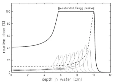
Figure 1.2.4: Construction of an extended Bragg peak by superposition of single Bragg peaks of different energy. [31].
It is important to note that, from the point of view of practical appli-cations in radiotherapy, a monoenergetic beam with a narrow Bragg peak makes possible to irradiate a very small and localized region within the body, with an entrance dose lower than that in the peak region [32]. Indeed, the tumour volume to be treated is normally much larger than the width of the Bragg peak and the lateral spot of the particle beam. In order to fill the target volume with the necessary amount of stopping particles, the peak has to be “spread out” in the longitudinal direction. This is achieved by superimposing several Bragg peaks at different depths obtained by suitably selecting the projectiles energy distributions. The resulting depth-dose pro-file, known as Spread-Out Bragg Peak (SOBP), shows an “extended” Bragg peak area which has to accurately overlap the target volume (see Figure
16 1.2 Physical aspects of radiotherapy with ion beams
1.2.4.).
Even if the peak-plateau ratio decreases for SOBP with respect to a pris-tine Bragg peak, the final dose distribution allows tumor conform treatments of enhanced quality with respect to those obtained by applying photons [31]. Radiotherapy of deep-seated tumors requires, tipically, ion beam ranges in tissue of up to 30 cm, corresponding to specific energies up to 220 AMeV for protons and 430 AMeV for carbon ions, with particle velocities β ≡ v/c ≈ 0.6 and 0.7 respectively. At these velocities the energy-loss rate dE/dx in the slowing-down process is dominated by inelastic collisions with the tar-get electrons (electronic stopping power ) and can be well described by the Bethe-Bloch formula [33][34][35],
−dE dx = Z
2
pf (v), (1.2.2)
with Zp and v the charge and velocity of the projectile respectively. The energy dependence of the specific energy loss in water for ions of interest in hadrontherapy is shown in Figure 1.2.5 [36].
Figure 1.2.5: Electronic (full lines) and nuclear (dashed lines) energy loss per unit path length dE/dx for ions of therapeutic interest in water. dE/dx values are calculated with the Stopping and Range of Ions in Matter code (SRIM) [36].
At non relativistic energies, dE/dx is dominated by the 1/v2 term and decreases with increasing velocity while, in the relativistic energy domain,
1.2 Physical aspects of radiotherapy with ion beams 17
near about v ≈ 0.96c, a minimum is reached. Particles at this point are known as minimum ionizing. The minimum value of dE/dx is almost the same for all particles of the same range. As the energy increases beyond this point, the term 1/β2 becomes almost constant and dE/dx rises again due to the logarithmic dependence.
In this energy domain, the atomic electrons are completely stripped off and the projectile charge is equal to the atomic charge number Zp. For energy below the minimum ionizing value, each particle exhibits a dE/dx curve which, in most cases, is distinct from the other particle types. At very low energy, the stopping power reaches a maximum and then drops rapidly, due to the tendency of particles to pick up electrons (see Figure 1.2.5). As a result, for light ions below about 10 AMeV, the mean charge state decreases due to the interplay of ionization and recombination processes and Zp in equation 1.2.2 has to be replaced by the effective charge Zef f, which is well described by the Barkas empirical formula [37]:
Zef f = Zp 1 − e−125βZ 2 3 p . (1.2.3)
Considering these dependences, at not relativistic energies, the energy loss rate grows up as the kinetic energy of the projectile decreases along the penetration depth, particularly in the last few millimetres of the particle path where it shows a much steeper rise. For this reason the distribution of the ionizing density produced by the charged particle along the track is characterized by a rather constant plateau, followed by a sharp maximum towards the end, where gives rise at the Bragg peak. Nevertheless, at the end of the path the stopping power drops quickly to zero because of the rapid reduction of the effective charge Zef f for very low energy values.
The maximum energy loss rate, corresponding to the Bragg peak, is reached at a projectile velocity vp of:
vp≈ Z
2 3
pv0 (1.2.4)
where v0 = e2/¯h is the Bohr velocity. For12C ions this maximum occurs at a specific energy of Ep ≈ 0.35 AMeV. At still lower projectile energies (Ep ≤ 0.10 AMeV) elastic collisions with target nuclei begin to contribute significantly to the energy loss and dominate the stopping process at the very end of the particle path (the last few µm). This mechanism of ions energy loss is commonly called nuclear stopping power. Examples of this latter are reported in Figure 1.2.5 (dashed lines) for ions of therapeutic interest. The
18 1.2 Physical aspects of radiotherapy with ion beams
dose contribution associated to nuclear stopping power is very small and can be neglected in radiotherapy applications [38].
1.2.3 Range straggling and lateral scattering
Assuming that stopping power is known, it is possible to calculate the range R of an energetic charged particle traversing a medium, i.e. the dis-tance it travels before coming to rest. The total path length of the particle trajectory in the absorber is thus given by:
R(E0) = E0 0 dE dx −1 dE, (1.2.5)
where E0 is the ion incident energy. For heavy charged projectiles equa-tion 1.2.5 is a very close approximaequa-tion of the mean range R, i.e. the average traversed absorber thickness, because heavy ions are very little scattered and travel almost on a straight line.
Ranges of various ion beams in water are shown in Figure 1.2.6(a). One can observe that the range of ions with the same energy per nucleon scales with a factor of A/Z2, being A and Z the mass and the atomic number of the particle, respectively. As a consequence, protons and α particles show the same path in water when plotted as a function of their specific energy.
According to equation 1.2.2, the energy loss of a single carbon ion plotted as a function of the absorber depth would result in a very sharp peak near the stopping point. Actually, statistical fluctuations of the energy loss in the large number of collisions of the slowing-down process result in a broadening of the Bragg peak for an ion beam consisting of many particles. These fluc-tuations, responsible of a dispersion of the path length (range straggling), are described by the asymmetric Vavilov distribution [39] for charged par-ticles passing through a thin layer of matter (energy loss straggling). In the limit of many collisions the Vavilov distribution becomes a Gaussian f(∆E) given by [40] [41]:
f (∆E) = √1 2πσe
(∆E−⟨∆E⟩)2
2σ2 (1.2.6)
where σ is the straggling parameter which expresses the half-width at the (1/e)-th height. Hence, statistical fluctuations of energy loss cause a smearing of the range of the stopping particle beam and, consequently, a larger width of the Bragg peak experimentally measured. Range straggling effects for ion beams vary approximately inversely to the square-root of the atomic mass and increase as the penetration depth grows up. Indeed, at
1.2 Physical aspects of radiotherapy with ion beams 19
the same penetration depth heavier ions show narrower Bragg peaks and also a steeper distal fall-off, which has a positive effect on the final level of conformation of the radiation to the tumour, as shown in Figure 1.2.6(b).
(a)
(b)
Figure 1.2.6: (a) Mean range of heavy ions in water [36] (a) and (b) measured Bragg peaks of protons and12C ions having the same mean range in water [42].
For example, the relative range straggling in tissue amounts to about 1% of the mean range of protons and only to 0.3% for 12C ions [43]. In clinical practice, however, the profile of the Bragg peaks is broader, mainly due to the density inhomogeneities of the penetrated tissue.
At higher energies and longer penetration depths, the half width of the Bragg maximum becomes larger and the height smaller. Typical values for carbon ions are given in Table 1.1 [44].
20 1.2 Physical aspects of radiotherapy with ion beams
of heavy ions, but this aspect with its implications will be discussed in the next sections.
Energy (AMeV) 90 180 270 330
Range (mm) 21.3 82.8 144.3 200.5
FWHM (mm) 0.7 2.3 5.0 7.0
Table 1.1: Typical values for carbon ions Bragg curves [44].
The small lateral deflection of heavy ions penetrating through an ab-sorber is a particular advantage in comparison to protons and is of clinical revelance for treatments near organs at risk (OAR). Particularly, multiple scattering of an incident ion stems from the small angle deflection due to collisions with nuclei of the traversed material. Numerous small angle deflec-tions in an ion beam lead to lateral spreading of the incident ions away from the central trajectory resulting in larger divergence of the beam. Elastic Coulomb scattering dominates this process with a small strong-interaction scattering correction, while scattering due to electronic interactions, which dominate the stopping process, can be neglected. The angular distribution of the scattered particles is roughly Gaussian for small deflection angles, and the mean beam deflection is approximately proportional to the pene-tration depth. The Coulomb scattering of the projectiles is described very precisely in the theory of Moli`ere [45] [46]. Measurements of proton scat-tering confirmed this theory [47] and a parameterization for small angles angular distribution f(α) [48] is given by:
f (α) = √ 1 2πσα e−2σαα2 (1.2.7) with: σα= 14.1M eV βpc Zp d Lrad 1 +1 9log10 d Lrad (1.2.8) where σα is the standard deviation, p the momentum, Lrad the radiation length and d the absorber thickness. Values of Lrad for common materials can be found in [49] and can be easily computed also for compounds (e.g. water 36.08, Al 24.01, Fe 13.83, Pb 6.37 g/cm2). Targets containing heavy elements cause a larger angular spread than targets of light elements with
1.2 Physical aspects of radiotherapy with ion beams 21
the same thickness and the angular spread for heavy charged particles is small (of the order of 1 mrad for thin target) but increases significantly towards low energies due to the βpc term in the denominator of equation 1.2.8.
(a)
(b)
Figure 1.2.7: (a) Calculated beam spread for 12C ions and protons in a typical treatment beam line [29] and (b) comparison between lateral distribution of dose deposited by proton and carbon ion beams having approximately the same range; the comparison clearly shows the improved selectivity of carbon ion beams respect to protons [50].
22 1.3 The problem of nuclear fragmentation
Comparison of beams with the same range in water (e.g. 150 MeV pro-tons and 285 AMeV 12C ions with R = 15.6 cm) shows that the angular spread (σα) for protons is more than three times larger than that for 12C ions (see Figure 1.2.7(a)). This fact represents a further advantage of the clinical use of carbon ion beams and it also contributes to an enhanced bal-listic precision.
Effects on lateral broadening are much more evident looking at the so called apparent penumbra, which is the sharpness of the lateral dose fall-off [50]. Heavier ion beams exhibit sharper lateral dose fall-offs at the field boundary than lighter ions: in Figure 1.2.7(b) the penumbras of proton and carbon beams are compared. The penumbra width increases essentially linearly with the penetration depth of the beam. For low-Z ions, such as protons, sharpest dose fall-offs are obtained when the final collimator is at the surface of the patient. For higher-Z ion beams, such as carbon ion beams, active scanning techniques without collimations will produce narrow penumbras.
1.3
The problem of nuclear fragmentation
While the stopping process of high-energy ions penetrating a thick ab-sorber is governed by collisions with atomic electrons, the probability of nuclear reactions is much smaller, but leads to significant effects at large penetration depths. Particularly, beam fragmentation represents the main disadvantage of using carbon ion beams for tumour treatments. Ion beams suffer nuclear reactions by interacting with the elements placed along the beam line as well as inside the tissue itself. The first contribution can be op-portunely reduced, and it is strictly dependent on the beam delivery system used (the best results are obtained when active systems are applied). The second one is an intrinsic contribution and therefore not eliminable, but it is important to know in details the effects on the delivered dose.
At energies of several hundred AMeV, the most frequently occurring nu-clear reactions are peripheral collisions where the beam particles may lose one or several nucleons. This process can be described by the abrasion-ablation model [51] as illustrated in Figure 1.3.1.
Nucleons in the overlapping zone of the interacting projectile and target nuclei are abraded and form the hot reaction zone (fireball ), whereas the outer nucleons (spectators) are only slightly affected by the collision. In the second step (ablation), the remaining projectile and target fragments as well
1.3 The problem of nuclear fragmentation 23
Figure 1.3.1: A simplified model of the nuclear fragmentation due to peripheral collisions of projectile and target nucleus as described by Serber [51].
as the fireball de-excite by evaporating nucleons and light clusters. Those emitted from the projectile fragments appear forward peaked in the labo-ratory frame due to the high velocity of the projectile. The projectile-like fragments continue travelling with nearly the same velocity and direction, and contribute to the dose deposition until they are completely slowed down or undergo further nuclear reactions. Neutrons and clusters from target-like fragments are emitted isotropically and with much lower velocities. The particles ablated from the fireball cover the range between the projectile and target emission.
Nuclear fragmentation reactions lead to an attenuation of the primary beam flux and a build-up of lower-Z fragments with increasing penetration depth. As far as the range of particles (at the same velocity) scales with A/Z2 (see section 1.2), the lower-Z fragments have longer ranges than the primary ions and, thus, are responsible for the undesired dose behind the Bragg peak, usually called tail. In Figure 1.3.2(a), the normalized depth-dose distributions in case of SOBP are showed for proton, carbon and neon ion beams having the same range and the tails are clearly visible for ions.
As shown, the increasing of dose just beyond the peak strongly depends on the mass of the ion: in this specific case, it approximates 15% of dose in the SOBP for ions like carbon and oxygen, while it can reach 30% in case of neon ions. This is one of the reasons why, at least from the physical point of view, it is not justified to use ions heavier than oxygen for a really conformal therapy. Moreover, also biological reasons can be address for the exclusion of very heavy ions, as discussed in the next section. Considering also the percentage of surface dose in the plateau region, carbon ions represent a
24 1.3 The problem of nuclear fragmentation
good compromise.
(a)
(b)
Figure 1.3.2: (a) Comparison of spread out Bragg peak (SOBP) for proton, carbon and neon beams with the same range in water. Tails due to fragmentation are evident for ion beams and more dramatic for neon ion beams [27]. (b) Measured Bragg curves of12C ions stopping in water [42].
Nevertheless, the effects of fragmentation have to be carefully taken into account in treatment planning also because of the different biological effects characterizing the secondary particles produced, which give rise to a mixed radiation field.
1.3 The problem of nuclear fragmentation 25
the fragments contribution in terms of dose and ionization density is a key point in hadrontherapy and it represents the main concern of the present work.
Indeed, for a volume irradiated by a parallel beam of ions, the absorbed dose D can be expressed as function of the ion fluence Φ and the stopping power (-dE/dx ) by:
D = Φ(x) ρ −dE dx , (1.3.1)
where ρ is the density of the stopping material. Because of the fragmen-tation processes, the particle fluence decreases with the penetration distance according to the relation:
Φ(x) = Φ(0)e−µx, (1.3.2)
where Φ(0) is the entrance fluence and µ is the linear attenuation co-efficient, proportional to the total microscopic reaction cross-section σ for the ion-tissue interaction [52]. In principle, the dose distribution from each beam could be summed to obtain the total dose distribution. In practical radiotherapy it is not possible, because the absorbed dose must be modified by a radiation weighting factor that is energy dependent and changes ac-cording to the ion species considered.
The impact of nuclear fragmentation on carbon ions depth-dose profile is shown in Figure 1.3.2(b). With increasing penetration depth the peak-to-entrance dose ratio becomes gradually smaller, mainly caused by the expo-nentially diminishing flux of primary ions (see equation 1.3.2). The build-up of lower-Z fragments is clearly visible in the dose tail behind the Bragg peak at larger depths.
The composition of this very complex particle field has to be known for dose optimization in heavy ion therapy, in order to take correctly into ac-count the global biological effect in the tissue, due to secondary as well as primary particles. As an example, measured build-up curves for charged fragments of primary12C ions with Z =1 to 5 are shown in Figure 1.3.3 [42]. Hydrogen and helium fragments are the most abundantly produced. The heavier fragments like boron are slowed down shortly after the Bragg peak, while hydrogen and helium fragments with much longer ranges produce the longer part of the dose tail.
As far as carbon ion therapy is concern, both nuclear fragmentation cross-sections, as well as algorithms that deal with the transport of charged particle in matter, are essential for accurate treatment planning, as only
26 1.3 The problem of nuclear fragmentation
roughly 50% of the heavy ions directed to the patient actually reach a deep-seated tumor [53].
Figure 1.3.3: Top: Measured depth-dose profile of a 200 AMeV 12C ion beam
in water is shown with the associated contribution of primary ions and fragments as calculated by the Monte Carlo code PHITS (Particle and Heavy Ion Transport code System). Bottom: The magnified ordinate scale shows the contribution of fragments with different atomic numbers Z as calculated with PHITS [42].
Treatment plans actually rely on relatively fast deterministic codes, such as TRiP, developed at GSI [54], [55] or HIBRAC [56], in which theoretical and/or semi-empirical transport models are implemented. The predictive capability of these models in case of mixed radiation fields and complex ge-ometries does not seem adequate for all practical purposes. Reliable tools to overcome the shortcomings of analytical calculations are represented by the Monte Carlo transport codes, such as GEANT4 [57] [58], FLUKA [59]
1.3 The problem of nuclear fragmentation 27
[60], MCNPX [61] [62], SHIELD/SHIELD-HIT [63] [64] and PHITS [65]. Specifically, the high accuracy (≤ 3%) required for medical treatment plan-ning and sparing of normal tissues surrounding the tumors makes necessary several inter-comparisons of the analytical codes with MC calculations [66] [67].
Concerning the carbon ion therapy, the nuclear reaction models imple-mented in FLUKA and GEANT4 Monte Carlo codes have been compared with experimental data in some recent studies [68] [69] [70] showing a modest agreement, but these studies are still not sufficient for the intended purposes, since the comparisons were limited to fragments build-up curves and yields. Moreover, in one of the most recent study [67], discrepancies in the order of some tens of percent between Monte Carlo model predictions and experi-mental data were found. The only way to improve the codes performances is by adjusting the Monte Carlo models on further experimental fragmentation data, which are currently a very limited set.
As a result, the production of light fragments and their angular distri-bution are still affected by large uncertainties and various codes may differ up to an order of magnitude in their predictions [71]. Similar problems are found in codes used for space radiation transport in shielding materials.
In particular, experimental data are missing, including double-differential cross-sections for carbon ions in the energy range 60-400 AMeV, which are those needed for improving treatment planning in therapy. In this context, the present work aims at filling the gap of information about the fragments build-up and their angular and energy distributions by presenting three car-bon fragmentation experiments, characterized by three different combina-tions of the beam incident energy and the target types and thicknesses.
Regarding the intermediate energy range, measurements of carbon frag-mentation at 62 AMeV have been performed on a thin carbon target and on different thick tissue-equivalent targets. For the relativistic energy domain, a measurement of a 400 AMeV carbon beam impinging on a thick carbon target has been performed. Although therapeutic carbon beams have ener-gies of the order of hundreds of AMeV, the primary ions lose their energy passing through the patient’s body so that the inelastic nuclear reactions may occur at energies much lower than the incident ones, hence different projectile energy have been investigated.
Moreover, fragmentation measurements performed with thin targets are of particular interest in benchmarking the nuclear interaction codes, since in these measurements the fragmentation of the secondary products and the electromagnetic physics effects in the target are not relevant. On the
28 1.3 The problem of nuclear fragmentation
other hand, fragmentation measurements on thick tissue-equivalent targets are essential in order to predict the surviving fraction of the primary ions and their contribution to dose after the target. The experiments will be treated in Chapter 2.
From a different point of view, an interesting potential for quality control arises from nuclear fragmentation, which by far compensate for the draw-backs, already discussed. The stripping of one or two neutrons from the12C projectile yields the positron emitting isotopes11C and 10C with half-lifes of 20 min and 19 sec (autoactivation), respectively. The stopping point of these isotopes can be monitored by measuring the coincident emission of the two annihilation gamma quanta following the β+ decay. In general, most of the lighter fragments have the same velocity as the primary ions at the collision point [72]. The range of these fragments is given by the formula:
Rf r = Rpr
Zpr2 Mf r Mf rZf r2
(1.3.3)
with R being the range, Z the atomic number, Mf r and Mpr the masses of the fragments and the projectiles, respectively [31]. Hence, the range of carbon isotopes is only slightly shorter than that of the primary particle:
R ∝ A Z2 → R 11 C = 11 12R 12 C . (1.3.4)
Thus, from the measured distribution of the annihilation quanta, the range of the stopping particles can be controlled and compared to the calcu-lated range in the treatment planning, providing an in-situ beam monitoring using Positron Emission Tomography (PET). In fact, it is further expected that the spatial distribution of β+-activity induced by heavy-ion beams is strongly correlated with the corresponding dose distribution. Even if carbon beams mostly produce11C and10C nuclei via projectile fragmentation, also 15O nuclei are produced from the target, with an half-life of 121.8 sec [73].
PET monitoring is of great interest in the hadrontherapy field because provides a direct measurement of the beam distribution inside the patient and it represents as well another great advantage coming from the exploita-tion of carbon ions in radiotherapy [74] [75]. The calculaexploita-tion of projectile range is, in fact, a critical point in treatment planning because human body is composed of a large variety of materials with different densities (bones, muscles, fat, air-filled cavities, etc).
1.4 Radiobiological rationale of ion beam therapy 29
1.4
Radiobiological rationale of ion beam therapy
1.4.1 Radiation damage by photons and heavy ions
A major advantage of heavy charged particle beams is their increased bi-ological effectiveness compared to photon beams, more precisely in the Bragg peak region and thus in the tumour volume. This increased effectiveness is due to the specific microscopic dose deposition pattern of charged particles. Indeed, a more favourable dose distributions is associated to heavier ions also with respect to protons. Particularly, carbon ions represent the best compromise between local control of the tumour and negative side effects. In the case of photons irradiation, the energy is transferred to the cell either by photo effect or by Compton effect, depending on the energy of the penetrating photon. Since the cross sections for these processes are rather low, the number of ionization events per incident photon within the volume of a cell is also small. Typically only a few electrons are ejected from target molecules possibly ionizing further molecules if they have received enough energy during their primary interaction. Due to this low number of events, many photons are required to deposit a relevant dose. Since these photons are randomly distributed, the resulting ionization density can be assumed to be homogenous over the entire cell volume thus giving rise to more repara-ble lesions. As a consequence, photons are referred to as sparsely ionizing radiation.
In contrast, the energy spatial distribution associated with ion beams is far more localized, thus resulting in a typically larger biological effect induced by particles, which are usually referred to as densely ionizing ra-diation. Indeed, concerning the biological response to ions irradiation, the track of the particle represents the key information. For an incident charged particle, the ionization occurs along its trajectory and most of the energy loss is transferred to the liberated electrons, which form a sort of “electron cloud” around the path of the primary ion, i.e. the ion track [76]. Finally, the action of these electrons determines the biological response together with the primary ionization. It is the higher electron-density, and consequently the ionization-density, that yields a larger biological effectiveness.
More in detail, the formation of a particle track can be regarded as a two-steps process: firstly, electrons are emitted in ion-atom or ion-molecule impact by means of Coulomb interaction of the projectile and the target, and secondly the liberated electrons are transported through the medium by elastic and inelastic collisions. Although the dominant ionization process can be described by the binary-encounter approximation assuming that the
30 1.4 Radiobiological rationale of ion beam therapy
projectile collides with a quasifree electron [30], another source of energetic electrons originates from the Auger effect, i.e. the expulsion of outer elec-trons in the process of filling inner-shell vacancies created by direct Coulomb collisions. A third important interaction mechanism produces the so-called cusp electrons. These electrons are either lost or picked up into unbound states of the projectile thus being sharply emitted into forward direction [29].
Interestingly, the ionization cross section of electrons in water exhibits its maximum at about 100 eV, which relates to a mean free path of a few nm. In other words, there is a high probability that two ionization events occur on each of the 2 nm separated, opposite strands of DNA (deoxyribonu-cleic acid ). The calculation of track structure is not a trivial task and it has been subject of many publications in which different approaches have been examined. All existing models, analytical [77] or Monte Carlo simulations [78] as well as experimental studies [79], show a steep radially symmet-ric dose distribution with a negative gradient for an increasing distance r, approximately following a 1/r2 dependence. Indeed, most of the induced electrons receive either only a small energy transfer or they are scattered in the forward direction, depositing most of the dose in the center of ion tracks. However, those electrons that are fast enough to leave the track core (δ rays) typically undergo a large number of interactions. Due to those fre-quent scattering processes, the initial preference of electrons in the forward direction diminishes, resulting in a broad angular distribution.
The diameter of a track depends on the range of the electrons and, conse-quently, on the velocity of the ion. At higher energy the track is wide and the energy loss is low, therefore the ionization events are well separated. With decreasing energy, the track narrows and the energy loss becomes larger. Consequently, the produced damage has a higher local density resulting in a diminished reparability of the lesion and, therefore, an increased biological effect.
The main target of the radiation attack is the DNA inside cells nuclei. DNA is a very complex system and its integrity is essential for cells survival. Therefore DNA is highly protected by an extremely elaborate repair system so that DNA violations like single strand breaks (SSB) or double strand breaks (DSB) are rapidly restored. But when DNA is exposed to very high local doses, where local refers to the scale of a few nanometres, the DNA lesions become concentrated or clustered and repair system fails to correct the damage, as shown in Figure 1.4.1(a).
1.4 Radiobiological rationale of ion beam therapy 31
(a)
(b)
Figure 1.4.1: (a) Schematic view of an undamaged part of DNA (A), two separated single strand breaks (B), a double strand break (C), and a clustered lesion (D). The (*) indicate a base damage. [31]. (b) The structure of a proton and a carbon track in nanometre resolution are compared with a schematic representation of a DNA molecule. The higher density of the secondary electrons, produced by carbon ions, creates a large amount of clustered DNA damage. [80].
From the quantitative point of view, the energy deposition along particles track in tissue is represented by the Linear Energy Transfer (LET), measured in keV/µm and defined as the ratio between the energy dE deposited by a charged particle in a track element and its length dx, considering only single collisions characterized by energy deposition within a specific value ∆:
L∆= dE dx ∆ . (1.4.1)
32 1.4 Radiobiological rationale of ion beam therapy
It is sometimes called also restricted energy loss or restricted LET. If no limitation in the amount of energy released in any single collision is consid-ered, it is called unrestricted energy loss and it is indicated with L∞.
Charged particle Energy(AMeV) LET (keV/µm)
M NZ Range = 262 mm 262 150 70 30 1 1 H+1 200.0 0.5 0.6 0.8 1.1 4.8 4 He+2 202.0 1.8 2.2 3.1 4.4 20.0 7Li+3 234.3 3.7 4.6 6.2 8.9 40.0 11 B+5 329.5 8.5 10.0 13.5 19.0 87.5 12C+6 390.7 11.0 13.5 17.5 24.5 112.0 14 N+7 430.5 14.5 17.5 22.5 31.5 142.0 12O+8 468.0 18.0 21.5 28.0 39.0 175.0
Table 1.2: LET values for different ion species at different residual ranges. The energies of column 2 correspond to a range of 26.2 cm in water [80].
Hence, the previous classification of radiations is strictly dependent on LET values: high LET radiations produce more microscopic damages and, thus, they are more biologically effective with respect to low LET radia-tions. Thus, in a high LET track the damage is extremely localized and consequently referred to as a clustered lesion which is, to a large amount, irreparable. Moreover, by considering a specific kind of particle, LET is sensibly variable with penetration depth.
Even if there is not a sharp limit between high and low LET values, for many cell systems the biological effectiveness starts being important if LET is larger than about 20 keV/µm. LET values for light ions are summarized in Table 1.2 for the range corresponding to 200 MeV protons (262 mm of water) [80].
One can see that the LET of carbon ions is larger than 20 keV/µm in the last 40 mm of their range in water, while in the initial part of an ap-proximately 20 cm range in matter (the so called “entrance channel”) LET is smaller that 15 keV/µm. Helium shows high LET values only in the last millimetre. For protons, the range of elevated effectiveness is restricted to a few micrometers at the end of the range, which is too small to have a significant clinical impact. For ions heavier than carbon the residual range of elevated LET starts too early and extends to the normal tissues located before the tumour. After the work done at Berkeley with neon and helium ions, in the beginning of the 1990’s, carbon ions were chosen as optimal for
![Figure 1.1.2: The original picture from Robert R. Wilson’s paper on protonther- protonther-apy [18].](https://thumb-eu.123doks.com/thumbv2/123dokorg/4503106.34187/14.892.293.672.319.594/figure-original-picture-robert-wilson-paper-protonther-protonther.webp)
![Figure 1.2.3: Depth-dose profiles of 60 Co radiation, megavolt photons, and 12 C ions in water [29].](https://thumb-eu.123doks.com/thumbv2/123dokorg/4503106.34187/19.892.239.577.588.929/figure-depth-dose-profiles-radiation-megavolt-photons-water.webp)


![Figure 1.7.1: Locations of the operative hadrontherapy facilities all around the world [96].](https://thumb-eu.123doks.com/thumbv2/123dokorg/4503106.34187/52.892.211.752.188.517/figure-locations-operative-hadrontherapy-facilities-world.webp)


![Figure 3.1.2: PIDN spectrum for all the telescopes superimposed to show the good quality of the identification procedure [118].](https://thumb-eu.123doks.com/thumbv2/123dokorg/4503106.34187/86.892.303.661.187.513/figure-pidn-spectrum-telescopes-superimposed-quality-identification-procedure.webp)
