Ph.D. in Chemical Sciences - XXX Cycle
F
UNCTIONAL
C
HARACTERIZATION OF
S
YNTHETIC
M
ETALLOPORPHYRIN
-C
ONTAINING
E
NZYMES
Vincenzo Firpo
ADVISOR: Prof. Angela Lombardi EXAMINER: Prof. Pietro Pucci COORDINATOR: Prof. Luigi Paduano
S
UMMARY
A growing attention is devoted to metalloenzymes has received increasing attention, due to the variety of complex chemical transformations that these molecules are able to perform. The excellent catalytic potential of metalloenzymes derives from their aptitude to form highly reactive intermediates, resulting from the accessibility metal ion to different oxidation states, required for the catalysis. In the effort to resemble the catalytic profile of natural metalloenzymes while improving their chemical stability, a wide variety of bioinspired systems have been devised, obtained modifying either the metal cofactor or the protein matrix. In this context, the miniaturization of heme-containing metalloenzymes has been conveniently developed, to identify the minimal structural elements required for catalytic activity, as well as to build up fully synthetic metalloenzymes enabling new opportunities in catalysis. Among heme-protein models with catalytic potential, metalloporphyrin-peptide conjugates (such as those known as “Mimochromes”) have emerged as an important class of artificial enzymes. The structure of Mimochromes – composed of a porphyrin core covalently linked to two peptide chains, resulting into helix-heme-helix sandwich structures – has been devised to closely resemble the active site of natural peroxidases. The characterization of these compounds has shed new light on our understanding of the structure-function relationships in heme-proteins. Most important, because of their considerable chemical robustness and versatility, Mimochromes have demonstrated to represent promising candidates in catalysis.
Aims. The development of a novel artificial heme-enzyme belonging to the Mimochrome family (Mimochrome VIa or MC6a) and the evaluation of its catalytic potential have been explored during this PhD project. First of all, the design, synthesis and characterization of FeIII-containing MC6a (FeIII
-MC6a) have been performed. The substitution of specific amino acid residues has been carried out by rational design, with the aim to evaluate the activity of the molecule as a peroxidase biomimetic, investigating its catalytic properties in a specific model reaction. Furthermore, the analysis of the reactivity of MC6a containing a CoIII ion (Co-MC6a) has been carried out.
These studies, which were carried out in collaboration with Prof. K. L. Bren (University of Rochester, NY), were aimed to investigate the potential MC6a in hydrogen evolution reaction (HER), in order to employ the catalyst in water splitting. Co-MC6a is intended to represent an alternative to natural hydrogenases, whose use as catalysts for HER is limited by their sensitivity to molecular oxygen.
Results. FeIII-MC6a as a peroxidase-like catalyst. Compared to the best
candidate among previous Mimochrome members, FeIII-MC6a
was found to act as an even more efficient peroxidase-like catalyst, since it was able to oxidize the model substrate ABTS with a much higher turnover number (from 5900 to 14000) and turnover frequency (from 2300 to 5900 s-1). This feature has been
correlated with the presence of two Aib residues in place of Gln3
e Ser7 on the distal peptide (D chain). Indeed, this substitution
has been found to significantly increase the conformational rigidity of the peptide chain, contributing to a higher preorganization of the molecule toward protein folding. Furthermore, the catalyst was endowed with a higher robustness
if compared to all previous Mimochrome members (up to a two-fold increase), although at the cost of a minor affinity toward H2O2 (up to a three-fold increase of KM value). In this case, it has
been supposed that the larger hydrophobic character of the (D) chain, due to the presence of Aib residues, may have resulted into additional interactions between the peptide chain and the porphyrin ring, thereby providing a more compact structure, which protects the metal cofactor from H2O2-mediated
bleaching.
CoIII-MC6a as a hydrogenase-like catalyst. Differently from most
natural enzymes or synthetic organometallic biomimetics, Co-MC6a was found to work as a water-soluble catalyst in the HER, active at neutral pH and in the presence of molecular oxygen. In addition, compared to previous Co-porphyrin based synthetic enzymes (e.g. Co-MP11), Co-MC6a shows similar turnover frequency and overpotential values but with much higher turnover numbers (up to 300000), retaining its activity even after several hours. Interestingly, the comparative analysis of Co-MP11 with Co-MC6a enabled to correlate the overpotential value in HER with the enzyme folding, since the latter beneficially affects the catalytic performance. In addition, preliminary SAR studies have been undertaken, suggesting that a correlation exists between primary coordination shell of the enzyme and its efficacy in the HER. The result of this study will allow exploring the chemical space enabling further improvement in the enzyme-like properties of the catalyst.
Conclusions. The results obtained during this PhD thesis represent a significant improvement in our knowledge of peptide-based artificial metalloenzymes, as they have contributed to provide the
most advanced candidate with catalytic potential within the Mimochrome family. Future endeavors will be addressed to an in-depth study of the structural requirements for enzymatic activity, in order to develop new catalysts, even higher performing respect to natural metalloenzymes. Furthermore, given the possibility to easily change the metal cofactors as well as their coordination spheres, efforts will be focused in widening the repertoire of reactions and thereby the potential applications of these enzymes.
Work outline. This PhD thesis has been subdivided in two major sections. The first section contains a general overview on natural heme-enzymes (their occurrence, structure and catalytic activity) and on the corresponding bio-inspired mimetics with catalytic activity. Herein, the results concerning the design, synthesis, characterization and evaluation of the catalytic properties of Fe-MC6a are presented. The second section is focused on the water splitting reaction, paying main attention on the role of natural hydrogenases and synthetic biomimetics as catalysts in the HER. The results related to the catalytic potential of Co-MC6a are eventually reported, and compared with those obtained for other Co-containing catalysts.
T
ABLE OF
C
ONTENTS
P
REFACEi
SECTION
1
Natural and Artificial Heme-Proteins:
Structure and Catalytic Activity
1
C
HAPTER1.1
Introduction to Natural Peroxidases
2
1.1.1 Natural Heme Peroxidases
3
1.1.2 Horseradish Peroxidase (HRP)
9
C
HAPTER1.2
Introduction to Heme-Protein Models
15
1.2.1 Artificial Metalloenzymes: Bioinspired Iron
Porphyrins with Catalytic Potential
16
1.2.2 Mimochromes: Miniaturized Heme-Protein
Models
24
1.2.3 Catalytically Active Mimochromes: MC6
and its Congeners
30
C
HAPTER1.3
A Novel Artificial Metalloprotein with
Peroxidase-Like Activity
35
1.3.1 Design and Synthesis of Fe
III-MC6a
36
-MC6a
43
1.3.3 CD Characterization of Fe
III-MC6a
48
1.3.4 Peroxidase-Like Activity of Fe
III-MC6a
52
1.3.5 pH and Folding Effect on the Catalytic activity of
FeIII-MC6a
55
C
HAPTER1.4
Concluding Remarks and Outlook
61
C
HAPTER1.5
Materials and Methods
63
1.5.1 Materials and Methods
64
1.5.2 Solid-Phase Peptide Synthesis
67
1.5.3 Solution-phase Synthesis of apo-MC6a
69
1.5.4 Metal Insertion: General Procedure
75
1.5.5 Stopped Flow Kinetic Studies
76
SECTION
2
Natural and Artificial Hydrogenases
in Water Splitting Reaction
84
C
HAPTER2.1
Introduction to the Water Splitting Reaction
85
2.1.1 Water Splitting: the “Green”
Way to Energy Storage
74
2.1.2 Native Enzymes for Proton
Reduction: Hydrogenases
91
2.1.3 [NiFe] Hydrogenases
93
2.1.4 [FeFe] Hydrogenases
96
2.1.5 [Fe] Hydrogenases
99
2.1.6 Artificial Hydrogenases
101
2.1.7 Insight into the HER Mechanism
Catalyzed by Co-Porphyrins
107
C
HAPTER2.2
Hydrogen Evolution Catalyzed by a
Water-Soluble Metalloprotein Model
109
2.2.1 Co
III-MC6a: Synthesis and Characterization
110
2.2.2 Evaluation of the Catalytic Potential of Co
III-MC6a in Hydrogen Evolution
101
2.2.3 New Co
III-MC6a Derivatives as
C
HAPTER2.3
Concluding Remarks and Outlook
129
C
HAPTER2.4
Materials and Methods
132
2.4 Materials and Methods
133
R
EFERENCES135
L
IST OF
A
BBREVIATIONS
Abs: Absorbance
ABTS: 2,2'-azino-bis(3-ethylbenzthiazoline-6-sulphonic acid) Ac: Acetyl
ACN: Acetonitrile
Aib: 2-amino-2-methylpropanoic acid Arg: Arginine
Asn: Asparagine Asp: Aspartic acid Boc: t-Butoxycarbonyl CD: Circular dichroism CDL: Curve Desolvation Line CT: Charge Transfer
CTM:c-type Cytochrome Maquette CV: Cyclic Voltammetry
Cys: Cysteine D: Decapeptide
Dab: 2,4-Diaminobutyric acid DCM: Dichloromethane DIEA: Ethyldiisopropylamine DMF: N,N-Dimethylformamide DPIX: Deuteroporphyrin IX EDT: 1,2-Ethanedithiol ee: Enantiomeric Excess
EPR: Electron paramagnetic resonance Eq.: equivalent
ESI-MS: Electrospray ionization mass spectrometry Fmoc: 9-Fluorenylmethoxycarbonyl
GC: Gas Chromatography Gln: Glutamine
Glu: Glutamic acid GP: Guanylylpyrodinol
HATU: N-[(Dimethylamino)-1H-1,2,3-triazolo-[4,5-b] pyridin- 1-ylmethylene]-methylmethanaminium hexafluorophosphate N-oxide
HER: Hydrogen Evolution Reaction HFIP: 1,1,1,3,3,3-Hexafluoro-2-propanol His: Histidine
HMDE: Hanging Mercury Drop Electrode HOBt: N-hydroxybenzotriazole
HPLC: High Performance Liquid Chromatography HRP: Horseradish peroxidase
HS: High spin Ile: Isoleucine
IUPAC: International Union of Pure and Applied Chemistry Km: Michaelis-Menten Costant
KPi: Potassium Phosphate Leu: Leucine
Lys: Lysine
MC6a: Mimochrome VIa
MCD: Magnetic circular dichroism Met: Methionine
Methenyl-H4MPT:Methenyltetrahydromethanpterin Mmt: (4-methoxyphenyl)diphenylmethyl
MP: Microperoxidase MTBE: Methyl-t-butyl ether
Mtt: (4-methylphenyl)diphenylmethyl NMP: N-Methyl-2-pyrrolidone NMR: Nuclear magnetic resonance Orn: 2,5-Diaminopentanoic acid
Pbf: 2,2,4,6,7-Pentamethyldihydrobenzofuran-5-sulfonyl PCET: Proton Coupled Electron Transfer
Pro: Proline
PyBop: (Benzotriazol-1-yloxy)tripyrrolidinophosphonium hexafluorophosphate
QS: Quantum-mechanical admixed spin Rf: Retention factor
RMD: Restrained molecular dynamics
RP-HPLC: Reverse Phase - High Performance Liquid Chromatography
RP: Reverse Phase Rt: Retention time
RT: Room Temperature
SAR: Structure and Activity Relationship SCE: Saturated Calomel Electrode Ser: Serine
T.O.N.: Turnover number tBu: tert-butyl
TD: Tetradecapeptide TES: Triethylsilane Tf: Triflic
TFA: Trifluoroacetic acid TFE: 2,2,2-trifluoroethanol Thr: Threonine
TIS: Triisopropylsilane
TLC: Thin Layer Chromatography Trt: Trityl
Tyr: Tyrosine
P
REFACE
Metalloproteins constitute a huge family of proteins that contain a metal ion cofactor. It has been estimated that approximately the fifty percent of all known proteins are metalloproteins.1 Over
3-billion years of evolution allowed nature to select specific metals, based on their properties and bioavailability. In proteins, metal ions play a crucial role in performing a wide range of fundamental biological functions, ranging from structure stabilization, signal transduction, oxidation reactions, photochemical reactions, or the protection against toxic and mutagenic agents.2 Members of metalloprotein family that have
enzymatic functions are called metalloenzymes. Transition metals are commonly found in metalloproteins. Their peculiarity is due to the partially filled d orbitals; this contributes to the rich variety of spectroscopic, magnetic, and catalytic properties exhibited by the complexes of transition metals, which are able to form complexes with a wide type of ligands.3
Ligands can be defined as atoms or groups of atoms capable to donate an electron pair (Lewis base) to the metal (Lewis acid). They are classified as monodentate (or polydentate) ligands if one (or more) electron pair is donated to the metal ion. In metalloproteins the polypeptide chain provides ligands to metal ions: these are named endogenous ligands. Amino acids typically participate in coordination through their side chains containing sulfur, oxygen or nitrogen as donor atoms. Common coordinating amino acid are histidine (His), aspartic acid (Asp), cysteine (Cys), glutamic acid (Glu), methionine (Met). Vice versa, tyrosine (Tyr), serine (Ser), lysine (Lys), glutamine (Gln), asparagine (Asn), threonine (Thr), are less commonly present.
Metal ions in proteins can be also coordinated by exogenous ligands, which are not delivered by polypeptide chains. They comprise small inorganic entities, such as oxide, water, hydroxide as well as organic macrocyclic ligands, such as porphyrins.4
Porphyrins are a group of aromatic macrocycles, composed of four modified pyrrole subunits interconnected at their a carbon atoms via methine bridges. The porphyrin ring presents two imidazole-like nitrogens and two pyrrole-like nitrogens, for a total of four coordinating positions. Metal is enclosed at the center of the porphyrin ring (Figure i).
Figure i. Chemical structure and numbering scheme, according to IUPAC
nomenclature of the porphyrin macrocycle. The 2, 3, 7, 8, 12, 13, 17 and 18 positions are defined beta positions, whereas 5, 10, 15 and 20 are meso positions.
The prosthetic group is defined as the non-amino acid portion of a conjugated protein. Examples include cofactors such as flavines of cytochromes as well as lipids and polysaccharides, which are the prosthetic groups of lipoproteins and glycoproteins, respectively.5
Cofactors are organic molecules or ions (usually metal ions) that are required by an enzyme for its activity. They may be attached
either loosely or tightly to the enzyme. A cofactor binds with its associated protein (apo-enzymes), which is functionally inactive, to form the active enzyme (holo-enzyme).5
The second coordination sphere contains those groups interacting with the first coordination sphere ligands. The second coordination sphere modulates metal center activity tuning redox potential, assisting substrate binding/activation, and can provide extra charges for metal stabilization. The number of atoms involved in coordination spheres depends on the size of the metal ion and the sizes of the ligand atoms.2 Several coordination
numbers and geometries are accessible among the metal ions; however, the most found coordination numbers are four and six. Coordination numbers and geometry in metalloproteins are related to the function that the metal ion achieves. For example, structural sites, which generally stabilize or direct the folding of the protein, exhibit common, coordinately saturated geometries that are well preceded in small-molecule/metal ion complexes. By contrast, functional metal-binding sites often show more unusual ligation geometries, which are largely pre-organized in the folded apo-protein.
The biological functions of metalloproteins are strictly related to protein composition, architecture, and to the metal ion. An important feature of metal ions is that each metal presents a characteristic chemistry and then metalloprotein characteristics depend on it.6 Moreover metal properties can be modulated by
the protein environment. This fine modulation is realized through the coordination of different amino acids to the metal, or through the variation of some behaviors of the site, such as the charge distribution, hydrophobicity or hydrogen bond interaction.
The metal binding site, composed by one or more metal ions, coordinating protein side chains, and exogenous ligands define the first coordination sphere. Metal cofactors in proteins play five main roles:
§ structural - stabilization of protein tertiary and/or quaternary structure;
§ metal storage - absorption, binding, and release of metals;
§ electron transfer - uptake, release;
§ carrier – small molecules binding, storage and release;
§ catalytic - substrate binding, activation, and turnover.
SECTION
1
N
ATURAL AND
A
RTIFICIAL
H
EME
-P
ROTEINS
:
S
TRUCTURE AND
C
HAPTER
1.1
1.1.1 N
ATURAL
H
EME
P
EROXIDASES
Heme-proteins are an extremely important class of macromolecules, pervasive in biological systems, characterized by the prosthetic group heme. The heme consists of an iron ion, usually in the +2 or +3 oxidation states, coordinated by the porphyrin.
Hemoproteins are involved in a wide variety of different biological functions, spanning from electron transfer,7,8 dioxygen
transport and activation,9 ligand sensing,10 steroid biosynthesis,11
photo-assisted synthesis,12 gene expression regulation,13
xenobiotic metabolism.14
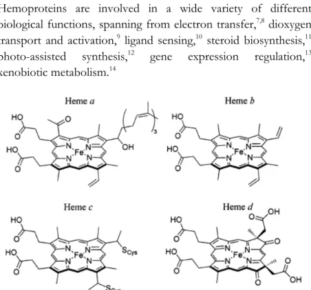
Figure 1.1. Chemical structures of naturally occurring hemes a, b, c and d
[reproduced from ref. 12].
Heme-proteins are classified in different subcategories depending on the type of porphyrin and how it is anchored to the protein (Figure 1.1). The most common heme is iron (II) protoporphyrin IX, known as heme b or “protoheme”. It presents vinyl groups
at positions 8 and 13, methyl groups at positions 3, 7, 12 and 17, and propionates at positions 2 and 18 of the porphyrin. Heme b is anchored to the protein scaffold through the combination of the axial coordinating residues to the iron ion, hydrophobic interactions with the heme macrocycle, and polar interactions with the porphyrin propionic acids.15
Heme c is anchored to proteins via thioether bonds between cysteine residues and the heme vinyl groups. Cytochromes c and f, in which the heme c is found, typically contain a CXXCH sequence motif, in which the two cysteine residues are covalently attached to the porphyrin, while the histidine coordinates the encapsulated iron.15,16
Heme a is synthesized from heme b by conversion of the vinyl group at position 8 into a hydroxy-ethylfarnesyl group, followed by oxidation of the methyl at position 3 to formyl group. Heme a is both more hydrophobic and more electron-withdrawing with respect to its precursor. It is found only in terminal oxidases, such as mammalian cytochrome c oxidase.15,17
Other less common heme architectures include heme d,18 heme
P-460,19 siroheme,20 and chloro-cruoroheme.21
The reactivity of the heme is deeply modulated by the protein matrix. Indeed, there are several classes of proteins with different catalytic functions, even though they contain the same prosthetic heme group. Protein composition and structural organization of the peptide fine-tune the environment of the heme cofactor, selecting one reaction as the only or predominant chemical transformation.22
Structural peptide organization, amino acid composition, protein tridimensional structure dictate the properties of the primary (metal coordination geometry, number, type and donor
properties of the axial ligands) and secondary (local dielectric constant, hydrophobicity, hydrogen bonding interactions near to the active site) coordination shells. Moreover, the protein directs long-range interactions. All these factors modulate heme-protein redox potentials, electronic structure, spin states, and catalytic properties.22
Different amino acids can serve as axial or proximal ligands to the heme. In a data set of non-homologous hemoproteins (Figure 1.2) histidine predominates as an axial ligand, especially in multi-heme proteins. Other residues with side chains acting as axial ligands are methionine, cysteine, and tyrosine.23In two cases
a change in the axial ligand is observed on reduction: in CO-sensing proteins, the axial ligand Cys75 is replaced by His77 in the reduced state,24 while in cytochrome cd1 nitrite reductase one
of the axial ligands (a tyrosine residue) exits from the coordination sphere on reduction, leaving a vacant site.25
Figure 1.2. Occurrence of axial ligands to the heme iron ion in a data set of
non-homologous heme-proteins; heme b (HEM), heme c (HEC), and heme b and c (ALL) [data taken from ref. 26].
The coordination number five is frequently found; this is achieved by the protein scaffold that provides only one histidine
as the fifth axial ligand, while the sixth coordination position is left empty to accommodate exogenous molecules. This geometry is commonly found in globins and peroxidases. The second most common coordination motif is six-coordinated, since two His residues bind the metal ion from the opposite faces of the porphyrin ring. This coordination geometry is found, as an example, in electron carrier proteins.
In addition to His, Met and Cys frequently act as sixth axial ligands to the heme. Indeed, a typical six-coordinated ligation motif for heme-proteins consists of a His and Met (e.g. in cytochrome c)27 or bis-methionine (e.g. in bacterioferritin).28
Conversely, Cys as the axial ligand is mainly observed in five-coordinated heme-proteins, such as cytochrome P-450.29 Other
few amino acid ligands are found in six-coordinated heme-proteins in combination with His (e.g. tyrosine in cytochrome cd1 nitrite reductase).25 In a very few structures, some residues
coordinate the heme iron through their neutral N-terminal amino group, such as a tyrosine in cytochrome f and a proline in CO-sensing protein. 30
Li et al. pointed out that heme binding pockets are enriched in aromatic and non-polar amino acids.26 Through bioinformatic
tools, they analyzed the occurrence of amino acid residues in a non-redundant data set of 125 heme-proteins. (Figure 1.3)
Figure 1.3. Relative frequency of the residues in heme b (HEM), heme c (HEC), and heme b and c (ALL) [reproduced from ref. 26].
In heme c proteins, the occurrence of Cys residues is very common, due to the large presence of the classic CXXCH binding motif, as mentioned above.31
The aromatic residues (Phe, Tyr, Trp) play important roles in protein-heme interactions through p-stacking interactions with the porphyrin. Aliphatic residues (Leu, Ile, Val) are slightly increased over the background frequencies: they make hydrophobic interactions with the heme macrocycle. The residues with the fewest occurrences (Asp, Glu, Lys) are charged residues, suggesting that the heme binding pocket is mainly placed within a hydrophobic environment.
Analyzing the heme-protein secondary structures (Figure 1.4), the dominance of α-helical structures for the three-quarters was observed (77%), although heme-proteins composed of β-sheets or mixed α/β structures are not uncommon (13% and 10%, respectively). This analysis has been performed using the CATH database, which hierarchically categorized protein structures by class, architecture, topology and homologous superfamily.15
Figure 1.4. A CATH wheel representation of natural heme-protein secondary
structures: α-helical in red, β-sheet in green, mixed α/β in yellow. At the bottom, an example for each type: myoglobin in red, nitrophorin in green, and FixL in yellow. Reproduced from ref. 15.
1.1.2
H
ORSERADISH
P
EROXIDASE
(HRP)
Peroxidases are a large family of ubiquitous enzymes that catalyze the oxidation of different organic and inorganic substrates, such as aromatic compounds (amine, phenols, thioanisole), using hydrogen peroxide (H2O2) as oxidizing agent. Peroxidases are
widely studied enzymes, due to their relevant position in chemistry, biotechnology and medicine. All peroxidases contain FeIII-protoporphyrin IX as the prosthetic group, except for
myeloperoxidase, which contains also halides (Cl-, Br-, I-) and
pseudohalides, such as [SCN]-32. In the resting state of the
enzyme, the Fe ion is invariantly penta-coordinated: four ligands are provided by the porphyrin (four nitrogen of the heme in equatorial positions), whereas His is coordinated as the axial fifth ligand (namely, the proximal His).33 The sixth coordination
position is occupied by the solvent, thus iron is in a high spin S = 5/2 state.
As all peroxidases, HRP belongs to a large family of different enzymes, named isoenzymes, which catalyze the same reactions but they have different physical, chemical and kinetic properties, because of their small differences in amino acid sequences. In the Armoracia rusticana root extract, several horseradish peroxidases have been found; among them, isoenzyme C (HRP C) is the most abundant enzyme. HRP C comprises a single polypeptide chain of 308 amino acid residues, with the N-terminal blocked by pyroglutamate and a heterogeneous C-terminus, with some molecules lacking the terminal residue (Ser308).34 Nine potential N-glycosylation sites can be recognized
in the primary sequence from the motif N-x-S/T and of these, eight are occupied. The total carbohydrate content of HRP C is
not constant and it depends on the provenance of the enzyme. Values between 18% and 22% are typically found. 35
HRP C contains three different metallic cofactors, FeIII-protoporphyrin IX and two calcium atoms; all of them are
essential for the structural and functional integrity of the enzyme.36
The heme group is attached to the enzyme by a coordination bond with the Nε of histidine 170 (proximal His). The second
axial coordination position on the heme distal side, is unoccupied and this represent the hydrogen peroxide binding site during catalysis.
HRP tertiary structure is composed of 13 a-helices and three β-sheets.37 Protein conformation is stabilized by four disulfide
bonds (Cys 11–91, Cys 44-49, Cys 97–301, and Cys 177–209). The protein has a quaternary structure composed of two domains, one distal and one proximal; between them, the heme group is located (Figure 1.5).
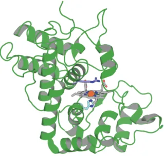
Figure 1.5. Three-dimensional representation of the X-ray crystal structure of
HRP C (PDB entry ID: 1H57).
Most reactions catalyzed by HRP C and other horseradish peroxidase isoenzymes can be expressed by the following equation:
H2O2 + 2AH HRP 2H2O + 2A·
in which AH and A· represent respectively the reducing
substrates (such as phenols, aromatic acids, aniline derivate, sulfides) and its radical product.
The catalysis occurs in a three-step process, in which the enzyme is first oxidized by H2O2 and then reduced back to the resting
state by two sequential steps, passing through two enzyme intermediates, Compounds I and II (Figure 1.6).38
Figure 1.6. Catalytic cycle of HRP.
The first step consists in the cleavage of the H2O2 peroxidic bond with the concomitant release of water and the incorporation of an oxygen atom to yield Compound-I, which contains an oxoferryl group, with the iron in the +4 oxidation state, and a porphyrin in π-radical cation.
Compound-I is able to oxidize a wide range of reducing substrates. By a single-electron transfer mechanism, the compound-I π-radical cation is reduced, leading the second enzyme intermediate, called Compound-II. This still contains an oxoferryl group, which is able to oxidize a second substrate molecule, going back to the native ferric enzyme form (Fe +3). During these double one-electron reductions, the oxygen accepts two protons to form a water molecule released from the heme. The presence of a strong hydrogen bond between the Nδ atom of the proximal histidine and an aspartate side chain (Asp247)
represents a key factor in the modulation of redox potential of the penta-coordinated complex. This hydrogen bond increases the basicity of the coordinating histidine, stabilizing the high oxidation state of the intermediates, such as the high valent oxoferryl center.39
Hydrogen peroxide bindings occur in the distal site, which is characterized by distal His and Arg residues (Figure 1.7). These two amino acids are invariant in all plant peroxidases and play a major role in the formation and stabilization of Compound I. Distal His (His42) acts mainly as a general acid–base catalyst: it assists the hydrogen peroxide deprotonation and the subsequent heterolytic cleavage of the peroxidic bond during Compound I formation.40 Distal Arg 38 is involved in charge stabilization,
mediated by its positively charged guanidinium group. Furthermore, once the O–O bond is cleaved, Arg contributes to stabilize the oxoferryl species via hydrogen bond formation.
41
Figure 1.7. Proximal and distal sites of HRP (PDB entry ID: 1H57).
The replacement of these highly conserved residues causes a drastic reduction of Compound I rate formation, and therefore of HRP catalytic activity. This is a proof of their foundamental role in catalysis. Treatment of the enzyme with an excess of hydrogen peroxide provides Compound III, whose structure is best described as a resonance of Fe2+-O
2 and the isoelectronic
Fe3+-O
2- form. Berglund and co-workers succeeded in obtaining a
three-dimensional movie of the X-ray-driven catalytic conversion of the bound dioxygen species to two molecules of water in the active site.42 The mechanism involves four successive,
one-electron reductions with the concomitant uptake of a proton, from Compound III to the ferrous HRP, via the intermediate forms of Compound I, Compound II and ferric HRP. The high-resolution crystal structure of Compound III shows dioxygen bound to heme iron in a bent conformation.42
Phe4 1 His42 Asn7 0 His170 Asp24 7 Arg3 8
C
HAPTER
1.2
1.2.1 A
RTIFICIAL
M
ETALLOENZYMES
:
B
IOINSPIRED
I
RON
P
ORPHYRINS
WITH
C
ATALYTIC
P
OTENTIAL
The most fundamental aspects of metal cofactor assembly and metalloprotein function have already been clarified, mainly through structural–functional studies on natural metalloproteins and related mutants.43 Despite these progresses, several questions
are still open. While the control of the functional specificity by the primary coordination sphere is well understood, the contribution of medium and long range interactions remains to be determined. Thus, it is not surprising that many efforts are being devoted to the development of metalloprotein mimics, with the aim to:
-‐‑ provide further insight for structure-activity relationship (SAR) studies;
-‐‑ understand the minimal requirements for function; -‐‑ reproduce the properties of the parent native proteins in
smaller molecules, with improved properties, such as higher stability and greater efficiency;
-‐‑ construct new, tailor-made molecules useful for biomedical, pharmaceutical and environmental applications.
Over the years, heme-protein models have been developed using quite different strategies. They differ in molecular structures, ranging from simple meso-substituted tetra-aryl metalloporphyrins to more complex peptide-porphyrin conjugates. A common feature in the compounds so far developed is the assembly,
around the porphyrin ring, of several different chemical components, which are intended to fulfill the features of the protein matrix and make the heme able to accomplish specific functions.
The first low molecular weight heme-protein models date back to the early 1970s. All these systems showed penta-coordination, obtained both by using hindered ligands and by covalently attaching the axial ligand to the porphyrin ring. A bulky moiety was needed to cover one face of the porphyrin ring, preventing the formation of a hexa-coordinated species.44The pioneering
works of Traylor,45,46, Momenteau,47 and Reed48 can be
considered fundamental for the development of dioxygen-binding molecules. Some representative examples are constituted by the picnic-basket49 and capped46 porphyrin systems, where the
coordination state of the iron(II) was achieved using hindered substituents on the meso or the pyrrole positions of a 5, 10, 15, 20-tetraphenylporphyrin. These systems can be considered as fundamental for the development of dioxygen binding molecules and have contributed to a better understanding, at a molecular level, of the properties of natural heme-proteins. Even though they displayed enhanced and interesting activity, their practical application was limited to the use in organic solvents.
Peptide-based models are fundamental in assisting our understanding of the factors governing the heme properties. They also offer the additional advantage of making the highly hydrophobic heme water-soluble, by simply accommodating the prosthetic group in a hydrophobic pocket of an amphiphilic structure. Being between small molecule models and large proteins, artificial peptide-based models are the best candidates to mimic both the structural characteristics and reactivity of natural systems. Their structures are simpler than those of natural
proteins, but they have sufficient size and chemical diversity to allow substrate recognition and to resemble the high chemo, regio, and stereoselectivity of natural hemoproteins.
The development of peptide-based models takes advantage from recent progress in both the design and the chemical synthesis of peptides and proteins. It is now possible to construct recognition and catalytic sites in a scaffold and to systematically vary the amino acid composition, allowing to (1) optimize the structural and functional properties of the initial target in terms of stability, catalytic activity and selectivity, (2) test how small changes in the sequence can affect heme properties and functions and (3) investigate the effect of variation in the first and second coordination spheres.5,
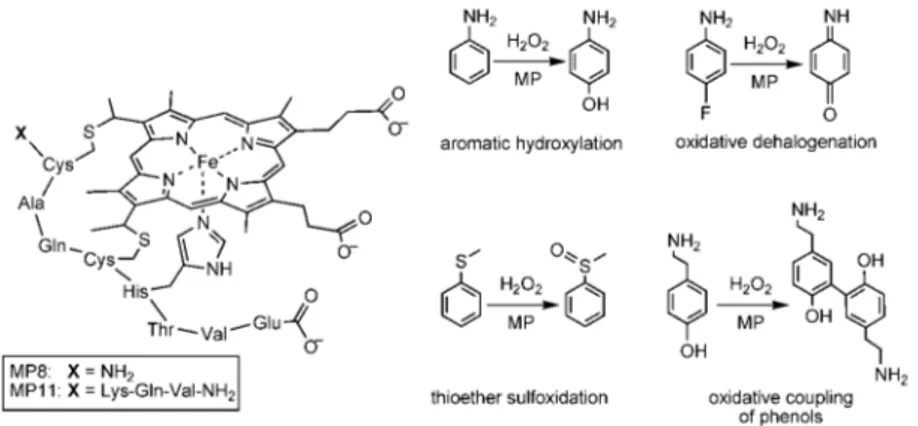
Microperoxidases (MPs) represent the first studied covalent heme-peptide fragments, which can be obtained by the proteolytic digestion of cytochromes c., The peptide moiety is
covalently linked to the heme through thioether bonds, by the Cys-(Xaa)2-Cys-His motif. The amino acid sequence of the
polypeptide chain in MPs is numbered according to the one of the parent cytochrome c. Selective proteolysis of cytochrome c affords various MPs, differing in the peptide chain length.50 The
most studied example among MPs is MP8, which is obtained from the tryptic digestion of horse heart cytochrome c; it retains the amino acid residues 14–21 of the starting protein. MP11 retaining residues 11–21, and MP9 retaining residues 14–22 have also been studied extensively. These MPs contain the minimal requirements for a heme-protein mimic, by retaining a His residue at position 18, which coordinates the heme iron and acts as a proximal ligand. In addition, the sixth coordination site to the heme is occupied by a water molecule, which is readily displaced by an entering exogenous ligand (Figure 1.8).
One of the most interesting features of the chemistry of MPs is that, despite their relatively small size, they are able to selectively oxidize a variety of organic substrates, including ABTS (2,2'-azino-bis(3-ethylbenzthiazoline-6-sulphonic acid)), anilines, naphthols, phenols.51 MP11 has been shown to oxidize sulfides
enantioselectively, although with only modest enantiomeric excess (ee) values (16–25%).52
Figure 1.8. left Structure of Microperoxidases; right and some of the
peroxidase and oxygenase-like reactions catalyzed by MPs.
It has been suggested that the peptide retains a conformation very similar to the amino acid residues of the parent protein. Molecular dynamics simulations of MP8 and MP11 suggests that the peptide only covers the proximal face of the porphyrin, while the distal face is totally open to the solvent.22
Despite their extensive use, no full atomic-resolution structure for any MP is available owing to the conformational flexibility of the peptide portion. Recently, a new MP analog, MP9cb562, was
obtained from the tryptic digestion of cytochrome cb562. The immobilization of MP9cb562 inside a protein lattice cage allowed
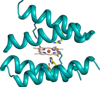
the first crystallographic structure determination for any member of the MP family (Figure 1.9).53
Figure 1.9. Crystal structure of MP9cb562, obtained from the tryptic digestion of cytochrome cb562, and the distal position is occupied by the solvent exposed His 73 from the co-crystallized cytochrome cb562 (PDB ID: 3m4c)
The structure of MP9cb562 reveals an a-helical peptide
conformation throughout its length that is slightly more expanded than that of the corresponding segment within the holo-protein. Nevertheless, the fact that a nine-residue long fragment can adopt a helical architecture outside the context of a folded protein suggests that c-type linkages may provide some bias to the formation of an a-helical structure toward both N- and C-terminal ends. 9
The application of MPs as catalysts is limited by their low stability under catalytic conditions. The accessibility of the distal side causes the degradation of the porphyrin ring during catalysis, either by the direct action of H2O2 or by intermolecular reactions
with another active iron–oxo species. Despite these limitations, MPs are still much more stable than simple protoporphyrin systems, indicating that the presence of the small peptide chain can play an important protective role and make the catalyst more robust.
The use of a-helical peptides able to cage the heme group, has been and remains the focus of the most of works in the field of peptide-based models, because the a-helix motif is a recurring structural motif that surrounds the heme cofactor in numerous natural hemeproteins.54 Over the years, a large number of helical
hemeprotein models have appeared in the literature. They differ in structural complexity, and can be grouped into two classes, according to the nature of the interactions between the heme and the helical peptides: 1) models in which peptide chains are covalently linked to the heme; 2) models in which peptide chains incorporate one or more heme groups, by non-covalent self-assembling around the heme.
The four-helix bundle motif has been widely used as protein cage for mono-heme and multi-heme binding. It should be highlighted that the assembly of a-helices into the four-helix bundle motifis a ubiquitous and functionally important architecture found in several hemeproteins, such as cytochrome c and cytochrome b562.
Excellent examples of covalent heme-peptide assemblies are the peptide sandwiched deuteroheme MP3. In a very recent paper, the construction of a de novo artificial heme-binding "maquette" able to efficiently catalyze oxidation reactions using H2O2 has
been reported.9,55.
MP3 has been designed by combining the excellent structural properties of four-helix bundle protein scaffolds with the activity of natural peroxidases56 (Figure 1.10). It houses two peptide
chains of different composition covalently linked the deuteroporphyrin IX through the side chain of two lysine residues, to obtain an asymmetric helix/loop/helix-heme-helix/loop/helix sandwich arrangement. MP3 is a penta-coordinated complex, characterized by: 1) a His residue on one
chain as the axial ligand to the heme iron; 2) an empty distal site, able to accommodate exogenous ligands or substrates; 3) an arginine residue in the distal site, that should assist in hydrogen peroxide activation, mimicking Arg38 in HRP.
Figure 1.10. Computer model of MP3. Tridimensional model of MP3
side-chains of key-residues are depicted as green sticks.
MP3 was synthesized and characterized as its Fe(III) complex. CD spectroscopy suggested that MP3 scaffold possessed high intrinsic helical propensity in water, even if complete folding of the helices occurred only in the presence of TFE (2,2,2-trifluoroethanol). UV/Vis, MCD and EPR spectroscopies gave insight into the coordination geometry and spin state of the metal, confirming mono-His coordination above pH 4. MP3 showed a high catalytic turnover in the oxidation ABTS by H2O2,
The maquette approach is based on the identification of a peptide sequence able to adopt a four-helix bundle. Maquettes-simple
4-α-helix bundles designed from first principles57 in particular
provide a blank canvas for tractable and iterative design processes, as demonstrated by the successful incorporation of sophisticated functions common to oxidoreductases such as intermolecular electron transfer57 and oxygen binding58. An
example of four helix bundle was reported by Anderson and co. workers. In early studies, the artificial maquettes were modified by the post-translational cytochrome c maturation system of E. Coli enabling covalent incorporation of the heme cofactor in vivo, giving rise to the "c-type cytochrome maquettes" (CTMs).59 In
this case, a heme bearing a His coordination as active site was introduced through a simple and few-steps design process.55
Figure 1.11: Key-steps in the developing of a catalytic c-type maquette (C45).
The resulting artificial enzyme (Figure 1.11), called C45, was endowed with very high catalytic activity regarding ABTS oxidation, even at high temperatures. Remarkably, C45 demonstrated high catalytic versatility, promoting a wide variety of oxidation reactions, including the oxidation of guaiacol, luminol, o-phenylenediamine, p-anisidine, 5-aminosalycilic acid and the oxidative dehalogenation of 2,4,6-trichlorophenol and its brominated and fluorinated congeners.
1.2.2 M
IMOCHROMES
:
M
INIATURIZED
H
EME
-P
ROTEIN
M
ODELS
The Artificial Metallo-Enzyme Group in Naples approached the challenge of constructing heme-protein models using a miniaturization process. By this strategy, a class of artificial heme-proteins has been developed, named Mimochromes.
Mimochromes are peptide-sandwiched deutero-heme systems, obtained by miniaturizing natural heme-proteins, with aim to resemble their structural and functional properties. Analyzing natural heme-protein structures, it emerges that the prosthetic group is almost completely embedded between two relatively small α-helical peptide fragments.
The natural motif helix-heme-helix was used as a template to engineer the family prototype, Mimochrome I.60,61 To design a
mini-heme protein, the copious interactions of the peptide moiety with the heme commonly found in natural proteins were replaced by a few strong local constraints. The smallest sequence required for a complete coating of one face of the heme, was identified in a nine-residue peptide, based on the F helix of hemoglobin β-chain (Figure 1.12).
Figure 1.12. Miniaturization design from hemoglobin (PDB ID: 2hhb) to
mimochrome I. (a and b) Active site identification and extraction. (c) Isolation of heme covering the nonapeptide. (d) Mutual approach of heme propionate towards Lys92 by proper rotamer selection. (e) Selection of solvent exposed residues and protoporphyrin IX to deuteroporphyrin mutation. (f) Symmetry-generated hexacoordinated deuteroheme. The structures were Symmetry-generated using PyMol.335
Mimochrome I peptide sequence contains: a central His residue to coordinate the heme iron; Leu residues at positions i-4 and i+4 relative to the His to hydrophobically interact with the heme macrocycle; a Lys residue to anchor the heme group; two alanine residues (that substitute Ser89 and Cys93), chosen for their high α-helical propensity; glutamine and asparagine residues (that substitute Glu90 and Asp94) to remove charge residues that could destabilize the target conformation (Figure 1.13).
Figure 1.13. Schematic structure of Mimochrome I and the comparison of the
peptide sequence with the L88-L96 fragment of the F helix in the β-chain of hemoglobin. Figure reproduced from ref. 61.
Deuteroporphyrin IX was preferred to the more common protoporphyrin IX to avoid the possibility of degradation of the sensitive vinyl substituents during the synthesis. Two identical copies of the peptide were covalently linked to the porphyrin propionic groups through the ε-amino function of Lys, obtaining a pseudo-C2-symmetric dimer
The insertion of cobalt ion into the porphyrin ring gave two diastereomers. In fact, linker flexibility between the peptide and the deuteroporphyrin ring allows each peptide chain to be positioned either above or below the porphyrin plane, producing enantiomeric configurations around the metal center. The presence of the substituents on the porphyrin ring and the chirality of the peptide chain contribute to establish a diasteromeric correlation between the Δ and Λ isomers of CoIII
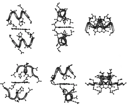
-Mimochrome I. This finding was confirmed by structural NMR characterization of the two previously separated Co(III) complexes62 (Figure 1.14).
Figure 1.14. Front, side and top views of the average molecular structures
obtained from NMR data and RMD calculations for Δ isomer (top) and Λ isomer (bottom) of Co(III)-Mimochrome I. Figure reproduced from ref. 55.
The information derived from Mimochrome I was applied for improving the design. Over the years, quite a few other models were developed, with same or different peptide chains linked to the deuteroporphyrin ring (Table 1.1).
Table 1.1. Peptide sequences of various Mimochrome members.
Mimochrome II63 presents two identical and elongated peptide
chains, modeled in an extended conformation, whose residues were carefully chosen to improve water solubility and to stabilize a unique diastereomer.
Later, another complex with identical peptide chains was designed: Mimochrome IV.64,65 This model considers
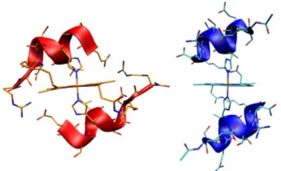
intra-molecular inter-helical interactions, provided by an ion pair between the carboxylate side chain of a Glu residue at position 1 of one peptide chain and the guanidine group of an Arg residue at position 9 of the other peptide chain (both side chains being modelled in an appropriate extended conformation). Moreover, the helix was further stabilized by a positively charged residue (Arg) at the C-terminus and a negatively charged residue (Glu) at the N-terminus, which reduced the intensity of the helix dipole. Mimochrome IV was the first molecule with a unique topology within the Mimochrome family. The corresponding Co(III)
complex was extensively characterized both in solution and in solid state, confirming the presence of the Λ topology (Figure 1.15). The structural characterization evidenced that in the solid-state crystal packing interactions strongly affected the molecular folding, with intra-chain Glu1–Arg9 ion pairs that were preferred over the designed, and experimentally found in solution, inter-chain interactions.
Figure 1.15. NMR (on the left) and X-ray (on the right) structures of
1.2.3 C
ATALYTICALLY
A
CTIVE
M
IMOCHROMES
:
MC6
AND
ITS
C
ONGENERS
Since Mimochrome IV, despite its small size, adopted a unique well-defined secondary and tertiary structure, the successive goal was to introduce peroxidase-like activity into Mimochrome’s architecture, with the perspective to obtain enzyme-mimicking molecules having a higher stability and even with improved catalytic properties compared to native hemoproteins.
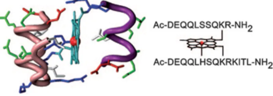
Among the Mimochrome family, Mimochrome VI was the first model showing catalytic potential.66 It was composed by a
14-residue peptide, with a His 14-residue at position 6 acting as axial ligand of the heme (proximal site) and a 10-residue peptide lacking a heme-coordinating moiety, to create a cavity around the metal ion (distal site). This unsymmetrical penta-coordinated mono-His model was designed starting from the NMR solution structures of both the symmetric bis-His Mimochrome II and Mimochrome IV.
The tetradecapeptide was designed to adopt a short helical conformation (residues 1-9), a loop γβD (residues 10 and 11) and
a short β-strand (residues 12-14) that folded back to interact with the helical part. The decapeptide chain also was designed to adopt a helical conformation (residues 1-8). Stabilization of the secondary structure was also given by the positively charged Arg10 and the negatively charged Glu2 at the C-terminal and N-terminal ends, respectively, with the opposite sign relative to the helix dipole (Figure 1.16).
The tertiary structure could be viewed as a sandwich, which is a characteristic trait of the Mimochrome family members, in which the two peptide chains embraced the metalloporphyrin; peptide helices were antiparallel to each other and the helix axes were conceived to be about parallel to the porphyrin plane. Stabilization of the tertiary structure was enhanced by inter-chain ion pairs between the carboxylate side chains of a glutamate residue (Glu2) on one helix and the guanidine group of an arginine residue (Arg10) on the other helix. Finally, several glutamines (Gln3, 4 and 8) and a serine (Ser7) were introduced in the solvent exposed positions to promote water solubility.
Figure 1.16. Molecular model and amino acid sequence of FeIII-Mimochrome VI. Figure reproduced from ref. 65.
Catalytic activity studies showed that FeIII-Mimochrome VI
(FeMC6) acted as an artificial peroxidase, since it catalysed oxidation reactions resulting from H2O2 activation.65
FeMC6 showed a catalytic performance comparable to that of the natural enzyme, with a kcat value only 11-fold lower than that
Table 1.2 Catalytic parameters for the oxidation of ABTS (AH2) by H2O2 activation for Mimochrome VI and HRP in the experimental conditions for maximal activity for each enzyme.
When evaluated at near neutral pH (pH 6.5), FeMC6 exhibited more than 4000 turnovers within 10 min in the ABTS oxidation. HRP worked with higher catalytic efficiency at more acidic pH (pH 4.5), this is notable if the catalyst should be used for wastewater depuration. FeMC6 also catalyzed: (a) the oxidation of guaiacol, with a kcat value 50-fold lower than that of HRP; (b)
the conversion of phenols to 4- and 2-nitrophenol in presence of NO2 and H2O2 in high yields; (c) the coupling reaction of
tyrosine residues.
In the following stage of design, attention was focused on Glu2 and Arg10 in the decapeptide and tetradecapeptide chains, since both peptides were involved in finely tune the reactivity.
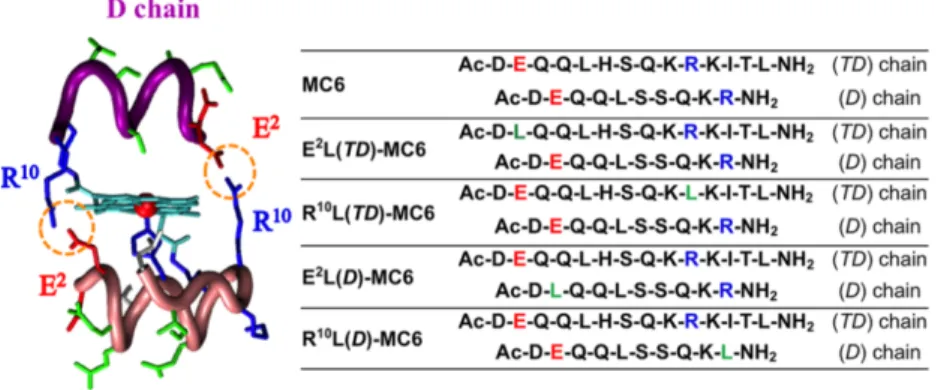
In Mimochrome VI design, these residues were placed at the N- and C- termini, respectively, to contribute to stabilize both secondary and tertiary structures. By individually replacing Glu2 and Arg10 on both peptide chains with a Leu residue, four analogues were obtained (Figure 1.17), which were screened for peroxidase-like activity.67
Figure 1.17. Left: molecular model of FeIII-Mimochrome VI, highlighting the R10-E2 ion pair interactions. Right: peptide sequences of FeIII-Mimochrome VI (MC6) and its analogues: acid, basic and non-polar residues in position 2 and 10 are indicated in red, blue and green, respectively. Image reproduced from ref. 66.
The apparent catalytic constant of the analogues substituted on the tetradeca chain was about 2-fold higher with respect to MC6. The best performances were obtained for E2L(TD)-MC6, which displayed an improvement in the apparent catalytic constant (kcat
= 7.8·102 s-1), and catalytic efficiency towards both H
2O2 and
ABTS (kcat/Km = 25 mM-1 s-1, and 16·103 mM-1 s-1, respectively).
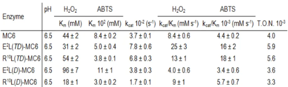
In contrast, analogues with an altered decapeptide chain showed an almost unmodified reactivity compared with MC6, the worst catalyst being the E2L(D)-MC6 congener. All MC6 members could perform thousands of turnovers, without bleaching. The turnover numbers (T.O.N.) reflected the trend of the catalytic efficiency, being higher in E2L(TD)-MC6 and lower in R10L(TD)-MC6 (Table 1.3).
Table 1.3 Steady-state kinetic parameters for FeIII-Mimochrome VI (MC6) and its analogues (reproduced from ref. 66).
A detailed spectroscopic characterization allowed to hypothesize the catalytic role of Arg10 on the decapeptide chain. Arg10 was suggested to approach the heme, contributing to the stabilization of compound I and assisting ligand binding, similarly to the conserved distal arginine (Arg38) in HRP. Indeed, there is experimental evidence that the catalytic cycle of E2L(TD)-MC6 occurred with a peroxidase-like mechanism, through the formation of compound I, proving its similarity with natural peroxidases, whose reactivity is influenced by the proximal and distal heme environments.66
C
HAPTER
1.3
A Novel Artificial Metalloprotein
with Peroxidase-Like Activity
1.3.1 D
ESIGN AND
S
YNTHESIS
OF
Fe
III-MC6a
Based on literature data discussed in Chapter 1.1 and 1.2, one of the focus of this PhD project has been the development of a novel artificial enzyme belonging to the Mimochrome family (Mimochrome VIa or MC6a; Figure 1.18).
Figure 1.18. Schematic representation of FeIII-MC6a.
The newly developed Mimochrome has been designed to introduce some new structural constraints, with the aim to stabilize the three-dimensional sandwich structure, typical of Mimochromes, while preserving or even enhancing its functional features. In the previous FeIII-Mimochrome VI (FeIII-MC6), the
catalytic performance of the enzyme was demonstrated to be modulated by mutating even one single residue at the N- or C-terminus on either the tetradecapeptide (TD) or the decapeptide (D) chain.66 In particular, the substitution of a glutamic acid with
a leucine on the TD chain to give (E2L(TD)-MC6) doubles the
kcat and quadruples the catalytic efficiency (kcat/KM), by allowing the arginine residue on the D chain to assist the catalysis as
observed in natural peroxidases. FeIII-MC6 showed a compact
structure that allowed accomplishing several thousands of turnovers in water without bleaching. Moreover, the D chain was observed to provide a major contribution to chemical stability by protecting the high-valence intermediate. Indeed, a lower number of turnovers (TON) has been observed with the compound lacking the D chain. Accordingly, during this PhD thesis we studied the relationship between the rigidity of the D chain as well as its hydrophobicity, catalyst robustness and catalytic efficiency, as resulting by introducing two 2-amino isobutyric acid (Aib, U) residues at positions 4 and 9. Aib is a natural, non-coded amino acid, with a high helical propensity, commonly found in peptides produced by microbes.68 The replacement of hydrogen
atoms with two methyl groups at Cα of the glycine produces severe restrictions on its conformational freedom: for Aib-containing peptides, the formation of α-helix secondary structures is strongly favored. Moreover, the equilibrium between the α-helix and 310-helix in Aib-containing peptides has been
observed to be dependent on several parameters, mainly including the peptide length and the Aib content.69
In order to find the best positions for Aib mutation in the (D) sequence, a search based on the helix propensity was performed.70 Five positions were excluded from this search: (1)
Asp1 at the N-terminus; (2) Glu2 and Arg10 for their established
contribution to catalysis;1 (3) Ser6 as it corresponds to the active
site cleft; (4) Lys9 because it is involved in the covalent binding
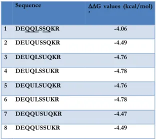
with the porphyrin. Four out of eight possible sequences gave the best score (Table 1.4). The sequence containing Q3U and S7U
was selected for synthesis and characterization. Indeed, according to our model, in the new compound both Aib residues face towards the porphyrin, offering a tiny hydrophobic patch for the substrate binding.
Table 1.4. Energy in Kcal/mol of the possible sequences for the new
Decapeptide chain.
The amino acid sequence of the full protected decapeptide chain was therefore:
Ac-Asp1(tBuO)-Glu2(OtBu)-Aib3-Gln4(Trt)-Leu5-Ser6
(tBu)-Aib7-Gln8(Trt)- Lys9(Mmt)-Arg10(Pbf)-NH 2
The amino acid sequence of the full protected tetradecapeptide chain was:
Ac-Asp1(tBuO)-Leu2-Gln3(Trt)-Gln4(Trt)-Leu5-His6
(Trt)-Ser7(tBu)-Gln8(Trt)-Lys9(Mmt)-Arg10(Pbf)-Lys(Boc)11-Ile12
-Thr(tBu)13-Leu14-NH 2
The synthesis of FeIII-MC6a was carried out according to the
following Scheme 1.
Sequence !!G values (kcal/mol)
* 1 DEQQLSSQKR -4.06 2 DEUQUSSQKR -4.49 3 DEUQLSUQKR -4.76 4 DEUQLSSUKR -4.78 5 DEQULSUQKR -4.76 6 DEQULSSUKR -4.78 7 DEQQUSUQKR -4.47 8 DEQQUSSUKR -4.49 !
Scheme 1. Schematic representation of the synthetic strategy.
In line with the synthesis of previous catalysts,67 Mimochrome
VIa was first prepared on a small scale (20 µmol). An automated peptide synthesizer was used for the synthesis of the two peptides chains. The coupling steps of the peptides to the porphyrin and the final deprotection were performed in solution. Subsequently, the synthesis was applied to a medium scale (20 mmol), using (as the only exception between the two procedures) a medium size reactor for solid-phase synthesis during peptide assembly. In both cases, peptide synthesis was performed using 9-fluorenylmethoxycarbonyl (Fmoc) chemistry, using a super acid labile resin. Regarding the two Lys residues involved in amide bond with porphyrin, ((4-methoxyphenyl)diphenylmethyl (Mmt) was chosen as the protecting group. Mmt was easily removed under mild acid conditions,71 leaving all the remaining protecting
groups unreacted.
Once the synthesis was completed, a small amount of peptidyl-resin was fully deprotected and the product was analyzed by TANDEM mass spectrometry coupled to liquid chromatography. A single peak, corresponding to the desired peptide, was found and its product ion scan showed the expected transitions (Table 1.5 and 1.6).
Table 1.5. Mass table of Decapeptide fragmentation. Green numbers
represent the theoretical fragments found.
Table 1.6. Mass table of Tetradecapeptide fragmentation.
The protected TD chain was first coupled to the deuteroporphyrin. To direct the (TD) peptide coupling toward the mono-substituted product (TD-DPIX) and to overcome the undesired homo di-substituted product (TD-DPIX-TD), the tetradecapeptide chain was slowly added to an excess of deuteroporphyrin IX, the reaction was monitored by HPLC (Figure 1.19). The chromatogram shows the presence of a main peak at 29.36 min, corresponding to the desired product, and two minor peaks: one corresponding to the homo di adduct (Rt 30.67
min), the second one (Rt 27.14 min) was not characterize. Once
the complete consumption of TD chian was confirmed, the crude # Immon. b b++ b+++ Seq. y y++ y+++ # 1 88.10 D 1215.34 608.17 405.78 10 2 102.13 287.25 144.13 96.42 E 1058.21 529.61 353.41 9 3 58.12 372.32 186.68 124.79 U 929.10 465.05 310.37 8 4 101.14 500.48 250.74 167.50 Q 843.99 422.50 282.00 7 5 86.17 613.64 307.32 205.22 L 715.86 358.44 239.29 6 6 60.09 700.71 350.86 234.24 S 602.71 301.86 201.57 5 7 58.12 785.82 393.41 262.61 U 515.63 258.32 172.55 4 8 101.14 913.95 457.48 305.32 Q 430.52 215.77 144.18 3 9 101.19 1042.12 521.56 348.05 K 302.40 151.70 101.47 2 10 129.20 R 1 # Immon. b b++ b+++ Seq. y y++ y+++ # 1 88.04 D 1749.01 875.01 583.68 14 2 86.10 271.13 136.07 91.05 L 1591.97 796.49 531.33 13 3 101.07 399.19 200.10 133.73 Q 1478.89 739.95 493.64 12 4 101.07 527.25 264.13 176.42 Q 1350.83 675.92 450.95 11 5 86.10 640.33 320.67 214.12 L 1222.77 611.89 408.26 10 6 110.07 777.39 389.20 259.80 H 1109.69 555.35 370.57 9 7 60.04 864.42 432.71 288.81 S 972.63 486.82 324.88 8 8 101.07 992.48 496.74 331.50 Q 885.60 443.30 295.87 7 9 101.11 1120.58 560.79 374.20 K 757.54 379.27 253.18 6 10 129.11 1276.68 638.84 426.23 R 629.45 315.23 210.49 5 11 101.11 1404.77 702.89 468.93 K 473.34 237.18 158.45 4 12 86.10 1517.86 759.43 506.62 I 345.25 173.13 115.75 3 13 74.06 1618.90 809.96 540.31 T 232.17 116.59 78.06 2 14 86.10 L 131.12 1