Microcirculatory perfusion-based approach
to the critically ill patient: bringing a
research tool technology to the bedside
Tutor:
PhD Candidate:
Prof. Abele Donati
Dr. Claudia Scorcella
Academic Year
2017-2018
UNIVERSITÀ POLITECNICA DELLE MARCHE
FACOLTÀ DI MEDICINA E CHIRURGIA
_________________________________________________________
Dottorato di Ricerca
“Rifiutate di accedere ad una carriera solo perché vi assicura una
pensione. La migliore pensione è il possesso di un cervello in piena
attività, che vi permetta di continuare a pensare usque ad finem,
fino alla fine.”
A me stessa,
alla mia testardaggine,
CONTENTS
Chapter 1
Introduction and outline of the thesis
Chapter 2
Cytocam-IDF (incident dark field illumination) imaging for bed-side
monitoring of the microcirculation.
Chapter 3
Impact of microcirculatory video quality on the evaluation of sublingual
microcirculation in critically ill patients
Chapter 4
Near-infrared spectroscopy for assessing tissue oxygenation and
microvascular reactivity in critically ill patients: a prospective observational
study
Chapter 5
MicroDAIMON: Microcirculatory DAIly MONitoring in critically ill patients
Chapter 6
Sublingual microcirculation and tissue perfusion as predictors of organ
failure in major trauma: a subgroup analysis of a prospective observational
study
Chapter 7
Effects of ketanserin on microcirculatory alterations in septic shock: an
open-label pilot study
Chapter 8
Conclusion and future perspectives
Chapter 1
INTRODUCTION
The microcirculation is defined as the intricated net of vessels smaller then 100 m, including thus arterioles, venules and capillaries. This is an essential functional unit of the organs, representing the site of gasses and nutrients exchange between blood and tissues.
After pioneer studies carried out by Leeuwenhoek and Krogh between 1700 and 1950 about the physiologic aspects of microcirculation, especially in animals [1,2], the research in this field gained an enthusiast attention.
The study of microcirculation in human beings by using techniques of intravital microscopy is almost 20 years old: in 1999 Groner et al [3] gave a first insight into this fascinating and extremely complex world, giving the birth to the first non invasive imaging technique to observe microcirculation in vivo and real-time: the orthogonal polarization spectral imaging (OPS). This technique allowed to image the microcirculation in a large diversity of organs and tissues surfaces in multiple surgical and clinical settings by condensing the mechanical and optical components in microscopes of smaller dimensions, capable to configure an instrument usable at the bedside. [4,5] In 2007 the second-generation technology, the Sidestream Dark Field (SDF), takes its place, with the purpose of solving some of the major pitfalls of the OPS system. [6] A system of green light LED (530 nm wavelength), corresponding to the adsorbing spectrum of the hemoglobin, disposed in a ring form, produce a pulsatile illumination and an analogic video camera captures the reflecting light and produces video imaging of the microcirculation, which could be directly send to a monitor and visualized in real time. Brightness and focus can be manually adjusted, such in a conventional camera. The main advancements of this technique were in better video quality (ameliorated optical system which leads to less disturbances in signal) and maneuverability (including smaller size and inner battery) both in experimental and clinical settings. The probe of this device, covered by a sterile disposable cap, can be gently applied to organs or mucosal surfaces to observe the microcirculation.
The sublingual region is the most investigated by researchers: it is a site covered by a thin lay of epithelial cells and its perfusion derives from extern carotid artery and lingual and sublingual arteries and share the embryogenetic origin with intestine and splanchnic circulation [7] and warrants a rapid and easily accessible window to evaluate human microcirculation, also in critical conditions, without performing invasive maneuvers. However, many other sites have been investigated such as conjunctiva, mucosa of the intestinal tract, stomas, vaginal mucosae and, in surgical settings, also cerebral cortex [8-11].
Next to in vivo videomicroscopy, other technologies give information about the status of microcirculation. Near Infrared Spectroscopy (NIRS) is one of them. Apply in conjunction with a vascular occlusion test of the forearm, it enables the assessment of tissue oxygenation and microvascular reactivity on the skeletal muscle [12].
The development of such instruments of direct in vivo visualization of the microcirculation, lead to a rapid increasing in production of research studies aiming to clarify the mechanisms underlying shock, hemodynamic impairment and many other conditions of organ dysfunction and VO2/DO2 mismatch. Seen the high complexity and severity, the critically ill patient represents the most investigated subject in the field of microcirculation.
In recent studies, many selected subpopulations of critically ill patients showed a consistent incidence of microcirculatory abnormalities when observed with the previously described techniques and were associated with unfavorable clinical evolution, higher morbidity and mortality. [13-22]
The recent microSOAP study, provides the largest prospective observation of the point-prevalence of microcirculatory alterations in a mixed population of 501 adult critically ill patients, involving 36 ICUs all over the world. [23] In such an heterogeneous population, observed in a single time-point in all the participating centers, 17% of the patients showed abnormal microvascular flow (identified as a Microvascular Flow Index inferior to 2.6 AU); even if it did not resulted to be associated to adverse outcome in the overall population, it seems to maintain prognostic significance in a subgroup of high risk patients, identified by the presence of tachycardia (heart rate > 90 beats per minute).
The most extensively investigated clinical condition is sepsis. Oxidative stress, inflammatory mediators and cytokines during systemic inflammation induce alterations in the physiologic endothelial function: disruption in the glycocalyx barrier, increased vascular permeability with generation of tissue oedema, enhanced endothelium-leucocyte interaction, formation of red blood cell aggregates and microthrombi with disseminated intravascular coagulation, resulting in impaired microvascular blood flow and hypoperfusion [24-25]. This leads to the typical pattern of sublingual microcirculation during sepsis: an extremely heterogeneous flow due to a patchy distribution of capillaries with no flow next to areas of normal flow associated with a varying vessel density causing an important impairment of convective and diffusive oxygen transport to the cells and, ultimately, organ dysfunction.
These alterations are barely detected by macro-hemodynamic variables and, conversely, the normalization of macro-hemodynamic variables with standards treatment (such as blood products [26], vasopressors [27], inotropes [28], etc) cannot always restore the microcirculatory perfusion,
thus failing to achieve the final goal of restoring tissue oxygenation and configurating the loss of hemodynamic coherence [29], especially in septic patients. For all these reasons, it is important to understand the microvascular response to commonly applied interventions, as well as the potential role of the microcirculation as a therapeutic target.
Many efforts have been made to bring this more physiologic approach to the bedside, especially within the intensive care units, but the step has not been made yet due to a series of technical limitations, time-consuming off-line analysis of the videos to obtain most of the microcirculatory variables of flow and vessel density, and concerns about the reproducibility of the results because of inter-observer variability. Moreover, many studies intended to monitoring the effects of common treatments on microcirculation, shown conflicting results, partly due to great variability in patients’ selection and stratification, quantification of responses and timing. Finally, it remains to be elucidated weather an improvement of microcirculatory dysfunction can lead to an improvement of patient outcome.
OUTLINE OF THE THESIS
This research project aimed to explore the advances in present technology for microcirculatory imaging, depicting its advantages and pitfalls in developing knowledges in this fields and in connecting research to clinical practice. For this purpose, all the work was focused on trying to answer these questions: How could microcirculatory monitoring improve the management (diagnostics, treatment and, thus, prognosis) of our critically ill patients? Are we ready to transform a pure research tools in a clinical instrument to use at the bedside in the daily practice? The clinical studies presented in this manuscript represent part of the research work within the Hemodynamic Research Group, operating in the Clinic of General, Respiratory and Major Trauma Intensive Care of AOU Ospedali Riuniti di Ancona. Two studies are products of the cooperation with the researchers of the Intensive Care Unit of Medisch Centrum in Leeuwarden, the Netherlands. during a year stage.
Chapter 2 presents the validation study for the newest technology for microcirculatory imaging, the Incident Dark Field illumination (IDF), providing a detailed comparison with the gold standard Sidestream Dark Field illumination (SDF) in a population of healthy volunteers.
Chapter 3 discusses the technical and executional issues which potentially affect the microcirculatory imaging and the interpretation of derived data. This represents the first
systematical analysis on SDF videomicroscopy video quality and describe the potential influence of this aspect in evaluating and interpreting microvascular density and perfusion.
Chapter 4 shows the results of a prospective observational study where NIRS monitoring was performed daily in a population of critically ill patients along their entire admission to the Intensive Care Unit, exploring the relationship between NIRS-derived parameters and outcome.
The MicroDAIMON study, is currently the largest prospective longitudinal observational study to describe the incidence of microcirculatory derangements among a mixed group of critically ill patients, offering a day-by-day follow-up. In Chapter 5, this study, shows the results of a microcirculatory daily monitoring and investigate the relationship between microcirculatory abnormalities and outcome, exploring the integration of data about capillary density and perfusion into a set of macroscopic clinical variables to predict mortality.
In a subgroup analysis of the MicroDAIMON study, Chapter 6 focused the attention on microcirculatory monitoring in multiple trauma patients, correlating microvascular variables and tissue perfusion to the evolution of organ dysfunction in the first 4 days of admission to the Intensive Care Unit. This analysis aimed to explore if adequately resuscitated traumatic patients, who still present impaired microvascular perfusion or altered tissues oxygenation, could develop more severe organ dysfunction or unfavorable prognosis.
In previous studies, many pharmacologic approaches have been proposed to improve microvascular density and perfusion. More recently, some vasodilatory agents have been investigated for their capacity to recruit impaired microcirculation. Chapter 7 explore in an open-label pilot study, the effects of ketanserin, a serotonin receptor antagonist, which represent an interesting molecule, seen its vasodilatory, antithrombotic and anti-inflammatory properties which could produce pleiotropic effects on microcirculation.
Chapter 8 gives an overview on the entire research project and describes the future perspectives in research in the field of microcirculation.
REFERENCES
1. A. Krogh: Studies on the capillometer mechanism: the reaction to stimuli and the innervation of the blood vessels in the tongue of the frog. J Physiol 1920, 53 (6): 399-419. 2. Nobel Prize in Physiology or Medicine 1920
[http://www.nobelprize.org/nobel_prizes/medicine/laureates/1920/]
3. Groner W., JW Winkelman, AG Harris, C. Ince, GJ Bouma, K Messmer, RG Nadeau. Orthogonal polarization spectral imaging: a new method for study of the microcirculation. Nat Med 1999; 5: 1209-1212.
4. KR Mathura, KC Vollebregt, K Boer, JC de Graaf, DT Ubbink, C Ince. Comparison of OPS imaging and conventional capillary microscopy to study the human microcirculation. J Appl Physiol. 2001; 91(1): 74-8.
5. KR Mathura, GJ Bouma, C. Ince. Abnormal microcirculation in brain tumors during surgery. Lancet 2001; 358(9294):1698-9.
6. Goedhart PT, Khalizada M, Bezemer R et al.: Sidestream Dark Field imaging: a novel stroboscopic LED ring-based imaging modality for clinical assessment of the microcirculation. Optics Express 2007, 15(23): 15101-15114.
7. Maboroda IN, Shapovalova IA: The formation of the microcirculatory bed of the neuromuscular system of the tongue in human prenatal ontogeny. Arkiv anatomii, gistologii I embriologii, May 1991; 100(5): 41-47.
8. Boerma EC, Kaiferova K, Konijn AJM et al: Rectal microcirculatory alterations after elective on-pump cardiac surgery. Minerva Anestesiol 2011, 77(7):698-703.
9. Pennings FA, Bouma GJ, Ince C: Direct observation of the human cerebral microcirculation during aneurysm surgery reveals increased arteriolar contractility. Stroke 2004, 35(6): 1284-1288.
10. Van Zijderveld R, Ince C, Schlingemann RO: Orthogonal polarization spectral imaging of conjunctival microcirculation. Graefes Arch Clin Exp Ophtalmol 2014, 252(5): 773-779. 11. Boerma EC, van der Voort PHJ, Spronk PE et al: Relationship between sublingual and
intestinal microcirculatory perfusion in patients with abdominal sepsis. Crit Care Med 2007, 35(4): 1055-1060.
12. Gómez H, Mesquida J, Simon P, Kook Kim H, Puyana JC, Ince C, Pinsky MR: Characterization of tissue oxygen saturation and the vascular occlusion test: influence of measurement sites, probe sizes and deflation thresholds. Crit Care 2009; 13: S3.
13. Tachon G, Harrois A, Tanaka S, Kato H, Huet O, Pottecher J, Vicaut E, Duranteau J: Microcirculatory alterations in traumatic hemorrhagic shock. Crit Care Med 2014; 42: 1433-41.
14. den Uil CA, Lagrand WK, van der Ent M, Jewbali LS, Cheng JM, Spronk PE, Simoons ML: Impaired microcirculation predicts poor outcome of patients with acute myocardial infarction complicated bycardiogenic shock. Eur Heart J 2010; 31: 3032-9.
15. Hernandez G, Boerma EC, Dubin A, Bruhn A, Koopmans M, Edul VK, Ruiz C, Castro R, Pozo MO, Pedreros C, Veas E, Fuentealba A, Kattan E, Rovegno M, Ince C: Severe abnormalities in microvascular perfused vessel density are associated to organ dysfunctions and mortality and can be predicted by hyperlactatemia and norepinephrine requirements in septic shock patients. J Crit Care 2013; 28: 538.e9-14.
16. Omar YG, Massey M, Andersen LW, Giberson TA, Berg K, Cocchi MN, Shapiro NI, Donnino MW: Sublingual microcirculation is impaired in post-cardiac arrest patients. Resuscitation 2013; 84: 1717-22.
17. De Backer D, Creteur J, Preiser JC et al: Microvascular blood flow is altered in patients with sepsis. Am J Respir Crit Care Med 2002, 166(1):98-104.
18. De Backer D, Creteur J, Dubois MJ et al: Microvascular alteration in patients with acute severe heart failure and cardiogenic shock. Am Heart J 2004, 147(1): 91-99.
19. Donadello K, Favory R, Salgado-Ribeiro D et al: Sublingual and muscular microcirculatory alterations after cardiac arrest: a pilot study. Resuscitation 2011, 82(6): 690-695.
20. De Backer D, Donadello K, Sakr Y et al: Microcirculatory Alterations in Patients with Severe Sepsis: impact of time assessment and Relationship with Outcome. Crit Care Med 2013, 41(3): 791-799.
21. Hernandez G, Boerma E, Dubin A et al: The relationship between microcirculatory flow abnormalities and systemic hemodynamic variable in septic shock patients. A multicenter cross-sectional study. Intensive Care Med 2011, 37 (Suppl 1): S91.
22. Jhanji S, Lee C, Watson D et al: Microvascular flow and tissue oxygenation after major abdominal surgery: association with post-operative complications. Intensive Care Med 2009, 35(4): 671-677.
23. Vellinga NAR, Boerma EC, Koopmans M et al: International study on microcirculatory shock occurrence in acutely ill patients. Crit Care Med 2015, 43(1):48-56.
24. Ince C: Microcirculation is the motor of sepsis. Crit Care 2005, 9: 13-19.
26. Sakr Y, Chierego M, Piagnerelli M, Verdant C, Dubois MJ, Koch M, Creteur J, Gullo A, Vincent JL, De Backer D: Microvascular response to red blood cell transfusion in patients with severe sepsis. Crit Care Med 2007; 35: 1639-44.
27. Dubin A, Pozo MO, Casabella CA, Pálizas F Jr, Murias G, Moseinco MC, Kanoore Edul VS, Pálizas F, Estenssoro E, Ince C: Increasing arterial blood pressure with norepinephrine does not improve microcirculatory blood flow: a prospective study. Crit Care. 2009; 13: R92.
28. Enrico C, Kanoore Edul VS, Vazquez AR, Pein MC, Pérez de la Hoz RA, Ince C, Dubin A: Systemic and microcirculatory effects of dobutamine in patients with septic shock. J Crit Care 2012; 27: 630-8.
29. Ince C: Hemodynamic coherence and the rationale for monitoring the microcirculation. Crit Care 2015 19:S8.
Chapter 2
Cytocam-IDF (incident dark field illumination)
imaging for bed-side monitoring of the
microcirculation
Cytocam-IDF (incident dark field illumination)
imaging for bed-side monitoring of the
microcirculation.
Guclu Aykut
1,3*, Gerke Veenstra
1,2*, Claudia Scorcella
2, Can Ince
1,
Christiaan Boerma
2Intensive Care Medicine Experimental (2015) 3:4
DOI 10.1186/s40635-015-0040-7
1. Department of Intensive Care, Erasmus MC University Medical Center, Dr. Molewaterplein 50, Rotterdam 3015 GE, The Netherlands.
2. Department of Intensive Care, Medical Centre Leeuwarden, PO Box 888, 8901BR Leeuwarden, The Netherlands.
3. Department of Anesthesiology, University Hospital Heidelberg, Im Neuenheimer Feld 110, 69120 Heidelberg, Germany.
ABSTRACT:
Background: Orthogonal polarized spectral (OPS) and sidestream dark field (SDF) imaging video microscope devices were introduced for observation of the microcirculation but, due to technical limitations, have remained as research tools. Recently, a novel handheld microscope based on incident dark field illumination (IDF) has been introduced for clinical use. The Cytocam-IDF imaging device consists of a pen-like probe incorporating Cytocam-IDF illumination with a set of high-resolution lenses projecting images on to a computer controlled image sensor synchronized with very short pulsed illumination light. This study was performed to validate Cytocam-IDF imaging by comparison to SDF imaging in volunteers.
Methods: This study is a prospective, observational study. The subjects consist of 25 volunteers. Results: Sublingual microcirculation was evaluated using both techniques. The main result was that Cytocam-IDF imaging provided better quality images and was able to detect 30% more capillaries than SDF imaging (total vessels density Cytocam-IDF: 21.60 ± 4.30 mm/mm2 vs SDF: 16.35 ± 2.78 mm/mm2, p < 0.0001). Comparison of the images showed increased contrast, sharpness, and image quality of both venules and capillaries.
Conclusions: Cytocam-IDF imaging detected more capillaries and provided better image quality than SDF imaging. It is concluded that Cytocam-IDF imaging may provide a new improved imaging modality for clinical assessment of microcirculatory alterations.
KEY WORDS:
BACKGROUND
Microcirculation is the main means of oxygen delivery to tissue cells and is essential for the maintenance of cellular life and function. Its function relies on the complex interaction of its component cellular systems, including red and white blood cells, endothelial, smooth muscle, and parenchymal cells. The function of the organs is directly dependent on the function of their respective microcirculation, and achievement of good microcirculatory function can be considered to be the prime goal of the cardiovascular system and of particular importance to critically ill patients, especially ones who are in shock [1]. Many studies have demonstrated that persistent microcirculation alterations that are unresponsive to therapy are independently associated with adverse outcome, especially in septic patients [1-5]. Additionally, these microcirculatory alterations have been shown in various studies to be independent of systemic hemodynamic variables, making the observation of microcirculation a potentially important extension of the conventional systemic hemodynamic monitoring of critically ill patients [3,4].
In the early 20th century, direct intravital observation of human microcirculation was limited to the use of bulky capillary microscopes, which were mainly applied to the nailfold capillary bed. In 1964, Krahl made use of incident light directed at an oblique angle to the study tissue surfaces [6]. In 1971, Sherman et al. introduced a new method of microcirculation observations called incident dark field illumination (IDF) microscopy. This method enabled observations of organ surface microcirculation using epi-illumination, without the need for transillumination of the tissue from below [7]. An alternative method to observe microcirculation using epi-illumination was introduced by Slaaf et al., enabling the imaging of subsurfaces using cross polarized light microscopy [8]. In the late 1990s, Groner et al. adapted the Slaaf et al. technique to a handheld video microscope [9]. This method was called orthogonal polarization spectral (OPS) imaging. We validated and introduced this technique to patients and were able for the first time to produce organ surface microcirculation images in surgical patients [10,11]. This technique opened up the field of studying human microcirculation in organ and tissue surfaces at the bedside especially in critically ill patients.
OPS imaging can be regarded as the first generation handheld bedside imaging instrument to be applied to critically ill patients, resulting in general recognition that microcirculation is an important physiological process that is compromised during critical illness and needs to be monitored in a clinical environment [12,13]. OPS imaging was improved upon by our development of a second generation handheld analogue video microscopes based on sidestream dark field (SDF) imaging [14]. Its advancement was that it provided better images than OPS imaging and allowed
battery operation, making the device more mobile than its predecessor. A device similar to SDF imaging device fitted with a USB extension called the Capiscope was also recently introduced [15]. These devices, however, remained research tools mainly due to the technological limitations preventing operator independent reproducible measurements and the inability to achieve automatic microcirculation analysis for quantification needed for clinical decision making [16-18]. Analysis of the images to extract relevant functional microcirculatory parameters required time-consuming off-line analysis [16] precluding their use in bedside clinical decision making and in titrating therapy to reach microcirculatory end points [19].
Cytocam-IDF imaging can be regarded as third generation handheld microscope because it employs a completely new hardware platform where a high density pixel- based imaging chip and short pulsed illumination source under computer control synchronizes and controls illumination and image acquisition. The device consists of a pen-like probe incorporating IDF illumination with a set of high-resolution lenses projecting images on to a computer controlled high-density image sensor synchronized to an illumination unit. The probe is covered by a sterilizable cap. Cytocam-IDF imaging is based on the Cytocam-IDF, a principle originally introduced by Sherman and Cook [7]. It further incorporates a stepping motor for quantitative focusing as well as high-resolution optics. In the first part of this study, Cytocam-IDF imaging is validated by quantitative comparison of microcirculatory parameters to SDF imaging in sublingual tissue using specialized image processing software developed earlier by us [20]. In addition, Cytocam-IDF and SDF images of one and the same sublingual microcirculatory area were obtained to directly compare the image quality to each other in the second part of the study. This feature allows serial measurements to be made without the need to refocus, an important feature with respect to previous generation devices which require time-consuming manual adjustment of focus dials.
Subjects:
Twenty-five healthy volunteers (8 male and 17 female) between the ages 23 and 55 were recruited. None of the subjects had history or evidence of disease or were taking drugs that are known to affect microcirculatory function.
METHODS
SDF imaging:
In SDF imaging (Microscan, MicroVision Medical, Amsterdam, The Netherlands), illumination is provided by surrounding a central light guide with concentrically placed light-emitting diodes
(LEDs) to provide sidestream dark field illumination [14]. The magnification lens in the core of the light guide is optically isolated from the illuminating outer ring, thus preventing tissue surface reflections. Light from the illuminating outer core of the SDF probe penetrates the tissue and illuminates the tissue-embedded microcirculation by scattering. The LEDs use green light (530 nm wavelength) corresponding to an isobestic point in the absorption spectra of oxyhemoglobin and de-oxyhemoglobin. The LEDs provide pulsed illumination to overcome the inter- lacing of the analogue video cameras used. The SDF device with a total weight of 320 g is fitted with a 5× objective lens. It is based on an analogue video camera which allows its output to be directly connected to a television monitor. For analysis of the video sequences, images need to be digitized using an external analogue to digital converting device and then analyzed off-line using specialized image-processing software [20]. Illumination intensity and image focus are adjusted manually by a dial on the devices. The probe, covered by a sterile disposable cap, can be placed on organ and tissue surfaces to observe the microcirculation.
Cytocam-IDF imaging:
Cytocam-IDF imaging (Braedius Medical, Huizen, The Netherlands) consists of a combination of IDF illumination with optical and technical features optimized for visualization of the microcirculation on organ surfaces. It uses incident dark field illumination [7] with high-brightness LEDs with a very short illumination pulse time of 2 ms. The image acquisition and sensor are under computer control and electronically synchronized to the illumination pulses. This feature, in combination with a specialized set of lenses, projects images onto a computer controlled image sensor and results in high penetration sharp contour visualization of the microcirculation showing flowing red and white blood cells. The device is constructed of aluminum and titanium, resulting in a lightweight (120 g) and pen-like instrument (length 220 mm, diameter 23 mm). The cam- era is fully digital with a high-resolution sensor, which is used in binning mode, resulting in a 3.5 megapixel frame size. The combination of an optical magnification factor of 4 and the large image area of the sensor provides a field of view of 1.55 × 1.16 mm about three times larger than the field of view of previous devices (see Figure 1).
The camera is connected to a device controller based on a powerful medical grade computer that is used for image storage and analysis. The device controller includes a camera adapter with a dedicated microprocessor for controlling the camera. Additionally, the camera adapter enables high-speed data transfer between the camera and controller. The Cytocam-IDF imaging device is supplied with an analysis application for quantification of microcirculatory parameters. The digitally recorded images can be analyzed automatically. It is also possible to analyze the recorded files off-line, as we did for this study. A novel feature of the device is its quantitative focusing mechanism, using a piezo linear motor with an integrated distance measuring system to position the sensor within 2 μm. Investigation has shown that each person has a characteristic depth of focus, which allows serial measurements to be made by pre-setting the characteristic focused depth [21].
Protocol:
Comparison of microcirculatory parameters:
The volunteers were evaluated in a supine, 30° head-up position. Room temperature was kept between 19°C and 22°C. Demographic data (age, gender, weight and length), blood pressure, and heart rate were recorded. Blood pressure was measured noninvasively, and heart rate was recorded by plethysmography. The microvascular measurements were obtained as a single measurement in the sublingual mucosa in three different areas with SDF and Cytocam-IDF imaging without special preparation of the mouth. The probe was handheld and adjusted by experienced operators (GA and GV) to obtain optimal image quality. With the SDF technique, after adequate focus and contrast adjustment, steady images of at least 15 s were acquired and recorded on a digital videotape (Sony Video Walkman GV-D 1000E; Sony, Tokyo, Japan), which digitizes the analogue SDF images prior to video storage. The images were captured in representative AVI format video clips (Sony DVgate; Sony, Tokyo, Japan) to allow off-line computerized image analysis using specialized software we had previously developed for this purpose [20]. To use the Cytocam-IDF imaging device, the optimal focus depth and contrast were first adjusted. Steady images of 6 s were acquired and computer stored. The image clips were exported for analysis using the same image analysis software used for the SDF images [20]. Analysis took into account the different magnification of the images by the two different techniques (4× magnification in Cytocam-IDF imaging versus 5× magnification in SDF imaging.
Analysis:
Comparison of microcirculatory parameters:
The perfusion of a tissue depends on the number, distribution, and diameters of the capillaries in combination with blood viscosity and driving pressure across the capillaries. There are two main hemodynamic principles governing how oxygen in red blood cells reaches the tissue cells; the first is the convection based on red blood cell flow, and the second is the diffusion distance oxygen must travel from the red blood cells in the capillaries to the parenchymal cells [19]. Convection is quantified by measurement of flow in the microvessels, and diffusion is quantified by the density of the perfused microvessels (functional capillary density).
Subsequent image analysis was performed using microvascular density (total or perfused vessel density) and microvascular perfusion (proportion of perfused vessels and microcirculatory flow index) parameters in line with international consensus [22]. Software assisted analysis (AVA 3.0; Automated Vascular Analysis, Academic Medical Center, University of Amsterdam) was used on
the images [20]. The analysis of the microvascular density was restricted to vessels with a diameter <20 μm.
The total vessel density (TVD; mm/mm2) was determined using the AVA soft- ware. A semiquantitative analysis previously validated [23] but assisted by the AVA software was performed in individual vessels that distinguished among no flow (0), intermittent flow (1), sluggish flow (2), and continuous flow (3). A value was assigned for each vessel. The overall score, called the microvascular flow index (MFI), is the average of the individual values [24]. The proportion of perfused vessels (PPV) was calculated as the number of vessels with flow values of 2 and 3 divided by the total number of vessels. Perfused vessel density (PVD) was determined as the total vessel density multiplied by the fraction of perfused vessels [22]. Analyses of all images were done off-line and blinded to the investigators.
Sublingual microcirculatory image contrast and sharpness analysis:
In the same way we had compared OPS imaging to SDF imaging [14], we evaluated image contrast and sharpness using image analysis software (ImageJ; developed at the US National Institutes of Health, www.nih.gov). Five venules and six capillaries found in one sublingual location, measured sublingually by the two cameras; the capillary and venular contrast, sharpness, and quality were calculated. To determine capillary contrast with respect to the surrounding tissue, cross-sectional grayscale histograms (grayscale value 0 corresponds to black, and 255 corresponds to white) were obtained. The lowest gray value in the capillaries (I min) and the highest gray value in the tissue left (I max, left) and right (I max, right) of the capillaries were measured. The increase of the maximum slope angles α left and α right of the slopes of the gray value at the capillary-tissue interfaces was calculated. Histogram points taken for analyses are presented in Figure 2.
Statistical analysis
Statistical analyses were performed using SPSS statistical software, version 21 (version 18/21, SPSS Inc., USA). The Kolmogorov-Smirnov test was used to test whether the data were distributed normally. After identifying a normal distribution, the density parameters were compared by the student’s t test. As the perfusion parameters did not show a normal distribution, a nonparametric test (Mann-Whitney U) was used to compare these parameters. Data are presented as the mean and standard deviation unless otherwise specified. A p value < 0.05 was considered statistically significant.
RESULTS:
Baseline characteristics are presented in Table 1. Tests for normality showed that TVD and PVD had normal distribution. The vascular density parameters TVD and PVD were significantly higher with Cytocam-IDF imaging than with SDF imaging (TVD-Cytocam-IDF: 21.60 mm/mm2 ± 4.30 mm/mm2 vs TVD-SDF: 16.35 mm/mm2 ± 2.78 mm/ mm2, p < 0.0001 and PVD-Cytocam-IDF: 21.50 mm/mm2 ± 4,38 mm/mm2 vs PVD-SDF: 16.24 mm/mm2 ± 2.81 mm/mm2, p < 0.0001). Boxplots are presented in Figure 3.
The perfusion parameters MFI and PPV did not differ significantly between the two techniques (Table 2), and the Bland-Altman plot showed no clinically significant bias. Bland-Altman plots are included in the Additional file 1.
Table 1: Baseline caracteristics.
Variable Results
Age, [years] 33 [27.5-46.0]
Gender, male [n] 8
Systolic blood pressure, [mm Hg] 130 [119-141]
Diastolic blood pressure, [mm Hg] 74 [66-87]
Mean arterial blood pressure, [mmHg] 92 [84-102]
Heart rate, [beats per minute] 70 [65-77]
Weight, [kg] 73 [63-78]
Height, [cm] 173 [170-180]
Sublingual microcirculatory image contrast and sharpness analysis.
Cytocam-IDF image quality from the sublingual area is significantly better in the capillaries and venules than the SDF image quality. (Table 3). The quality score was obtained based on contrast and sharpness, both of which are significantly improved with the Cytocam-IDF imaging in both capillaries and venules.
Cytocam SDF p MFI small 3.0 [3.0-3.0] 3.0 [2.96-3.00] 0.289 MFI large 3.0 [3.0-3.0] 3.0 [2.96-3.00] 0.494 TVD, mm/mm2 21.60 ± 4.30 16.35 ± 2,78 < 0.0001 * PPV, % 100 [99-100] 99 [99-100] 0.368 PVD, mm/mm2 21.50 ± 4.38 16.24 ± 2.81 < 0.0001 *
MFI small: < 20 um; MFI large: > 20um. * p < 0.05 is the cut off value for statistical significance.
DISCUSSION:
In this study, we validated Cytocam-IDF imaging, a third generation novel lightweight computer-controlled imaging sensor-based handheld microscope, by comparing it to a second generation device, SDF imaging. Our results showed that Cytocam-IDF imaging visualized more (30%) microvessels as quantified by measurement of total vascular density parameters in the sublingual microcirculation than did SDF imaging. In addition, our study showed that Cytocam-IDF imaging provided improved image quality with respect to SDF imaging in terms of contrast and image sharpness. Similar results were found in a recent different preliminary validation study comparing Cytocam-IDF imaging to SDF imaging in neonates [25].
It is likely that the significantly higher vascular density, as observed with the new Cytocam-IDF technique in comparison to SDF imaging, is the direct result of the observed increase in contrast and sharpness, due to the improved magnification lens and high-resolution sensor in combination with more precise quantitative focusing. Furthermore, since the new device is fully digitalized, there is no loss of image quality in the conversion process from analogue to digital. Analogue cameras have the disadvantage of alternatively scanning odd and even video lines, resulting in a loss of resolution in the time domain as is the case in the SDF camera. An alternative explanation could be the reduction in pressure artefacts in the lighter Cytocam-IDF device in comparison to the much heavier SDF device resulting in compressed microvessels becoming now more visible. We think, however, that this is unlikely since pressure artefacts are mainly reflected in a reduction in red blood cell velocity characteristically in the larger, venular, vessels. Since this study was performed in healthy individuals, such flow abnormalities should be absent, as is the case in the observations in both systems. Therefore, it seems likely that the observed difference in capillary density is not related to reduction pressure artefacts.
This conclusion is in line with the second important observation of the study, the absence of difference in variables of red blood cell velocity, such as MFI and PPV. Previous publications report a ‘normal’ MFI in healthy volunteers, equal or close to 3, and a PPV close to 100% [4,5,26,27]. Therefore, the absence of difference in MFI and PPV between the two methods can be considered as an important quality check of the observations found in the present study. Although the visualization of vessels with the new device is based on the same physical principal of indirect background illumination applied in all such devices, there are clearly new features with relevance to the development of research in this field. The high-resolution sensor combined with lenses especially made for microcirculatory imaging makes the optical resolution higher than the SDF system. As a result, more capillaries become visible (Figure 2), with substantial implications
for the observation of the microcirculation in several disease states. This also has the potential to observe smaller structures such as vessel wall abnormalities and possibly the endothelial glycocalyx. The new quantitative focusing mechanism not only allows more precise focusing but also maintains optimal focusing depth throughout a measurement allowing multiple observations to be made over time without the need to refocus each time a measurement is made. Focus of ongoing research, i.e., measurements for extended periods of time on one and the same spot, is currently not possible, especially in the non-sedated patients. As such, the potential to maintain optimal focusing depth reduces the variability in observations. The potential for development of treatment based on the on- line analysis of the fully digitalized images and increase in frame rate are outside the scope of this article.
Two studies [4,27] have reported sublingual vascular density values in mm/mm2 in volunteers using SDF imaging. Ours are in exact agreement with the values found by Edul et al. who used similar AVA software. However, SDF-derived TVD in our experiment is lower than in comparison to those found by Hubble et al., but this may be explained by a difference in software analysis (Capiscope, KK Technology, Axminster, UK). Agreement was found however between the three studies on the finding that in volunteers almost all vessels exhibit flow.
Clearly, the results of this study are limited to healthy volunteers, and further validation is needed in the clinical setting. However, heterogeneity of blood flow within the catchment area of the device has now been recognized as a key characteristic in many disease states [5]. To this end, the consensus paper decided to obtain three to five video clips per observation and report the average of the variables [28]. It was key to our experiment to exclude this heterogeneity.
Secondly, our data do not deal with the potential influence of intra-observer variability. Although a substantial variability has been reported in the jejunal mucosal microcirculation of pigs [29], multiple studies have confirmed the excellent reproducibility in the human sublingual area [4,5,26].
CONCLUSION:
In this study, we validated a third generation novel lightweight computer-controlled imaging sensor-based handheld microscope called the Cytocam-IDF imaging by comparing it to a second generation device, SDF imaging. Our results showed that Cytocam-IDF imaging was able to detect more capillaries in terms of density and provided improved quality image in the sublingual microcirculation. Considering the improved image quality along with its light weight and ability
to automatically analyze images, we expect it to contribute to the clinical assessment of microcirculation alterations in various clinical scenarios.
Competing interests
Dr Ince has developed SDF imaging and is listed as inventor on related patents commercialized by MicroVision Medical (MVM) under a license from the Academic Medical Center (AMC). He has been a consultant for MVM in the past, but has not been involved with this company for more than five years now, except that he still holds shares. Braedius Medical, a company owned by a relative of Dr Ince, has developed and designed a hand-held microscope called CytoCam-IDF imaging. Dr Ince has no financial relation with Braedius Medical of any sort, i.e., never owned shares, or received consultancy or speaker fees from Braedius Medical. The other authors have no conflict of interest.
Authors' contributions
GA and GV coordinated the study, performed the SDF and IDF imaging, performed analysis and participated in the draft of the manuscript. CI and CB participated in the design of the study and the draft of the manuscript.
REFERENCES
1. D. De Backer, K. Donadello, Y. Sakr, G. Ospina-Tascon, D. Salgado, S. Scolletta. Microcirculatory alterations in patients with severe sepsis: impact of time of assessment and relationship with outcome. Crit Care Med. 2013;41(3):791-9.
2. C. Ince. The microcirculation is the motor of sepsis. Crit Care. 2005;9 Suppl 4: S13-9.
3. A.P.C. Top, C. Ince, N. de Meij, M. van Dijk, D. Tibboel. Persistent low microcirculatory vessel density in non-survivors of sepsis in pediatric intensive care. Crit Care Med. 2011;39(1):8-13. 4. V.S.K. Edul, C. Enrico, B. Laviolle, A.R. Vazquez, C. Ince, A. Dubin. Quantitative assessment
of the microcirculation in healthy volunteers and in patients with septic shock. Crit Care Med. 2012;40(5):1443-8.
5. S. Trzeciak, R.P. Dellinger, J.E. Parrillo, M. Guglielmi, J. Bajaj, N.L. Abate. Early microcirculatory perfusion derangements in patients with severe sepsis and septic shock: relationship to hemodynamics, oxygen transport, and survival. Ann Emerg Med. 2007;49(1):88-98, 98. e1-2.
6. Krahl VE (1962). Observations on the pulmonary alveolus and its capillary circulation in the living rabbit. Anat Rec 142: 350. Anat Rec. 1962;(142):350.
7. H. Sherman, S. Klausner, W.A. Cook. Incident dark-field illumination: a new method for microcirculatory study. Angiology. 1971;22(5):295-303.
8. D.W. Slaaf, G.J. Tangelder, R.S. Reneman, K. Jäger, A. Bollinger. A versatile incident illuminator for intravital microscopy. Int J Microcirc Clin Exp. 1987;6(4):391-7.
9. W. Groner, J.W. Winkelman, A.G. Harris, C. Ince, G.J. Bouma, K. Messmer. Orthogonal polarization spectral imaging: a new method for study of the microcirculation. Nat Med. 1999;5(10):1209-12.
10. K.R. Mathura, K.C. Vollebregt, K. Boer, J.C. De Graaff, D.T. Ubbink, C. Ince. Comparison of OPS imaging and conventional capillary microscopy to study the human microcirculation. J Appl Physiol. 2001;91(1):74-8.
11. K.R. Mathura, G.J. Bouma, C. Ince. Abnormal microcirculation in brain tumors during surgery. Lancet. 2001;358(9294):1698-9.
12. D. De Backer, J. Creteur, J.C. Preiser, M.J. Dubois, J.L. Vincent. Microvascular blood flow is altered in patients with sepsis. Am J Respir Crit Care Med. 2002;166(1):98-104.
13. P.E. Spronk, C. Ince, M.J. Gardien, K.R. Mathura, H.M. Oudemans-van Straaten, D.F. Zandstra. Nitroglycerin in septic shock after intravascular volume resuscitation. Lancet. 2002;360(9343):1395-6.
14. P.T. Goedhart, M. Khalilzada, R. Bezemer, J. Merza, C. Ince. Sidestream Dark Field (SDF) imaging: a novel stroboscopic LED ring-based imaging modality for clinical assessment of the microcirculation. Optics express. 2007;15(23):15101-14.
15. L. Dababneh, F. Cikach, L. Alkukhun, R.A. Dweik, A.R. Tonelli. Sublingual microcirculation in pulmonary arterial hypertension. Annals of the American Thoracic Society. 2014;11(4):504-12.
16. E.G. Mik, T. Johannes, M. Fries. Clinical microvascular monitoring: a bright future without a future? Crit Care Med. 2009;37(11):2980-1.
17. M. Sallisalmi, N. Oksala, V. Pettilä, J. Tenhunen. Evaluation of sublingual microcirculatory blood flow in the critically ill. Acta Anaesthesiol Scand. 2012;56(3):298-306.
18. R. Bezemer, S.A. Bartels, J. Bakker, C. Ince. Clinical review: Clinical imaging of the sublingual microcirculation in the critically ill - where do we stand? Crit Care. 2012;16(3):224. 19. C. Ince. The rationale for microcirculatory guided fluid therapy. Curr Opin Crit Care.
2014;20(3):301-8.
20. J.G.G. Dobbe, G.J. Streekstra, B. Atasever, R. van Zijderveld, C. Ince. Measurement of functional microcirculatory geometry and velocity distributions using automated image analysis. Med Biol Eng Comput. 2008;46(7):659-70.
21. D. Milstein, E. Romay, C. Ince. A novel computer-controlled high resolution video microscopy imaging system enables measuring mucosal subsurface focal depth for rapid acquisition of oral microcirculation video images. Intensive Care Med. 2012;(38): S271.
22. D. De Backer, S. Hollenberg, C. Boerma, P. Goedhart, G. Büchele, G. Ospina-Tascon. How to evaluate the microcirculation: report of a round table conference. Critical Care. 2007;11(5): R101.
23. A. Dubin, M.O. Pozo, G. Ferrara, G. Murias, E. Martins, C. Canullán. Systemic and microcirculatory responses to progressive hemorrhage. Intensive Care Med. 2009;35(3):556-64.
24. E.C. Boerma, K.R. Mathura, P.H.J. van der Voort, P.E. Spronk, C. Ince. Quantifying bedside-derived imaging of microcirculatory abnormalities in septic patients: a prospective validation study. Crit Care. 2005;9(6): R601-6.
25. H. van Elteren, V. van den Berg, R. de Jonge, C. Ince, I. Reiss. Cutaneous microcirculation in preterm neonates: comparison between sidestream darkfield (SDF) and incident darkfield (IDF) imaging. Pediatr Crit Care Med. 2014;15(4):130-131.
26. D. De Backer, J. Creteur, M.J. Dubois, Y. Sakr, J.L. Vincent. Microvascular alterations in patients with acute severe heart failure and cardiogenic shock. Am Heart J. 2004;147(1):91-9.
27. S.M.A. Hubble, H.L. Kyte, K. Gooding, A.C. Shore. Variability in sublingual microvessel density and flow measurements in healthy volunteers. Microcirculation. 2009;16(2):183-91. 28. D. De Backer, S. Hollenberg, C. Boerma, P. Goedhart, G. Büchele, G. Ospina-Tascon. How
to evaluate the microcirculation: report of a round table conference. Crit Care. 2007;11(5): R101.
29. H. Bracht, V. Krejci, L. Hiltebrand, S. Brandt, G. Sigurdsson, S.Z. Ali. Orthogonal polarization spectroscopy to detect mesenteric hypoperfusion. Intensive Care Med. 2008;34(10):1883-90.
Additional file 1: Bland-Altman MFI and PPV. Due to substantial overlap multiple
observations may be represented as one dot.
Bland-Altman MFI 2.7 2.8 2.9 3.0 3.1 3.2 -0.10 -0.05 0.00 0.05 0.10 +1.96SD -1.96SD Di ff e re n c e v s a v e ra g e Average Bland-Altman PPV 0.95 1.00 1.05 1.10 -0.04 -0.02 0.00 0.02 0.04 0.06 Di ff e re n c e v s a v e ra g e +1.96SD -1.96SD Average
Chapter 3
Impact of microcirculatory video quality on the
evaluation of sublingual microcirculation in
Impact of microcirculatory video quality on the
evaluation of sublingual microcirculation in critically
ill patients Cytocam-IDF
Elisa Damiani
1, Can Ince
2, Claudia Scorcella
1, Roberta Domizi
1,
Andrea Carsetti
1, Nicoletta Mininno
1, Silvia Pierantozzi
1, Erica
Adrario
1, Rocco Romano
1, Paolo Pelaia
1, Abele Donati
1.
Journal of Clinical Monitoring and Computing 2017 Oct;31(5):981-988
DOI 10.1007/s10877-016-9924-7.
1 Anaesthesia and Intensive Care Unit, Department of Biomedical Sciences and Public Health, Università
Politecnica delle Marche, Ancona, Italy
2 Department of Translational Physiology, Academic Medical Centre, University of Amsterdam,
Amsterdam, The Netherlands
Corresponding author: Abele Donati, Anaesthesia and Intensive Care Unit, Department of
Biomedical Sciences and Public Health, Università Politecnica delle Marche, via Tronto 10/a 60126, Ancona, Italy. Email: [email protected]. Phone: +390715964603.
Acknowledgements
The authors wish to thank all those who participated in the data collection and analysis, and the medical and nurse staff of the Intensive Care Unit of Azienda Ospedaliera Universitaria “Ospedali
Riuniti” of Ancona (Italy) for their support in the realization of this work. No external funding was received for the realization of this work.
ABSTRACT
Purpose: To assess the impact of image quality on microcirculatory evaluation with sidestream dark-field (SDF) videomicroscopy in critically ill patients and explore factors associated with low video quality.
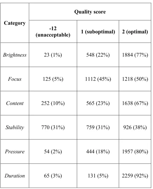
Methods: Retrospective analysis of a single-centre prospective observational study. Videos of the sublingual microcirculation were recorded using SDF videomicroscopy in 100 adult patients within 12 hours from admittance in the Intensive Care Unit and every 24 hours until discharge/death. Parameters of vessel density and perfusion were calculated offline for small vessels. For all videos, a quality score (-12=unacceptable, 1=suboptimal, 2=optimal) was assigned for brightness, focus, content, stability, pressure and duration. Videos with a total score ≤8 were deemed as unacceptable.
Results: A total of 2,455 videos (853 triplets) was analysed. Quality was acceptable in 56% of videos. Lower quality was associated with worse microvascular density and perfusion. Unreliable triplets (≥1 unacceptable or missing video, 65% of total) showed lower vessel density, worse perfusion and higher flow heterogeneity as compared to reliable triplets (p<0.001). Quality was higher among triplets collected by an extensively-experienced investigator or in patients receiving sedation or mechanical ventilation. Perfused vessel density was higher in patients with Glasgow Coma Scale (GCS) ≤8 (18.9 ± 4.5 versus 17.0 ± 3.9 mm/mm2 in those with GCS>8, p<0.001) or
requiring mechanical ventilation (18.0 ± 4.5 versus 17.2 ± 3.8 mm/mm2 in not mechanically
ventilated patients, p=0.059).
Conclusions: SDF video quality depends on both the operator’s experience and patient’s cooperation. Low-quality videos may produce spurious data, leading to an overestimation of microvascular alterations.
Keywords: microcirculation, sidestream dark field imaging, microcirculatory image quality, critically ill patients
BACKGROUND
The development of non-invasive videomicroscopy techniques, such as sidestream dark-field (SDF) imaging, has enabled the in vivo assessment of microcirculatory blood flow at the bedside, using the sublingual region as an accessible window to the microcirculation of inner organs [1]. SDF technology is incorporated into a hand-held video microscope system which epi-illuminates the tissue with green (530 nm) light-emitting diodes [2]. As this light is absorbed by hemoglobin, erythrocytes appear as dark globules against a grayish background. Sublingual microcirculatory alterations have been reported in different patient subsets, especially during sepsis [3, 4], and were associated with morbidity and mortality [5-7]. A number of studies have shown a dissociation between macro-hemodynamics and microcirculatory response to several interventions, including fluid infusion [8] and vasopressor administration [9]. These data would encourage the introduction of microcirculatory monitoring in the clinical practice as an additional target for therapy. Nonetheless, there are practical challenges to the widespread adoption of this technique in Intensive Care Units (ICUs), mainly the time-consuming offline video analysis and a long learning curve for good-quality image acquisition.
In 2007 a roundtable consensus conference indicated five key-points for optimal image acquisition (sampling 5 sites per organ, avoidance of pressure artefacts, elimination of secretion, adequate focus and contrast adjustment and high quality recording) [10]. However, obtaining good-quality videos may be challenging due to operator’s inexperience or poor patient’s cooperation. In a study by Sallisalmi et al., excellent technical quality was reported in only 30% of SDF videos [11]. Inadequate video quality may produce spurious microcirculatory data: blood flow may be artificially obstructed due to excessive pressure applied on the sublingual mucosa; inadequate focus and contrast or occluding artefacts (saliva bubbles or blood) may prevent the visualization of some blood vessels. Massey et al. have proposed a microcirculatory image quality score considering 6 domains of video quality: illumination, duration, focus, content, stability and pressure [12]. This score has never been used systematically and studies evaluating the microcirculation do not generally report any assessment of image quality. To our knowledge, no study has previously investigated to what extent a low microcirculatory image quality will affect the reliability of microcirculatory assessment.
We hypothesized that SDF video quality depends on both operator- and patient-related factors. Patient’s compliance may contribute crucially to high-quality video acquisition: as a result, more severe patients requiring sedation and/or mechanical ventilation could paradoxically show an apparently better microvascular perfusion as compared to less severe patients, merely due to an easier video recording. In this study, we investigated the relationship between SDF video quality
and the microcirculatory parameters obtained, and evaluated operator- or patient-related factors potentially influencing microcirculatory video quality.
METHODS
This is a retrospective analysis of data collected in a single-centre prospective observational study, the MICROcirculatory DAIly MONitoring in the ICU (MICRODAIMON-ICU) Study (NCT02649088, www.clinicaltrails.gov). A total of 100 adult (>18-year old) patients admitted to our 12-bed ICU between 1st April and 31st December 2013 were included in the study. Exclusion
criteria were factors impeding the sublingual microvascular evaluation (i.e. maxillo-facial trauma or surgery) and enrolment in the same study during a previous admission. The study protocol was approved by the local ethics committee of Azienda Ospedaliera Universitaria “Ospedali Riuniti” of Ancona, Italy. Written informed consent was obtained by the patients or their next of kin.
Microcirculatory image acquisition
The sublingual microcirculation was assessed using SDF imaging (Microscan, Microvision Medical, Amsterdam, The Netherlands) [2] within the first 12 hours of admission to the ICU and every 24 hours until discharge or death. In order to minimize variability due to operator’s experience with SDF technology, whenever possible video recording sessions were performed by one principal investigator (PI) with extensive (3 years approximately) clinical experience in SDF monitoring. In case of unavailability of the PI, video recording was performed by a group of adequately trained operators (6 to 12-month experience, on average). No additional sedation was provided during image acquisition. Secretions were gently removed with a gauze. Every effort was made to optimize contrast and focus and avoid pressure artefacts. Stable videos of at least 5 seconds’ duration [12] from 5 different sites of sublingual mucosa were recorded during each session. When adequate stability was difficult to achieve without increasing the pressure on the probe, priority was given to avoidance of pressure artefacts and videos shorter than 5s were accepted.
Analysis of microvascular density and perfusion
A random number was assigned to each video sequence through a random number generator. Three videos of the best available quality were selected from each sequence and analysed using the Automated Vascular Analysis software (Microvision Medical, Amsterdam, The Netherlands) by a group of 4 experienced investigators who had not participated in the video acquisition. The image was divided by three equidistant horizontal lines and three equidistant vertical lines for the
calculation of the De Backer score: this resulted from the number of vessels crossing the lines, divided by the total length of the lines [13]. For each vessel crossing the lines, perfusion was categorized as continuous, sluggish, intermittent or absent. The percentage of perfused vessels (PPV) was estimated as follows: 100 × [(total number of grid crossings - [no flow + intermittent flow])/total number of grid crossings]. Total vessel density (TVD) was calculated as the total length of vessels divided by the total area of the image [10]. The perfused vessel density (PVD) was estimated by multiplying TVD by PPV as estimated with the De Backer method [14]. Microvascular flow index (MFI) was calculated semiquantitatively as described elsewhere [15]. For each video sequence, values obtained from 3 sites were averaged. The flow heterogeneity index (FHI) was calculated as the highest MFI minus the lowest MFI, divided by the mean MFI of all sublingual sites [10]. For this study, analyses were focused on small vessels (≤20 µ in diameter).
Analysis of microcirculatory video quality
This was performed by the same investigators before the analysis of microcirculatory parameters. The microcirculatory image quality score used in this study was adapted from the one proposed by Massey et al. [12] (Table 1). For each video, a score of -12 (unacceptable), 1 (suboptimal but acceptable) or 2 (optimal) was given for each category of quality (brightness, focus, content, stability, pressure, duration). A video was defined as unacceptable if it scored any “-12” in any category or “1” in at least 4 categories (total score ≤8). Among unacceptable videos, quality was categorized as bad (“-12” for at least three categories, total score ≤-30) or poor (“-12” in less than three categories, total score between -29 and 8). Among acceptable videos, quality was defined as
good (total score of 9-11) or excellent (“2” for all categories, total score of 12). A video triplet was
defined as reliable if it was of excellent (all videos of excellent quality) or acceptable (3 acceptable or excellent videos) quality, unreliable if it included at least one video of unacceptable quality or if less than three videos had been suitable for analysis.
Statistical analysis
This was performed using GraphPad Prism Version 6 (GraphPad Software, La Jolla, CA, USA). Normality of distribution was checked using the Kolmogorov-Smirnov test. Unpaired t-test or Mann Whitney U-test were used for comparisons of continuous variables, as appropriate. One-way analysis of variance (ANOVA) or Kruskal-Wallis test with Bonferroni or Dunn’s post-hoc testing were used for comparisons between more than two groups, as appropriate. The chi-square
test was used for comparison of proportions. The alpha level of significance was set a priori at 0.05.
RESULTS
A median of 6 [3-12] video recording sessions per patient was performed. A total of 2,455 microcirculatory videos from 100 different patients were analyzed. This corresponded to a total of 853 video triplets. Of these, 583 (68%) had been recorded by the PI. Quality score for each single domain is reported in Table 2. Based on our predefined criteria, only 1,365 videos out of 2,455 (56%) were defined as acceptable, 27% of these being of excellent quality (Figure 1). If considering video triplets, only 301 triplets out of 853 (35%) were judged as reliable, with 4% of total being of excellent quality, while 552 (65%) were of unacceptable quality. Of these, 72 (8% of total) were incomplete triplets where less than three videos had been suitable for analysis (104 videos lacking in total). As a result, 67 [44-89] % of microcirculatory assessments performed in each patient were deemed unreliable. Only 8 patients out of 100 had reliable microcirculatory data for all the sessions performed, while we were unable to take any reliable video triplets in 20 patients.
Video quality and microcirculatory parameters
Videos of lower quality for each category were generally associated with lower vessel density, PPVs and MFIs as compared to videos of optimal quality (Electronic Supplementary Material, ESM 1 and 2). This trend was less pronounced for the category “stability”, for which unacceptable videos showed slightly lower vessel densities but higher PPVs, without any differences in MFIs depending on video quality. Vessel density, PPVs and MFIs were progressively lower with decreasing overall video quality (Figure 2). All microcirculatory parameters varied significantly between reliable and unreliable video triplets, with unreliable triplets indicating worse vessel density and perfusion and higher flow heterogeneity (Electronic Supplementary Material, ESM 3).
Influence of operator and patient-related factors on video quality and microcirculatory assessment
Triplets recorded by the PI were less likely to be of unacceptable quality (60% versus 74.8% among triplets collected by other investigators, p=0.041, Electronic Supplementary Material, ESM 4) and yielded higher PVD (18.2 ± 4.1 versus 17.4 ± 4.8 mm/mm2, p=0.001) as compared to those
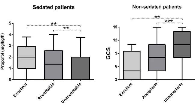
recorded by other operators. The vast majority of triplets recorded in non-sedated or not mechanically ventilated patients were of unacceptable quality (81% and 96% respectively), while the percentage of unreliable video triplets was lower among sedated or mechanically ventilated patients (55% and 63% respectively) (p<0.001, Electronic Supplementary Material, ESM 5). Among sedated patients, a higher video quality was associated with higher doses of propofol being administered at the time of microcirculatory assessment (Figure 3). Among non-sedated patients, a lower video quality was associated with higher values of Glasgow Coma Scale (GCS) (Figure 3), and those with GCS ≤8 showed higher PVD as compared to those with GCS >8 (18.9 ± 4.5
versus 17.0 ± 3.9 mm/mm2, p<0.001). Similarly, mechanically ventilated patients tended to show
a higher PVD as compared to those not mechanically ventilated (18.0 ± 4.5 versus 17.2 ± 3.8 mm/mm2, p=0.059).
DISCUSSION
This is the first study that evaluated the impact of microcirculatory image quality on the assessment of sublingual microvascular density and perfusion, using one of the largest existing databases and including a heterogeneous population of 100 critically ill patients monitored in different moments over the course of their acute condition. Our first finding was that almost half of the videos collected were of unacceptable quality, resulting in 65% of video triplets being unreliable for a clinical assessment of microvascular perfusion. Secondly, parameters of microvascular density and flow varied together with image quality, with videos of lower quality suggesting lower vessel densities and a more altered perfusion. Thirdly, not only the investigator’s experience in SDF technology, but also patient-related factors (mainly the neurological state) played a role in determining a good-quality video recording.
Since the first appearance of Orthogonal Polarization Spectral imaging more than 15 years ago [16], an increasing number of studies has explored the sublingual microcirculation and its response to several interventions in different patient populations [17]. Several authors supported the potential role of the microcirculation as a target for therapy [18-20]. Nonetheless, before introducing any monitoring device in the clinical practice, it is necessary to understand the potential limitations of the technique in order to obtain reliable information. Only a few studies addressed the limitations of sublingual videomicroscopy [11, 12]. Our study highlights the technical challenges to the widespread adoption of sublingual microcirculation monitoring in ICU patients. A substantial proportion of the videos recorded in each patient was of unacceptable quality. In 20 patients out of 100 we failed to perform any reliable microcirculatory assessment during their stay in the ICU. These data add to the results of Sallisalmi et al. [11], who reported a success rate as low as 8.5% of video recording sessions in creating video triplets of adequate quality. More importantly, our study demonstrates that low-quality videos can introduce a substantial bias in microcirculatory assessment. The analysis of videos with defects in focus or brightness, as well as artefacts such as secretions or blood, yielded significantly lower vessel densities, PPV and MFI. Similarly, the presence of pressure artefacts was associated with lower vessel density and worse perfusion. Even if they can be recognized and distinguished from real flow alterations by a well-trained investigator during video analysis, pressure artefacts preclude a reliable evaluation of microvascular flow. The first step for the introduction of the microcirculation

![Table 1 – Microcirculatory image quality score, as adapted from Massey et al. [12].](https://thumb-eu.123doks.com/thumbv2/123dokorg/2967625.27040/50.892.108.822.163.1146/table-microcirculatory-image-quality-score-adapted-massey-et.webp)