UNIVERSITA’ POLITECNICA DELLE MARCHE
SCUOLA DI DOTTORATO DI RICERCA - FACOLTÀ DI MEDICINA E CHIRURGIA DIPARTIMENTO DI SCIENZE CLINICHE E MOLECOLARI
Osteoarticular damage in the knee: new frontiers in joint
regeneration and substitution
Tesi di Dottorato di:
Dott. Davide Enea
Tutor:
Chiarr.mo Prof. Antonio Gigante
Coordinatore del Curriculum:
Chiarr.mo Prof. Roberto Di Primio
Abstract
Acute cartilage injury represents a major clinical problem affetting patients younger and younger since competitive sport activity has become largely widespread. The so-called biological resurfacing has moved its first steps in the early 90’s with the aim to restore native cartilage and to avoid or delay the need for joint substitution. Since then, great technical and medical progress has occurred. However the target to reproducibly restore native hyaline (or even hyaline –like) cartilage with good mechanical properties has still been missed. In fact most of the cartilage we are able to regenerate is fibrocartilage, with scarce mechanical properties, which, does not guarantee long-term durability and prevention of osteoarthrosis. Therefore the efforts of the scientific community are still directed to improve the quality of the regenerated cartilage. In the last few years the possible combination of multipotent, autologous mesechimal cells and scaffolds has emerged as viable option to target hyaline cartilage regeneration.
The first part of this thesis focuses on the association of a source of mesechchimal stem cells - the bone marrow concentrate - and two different scaffolds; one made of collagen and the other made of a combination of hyaluronan and polyglycolic acid. The scaffold and the cells are associated in two novel surgical techniques for cartilage repair in the knee. The outcomes of these techniques have been analysed and compared from the functional arthroscopic and histological point of view. We hypothesize that both these techniques have the potential to regenerate hyaline or hyaline-like cartilage and painless motion.
Chronic joint degeneration is a major social problem since the average age of western countries is increasing year after year and so is the need to move and be active even later in life. Osteoarthritis of the knee is one of the first causes of pain and disability among the elderly people. Knee substitution (i.e. total knee replacement) aims to re-establish knee function and painless motion. In the past forty years knee prosthesis have made huge progress in terms of design improvments, material developments and clinical outcome. Even though most of the patients who have undergone a knee substitution have good or excellent results, it remains a percentage of patients scarcely satisfied because of pain, instability, tightness or failure of the implant. Therefore there is still room to improve this highly successful procedure.
Among the different prosthetic designs the conforming, mobile bearing designs are the ones that guarantee low shear stress, wear and debris production and therefore maximum durabiliry of the implant. Fully conforming mobile bearing prosthetic design may be implanted both preserving and resecting the posterior cruciate ligament. The role of the posterior cruciate ligament in mobile bearing prosthesis is unclear since there are prosthetic designs which require its sacrifice and others that allow its preservation.
The second part of this thesis focuses on the clinical and functional outcomes of patients who underwent total knee replacement with a mobile bearing prostetic design either with or without posterior cruciate ligament sacrifice. We hypotesize that patients with posterior cruciate ligament preservation may have better knee motion and feeling due to increased proprioception linked to the ligament retainment.
List of peer-reviewed publications arising from this thesis
1. Single-stage cartilage repair in the knee with microfracture covered with a resorbable
polymer-based matrix and autologous bone marrow concentrate.
Enea D, Cecconi S, Calcagno S, Busilacchi A, Manzotti S, Kaps C, Gigante A.
Knee. 2013 Dec;20(6):562-9
2
.
One-step cartilage repair in the knee: collagen-covered microfracture and autologous bone marrow concentrate. A pilot study.Enea D, Cecconi S, Calcagno S, Busilacchi A, Manzotti S, Gigante A.
Knee. 2015 Jan;22(1):30-5
3. Retained versus resected posterior cruciate ligament in mobile-bearing total knee replacement: a
retrospective, clinical and functional assessment.
Enea D, Cigna V, Sgolacchia C, Tozzi L, Verdenelli A, Gigante A.
Table of contents
PART I NEW FRONTIERS IN JOINT REGENERATION
1. CARTILAGE AND CARTILAGE REPAIR STRATEGIES 10
1.1 INTRODUCTION 10
1.2 COMPOSITION OF ARTICULAR CARTILAGE 10
1.3 TYPES OF CARTILAGE INJURY 12
1.4 THE ROLE OF MAGNETIC RESONACE IMAGING 13
1.5 SURGICAL TECHNIQUES FOR CARTILAGE REPAIR 14
1.5.1 LAVAGE AND DEBRIDEMENT 14
1.5.2 MICROFRACTURE AND PERFORATION 14
1.5.3 MICROFRACTURE AND SCAFFOLD 16
1.5.4 OSTEOCHONDRAL AUTOGRAFT TRANSFER (OAT) -‐ MOSAICPLASTY 16 1.5.5 AUTOLOGOUS CHONDROCYTE IMPLANTATION (ACI) 18 1.5.6 MATRIX ASSISTED CHONDROCYTE IMPLANTATION (MACI) 19
1.5.7 OSTEOTOMIES AND MENISCAL TRANSPLANTS 21
1.5.8 HYALURONIC ACID 22
1.5.9 POSTOPERATIVE CARE 23
1.6 CONCLUSION 23
2. ARTICULAR CARTILAGE ENGINEERING 24
2.1 TISSUE ENGINEERING CONCEPT 24
2.2 SCAFFOLDS 24
2.2.1 NATURAL POLYMERS 25
2.2.2 SYNTHETIC POLYMERS 26
2.3 CELLS 27
2.4 GROWTH FACTORS 30
3. ONE-‐STEP CARTILAGE REPAIR WITH POLYGLYCOLIC ACID/HYALURONAN MEMBRANE ASSOCIATED WITH MICROFRACTURE AND BONE MARROW CONCENTRATE 31
3.1 INTRODUCTION 31
3.2 MATERIALS AND METHODS 33
3.2.1 STUDY DESIGN 33
3.2.2 SURGICAL TECHNIQUE 33
3.2.3 SECOND-‐LOOK ARTHROSCOPY 36
3.2.4 HISTOLOGY 36
3.2.5 STATISTICAL ANALYSIS 36
3.3 RESULTS 37
3.3.1 CLINICAL OUTCOME 37
3.3.2 ARTHROSCOPIC AND MRI EVALUATION 39
3.3.3 HISTOLOGICAL EVALUATION 39
3.4 DISCUSSION 40
4. ONE-‐STEP CARTILAGE REPAIR WITH COLLAGEN MEMBRANE ASSOCIATED WITH
MICROFRACTURE AND BONE MARROW CONCENTRATE 45
4.1 INTRODUCTION 45
4.2 MATERIALS AND METHODS 47
4.2.1 STUDY DESIGN 47
PhD Thesis 2015 5 Dr. D. Enea
4.2.3 SECOND-‐LOOK ARTHROSCOPY 50
4.2.4 HISTOLOGY 51 4.2.5 STATISTICAL ANALYSIS 51 4.3 RESULTS 51 4.3.1 CLINICAL OUTCOME 51 4.3.2 MRI EVALUATION 52 4.3.3 ARTHROSCOPIC EVALUATION 54 4.3.4 HISTOLOGICAL EVALUATION 54 4.4 DISCUSSION 55
5. COLLAGEN-‐COVERED VERSUS POLYGLYCOLIC ACID/HYALURONAN-‐COVERED
MICROFRACTURE AND BONE MARROW CONCENTRATE. 59
5.1 RESULTS 59 5.1.1 CLINICAL OUTCOME 59 5.1.2 ARTHROSCOPIC EVALUATION 60 5.1.3 HISTOLOGICAL EVALUATION 61 5.2 DISCUSSION 64 5.3 CONCLUSIONS 67 PART II NEW FRONTIERS IN JOINT SUBSTITUTION
6. BRIEF HISTORY OF CONDYLAR KNEE REPLACEMENT 69
6.1 INTRODUCTION 69
6.2 ANATOMICAL APPROACH 72
6.3. FUNCTIONAL APPROACH 78
6.4 MOBILE BEARING (MB) KNEE 81
6.4.1 PARTIALLY CONFORMING MB 82
6.4.2 FULLY CONFORMING MOBILE BEARING 84
6.4.3 POSTERIOR STABILIZED MB 85
6.5 FUTURE PERSPECTIVE 86
7. THE ROLE OF POSTERIOR CRUCIATE LIGAMENT IN MOBILE-‐BEARING TOTAL KNEE
REPLACEMENT 87
7.1 AIM OF THE STUDY 87
7.2 INTRODUCTION 87
7.3 MATERIAL AND METHODS 90
7.4 RESULTS 92 7.5 DISCUSSION 94 7.6 CONCLUSIONS 97 REFERENCES 98 IV
List of Figures
Fig. 1.1. The zonal architecture of articular cartilage Fig. 1.5.2.1 The microfracture technique
Fig. 1.5.2.2 Autologous matrix induced chondrogenesis (AMIC) Fig. 1.5.4.1 Mosaicplasty
Fig. 1.5.5.1 Autologous chondrocyte implantation (ACI)
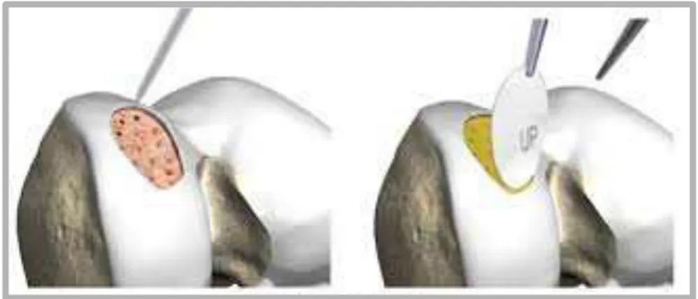
Fig. 1.5.6.1 The matrix assisted chondrocite implantation (MACI) on the patella. Fig. 3.1 Arthroscopic CMBMC technique
Fig. 4.1 Harvest phases
Fig. 4.2 Arthroscopic technique
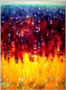
Fig. 5.1 : Biopsies stained with safranin-O
Fig 6.1.1 Gluck’s ivory knee
Fig. 6.1.2 Campbell’s interposition device Fig 6.1.3 McKeever tibial replacement
Fig. 6.1.4 Freeman-Swanson knee and a postoperative ICLH radiograph Fig. 6.1.5 Polycentric knee
Fig 6.1.6 Geomedic knee Fig. 6.1.7 Duocondilar knee
Fig. 6.2.1 Kodama-Yamamoto knee Fig. 6.2.2 The UCI knee
Fig. 6.2.3 The Anatomical Knee Fig 6.2.4 The Leeds Knee Fig. 6.2.5 Duopatella knee Fig. 6.2.6 PFC Knee
Fig 6.2.7 The Kinematik Stabilizer knee Fig. 6.2.8 The PCA knee
Fig. 6.2.9 The Miller-Galante knee Fig. 6.3.1 Total Condylar knee
Fig. 6.3.2 IBPS metal backed (IBPS-II) and allpoly (IPBS-I)
Fig. 6.3.3 The Bisurface knee prosthesis Fig. 6.3.4 The Medial Pivot Knee
Fig. 6.3.5 LCS rotating platform (low contact stress) knee Fig. 6.4.1 The LCS meniscal bearing knee
Fig. 6.4.1.1 The Self Alining MB knee Fig. 6.4.1.2 The TACK knee
Fig. 6.4.1.3 A radiograph of the Interax Integrated Secure Asymmetric knee Fig. 6.4.2.1 The Rotaglide total knee system
Fig. 6.4.2.2 The Medially Biased Kinematics Knee (MBK)
Fig. 6.4.3.1 The PFC sigma RPF knee Fig. 6.4.3.2 The LPS mobile flex knee Fig. 7.1 The GKS Prime
List of Tables
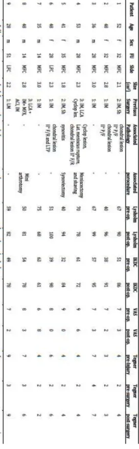
Table 3.1 Characteristics of patients prior to surgery
Table 3.2 Outcome data
Table 4.1 Patient characteristics and outcome data
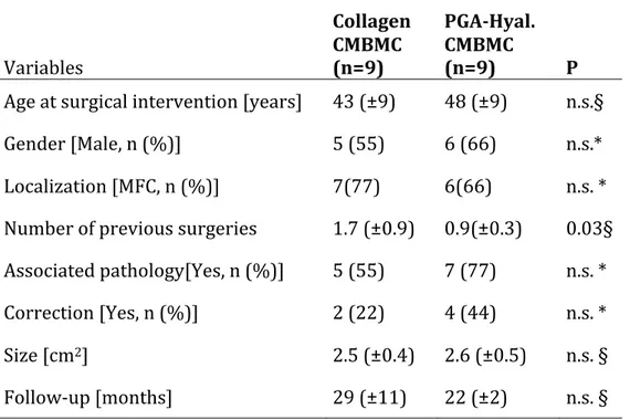
Table 5.1 Baseline characteristics of patients according to treatment
Table 5.2 Patient-reported outcome data
Table 7.1 Incomplete list of mobile-‐bearing prosthetic designs
Table 7.2 Preoperative demographics
List of abbreviation
AC = Articular cartilageACI = Autologous chondrocyte implantation ACL = Anterior cruciate ligament
AMIC = Autologous matrix induced chondrogenesis BMC = Bone marrow concentrate
BMP’s = Bone morphogenetic proteins CCI = Characterised chondrocyte implantation
C-CMBCM = collagen-covered microfracture and bone marrow concentrate COMP = Cartilage oligomeric protein
CPM = Continuous passive motion CR = Cruciate retaining
CS = Cruciate sacrificing
CMBMC = covered microfracture and bone marrow concentrate ECM= Extra cellular matrix
FB = Fixed bearing
FDA = Food and drug administration FGF-2 = Fibroblast growth factor 2 GKS = Global knee system
FSE = Fast Spin-Echo GRE = Gradient echo
IBPS = Insall-Burstein posterior stabilized IGF-1 = Insulin-like Growth Factor-1 KSS = Knee society score
IKDC = international knee documentation committee LCS = Low Contact Stress
OA = osteoarthritis
MACI = Matrix assisted chondrocyte implantation MB= Mobile bearing
MBK = Medially biased kinematics MFX = Microfracture
MRI= Magnetic resonance imaging MSC = Mesenchymal stem cell OA = Osteoarthritis
OAT = Osteochondral autograft transfer PCA = Porous-coated anatomical knee PCL = Posterior cruciate ligament PDGF = Platelet derived growth factor
PGA-HA-CMBCM = Polyglycolic acid/hyaluronan-covered microfracture and bone marrow concentrate PRP =Platelet Rich Plasma
PS = Postero-stabilized ROM = Range of motion VAS = Visual analogic scale ST = Standard deviation TC = Total condylar
TGF-β = Transforming growth factor beta TKR = Total knee replacement
TRAC = Two Radii Area Contact
Part I
New Frontiers in
Joint Regeneration
1.
Cartilage and cartilage repair strategies
1.1 INTRODUCTION
Lesions of the articular cartilage (AC) are a common pathology of joints which can affect people of all ages resulting in a wide range of clinical presentations. Articular cartilage defects at the time of arthroscopy are present in about 60% [1] of knees however, it is unclear how many of these defects are actually symptomatic and require surgical intervention. The population continues to live longer and remains active into later life and as a result the number of cartilage surgeries performed is increasing, (more tha half a million procedures performed yearly in the US).
Hunter observed in 1743 that “cartilage once destroyed never heals”. Nowdays this is not entirely true. Although AC has a poor ability to regenerate itself, there is potential for repair. Understanding of AC pathophysiology is improving, yet the mechanism of regeneration of hyaline cartilage continues to elude us. What is clear is the distinction between traumatic and degenerative lesions (i.e. osteoarthritis). Any particular type of lesion may require a specific form of treatment.
1.2 COMPOSITION OF ARTICULAR CARTILAGE
Hyaline cartilage is a highly specialised tissue with a regional organisation. Figure 1.1 shows the zonal architecture of hyaline cartilage. The osteochondral unit is made up of subchondral bone, calcified cartilage and the, radial, transitional and tangential or superficial zones of the articular cartilage itself.
Fig. 1.1. The zonal architecture of articular cartilage
It develops along a path of bone morphogenesis therefore it’s production is under similar conditions and signalling factors as bone [2]. The growth plate separates cartilage from bone via a line of proliferating chondrocytes which go on to hypertrophy and form bone. It is at this stage that the cell numbers in AC do not multiply, instead the bulk of the tissue is made up of extracellular matrix (ECM).
AC contains 60-80% water [3]. The predominant cell type is the chondrocyte (5% wet weight) which is responsible for ECM production. They are spherical and surrounded by round small lacunae, but become more flattened as they get closer to the superficial zone, where they are fibroblastic in shape. They often group together in columns forming chondrons (2-4 cells), which are orientated along collagen fibres [4]. The ECM is made up of collagen fibres (25% wet weight) of which Type II predominates (95%), but also includes types VI, IX and XI with type X in the calcified layer. These fibres are anchored to the calcified layer then run perpendicularly to it crossing each other in arcs at the superficial zone. The cells within the superficial zone are thus orientated horizontally and along with the collagen network provide resistance to shear. They also
secrete lubricin (also known as Superficial Zone Protein), a molecule which is responsible for reducing the coefficient of friction and thus providing cartilage with such favourable tribiological properties .
Negatively charged hydrophilic proteoglycan molecules (mostly aggrecan and hyaluronan) protrude from the net of collagen fibers. The dense collagen network is somewhat restrictive to the hydration of these molecules thus they are only hydrated to about 40-60%. As a result, the swelling pressure which is generated provides the compressive stiffness of cartilage [5]. During the early degenerative process, when the collagen fibres are disrupted, the proteoglycans can become more hydrated thus causing the cartilage to become bulcky and soft.
Smaller glycoproteins also exist, fibronectin and cartilage oligomeric protein (COMP) which have a role in cell adhesion, and growth factors such as Bone Morphogenetic Proteins (BMP’s). Their role is under intense investigation and in many ways remain least understood [5].
Tissue turnover is mostly governed by a balance between the matrix metalloproteinases (MMP-3, MMP-8, MMP-9, MMP-13 and aggrecanases 4&5 predominating) and the Tissue Inhibitors of Metalloproteinases (TIMPS). Over expression of one or other is a likely contributor of OA [6]. AC is avascular and aneural and receives its nutrition from the synovial fluid, as a result of mechanical movement of the tissue producing a diffusion gradient. AC is immunopriveledged in that it does not contain immune cells. Therefore chondrocytes secrete lysozyme to counteract microorganisms [5].
1.3 TYPES OF CARTILAGE INJURY
Once the cartilage is damaged, a degenerative process begins. The aetiology of cartilage degeneration which leads to osteoarthritis is multifactorial and many different risk factors have been implicated. Mechanical factors such as direct trauma, instability, malalignment and loss of meniscal chondroprotection have a role, as do metabolic factors such as diabetes, alcohol abuse and obesity [7]. It is therefore difficult to target one specific cause.
The depth of the injury will influence the healing potential of cartilage. Cartilage defects which do not extend to the subchondral bone do not spontaneously for the small number of chondrocytes within the ECM, their inability to migrate to the zone of injury and their relative inability to regenerative large amounts of ECM. It means these defects will usually progress. Full thickness defects which penetrate the subchondral bone do have the potential for intrinsic repair due to the communication gained with the marrow cavity and the mesenchymal stem cell (MSC) population which allow repair tissue to be. However, this is not a regenerate tissue as the repair tends to be fibroblastic in origin [8]. Eearly OA behaves very differently. An increase in matrix molecule synthesis is often recognised. However, once loss of matrix eventually exceeds that which is deposited a net loss of ECM results. The chondrocytes are noted to proliferate and form clusters, and cell hypertrophy is often observed. Loss of chondrocytes in the superficial zone occurs followed by fibrillation, fissuring, erosion, subsequent denudation of bone and subchondral alteration [5].
1.4 THE ROLE OF MAGNETIC RESONACE IMAGING
Pain and mechanical symptoms such as locking or catching are characteristic of eraly stage cartilage injury. Loss of movement and reduced function predominate in the latest stages of OA. Magnetic Resonance Imaging is the gold standard for diagnostic imaging and treatment planning. A 1.5T magnet or stronger must be used with a dedicated extremity coil to be able to use MRI as an assessment tool. The main sequences employed are T2 weighted Fast Spin-Echo (FSE) with or without fat suppression and T1 weighted 3D gradient echo (GRE). The FSE images show cartilage to be dark in contrast to the high signal of synovial fluid and bone marrow. Thus surface and matrix irregularities will be shown with increased signal. GRE sequences produce high signal intensity in the cartilage compared to that low in bone and synovial fluid. The 3D nature of these images allows improved visualisation and volume measurements [9].
1.5 SURGICAL TECHNIQUES FOR CARTILAGE REPAIR
Prior to consider a cartilage repair procedure, the surgeon has to plan the treatment according to biology of the defect and the physical condition and requirements of the patient. The strong will of the patient to take part in an extended rehabilitation programme should be ascertained. The nature of the patient’s employment and sporting activities will have a significant impact on what procedure is chosen. Demotivated patients will have to be advised not to performe any cartilage repair procedure.
1.5.1 Lavage and Debridement
This procedure is a palliative and no tissue regeneration may be obtained with this technique. The procedure consists in the removal of unstable flaps of cartilage and loose bodies and should be performed if the patient exhibits mechanical symptoms. Early mobilisation and weight bearing as tolerated is encouraged and this is the only real strength of this procedure. A study which compared arthroscopic lavage to placebo found that neither arthroscopic lavage nor debridement were better than placebo [10]. Debridement should not be offered as a treatment apart from in patients who suffer mechanical symptoms and signs in whom more aggressive treatments are not acceptable.
1.5.2 Microfracture and Perforation
Microfracture or perforation of the subchondral bone represent the so-called “Marrow stimulation techniques”. They involve penetrating the subchondral bone plate to allow bone marrow blood in the joint cavity. This allows marrow stromal cells containing mesenchymal stem cells (MSC’s), platelets and other chemotactic factors to collect within the defect. The original techniques described such as Pridie drilling have gone out of favour for the more refined microfracture technique described by Steadman [11, 12] (Fig. 1.5.2.1). Following debridement of the perilesional cartilage producing a perpendicular shoulder of cartilage, 3-4 mm deep holes are made in the subchondral bone 2-3mm apart, starting in the periphery, working into the centre of the defect. Unlike Pridie drilling, this maintains greater mechanical stability of the subchondral bone and collapse is not seen as readily as in previous techniques.. The rehabilitation seems to form an
integral part of the procedure and the original authors maintain that the intensive rehab period must be adhered to for the procedure to be successful. The procedure is easy to perform, cheap, minimally invasive and well tolerated by the patient. Recently nanofracture have been introduced.
Fig. 1.5.2.1 The microfracture technique
Studies have shown microfracture is able to give a fibrocartilagenous reparative response which mostly provides symptom relief for up to 2 years. Steadman et al have published data on 72 patients with defects less than 4cm2 who underwent microfracture with follow-up of 7-17 years [11]. Eighty percent of patients improved with Lysholm scores improving from 59 to 89, and Tegner activity scores improving from 6 pre-operatively to 9 post-operatively. Gobbi reported similar results in a group of athletes but at 2 years noted that 80% of patients had a reduction in Tegner score [13]. This may be as a result of the less durable fibrocartilage which is produced but also the poor fill of the defect, particularly around the margins with poor integration with the native articular cartilage, which may also be related to the size of the defects treated. This was shown histologically by Dorotka et al. in an ovine model, with improved defect fill found when the clot was stabilised with a collagen membrane [14].
1.5.3 Microfracture and scaffold
To overcome the shortcomings of the original thechnique microfracture has been augmented with a collagen I/III matrix (Chondroguide, Geistlich Biomaterials) which soak up the blood clot from the subchondral holes, providing a scaffold for cells and chemotactic factors to reside (Fig. 1.5.3.1). The membrane can be fixed with fibrin glue or 6/0 vicryl sutures. Autologous Matrix Induced Chondrogenesis (AMIC) has shown promising results however we await long term data [15]. This may represent the future of marrow stimulation techniques. The first part of this thesis will focus on a novel surgical technique that represents an evolution of the original AMIC technique.
Fig. 1.5.2.2 Autologous matrix induced chondrogenesis (AMIC)
1.5.4 Osteochondral autograft transfer (OAT) -‐ Mosaicplasty
In this technique osteochondral plugs are harvested from a non-weight bearing articular surface (lateral trochlear ridge or notch) and transferred to a pre-prepared cylindrical hole within the defect. It is the only procedure which produces hyaline cartilage within the defect,. For single plug transfer, the size of the defect will be the limiting factor. Problems such as joint congruency and donor site availability are encountered hence mosaicplasty techniques have been developed.
Mosaicplasty involves the transfer of a number of smaller defects, allowing a congruent joint surface (Fig. 1.5.4.1). It is extremely technically demanding and donor site morbidity remains a problem (lateral trochlear ridge not truly non-weight bearing). Both techniques suffer from poor
graft incorporation, with mosaicplasty particularly suffering with poor integration in gaps between the plugs and native cartilage.
Fig. 1.5.4.1 Mosaicplasty
Mosaicplasty has shown mixed results with a number of studies showing superior outcomes to marrow stimulation techniques. Hangody et al showed that 87% of patients had good to excellent results at 5 years following mosaicplasty compared with 0-34% of patients who were randomised to 1 of 3 marrow stimulation techniques [16]. In a further study looking at a group of competitive athletes, the same group found 100% of patients had a good to excellent result at greater than one year with 63% returning to full sports [17]. Bentley et al have since shown improved outcomes with Autologous Chondrocyte Implantation over mosaicplasty in their RCT with 88% showing good to excellent results compared to 69% at a mean of 19 months [18].
Although good results have been reported the use of mosaicplasty seems to be less widely utilised due to the technical difficulty, problems with congruency and donor site morbidity. The use of a single osteochondral transfer in small isolated defects is more acceptable.
1.5.5 Autologous Chondrocyte Implantation (ACI)
The process of transplanting autologous chondrocytes in suspension to a cartilage defect was first described clinically by Brittberg et al in 1994 [19]. A biopsy of normal cartilage from the non weight bearing aspect of the ipsilateral knee is taken during the initial arthroscopic procedure. Chondrocytes are then isolated and expanded in-vitro. The cell suspension is then returned to the defect during the second stage procedure, and held in place with a periosteal patch, harvested from the proximal tibia, sutured over it to keep the cells in place. This procedure has since evolved to using a collagen membrane instead of periosteum: is called second generation ACI or ACI-C. Large defects (>4cm2) can be treated as can multiple or even in some cases, kissing lesions [20] (Fig. 1.5.5.1).
Good to excellent results have been reported in 85-92% of patients at 2 years in a number of observational cohort studies [21]. A number of randomised studies have been published comparing ACI to other cartilage repair procedures. Horas et al showed that an improvement in symptoms could be established with both ACI and OATS however the speed of recovery of ACI was slower[22]. Bentley et al compared ACI to mosaicplasty in 100 patients with similar demographics and lesion size. At a mean of 19 months, 89% of patients who had ACI showed good to excellent results compared with 69% in that of mosaicplasty [18]. But Dozin et al found an improvement in 88% of mosaicplasty patients in their study compared to 68% in the ACI group. The numbers in this study are small, and interestingly, 31.4% of patients were excluded due to improvement in symptoms following debridement at the time of the arthroscopic assessment/biopsy.They were not included in the final analysis .
There are randomised controlled trials comparing ACI to microfracture [23, 24]. The study by Knutsen et al. has shown no difference in clinical and radiographic outcome between the groups at 5 years, however, the study is underpowered. No correlation was found between histology grade and clinical outcome however those with the best histology at two years did not exhibit any failures. The more recent study by Saris et al. incorporates a novel chondrocyte characterisation system which supposedly correlates with improved histology and outcome. One year results favour ACI over microfracture in terms of histomorphometry however functional outcome as measured by the KOOS score is similar in both groups at 12-18 months assessment.
1.5.6 Matrix Assisted Chondrocyte Implantation (MACI)
Matrix assisted chondrocyte implantation (MACI) (Genzyme, Cambridge, MA) was among the first variations from ACI original technique and has been in clinical use for a number of years. This technique is also numbere among the so-colled third-generation ACI (Fig. 1.5.4.1). MACI employs a collagen matrix on which chondrocytes are expanded in-vitro and then transferred into the defect. The membrane can be held with fibrin glue or sutures. Benefits of this include the ease of
application of the membrane and also the possibility of performing the procedure totally arthroscopically.
Two further randomised trials, the first by Bartlett et al has shown ACI and MACI to be comparable at 1 year [25], and that done by Gooding et al [26] which has shown similar functional outcome at two years between ACI with periosteum patch compared to a collagen membrane. Of note, 36% of the periosteum group required debridement of the graft due to periosteal hypertrophy.
Other problems with this technique exist. Two operations are required, it is very expensive to culture cells and the repair tissue still is not reproducibly hyaline cartilage. However, in a histological study by Briggs et al examining ACI-C grafts one year following implantation, mRNA for type II collagen was found to be produced by chondrocytes, irrespective of the quality of the repair tissue[27]. Similarly, our group has shown that MACI can lead to the formation of hyaline-like cartilage tissue [28], and that patellar MACI can lead to good results when associated to tibial tuberosity trasposition [29]
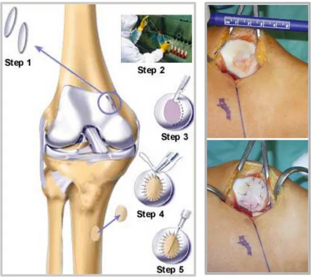
Fig. 1.5.6.1 The matrix assisted chondrocite implantation (MACI) on the patella. From Gigante et al 2009 [29].
Other products currently on the market which are similar to MACI include Hyalograft C (Fidia Farmaceutci, Italy), Bioseed-C (BioTissue Technologies, Freiburg, Germany), CaRes (Arthrokinetics, Macclesfield, UK), Cartipatch (TBF Tissue Engineering, Bron, France) and Novocart (TETEC AG, Reutlingen, Germany). Each utilises in-vitro culture of chondrocytes in the
scaffold matrix followed by implantation. This enables 3D culture of chondrocytes preventing dedifferentiation down a fibroblastic lineage and loss of phenotype. Novel methods of implantation and fixation are developing allowing minimally invasive and arthroscopic techniques to be used. Although many of these products have produced satisfactory clinical outcomes, we wait to see if they provide any benefit over other aforementioned cartilage repair techniques.
1.5.7 Osteotomies and meniscal transplants
Joint environment preservation is essential to successful cartilage repair. If mechanical imbalance malalignment, instability or the lack of meniscus is not corrected the repair will undergo abnormal mechanical loads. Combinations of distal femoral, high tibial or tibial tubercle osteotomy with anterior cruciate ligament reconstruction and meniscal allograft/collagen meniscal implant can be done simultaneously or staged, depending on the degree and amount of correction required.
Varus/Valgus osteotomy: In physiological loading of the knee 60% is transmitted through the
medial compartment and 40% through the lateral. Although there is no good clinical evidence to support it, more authors suggest performing staged or simultaneous osteotomy to unload a defect/repair if the mechanical axis falls within the affected compartment. Mina et al have demonstrated in a cadaveric model that load is equally distributed in both compartments if a corrective osteotomy is performed to 0-4 degrees valgus. Complete unloading of the medial compartment can be achieved with 6-10 degrees of valgus [30]. The problem with trying to prove this concept is that if procedures are performed simultaneously it is difficult to ascertain which has the biggest impact on the functional outcome. To perform a randomised control trial would take large numbers and likely be very difficult to recruit to achieve statistical power. We are therefore left with published case series such as those by Minas who found an improvement in Cincinnati rating scale and SF-36 scores at two years in a group of 71 salvage cases where multiple defects were treated simultaneously with osteotomy and ACL reconstruction. Ninety percent of these salvage patients were happy with their treatment at last follow-up [31].
Tibial tubercle osteotomy: A similar concept is found within the patellofemoral joint (PFJ).
Increased loading of chondral defects associated with maltracking should be corrected following cartilage restoration procedures. Combinations of lateral release, medial patellofemoral ligament (MPFL) reconstruction and tibial tubercle osteotomy can be employed. The Fulkerson tibial tubercle osteotomy employs the anteromedialisation of the tibial tubercle which not only corrects maltracking but also reduced the load transmitted through the PFJ. Farr has shown improved results in complex PFJ defects treated with ACI and osteotomy +/- MPFL reconstruction with improvements seen in Cincinnati rating, Lysholm and VAS at a mean of 1.2 years [32]. Again, no studies have been performed to conclude which procedure has the greatest impact [29].
Meniscal allograft transplantation/Collagen Meniscal Implant: It is well documented how loss of
meniscus can lead to progressive degenerative change of the involved knee compatment. The chondroprotective role of the meniscus is recognised and thus transplantation with allograft (Fig. 2.16) or tissue engineered meniscus is an accepted procedure. Although good results have been reported with both allograft transplantation and Collagen Meniscal Implantation [33] alone, a study by Deie et al found an unsatisfactory outcome when allograft transplantation was performed in the presence of a medial femoral condyle articular cartilage defect [34]. We would therefore suggest that the procedures should be performed in combination so as to benefit from the load sharing role of the new meniscus tissue, thereby protecting the cartilage graft.
1.5.8 Hyaluronic Acid
There is objective evidence to show that HA can provide symptom relief in degenerative conditions. Further randomised controlled trial continue to be published looking at specific preparations and delivery to maximise its affect. Evidence pointing toward the role of HA in chondroprotection is less prevalent however a study by Tytherleigh-Strong et al demonstrated its potential positive role in cartilage repair when used in conjunction with osteochondral autograft transfer [35]. Further
evidence will be required before it should be incorporated as standard into cartilage repair procedures.
1.5.9 Postoperative care
Every rehab protocol applied to cartilage repair procedures will have to focus on the perfect balance of implant stimulation via mechanical load and prevention of implant detachment or delamination. It is well established from preclinical studies that chondrocytes require mechanical load to stimulate the production of ECM and remain in a chondrogenic phenotype. The type of surgery performed and the location of the implant will govern how early weight bearing can commence. The benefit of osteochondral grafting is that early weight bearing can be tolerated due to the stability of the graft. This is not the same scenario with ACI/MACI or microfracture as the repair graft has to be given time to embed in the subchondral bone. Mechanisms of fixation therefore also continue to evolve to allow early range of motion, weight bearing and hopefully a quicker return to function or sport.
1.6 CONCLUSION
No one procedure is best for all types of defect and for all the patients. Indeed, the procedure should also be tailored to the patient’s expectations and functional requirements. We therefore require a method of differentiating which procedures should be used and for which lesion and patient. The treatment algorithm has to take into account the anatomical location and size of defect and the mechanical environment to which it resides togheter with the specific skill and experience of the surgeon. To date the majority of current treatments are still unable to regenerate reproducibly hyaline cartilage. Biologics and tissue engineering will hopefully give us the opportunity to improve our tissue regeneration capabilities and ultimately patient outcome.
2.
Articular cartilage engineering
2.1 TISSUE ENGINEERING CONCEPT
Hunziker describes tissue engineering as “the art of reconstituting mammalian tissues, both structurally and functionally” [8]. Tissue engineering strategies are now being employed with the goal of improving the quality of the repair tissue, its longevity and ultimately patient outcome. The purpose of this chapter is to outline the different aspects of tissue engineering and how they may be utilised to good effect in AC repair.
Tissue engineering involves the delivery of cells and/or bioactive molecules on a scaffold to a defect to achieve tissue regeneration. Different combinations of cells and growth factors can be utilised with and without scaffolds. The pioneers Langer and Vacanti detailed this concept back in 1993 [36]. Since then it has been used in clinical practice for many tissues including dermal and neural regeneration. In AC research there has been extensive studies performed in-vitro however we are now seeing these concepts being brought through into clinical practice.
2.2 SCAFFOLDS
The ideal scaffold should mimic the 3D environment of the extracellular matrix (ECM), provide structural support to the regenerate and surrounding tissues and provide an increased surface area to volume ratio for cellular migration, adhesion and differentiation. At the same time it has to be biodegradable, reabsorbing with the right timing and without toxic by-products [37].
Is should have the right consistency to be fixed to the defect site, facilitate cell attachment and regulate cell expression [38]. Porosity and interconnectivity are important to allow cell migration and the passage of nutrients and waste products and may affected cell adhesion and proliferation in collagen/GAG scaffolds. The optimum pore size has been estimated to be between 100 and 500 µm [39]. Scaffold can be classified into natural and synthetic polymers.
2.2.1 Natural polymers
Natural scaffolds have the theoretical advantage of providing a more effective environment for cell adhesion and proliferation. They can be divided into protein based matrices such as collagen and fibrin, and carbohydrate based matrices such as alginate, agarose, chitosan and hyaluronan.
Collagen has been investigated extensively and is now used in clinical practice in many forms.
Collagen possess ligands which facilitate cell adhesion and can influence cell morphology, migration and differentiation. Although autologous chondrocyte implantation (ACI) was originally described using a periosteal patch over the defect, collagen is also now used [21]. An example is Chondro-Gide (Geistlich Biomaterials, Wolhausen) which is a mixture of Type I and Type III porcine collagen produced in a bilayered structure which allows chondrocyte adhesion while maintaining a watertight patch over the chondral defect. This has evolved further to matrix assisted chondrocyte implantation (MACI) where chondrocytes are expanded on the collagen membrane ex-vivo then re-implanted. A study by Bartlet et al has shown that both ACI and MACI produce similar results at two years [25]. Further developments have lead to a simplified procedure: the clinical use of Chondro-Gide as a scaffold augmentation of microfracture, the so called autologous matrix induced chondrogenesis (AMIC) [15]. Type II collagen, which is scarcely adopted on the market (no off-the-shelf product is adopting type II collagen), has shown good biocompatibility and potential for hyaline-like cartilage repair on rabbits [40].
Fibrin is a 3D natural net formed from a reaction between fibrinogen and thrombin which has
favourable porosity and degradation characteristics producing non-toxic physiological by-products. It successfully supported chondrocytes in an equine cartilage defect model in-vivo [41].
Alginate is an anionic polysaccharide which is derived from seaweed. Alginate cultures have been
shown to aid redifferentiation of dedifferentiated chondrocytes which have lost their phenotype due to monolayer expansion. Alginate has also been used successfully in-vivo. Diduch et al have demonstrated chondrogenesis in MSC’s supported within alginate beads in rabbit osteochondral
defects [42] however, there are concerns over biocompatibility of both alginate and agarose, therefore they are not widely employed in clinical practice.
Hyaluronan is a component of the ECM with the ability to stimulate chondrogenesis in MSC’s. In
order to be enough strong to be implanted and to be manipulated hyaluronan requires crosslinking thereby possibly compromising its biocompatibility [43]. Hyaff is an esterfied hyaluronan scaffold which has been use extensively in clinical AC repair practice. On the market as Hyalograft C (Fidia Farmaceutici, Italy), it has been associated with cultured chondrocytes and has shown good results at three years comparable to standard ACI [43]. Minimal exposure is required as it may be pres-fitted into the defect. Results in patellofemoral lesions have also been found to be satisfactory [44].
Chitosan is a bicopolymer of glucosamine and N-acetylglucosamine. Its degradation products are
non-toxic and are involved in the synthesis of AC, including chondroitin sulphate, dermatan sulphate, hyaluronic acid, keratin sulphate and glycosylated type II collagen. An example of its clinical use is BST Cargel (Biosyntech, Canada) which is a chitosan/glycerol copolymer hydrogel which is mixed with blood and injected into a chondral defect following microfracture. Recent results results in clinical practice have shown an improvement over microfracture alone [45].
2.2.2 Synthetic polymers
Synthetic polymers include polylactic acid (PLA), poly glycolic acid (PGA) and their derivatives
poly L-Lactide and poly(lactic-co-glycolic) acids (PLLA, PLGA). They have been used extensively
in-vitro and in-vivo partly due to their acceptance by the FDA for their use as suture material since early 80’s. They are easy to produce and their dissolution and degradation. May be easily controlled. However, they do not possess natural sites for cell adhesion therefore these often need to be added. They are broken down by a hydrolytic reaction thus high concentrations of acidic by-products and particulates can be released causing inflammation, giant cell reactions and chondrocyte death due to a reduction in pH. Verhaegen et al. have recently published a review of the literature in which the use Trufit CB osteochondral plug (Smith & Nephew, San Antonio,
Texas) is not adviced [46]. Trufit is a biphasic synthetic plug made of polylactide co- glycolide (PLG) which is supplemented with calcium sulphate. It has been designed as an ‘off the shelf’ osteochondral scaffold for the treatment of small, isolated full thickness osteochondral defects.
2.3 CELLS
It is still not clear if the presence of added cells is essential for cartilage regeneration and, which cells are most suitable. If opting for cell application literature has been focused mainly on chondrocytes or stem cells of different derivations (autogenic or allogeneic).
Chondrocytes: Since the first publication of the ACI technique [19], much attention has been
focused on the use of autologous chondrocytes in suspension under a watertight periosteum or collagen patch. However the ability to regenerate hyaline cartilage has not been achieved in a reproducible fashion by this method, two surgeries are required, and the procedure is very expensive. With regards to the eventual repair tissue associated with ACI, it is not clear from which cell source this is generated. Breinin et al have shown similar results in a canine model with ACI compared to a periosteum patch alone [47]. It may be the case that the periosteal progenitor population as well as communication with the bone marrow may provide the improved repair tissue. However, Dorotka et al have shown in a goat model that collagen membranes augmented with a homogenous population of chondrocytes added to a microfracture defect create significantly better repair tissue than microfracture and collagen alone (AMIC) in terms of defect tissue fill and histological grade [48]. These results suggest that adding chondrocytes to collagen matrices may be the key due to the availability of integrin binding sites. Bartlett et al have shown comparable results when chondrocytes are used in suspension with a collagen membrane (ACI-C) versus chondrocytes expanded in the collagen membrane ex-vivo (MACI) [25].
Another method of maintaining the chondrogenic phenotype is characterised chondrocyte implantation (CCI, Tigenix, Leuven Belgium). This involves a genetic marker profile predictive of the capacity to form hyaline-like cartilage in-vivo in a constant and reproducible manner. Saris et al
have compared this technique of cell expansion and implantation to microfracture and found superior histological scores [24].
For the purposes of tissue engineered cartilage repair, an ‘off the shelf’ option would be more appealing to both patient and surgeon. Allogeneic chondrocytes may provide this however little research has been published with their use. Encouraging results from a human pilot study and no adverse events have been reported [49].
Stem Cells: Stem cells by definition have the ‘capacity for self renewal or unlimited self-renewal
under controlled conditions’ and ‘they retain the potential to differentiate into a variety of more specialised cell types [50]. Thus, these are cells with multipotent differentiation capacity. It is the
adult mesenchymal stem cell which is of most interest in AC repair. They represent an autologous
supply of cells which can be easily harvested from a number of different tissues including bone marrow, adipose tissue, muscle, periosteum and synovium.
A number of different terms have been used referring to different populations of cells which have resulted in some confusion, such as marrow stromal cells, mesenchymal progenitor cells and mesenchymal stem cells (MSC). The International Society for Cellular Therapy have thus stated cells that qualify as MSC need to have plastic adherence in cell culture; positivity for surface antigens including CD105, CD73 and CD90, and negative for haemopoetic markers including CD45 and CD34 and the ability to differentiate into at least osteoblasts, adipocytes and chondroblasts in standard in-vitro conditions [51].
By analysing these characteristic it is possible to differentiate which cell type is being used in the literature. However it is important to notice that some haemopoetic cells such as granulocytes and monocytes often show adherence to plastic. Therefore, a sample of bone marrow separated by density centrifugation and plastic adherence remains a heterogeneous cell population. It is frequent that there is not a “pure” stem cell culture, but rather a culture in which a subset of cells are stem cells [52].
In-vitro, MSC’s have been shown the abilityto differentiate into chondrocytes under certain conditions. The application of growth factors such as fibroblast growth factor 2 (FGF-2) and transforming growth factor beta (TGF-β) have been particularly useful.
The majority of in-vivo research using MSC’s has used rabbits. Encouraging results have been shown with a number of studies, each using different carrier systems, with improvements in histological and biomechanical endpoints [53]. Most of the researchers employed culture expanded MSC’s; however, a fibronectin coated hyaluronan based sponge as a carrier for bone marrow into rabbit full thickness defects without the isolation and expansion of MSC’s has been used [54]. In this study no statistical difference in histology was seen between control and treatment groups. This may be due to the small number of MSC’s present within the constructs due to lack of concentration and/or expansion. In a recent study a specially designed centrifuge (SmartPReP 2, Harvest Technologies, Plymouth MA) which can concentrate the total nucleated cell number in a bone marrow aspirate by way of density centrifugation has been used. Full thickness cartilage lesions were treated in 12 horses. Improved scores were found in treatment over control groups and 8 months, both macroscopic and histological scores were significantly improved in treatment over control groups [55]. This is extremely important because it has been possible to concentrate MSC’s in the operating theatre, without the need for cell culture facilities.
A small number of studies using MSC’s in the human population have been published. Human patients underwent high tibial osteotomy and cartilage grafting on the medial femoral condyle with culture expand bone marrow derived MSC’s embedded in collagen type I gels [56]. Twelve had MSC seeded defects which at 42 weeks were found to have a cartilage like appearance with hyaline-like cartilage at biopsy. However, this was not statistically different to defects without cells. The same group published a case series using the same system treating patellofemoral defects [57]. Although knee outcome scores had improved with time, biopsy revealed fibrocartilage. Allogeneic MSC’s represent an ‘off-the shelf’ prospect for cartilage repair. Due to their potential immunopriveledged nature it is possible they can be implanted into defects without rejection. They
do not seem to initiate alloreactive lymphocyte proliferative responses. However, it may be that MSC’s require specific carrier materials and the addition of growth factors to stimulate chondrogenic differentiation.
2.4 GROWTH FACTORS
Both chondrocytes and MSC’s are influenced by signalling molecules within the ECM which include hormones, cytokines and growth factors. It has been shown that an imbalance between the anabolic and catabolic signalling factors has a significant impact on the development of osteoarthritis. This delicate balnace therefore also plays a significant role in the regenerative process. Growth factors work in the locale environment in an autocrine or paracrine fashion, influencing a variety of cell types. They are often secreted in an inactive form requiring activation. A number of different growth factors have been demonstrated to have an action on AC repair. A specific discussion of the single growth factor is not argument of this thesis, however a list of the main growth factors influencing AC repairis provided:
• Transforming Growth Factor Beta (TGF-β) • Bone morphogenetic proteins (BMP’s) • Insulin-like Growth Factor-1 (IGF-1 ) • Fibroblast growth factor (FGF)
• Platelet Derived Growth Factor (PDGF ) • Vascular Endothelial Growth Factor (VEGF)
Platelet Rich Plasma (PRP): The ability to concentrate platelets and apply them to local defects
providing a source of autologous growth factor has become of interest in orthopaedic surgery, particularly AC tissue engineering. It is a relatively cheap and easy way to provide PDGF, TGFβ-1, and FGF-2 directly to a defect without the need to use recombinant proteins which need to be passed through regulatory channels before clinical use.
3.
One-step cartilage repair with polyglycolic acid/hyaluronan membrane
associated with microfracture and bone marrow concentrate
3.1 INTRODUCTION
Cartilage lesions occur frequently and are a common cause of knee symptoms and disability, and may progress to severe osteoarthritis (OA) [58, 59]. Therefore, an ideal cartilage repair procedure should recreate hyaline-like cartilage, ultimately prevent OA [24] and restore the articular surface. Different surgical options are now available to treat cartilage defects, which have to be chosen mainly according to defect size, patient functional needs and expected cost-effectiveness. Among others, the microfracture (MFX) treatment is a commonly used and cost effective first-line treatment option for focal cartilage defects [12, 60]. In addition, autologous chondrocyte implantation (ACI) and matrix and/or scaffold-assisted ACI [25, 61-64] are regarded as second-line treatment for small and a first line option for defects larger than 2 to 4 cm2 [65].
The limits of the MFX treatment are with respect to lesion size and to long term functional improvements [24, 66]. However, high costs and the need for two interventions in ACI and ACI-related procedures [67] have prompted the search for new and improved single-stage cartilage repair methods. Autologous matrix-induced chondrogenesis (AMIC) has emerged as a new technique utilizing a porcine collagenic scaffold combined with fibrin glue, autologous serum and microfractures [68, 69]. Newer procedures favour synthetic polymer scaffolds like polyglycolic acid/hyaluronan (PGA-HA) scaffolds for covering of microfractured defects have shown the potential to regenerate hyaline-like cartilage [64, 70-72]. All these techniques have in common that the microfractures should allow for the in-growth of mesenchymal progenitor cells from the subchondral bone into the scaffolds, enrich the cells within the defect and guide them towards cartilaginous tissue formation [73].
Since the number of stem or progenitor cells may be reduced with age [74] and subchondral progenitors may show a low potential to form hyaline-like repair tissue in early osteoarthritis [75],
the enrichment of the defect with autologous BMC or bone marrow-derived cells seems to be attractive. In particular BMC from the iliac crest may be of interest, since twice the percentage of cells show mesenchymal stem cell markers compared to cells harvested from blood during the microfracture procedure [76]. Recently, it has been shown that intra-articular application of iliac crest BMC and marrow aspirate in hyaluronan improved the outcome of the microfracture treatment in full thickness cartilage defect, in the horse model [77] and in the goat model [78]. These findings, for instance, have led to modification of the original single-stage technique involving the addition of BMC to treat talar osteochondral lesions [79].
In this study chondral cartilage lesions have been treated with MFX and defects were covered with PGA-HA scaffolds immersed with autologous BMC from the iliac crest. The aim of this chapter is to analyze the clinical and histological outcome of PGA-HA-covered microfractures and bone marrow concentrate (PGA-HA-CMBMC) [80].
3.2 MATERIALS AND METHODS
3.2.1 Study design
From April to October 2010, nine consecutive patients with symptomatic chondral lesions of the knee underwent arthroscopic MFX and implantation of the PGA-HA matrix (Chondrotissue®, BioTissue AG, Zurich, Switzerland) seeded with autologous BMC from the iliac crest (PGA-HA-CMBMC). After ethical committee approval, full informed consent was obtained from each patient. Inclusion criteria were: lesion size ≥ 1,5 cm2, age ≤ 60, chondral defect Outerbridge type III or IV, full rehabilitation protocol compliance, full anamnesis available, signed consent, full surgeon report available. Exclusion criteria were tibia-femoral or patella-femoral mal-alignment, knee instability, kissing lesions, advanced OA, rheumatic arthritis, metabolic or neoplastic diseases. Every patient, after informed consent, was asked to undergo a second look arthroscopy with biopsy for assessing the state of the repair at 12 months follow-up. Every patient was also scheduled for a post-operative MRI with a 1.5 Tesla scanner. Failure was defined as the need of a new surgical procedure to treat persisting pain or effusion in the previously operated knee. Patients were retrospectively analyzed with standardized assessment tools such as the IKDC score [81], the Lysholm score [82], the VAS pain score and the Tegner activity scale [83].
3.2.2 Surgical technique
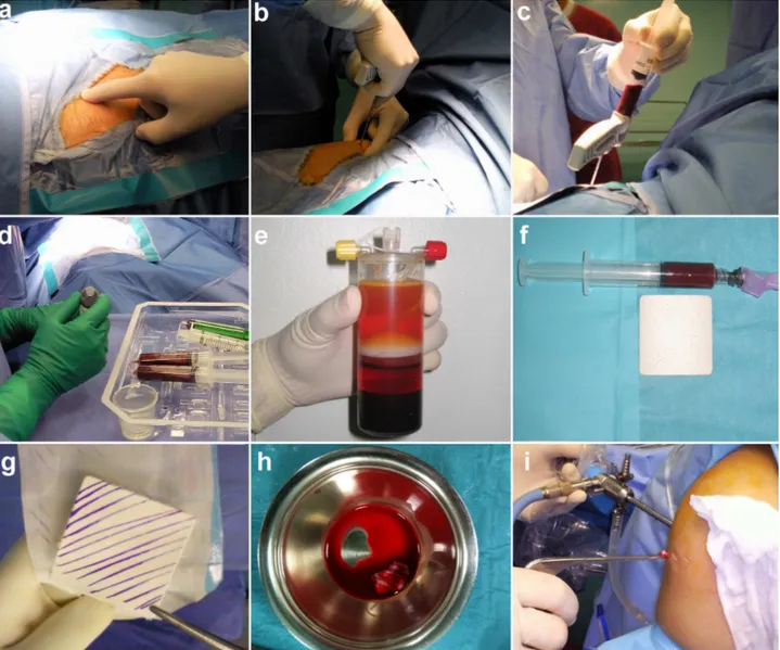
The CMBMC surgical technique has been described in detail by Gigante et al. [80]. Briefly, for bone marrow harvest, a small area over the iliac crest donor site was draped. A 2.5 mm Jamshidi needle was inserted percutaneously into the iliac crest, sixty ml of bone marrow blood were aspirated and processed with the MarrowStim Concentration kit (Biomet, Warsaw, IN) according to the manufacturer’s instructions, obtaining 3-4 ml of BMC. The PGA-HA matrix was immersed with the BMC and kept till implantation.
After diagnostic arthroscopy to confirm the indication for the procedure, the chondral lesion was debrided, measured and microfractures were performed using appropriate awls (Fog. 3.1). The measured size of the lesion was used to adjust a rubber template to the exact shape of the defect.
The PGA-HA matrix was cut to match the defect shape and size. The water flow was stopped and water was aspirated from the joint cavity. A 10:1 mixture of 1-2 mL fibrin glue and BMC was applied to the lesion bed using a long needle. The PGA-HA matrix immersed with BMC was inserted through the appropriate portal with a grasper and placed with a probe. Then an additional 2-3 mL of the fibrin glue-BMC mixture were dispersed over the matrix and allowed to solidify for 2-3 min. Finally, exceeding fibrin glue-BMC was removed and the knee repeatedly flexed and extended to check membrane stability.
For rehabilitation, the patients started continuous passive motion (CPM) on day 4-5 and partial weight-bearing at 3 weeks, progressing to full weight-bearing at 6 weeks. Isometric quadriceps and hamstrings training and straight leg raising were advised during the non-weight-bearing period. Light sports activities such as swimming, cycling or jogging on even soft ground were allowed at 6 months. Permission to participate in unrestricted sports activity was given after 12 months.
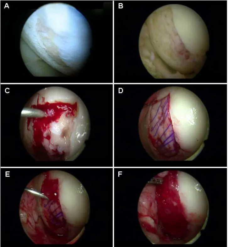
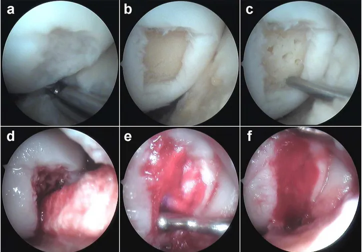
Fig. 3.1 Arthroscopic CMBMC technique. a The cartilage defect is identified; b debrided and microfracture is
performed c The water flow is stopped and the mixture of fibrin glue and BMC is deposited on the bed of the defect. d The scaffold immersed with BMC is set in place (in this picture a PGA-HA scaffold is represented); e and covered with the rest of the fibrin glue-BMC mixture injected through a long needle. f Final appearance of the repaired defect. (From Enea et al. 2012 [84])
3.2.3 Second-‐look arthroscopy
Two patients consented to second-look arthroscopy and biopsy harvest. Three additional patients consented to second-look arthroscopy but did not consent to biopsy. Biopsies were taken with a standard 2.5 mm diameter Jamshidi needle. The specimens were placed in 10% formalin and sent for histology processing.
3.2.4 Histology
Histological characteristics of the repair tissue were evaluated. Specimens were decalcified, paraffin-embedded and stained with Safranin-O to detect the presence of glycosaminoglycans. Polarized microscopy was used to discriminate between hyaline-like cartilage and fibro-cartilage. The International Cartilage Repair Society (ICRS) II Histology Scoring System [85] was used to evaluate the quality of the repair tissue. Histological evaluation was performed blindly by two different investigators and scores were averaged.
3.2.5 Statistical Analysis
The paired t-test was performed for the IKDC score, the Lysholm score and the VAS to compare pre- and postoperative values. Data are expressed as means with standard deviations. The nonparametric Wilcoxon-signed rank test was performed for the Tegner activity scale to compare pre- and postoperative values. Data are expressed as medians and interquartile ranges. For all tests, p<0.05 was considered significant. The statistical software SPSS (Version 17.0) was used for biometric analysis.
3.3 RESULTS
3.3.1 Clinical Outcome
Patients’ characteristics are shown in Table 3.1. Previous surgeries were: 4 meniscectomies, 3 articular debridement and 1 anterior cruciate ligament (ACL) reconstruction. Concomitant interventions at the time of surgery were 1 ACL calcification removal, 1 osteo-chondral fragment fixation, 1 meniscectomy and 1 trochlear resurfacing. No patient-related or device-related complications were encountered. All patients followed the standardized rehabilitation protocol. At 22 (+/-2) months follow-up, patients treated with PGA-HA-CMBMC showed significant (p<0.05) improvement in IKDC subjective score from 68 pre-operatively to 88 post-operatively, in Lysholm score from 52 to 86 and in VAS pain score from 7.4 pre-operatively to 1.5 post-operatively (3. 2). The Tegner activity scale showed no significant difference from pre-injury (4) to post-operative levels (4) at latest follow-up, but significant improvement in the activity level from post-injury (3) to post-operative activity levels (4).
The procedure failed in one patient, who needed a re-operation due to persisting pain. The patient (latest VAS=8) was subjected to second look arthroscopy that showed the persistence of the defect at the medial femoral condyle. This female patient, with a body mass index (BMI) of 33, is currently losing weight in order to undergo a new surgical intervention.
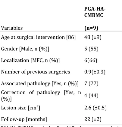
Table 3.1 Characteristics of patients prior to surgery Variables PGA-‐HA-‐ CMBMC (n=9)
Age at surgical intervention [86] 48 (±9) Gender [Male, n (%)] 5 (55) Localization [MFC, n (%)] 6(66) Number of previous surgeries 0.9(±0.3) Associated pathology [Yes, n (%)] 7 (77) Correction of pathology [Yes, n
(%)] 4 (44)
Lesion size [cm2] 2.6 (±0.5)
Follow-‐up [months] 22 (±2)
PGA-‐HA-‐CMBMC = polyglycolic acid/hyaluronan-‐covered microfracture and bone marrow concentrate; MFC = Medial Femoral Condyle
Table 3.2 Outcome data
Score PGA-‐HA-‐CMBMC (n=9) Lysholm Pre-‐op. 68 (±10) † Lysholm Post-‐op. 88 (±18) † IKDC Pre-‐op. 52 (±12)† IKDC Post-‐op. 86 (±15)† VAS Pre-‐op. 7.4 (±2.2) † VAS Post-‐op. 1.5 (±2.7) † Tegner Pre-‐injury 4 (4-‐6) α Tegner Post-‐ Injury 3 (2-‐3) α, β Tegner Post-‐op. 4 (3.5±6) β
PGA-‐HA-‐CMBMC = polyglycolic acid/hyaluronan-‐covered microfracture and bone marrow concentrate; Lysholm, IKDC and VAS are expressed as mean (±SD).Tegner is expressed as median (interquartile range).
†
Pre-‐op. statistically significantly different from Post-‐op. (t-‐test); α Pre-‐injury statistically significantly different
from Post-‐injury; β Post-‐injury statistically significantly different from Post-‐op. (Wilcoxon sum rank test). Post-‐
3.3.2 Arthroscopic and MRI Evaluation
At the time of the second-look arthroscopy all the patients but one were asymptomatic. According to the ICRS CRA evaluation, 1 out of 5 patients treated with PGA-HA-CMBMC was graded normal, 3 nearly normal (Fig. 3.2c, please note the lipid droplet due to biopsy harvest) and 1 abnormal (median 10, range 7-12). The patient scoring 7 was the one that failed.
Four MRI were performed with an average of 10±1.6 months follow-up (range 8-12 months). All patients showed complete defect and volume filling with resurfacing of the articular cartilage to the original cartilage level. Mild bone marrow edema and some subchondral irregularities were observed in all cases. Non-homogeneous cartilage signal was observed in 2 out of 4 cases; fissures were noted in 1 case, surface irregularities in 1 case and a slight hypertrophy of the repair tissue was observed in 1 case.
3.3.3 Histological Evaluation
Biopsies were obtained from two patients. Safranin O staining showed that the repair tissue was rich in proteoglycan and chondrocytic cells. In line with nearly normal MRI findings and improvement in clinical scores, the biopsy proofed hyaline-like repair tissue formation after the implantation of the PGA-HA matrix immersed with autologous BMC. The repair tissue formed in the patient with the failed treatment was rich in chondrocytes but thin and of a fibrocartilagineous appearance. There were no remnants of the PGA-HA matrix and no signs of foreign body reaction or necrosis. According to the ICRS II score they scored respectively an overall of 93 and 41, with a tissue morphology of 100 and 30.
![Fig. 1.5.6.1 The matrix assisted chondrocite implantation (MACI) on the patella. From Gigante et al 2009 [29]](https://thumb-eu.123doks.com/thumbv2/123dokorg/2969877.27203/20.892.122.562.648.920/fig-matrix-assisted-chondrocite-implantation-maci-patella-gigante.webp)