© The Author(s) 2018. Published by Oxford University Press for the Infectious Diseases Society of America. All rights reserved. For permissions, e-mail: [email protected].
A balanced pro-inflammatory and regulatory cytokine signature in young African children is associated with lower risk of clinical malaria
Authors
Carlota Dobaño1,2,3, Augusto J. Nhabomba2, Maria N. Manaca2, Tamara Berthoud1, Ruth Aguilar1,2,3, Llorenç Quintó1,2,3, Arnoldo Barbosa1,2, Mauricio H. Rodríguez1,2, Alfons Jiménez1,3, Penny L Groves4, Rebeca Santano1, Quique Bassat1,2,4, John J. Aponte1,2,3, Caterina Guinovart1,2,3, Denise L. Doolan5, Pedro L. Alonso1,2,3
Affiliations
1ISGlobal, Hospital Clínic - Universitat de Barcelona, Barcelona, Catalonia, Spain.
2Centro de Investigação em Saúde de Manhiça (CISM), Maputo, Mozambique.
3CIBER Epidemiología y Salud Pública (CIBERESP), Barcelona, Spain.
4ICREA, Pg. Lluís Companys 23, 08010 Barcelona, Spain
5QIMR Medical Research Institute, Brisbane, Australia.
Corresponding author. Carlota Dobaño. [email protected]. Carrer Roselló 153
(CEK building), E-08036 Barcelona, Catalonia, Spain.
Summary. Prevention of exposure to Plasmodium falciparum infection during infancy
significantly impacted antigen-specific TH1 and TH2 cytokine responses at age 2 years, and
protection from clinical malaria was associated with balanced pro-inflammatory and regulatory cytokine/chemokine signatures characteristic of innate immune cells.
Abstract
Background. The effect of timing of exposure to first Plasmodium falciparum infections
during early childhood on the induction of innate and adaptive cytokine responses and their contribution to the development of clinical malaria immunity is not well established.
Methods. As part of a double-blind randomized placebo-controlled trial in Mozambique using
monthly chemoprophylaxis with sulfadoxine-pyrimethamine plus artesunate to selectively control timing of malaria exposure during infancy, peripheral blood mononuclear cells collected at ages 2.5, 5.5, 10.5, 15 and 24 months were stimulated ex vivo with parasite schizont and erythrocyte lysates. Cytokine mRNA expressed in cell pellets and proteins secreted in supernatants were quantified by real time quantitative PCR and multiplex flow cytometry, respectively. Children were followed up for clinical malaria from birth until 4 years of age.
Results. Higher pro-inflammatory (IL-1, IL-6, TNF) and regulatory (IL-10) cytokine
concentrations during the second year of life were associated with reduced incidence of clinical malaria up to 4 years of age, adjusting by chemoprophylaxis and prior malaria exposure. Significantly lower concentrations of antigen-specific TH1 (IL-2, IL-12, IFN-) and TH2 (IL-4, IL-5) cytokines by 2 years of age were measured in children under chemoprophylaxis compared to children receiving placebo (p<0.03).
Conclusions. Selective chemoprophylaxis altering early natural exposure to malaria blood
stage antigens during infancy had a significant effect on TH lymphocyte cytokine production more than one year later. Importantly, a balanced pro-inflammatory and anti-inflammatory cytokine signature probably by innate cells around age 2 years was associated with protective clinical immunity during childhood.
Keywords. Plasmodium falciparum, cytokines, age, exposure, immunity.
INTRODUCTION
In endemic areas, clinical malaria primarily affects children under the age of 5 years [1]. Exposure to repeated Plasmodium falciparum infections from birth leads to the development of naturally acquired immunity, which is attained faster against the most severe forms of malaria, takes longer against milder forms but is never sterilizing [2]. Young children exposed to P. falciparum are at high risk from suffering malaria complications until they have developed partial clinical immunity, but the immune mechanisms involved and their determinants are not fully elucidated. Specifically, cellular immune correlates of protection against P. falciparum have been less characterized [3] in contrast to antibodies that are known to exert an anti-parasitic effect [4].
Cytokines and chemokines mediate cellular immune responses and contribute in part to some of the symptoms and pathological alterations during malarial disease [5]. The outcome of the infection depends on the regulation of a network of pro-inflammatory and regulatory immune responses, leading to protection or immunopathology [6, 7]. There are little field data available on the relevance of individual cytokines or chemokines in acquired immunity to malaria. To date, the factors shown to be potentially implicated in protective immunity include IFN-γ and TNF produced by T cells that may inhibit parasite development and destroy infected hepatocytes [8, 9]; IFN-γ and memory T cells that activate macrophages to phagocyte parasitized erythrocytes and merozoites [10]; and IL-10 produced by regulatory T lymphocytes and other cells that control pathogenesis [11]. Antigen-specific TH1 responses are clearly involved in protection against malaria in animal models [12], but human data are scarcer. IL-12, a key TH1 cytokine produced mainly by antigen presenting cells, induces and regulates dendritic cell maturation and function, in addition to promoting the activation and IFN-γ production of T cells and natural killer (NK) cells [13]. IFN-γ and IL-2 TH cell responses, as well as γδ T cells, are induced after P. falciparum experimental infection in naïve individuals; IFN-γ has been associated with malaria protection [14] and IL-2 may be key for the generation of effector responses to malaria [15]. Although pro-inflammatory and TH1
signatures correlate with immunity, it is not clear if they reflect innate rather than protective adaptive immune responses, particularly in the immature immune system of a child [16]. Re-exposure to P. falciparum has been associated with acquisition of antigen-specific IL-10 immunoregulatory responses that dampen pathogenic inflammation while enhancing anti-parasite effector mechanisms [11, 17].
Most previous studies of cytokine responses and malaria immunity have been done in newborn cord blood samples [18, 19], in adult populations [20] or in cross-sectional studies after the onset of clinical symptoms [21-23]. Few have investigated the early acquisition of P. falciparum-specific cytokine responses in asymptomatic or healthy young infants and their relationship with development of clinical immunity in prospective cohorts [7, 9, 11, 24, 25] or how they are affected by malaria chemoprevention or therapeutic tools [26, 27]. Data reported have not shown consistent patterns. For example, in Gambian children, chemoprophylaxis resulted in higher lymphoproliferative responses and IFN-γ production [28]. In Ugandan children, chemoprophylaxis was associated with higher production of IL-2 and TNF, which was associated with malaria protection, and lower production of IL-10 and IFN-γ, which was associated with malaria risk [27]. Also, Kenyan children sleeping under bednets had decreased production of pro-inflammatory cytokines TNF, IL-1 and IL-6 [29].
We conducted a double-blind randomized placebo-controlled trial in Mozambique administering monthly chemoprophylaxis with sulfadoxine-pyrimethamine (SP) plus artesunate (AS) to selectively control the age of first infection by blood stage P. falciparum during infancy, to understand the role of parasite exposure in the acquisition of immunity to malaria [30]. This study set out to elucidate the role of age and exposure to P. falciparum in the induction of cytokine responses and their role in immunity in young children. To this end, we measured cellular mediators produced by blood leukocytes after parasite antigen or mock stimulation to identify those associated with prospective risk of malaria.
MATERIALS AND METHODS
Study design
The study was conducted at the Centro de Investigação em Saúde de Manhiça (CISM), Maputo Province, Southern Mozambique, from September 2005 to March 2009, and has been described in detail elsewhere [30]. Briefly, it consisted on a double-blind randomized placebo-controlled trial (RCT) including 349 newborns from the Maragra village receiving monthly chemoprophylaxis with SP plus AS or placebo administered during different periods of the first year of life according to the randomization group (Supplementary methods and Figure 1): “Late Exposure”, “Early Exposure” or “Control”. Study participants were followed up until age 24 months as part of the RCT. Weekly active case detection (ACD) was conducted from birth to approximately age 10.5 months and monthly home visits from 10.5 to 24 months of age. Children presenting fever were taken to the Maragra Health Post (MHP), where they were examined and parasitaemia and hematocrit were determined. Additionally, passive case detection (PCD) was carried out at the MHP and Manhiça District Hospital (MDH) through the continuous morbidity surveillance system to monitor attendances to the outpatient clinics and admissions to hospital; data were analyzed until children were 4 years of age. One mL blood sample was collected into EDTA microtainers by finger-prick at the five cross-sectional visits by months [M] 2.5, 5.5, 10.5, 15 and 24, at the first clinical malaria episode (if any) and one month later (convalescence). Approval for the study was obtained from the National ethical review committee of Mozambique and the ethical review committee of Hospital Clínic in Barcelona, Spain. Children were enrolled in the study after their guardians provided written informed consent.
Laboratory procedures
Standard laboratory methods were used to assess parasitological and hematological parameters [30,31]. Peripheral blood mononuclear cells (PBMC) were isolated using a
Lymphoprep gradient and resuspended in complete medium. A total of 1.2 million fresh PBMC were stimulated with 20 μl of a P. falciparum (3D7 strain) schizont extract corresponding to lysate from 2 million synchronized infected red blood cells (iRBC) or with 20 μl of uninfected RBC (uRBC). After the incubation for 24 h, 48 h or 72 h, the supernatants were collected and frozen at 80ºC. The PBMC pellets were collected in trizol and frozen at -80ºC. At the end of the field study, supernatants were thawed and cytokine concentrations (IL-12p70, IFN-, IL-2, IL-10, IL-8, IL-6, IL-4, IL-5, IL-1, TNF, TNF-) were measured with the Bender MedSystems Human Th1/Th2 11plex FlowCytomix Multiplex Kit [32]. Cytokine mRNA levels (IL-2, IL-4, IL-6, IL-10, IFN-, TNF, IL-13) normalized to the reference gene RPL13a, were measured in a sub-sample by reverse transcriptase (RT) qPCR. A high level of cytokine production detected in culture supernatants of unstimulated infant samples are considered biologically relevant and were therefore not subtracted from the stimulated samples but shown side by side [19, 33]. Cytokine concentrations were analyzed in relation to plasma antibody responses to P. falciparum blood stage antigens [34].
Definitions and statistical methods
Clinical malaria was defined as axillary temperature ≥ 37.5 ºC or a reported fever in the preceding 24 h with any positive parasitaemia from the blood slide microscopy, using a case definition previously validated in the area for this age group [35]. Cytokine concentrations (pg/mL) were logarithmically transformed and average within the groups presented as geometric means (GM) plus 95% confidence intervals (CI). Differences among treatment groups at different timepoints were estimated by ANOVA and evaluated using a likelihood ratio test and a global p-value for significance. Correlations with antibody levels within and between visits were done by Spearman. To analyze factors independently associated with cytokine concentrations we used mixed-effects regression models including relevant covariates. Cytokine concentrations in relation to incidence of malaria were assessed by
negative binomial regressions, unadjusted and after adjusting by relevant covariates. Data analysis was performed using STATA 11 (StataCorp. 2007) and R studio. Statistical significance was defined at P<0.05.
RESULTS
A total of 1,712 blood samples were collected from children and processed during the field study: 318 at M2.5, 295 at M5.5, 290 at M10.5, 273 at M15, 301 at M24, 129 at first acute malaria episode, and 106 at convalescence. Data presented here are based on the analysis of a random subgroup of 643 sets of supernatants (iRBC and uRBC stimulated, M2.5=93, M5.5=95, M10.5=220, M15=89, M24=35, 59 acute and 52 convalescent). In addition, 316 sets of cell pellets in Trizol (iRBC and uRBC stimulated, 632 total) were processed for mRNA cytokine analysis (M2.5=59, M5.5=60, M10.5=192, M15=59, M24=11, 10 acute and 9 convalescent). A pilot study was conducted to select the optimal timepoint among 24, 48 and 72 h for cytokine detection in supernatants and in Trizol pellets after PBMC stimulation with iRBC using samples from 24 immune adults and 28 infants. Data indicated that the 24 h timepoint was the optimal for cytokine detection [19]. The correlations between mRNA expression in PBMC pellets and concentration in culture supernatants for quantifiable cytokines were moderate-high for IL-6, TNF and IL-10 that had the highest production but low for the others (Supplementary Figure 2).
Factors affecting cytokine concentrations
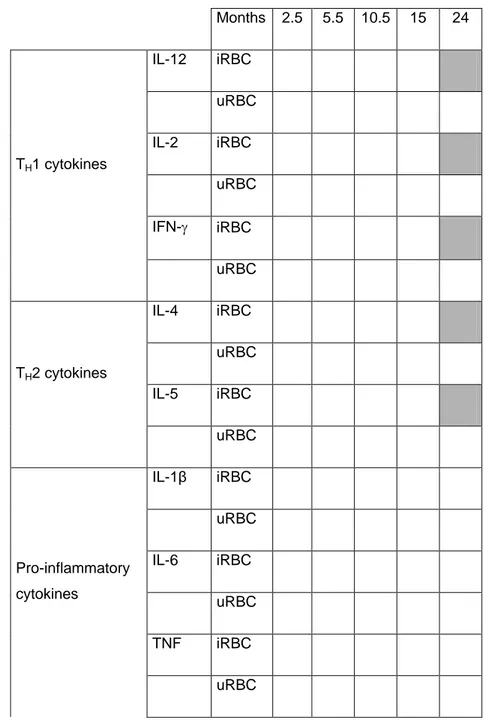
SP+AS chemoprophylaxis during M2.5-M5.5 or M5.5-M10.5 significantly affected the production of TH1 and TH2 cytokines in PBMC collected at M24 following iRBC antigen stimulation but not upon uRBC mock stimulation. Thus, children who had been chemo suppressed in year one had significantly lower supernatant concentrations of IL-12 (p=0.01), IFN- (p=0.002), IL-2 (p=0.03), IL-4 (p=0.005) and IL-5 (p=0.01) at M24 compared to
continuously exposed controls (Figure 1). Chemoprophylaxis had no significant impact on the pro-inflammatory (IL-1, IL-6, TNF, TNF-, IL-8) or the regulatory (IL-10) cytokine concentrations (Table 1).
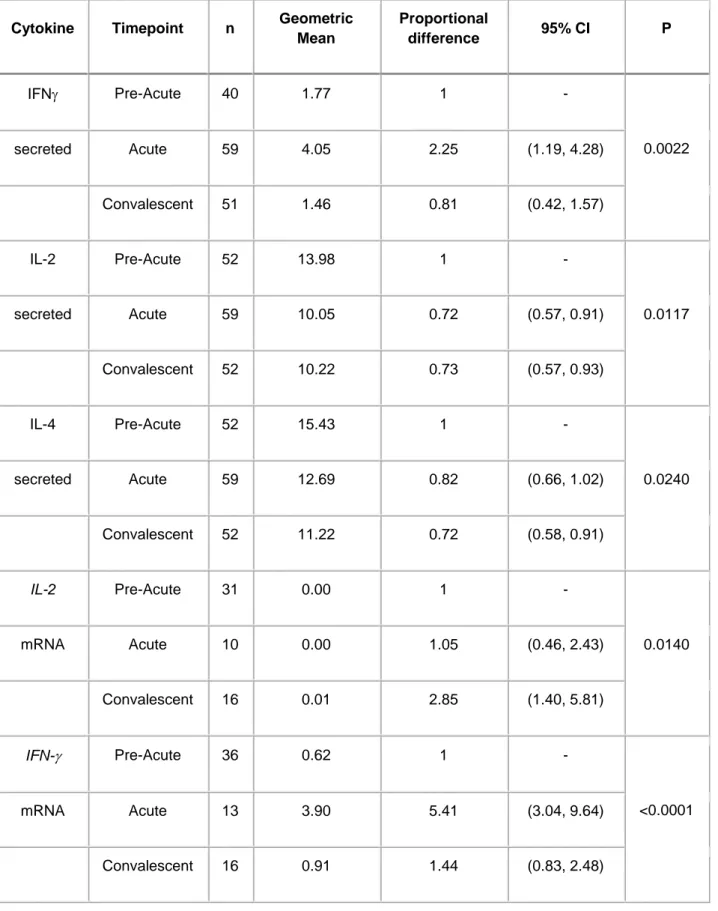
Clinical malaria episodes significantly affected the magnitude of some cytokine responses. Overall, pro-inflammatory cytokines and IL-10 were higher during the acute phase compared to levels preceding clinical disease and declined at convalescence. Differences were significant for the secreted proteins and mRNA transcripts (Table 2).
Other factors evaluated in relation to cytokine production (maternal infection, parity, season, birth weight, etc) did not show any specific or consistent association, except for weight-for-age Z-score that was negatively associated with levels of pro- and anti-inflammatory cytokines (Supplementary Table 1). Exposure to previous or current P. falciparum infection was not associated with cytokine concentrations. Age significantly affected the production of some cytokines during the first year of life. IL-6 levels were higher at M2.5 and declined gradually (Figure 1). IL-2 and IL-8 showed a steady increase with age, whereas IL-1 and TNF did not vary during most of the first year.
Effect of cytokine concentrations on clinical malaria
Higher concentrations of pro-inflammatory cytokines in PBMC supernatants by the end of the first year of life were associated with reduced incidence of clinical malaria up to M24. Table 3 shows the effect of a 2-fold increment in cytokine levels at M10.5 on the incidence of malaria up to M24, adjusted by treatment, season, neighborhood, malaria infection at visit and before visit, maternal infection, congenital infection, inflammation in the placenta, use of insecticide treated nets (ITN) and indoor residual spraying (IRS). This was statistically significant for TNF and a trend was observed for IL-1, IL-6 and IL-8. Furthermore, as part of an extended follow up analysis (Supplementary Figure 1), higher secretion of pro-inflammatory and also regulatory cytokines by PBMC at M24, either spontaneous production or following P.
falciparum antigen stimulation, was associated with lower risk of subsequent clinical malaria up to M36 and M48 (Table 3).
Correlations among cytokine and antibody responses
Different patterns were observed at each visit. Pro- and anti-inflammatory cytokines were highly and positively correlated from the younger ages, except for IL-8 that was negatively correlated to the rest, while TH1 and TH2 cytokines correlated positively among themselves only as age increased (Supplementary Figure 3). With time, weak-moderate correlations of inflammatory with TH cytokines transitioned from negative to positive. In addition, TNF and IL-6 at M10.5 weakly correlated positively with TH2 cytokines at M24 (Supplementary Figure 4).
We tested associations of cytokines affected by chemoprophylaxis or involved in malaria protection with antibody responses [34]. At M24, TH cytokines correlated positively with M5.5 IgM to P. falciparum lysate, and negatively with M5.5 IgG to MSP1 and EBA175, M10.5 IgG to VSA, and various anti-malarial antibodies at M15 (Supplementary Figure 5A-B) and M24, particularly IgG to VSA and EBA175 (Figure 2A).
Regarding inflammation cytokines, TNF, IL-1, IL-6 and IL-10 at M10.5 correlated negatively with IL-8 and IgM to malarial antigens at M5.5 (Supplementary Figure 5C). TNF correlated positively with M10.5 iRBC IgM while IL-6 correlated negatively with iRBC IgG
(Supplementary Figure 5D). At M24, IL-10 correlated negatively with M10.5 AMA1 IgG; TNF, IL-1, IL-6 and IL-10 correlated negatively with M15 iRBC antibodies (Supplementary Figure 5E-F), and M24 AMA1 and MSP1 IgMs (Figure 2B), while they correlated positively with M15 AMA1, MSP1 and EBA175 IgGs.
DISCUSSION
This study shows that a pro-inflammatory (IL-1, IL-6, TNF) and regulatory (IL-10) cytokine signature between 1 and 2 years of age is associated with less incidence of malaria up to ages 3 and 4 years, having adjusted by chemoprophylaxis and prior malaria exposure. In terms of what responses correlate with timing of first infection in infancy, children receiving 3- to 5-month malaria chemoprophylaxis in the first year of life (early and late exposure groups) had significantly lower concentrations of antigen-specific TH1 (IL-2, IL-12 and IFN-) and TH2 (IL-4, IL-5) cytokines at 2 years of age compared to children under continuous P. falciparum exposure. Since our analysis was done in a RCT with longitudinal design [30], selectively controlling by monthly chemoprophylaxis exposure to blood stage P. falciparum, results can more reliably shed light into the determinants of the acquisition of cellular immune responses in early childhood. However, the two subsets of cytokines were non-overlapping and thus we found no evidence that altering the timing of initial P. falciparum exposure impacts subsequent development of clinical immunity, consistent with the main trial results [30].
Remarkably, a chemoprophylactic intervention altering natural exposure to P. falciparum blood stages in infancy had an effect on the T cell adaptive response that was apparent over 1 year later. Lower TH cytokine concentrations at M24 correlated with higher anti-P. falciparum IgG levels at M24 and prior visits, and lower anti-P. falciparum IgM levels at M5.5, indicating that antibodies could be markers of exposure in children who received prophylaxis in year 1. Indeed, in the RCT, early and late exposure groups had lower incidence of malaria in year 1 but higher in year 2 than the control group [30]; although a potential malaria “rebound” in year 2 was not statistically significant, this could be reflected immunologically. Thus, higher malaria exposure between M10.5-M24 as a result of chemoprophylaxis between M2.5-10.5 could have dampened the production of TH cytokines at M24.
Higher concentrations of inflammatory cytokines at M10.5 and M24, associated here with malaria protection, correlated with lower IL-8 and lower IgM to P. falciparum antigens,
indicative of less recent/current exposure, at prior timepoint visits. The fact that the association between pro-inflammatory cytokines and protection was observed in iRBC- and uRBC-stimulated PBMC suggests that these responses were produced by innate rather than adaptive cells [36, 37]. In contrast, the TH signature at M24 was only detected in iRBC-stimulated PBMC, suggesting that these cytokines were produced by memory T cells. Infants recruited were initially naïve, and the non-specific innate immune response has been reported as the key defense mechanism in this population [38, 39]. TNF has correlated with immunity to clinical malaria in children [9], while IL-1 and IL-6 in the mothers from this cohort were also associated with less malaria [19].
Furthermore, the pro-inflammatory protective signature was accompanied by a regulatory protective IL-10 response at age 2 years, when TNF levels diminished, showing that effective immunity against clinical malaria requires a pro-inflammatory followed by an anti-inflammatory response. In previous studies, IL-10 was elevated as a result of poor immune regulatory ability typically seen in children, and as a result of the absence of acquired immunity due to a lack of previous exposure to malaria [22]. During acute malaria, pro-inflammatory and TH1 cytokines had higher concentrations, which were more significant for IFN-, TNF and IL-10. IFN- is produced by NK cells, -T cells, CD8+ and CD4+ T cells, therefore it is both an innate and adaptive cytokine. Increased IFN- during acute malaria is related to protection against malaria [8, 9, 14]. The fact that regulatory IL-10 was higher during acute phase and declined at convalescence further supports that regulatory balanced responses are acquired in adequate immunity to control excessive inflammation [11].
In contrast with antibody responses [34], age or prior/present infection had no prominent influence on the magnitude of the cytokine response, consistent with previous studies by our group [26]. Cytokine concentrations except IL-2 and IL-8 declined by M24. At the age of 2 years, when the spleen matures and the incidence of severe malaria declines [2], there appears to be an inflexion point in the acquisition of immunity. Indeed, key responses for immunity like VSA and EBA175 antibodies attained higher levels [34], which have been
associated with malaria protection in children from the same area [40]. A limitation of our study is that it was not designed to assess cytokines after M24, at a time when a protective adaptive TH signature correlated with immunity could have emerged. Another limitation is that we did not phenotype the cells producing the cytokines. Also, a larger sample size could have established more statistically conclusive associations between TH and inflammatory cytokines, and malaria.
Finally, this study could portray, in a small scale, the impact of drug usage during malaria elimination programs including partial or temporary interruption of exposure by artemisinin-based combination therapies, on acquisition of protective immunity to malaria. Suppressive doses of SP+AS during a defined period prevented infection and altered development of natural immunity but not completely. It remains to be assessed how quickly sustained chemoprophylaxis or mass drug administration would slow down or interrupt acquisition of immunity and/or could cause a loss of immunity at a population level [41].
Conclusions
A balanced pro-inflammatory and anti-inflammatory cytokine response at age 2 years may be required for the acquisition of protective clinical immunity to malaria in childhood. In addition, timing of malarial antigen immune priming in infancy did not impact development of clinical immunity within the first 2 years but it may affect the subsequent acquisition of adaptive T helper responses that may be relevant later in life.
Acknowledgements
We thank all children and their families for their participation in the study; the field workers, field supervisors, laboratory staff, data managers and other staff at CISM for their work during the study; Laura Puyol, Diana Barrios and Pau Cisteró for laboratory support; Jaume Ordi for the training and quality control for the placental histology readings; Lázaro Mussacate Quimice for reading all placental histologies; Gemma Moncunill for critically reviewing the manuscript; Sònia Tomàs and Patricia García for their work as project managers.
Funding
The study was funded by a EU Framework Programme 6 STREP project (Malaria age exposure, Project reference 18902), the Instituto de Salud Carlos III (Ayuda de incentivación a la participación en proyectos del Espacio Europeo de Investigación) and the Spanish Ministerio de Educación y Ciencia (Project reference A107190024). CD was supported by a Ramón y Cajal grant from the Spanish Ministerio de Ciencia e Innovación (RYC-2008-02631), MNM by a PhD Scholarship from Fundació Marfà, CG and QB by a grant from the Spanish Ministry of Health (Contrato post-Formación Sanitaria Especializada, Fondo de Investigaciones Sanitarias, Instituto de Salud Carlos III, ref. CM04/00028 and CM05/00134 respectively). DLD was supported by a Pfizer Australia Senior Research Fellowship. The Manhiça Health Research Centre receives core funding from the Spanish Agency for International Cooperation and Development (AECID). ISGlobal is a member of the CERCA Programme, Generalitat de Catalunya.
Conflict of interest
Authors report no potential conflicts.
References
1. WHO. World Malaria Report 2017. Available from:
http://wwwwhoint/malaria/publications/world-malaria-report-2017/en/ 2017.
2. Doolan DL, Dobaño C, Baird JK. Acquired immunity to malaria. Clin Microbiol Rev
2009; 22(1): 13-36, Table of Contents.
3. Hoffman SL, Oster CN, Mason C, et al. Human lymphocyte proliferative response to a sporozoite T cell epitope correlates with resistance to falciparum malaria. J Immunol
1989; 142(4): 1299-303.
4. Cohen S, Mc GI, Carrington S. Gamma-globulin and acquired immunity to human malaria. Nature 1961; 192: 733-7.
5. Clark IA, Budd AC, Alleva LM, Cowden WB. Human malarial disease: a consequence of inflammatory cytokine release. Malar J 2006; 5: 85.
6. Farrington L, Vance H, Rek J, et al. Both inflammatory and regulatory cytokine responses to malaria are blunted with increasing age in highly exposed children. Malar J 2017; 16(1): 499.
7. Dodoo D, Omer FM, Todd J, Akanmori BD, Koram KA, Riley EM. Absolute levels and ratios of proinflammatory and anti-inflammatory cytokine production in vitro predict clinical immunity to Plasmodium falciparum malaria. J Infect Dis 2002; 185(7): 971-9. 8. Luty AJ, Lell B, Schmidt-Ott R, et al. Interferon-gamma responses are associated with
resistance to reinfection with Plasmodium falciparum in young African children. J Infect Dis 1999; 179(4): 980-8.
9. Robinson LJ, D'Ombrain MC, Stanisic DI, et al. Cellular tumor necrosis factor, gamma interferon, and interleukin-6 responses as correlates of immunity and risk of clinical Plasmodium falciparum malaria in children from Papua New Guinea. Infect Immun
2009; 77(7): 3033-43.
10. Ockenhouse CF, Schulman S, Shear HL. Induction of crisis forms in the human malaria parasite Plasmodium falciparum by gamma-interferon-activated, monocyte-derived macrophages. J Immunol 1984; 133(3): 1601-8.
11. Boyle MJ, Jagannathan P, Bowen K, et al. The Development of Plasmodium falciparum-Specific IL10 CD4 T Cells and Protection from Malaria in Children in an Area of High Malaria Transmission. Front Immunol 2017; 8: 1329.
12. Doolan DL, Hoffman SL. The complexity of protective immunity against liver-stage malaria. J Immunol 2000; 165(3): 1453-62.
13. Torre D. Early production of gamma-interferon in clinical malaria: role of interleukin-18 and interleukin-12. Clin Infect Dis 2009; 48(10): 1481-2.
14. D'Ombrain MC, Robinson LJ, Stanisic DI, et al. Association of early interferon-gamma production with immunity to clinical malaria: a longitudinal study among Papua New Guinean children. Clin Infect Dis 2008; 47(11): 1380-7.
15. Horowitz A, Newman KC, Evans JH, Korbel DS, Davis DM, Riley EM. Cross-talk between T cells and NK cells generates rapid effector responses to Plasmodium falciparum-infected erythrocytes. J Immunol 2010; 184(11): 6043-52.
16. PrabhuDas M, Adkins B, Gans H, et al. Challenges in infant immunity: implications for responses to infection and vaccines. Nat Immunol 2011; 12(3): 189-94.
17. Portugal S, Moebius J, Skinner J, et al. Exposure-dependent control of malaria-induced inflammation in children. PLoS Pathog 2014; 10(4): e1004079.
18. Fievet N, Ringwald P, Bickii J, et al. Malaria cellular immune responses in neonates from Cameroon. Parasite Immunol 1996; 18(10): 483-90.
19. Dobaño C, Berthoud T, Manaca MN, et al. High production of pro-inflammatory cytokines by maternal blood mononuclear cells is associated with reduced maternal malaria but increased cord blood infection. Malar J 2018; 17(1): 177.
20. Anum D, Kusi KA, Ganeshan H, et al. Measuring naturally acquired ex vivo IFN-gamma responses to Plasmodium falciparum cell-traversal protein for ookinetes and sporozoites (CelTOS) in Ghanaian adults. Malar J 2015; 14: 20.
21. Wroczynska A, Nahorski W, Bakowska A, Pietkiewicz H. Cytokines and clinical manifestations of malaria in adults with severe and uncomplicated disease. Int Marit Health 2005; 56(1-4): 103-14.
22. Mandala WL, Msefula CL, Gondwe EN, Drayson MT, Molyneux ME, MacLennan CA. Cytokine Profiles in Malawian Children Presenting with Uncomplicated Malaria, Severe Malarial Anemia, and Cerebral Malaria. Clin Vaccine Immunol 2017; 24(4). 23. Rovira-Vallbona E, Moncunill G, Bassat Q, et al. Low antibodies against Plasmodium
falciparum and imbalanced pro-inflammatory cytokines are associated with severe malaria in Mozambican children: a case-control study. Malar J 2012; 11: 181. 24. Le Hesran JY, Fievet N, Thioulouse J, et al. Development of cellular immune
responses to Plasmodium falciparum blood stage antigens from birth to 36 months of age in Cameroon. Acta Trop 2006; 98(3): 261-9.
25. Schofield L, Ioannidis LJ, Karl S, et al. Synergistic effect of IL-12 and IL-18 induces TIM3 regulation of gammadelta T cell function and decreases the risk of clinical malaria in children living in Papua New Guinea. BMC Med 2017; 15(1): 114. 26. Quelhas D, Puyol L, Quinto L, et al. Intermittent preventive treatment with
sulfadoxine-pyrimethamine does not modify plasma cytokines and chemokines or intracellular cytokine responses to Plasmodium falciparum in Mozambican children. BMC Immunol 2012; 13: 5.
27. Jagannathan P, Bowen K, Nankya F, et al. Effective Antimalarial Chemoprevention in Childhood Enhances the Quality of CD4+ T Cells and Limits Their Production of Immunoregulatory Interleukin 10. J Infect Dis 2016; 214(2): 329-38.
28. Otoo LN, Riley EM, Menon A, Byass P, Greenwood BM. Cellular immune responses to Plasmodium falciparum antigens in children receiving long term anti-malarial chemoprophylaxis. Trans R Soc Trop Med Hyg 1989; 83(6): 778-82.
29. Friedman JF, Phillips-Howard PA, Hawley WA, et al. Impact of permethrin-treated bed nets on growth, nutritional status, and body composition of primary school children in western Kenya. Am J Trop Med Hyg 2003; 68(4 Suppl): 78-85. 30. Guinovart C, Dobaño C, Bassat Q, et al. The role of age and exposure to
Plasmodium falciparum in the rate of acquisition of naturally acquired immunity: a randomized controlled trial. PLoS One 2012; 7(3): e32362.
31. Mayor A, Serra-Casas E, Bardaji A, et al. Sub-microscopic infections and long-term recrudescence of Plasmodium falciparum in Mozambican pregnant women. Malar J
2009; 8: 9.
32. Berthoud TK, Manaca MN, Quelhas D, et al. Comparison of commercial kits to measure cytokine responses to Plasmodium falciparum by multiplex microsphere suspension array technology. Malar J 2011; 10: 115.
33. Kollmann TR, Crabtree J, Rein-Weston A, et al. Neonatal innate TLR-mediated responses are distinct from those of adults. J Immunol 2009; 183(11): 7150-60. 34. Nhabomba AJ, Guinovart C, Jimenez A, et al. Impact of age of first exposure to
Plasmodium falciparum on antibody responses to malaria in children: a randomized, controlled trial in Mozambique. Malar J 2014; 13: 121.
35. Saute F, Aponte J, Almeda J, et al. Malaria in southern Mozambique: malariometric indicators and malaria case definition in Manhica district. Trans R Soc Trop Med Hyg
2003; 97(6): 661-6.
36. McCall MB, Netea MG, Hermsen CC, et al. Plasmodium falciparum infection causes proinflammatory priming of human TLR responses. J Immunol 2007; 179(1): 162-71. 37. Walther M, Woodruff J, Edele F, et al. Innate immune responses to human malaria:
heterogeneous cytokine responses to blood-stage Plasmodium falciparum correlate with parasitological and clinical outcomes. J Immunol 2006; 177(8): 5736-45.
38. Kumar SK, Bhat BV. Distinct mechanisms of the newborn innate immunity. Immunol Lett 2016; 173: 42-54.
39. Schrum JE, Crabtree JN, Dobbs KR, et al. Cutting Edge: Plasmodium falciparum Induces Trained Innate Immunity. J Immunol 2018; 200(4): 1243-8.
40. Campo JJ, Dobaño C, Sacarlal J, et al. Impact of the RTS,S malaria vaccine
candidate on naturally acquired antibody responses to multiple asexual blood stage antigens. PLoS One 2011; 6(10): e25779.
41. Aponte JJ, Menendez C, Schellenberg D, et al. Age interactions in the development of naturally acquired immunity to Plasmodium falciparum and its clinical presentation. PLoS Med 2007;4(7):e242.
FIGURE LEGENDS
Figure 1. Weighted scattered plots of Plasmodium falciparum antigen-specific cytokine
concentrations (pg/mL) in each study group and at each cross sectional visit, showing
geometric means and 95% confidence intervals. A) TH1: IL-12, IL-2, IFN-B) TH2: IL-4, IL-5.
C) Pro-inflammatory and regulatory: IL-1, IL-6, TNF, IL-8 and IL-10. Table 1 shows the
outcomes of the statistical tests for the comparisons that had significantly different cytokine responses. The area of the symbol is proportional to the number of observations; in red those with previous or current P. falciparum infections. In the case of TH1 and TH2 cytokines at cross sectional visit 24 months, most concentration values were low or undetectable for the LE and EE groups (i.e. larger area of red and blue symbols at the bottom). Ctrol: control, LE: late exposure, EE: early exposure.
Figure 2. Heatmaps and scatter plots of the correlations between antibody levels and
cytokine concentrations after stimulation with P. falciparum schizont lysate (log-transformed) at the indicated study visits. A) Antibodies vs TH1 and TH2 cytokines at month 24. B)
Antibodies vs pro-inflammatory and regulatory cytokines at month 24. Spearman coefficients and p values: *<0.05, **<0.01, ***<0.001.
TABLES
Table 1. Effect of chemoprophylaxis on the magnitude of cytokine production at
cross-sectional visits. A) Heatmap of all cytokines, statistically significant differences in grey, non-significant differences in white. B) Statistically non-significant proportional differences (95% confidence intervals) in chemoprophylaxis groups compared to control group in the case of TH1 and TH2 cytokines at month 24.
A) Months 2.5 5.5 10.5 15 24 TH1 cytokines IL-12 iRBC uRBC IL-2 iRBC uRBC IFN- iRBC uRBC TH2 cytokines IL-4 iRBC uRBC IL-5 iRBC uRBC Pro-inflammatory cytokines IL-1β iRBC uRBC IL-6 iRBC uRBC TNF iRBC uRBC
TNF- iRBC uRBC Regulatory cytokines IL-10 iRBC uRBC Pro-inflammatory chemokine IL-8 iRBC uRBC
BB) Proportional difference (95% CI) vs Control
Late exposure Early exposure p value
TH1 IL-12 iRBC 0.25 (0.08-0.75) 0.29 (0.10-0.83) 0.0178 uRBC ns IL-2 iRBC 0.35 (0.15-0.84) 0.45 (0.20-1.01) 0.0337 uRBC ns IFN- iRBC 0.20 (0.06-0.66) 0.15 (0.05, 0.47) 0.0024 uRBC ns TH2 IL-4 iRBC 0.35 (0.16-0.77) 0.33 (0.16-0.70) 0.0055 uRBC ns IL-5 iRBC 0.38 (0.16-0.90) 0.31 (0.14-0.71) 0.0116
uRBC ns
p-value from linear regression model using Likelihood Ratio Test. ns = non-significant
Difference adjusted by clinical malaria episodes before visit
Table 2. Effect of a clinical malaria episode on the magnitude of cytokine production after
stimulation with Plasmodium falciparum lysate at the acute and convalescent visits. P-values from regression models using Likelihood Ratio Test. Only significant differences are shown.
Cytokine Timepoint n Geometric Mean Proportional difference 95% CI P IFN Pre-Acute 40 1.77 1 - 0.0022 secreted Acute 59 4.05 2.25 (1.19, 4.28) Convalescent 51 1.46 0.81 (0.42, 1.57) IL-2 Pre-Acute 52 13.98 1 - 0.0117 secreted Acute 59 10.05 0.72 (0.57, 0.91) Convalescent 52 10.22 0.73 (0.57, 0.93) IL-4 Pre-Acute 52 15.43 1 - 0.0240 secreted Acute 59 12.69 0.82 (0.66, 1.02) Convalescent 52 11.22 0.72 (0.58, 0.91) IL-2 Pre-Acute 31 0.00 1 - 0.0140 mRNA Acute 10 0.00 1.05 (0.46, 2.43) Convalescent 16 0.01 2.85 (1.40, 5.81) IFN- Pre-Acute 36 0.62 1 - <0.0001 mRNA Acute 13 3.90 5.41 (3.04, 9.64) Convalescent 16 0.91 1.44 (0.83, 2.48)
TNF Pre-Acute 37 3.29 1 - <0.0001 mRNA Acute 13 13.13 3.67 (2.18, 6.16) Convalescent 16 6.36 1.86 (1.15, 3.02) IL-10 Pre-Acute 37 8.11 1 - 0.0057 mRNA Acute 13 24.18 2.63 (1.41, 4.91) Convalescent 16 7.48 0.97 (0.54, 1.75)
Table 3. Effect of the magnitude of cytokine production at the month indicated on the first
raw (left-) on subsequent incidence of clinical malaria up to the follow up month indicated in the first row (-right). A) Heatmap of all cytokines and chemokines in supernatants. Statistically significant or trend differences indicating a reduction in the incidence of clinical malaria are marked in grey colors, and non-significant differences are in white. B) Incidence rate ratios (IRR) with 95% CI and p values for statistically significant or trend differences, for pro-inflammatory and regulatory cytokines and chemokines.
A) Months 2.5-24 5.5-24 10.5-24 15-24 24-36 24-48 TH1 cytokines IL-12 iRBC uRBC IL-2 iRBC uRBC IFN- iRBC uRBC TH2 cytokines IL4 iRBC uRBC IL5 iRBC uRBC Pro-inflammatory cytokines IL-1 iRBC uRBC IL-6 iRBC
uRBC TNF iRBC uRBC Pro-inflammatory chemokine IL-8 iRBC uRBC Regulatory cytokine IL-10 iRBC uRBC B) Cytokines at 10.5 months on malaria risk up to 24 months Cytokines at 24 months on malaria risk up to 36 months Cytokines at 24 months on malaria risk up to 48 months
IRR 95% CI p value IRR 95% CI p value IRR 95% CI p value
IL-1 iRBC 0.93 0.85-1.01 0.078 0.69 0.48-0.99 0.030 0.72 0.51-1.02 0.077 uRBC 0.92 0.84-1.01 0.072 0.74 0.58-0.95 0.018 0.76 0.59-0.98 0.049 IL-6 iRBC 0.93 0.84-1.03 0.152 0.73 0.54-1.00 0.034 0.77 0.56-1.04 0.112 uRBC 0.92 0.83-1.02 0.107 0.73 0.55-0.97 0.016 0.77 0.59-1.01 0.076 TNF iRBC 0.88 0.79-0.97 0.014 ns ns uRBC 0.89 0.80-0.99 0.037 0.6 0.41-0.88 0.006 0.62 0.43-0.89 0.022 IL-10 iRBC ns 0.82 0.67-1.00 0.044 0.85 0.72-1.00 0.089 uRBC ns 0.79 0.66-0.94 0.005 0.77 0.66-0.90 0.002 IL-8 iRBC 0.74 0.53-1.03 0.073 ns ns
uRBC 0.73 0.52-1.03 0.068 0.23 0.04-1.30 0.072 0.17 0.02-1.13 0.031
IRR = incidence rate ratio; CI = confidence interval; ns = non-significant
Figure 1
Figure 2