r
w*
Iffi@ffi
Iffi@t
:7
[Jniversitù dellu
Culubriu
Facoltù
di
Farmaciu
eScienze
dells
Nutrizione
edella
Salute
Dipartimento Furmaco-Biologico
(MED/04
PATOLOGIA GENERALE)
Dottorato
di
Ricerca
in
ooBiochimica
Cellulare
ed
Attività
dei
Farmaci
in
Oncologiaoo
(XXII
ciclo)
T3/TRPr
indaces
proltferutive
effects
on
pupillury
thyroid
cuncer cells
FB2
Coordinatore
Prof.
Diego
SISCI
{*l ,4J-,-+
1
INDEX
INTRODUCTION
1
MATERIALS AND METHODS
7
o Reagents
7
o
Cell culture
7
o Proliferation assay
8
o Reverse transcription-PCR (RT-PCR) assay 8
o
Immunoblotting
9
o Immunoprecipitation
10
o
PI3Kinase assay
10
o RNA interference (Rnai)
11
o Immunofluorescent microscopy
12
o
Statistical analysis
13
RESULTS
o Thyroid hormone receptor B1 is expressed in FB2 cells 14
o Thyroid hormone T3 induces a proliferative effect in human papillary
thyroid cancer FB-2 cell line 16
o
TRβ
1mediates T3 action on FB2 cells proliferation 18
o
TRβ
1receptor mediates the rapid activation of AKT induced by T3 in
FB2 cells
20
o
TRβ
1complexes with p85a subunit of PI3K in a ligand independent 23
o
T3 induces TRβ
1–associated PI3K activity and promotes the nuclear
2
o
Activation of PI3-Kinase and MAP-Kinase is involved in thyroid
hormone-induced proliferation in FB2 cells
26
DISCUSSION
28
BIBLIOGRAFY
34
Scientific Publications Performed during the Program
40
3
INTRODUCTION
A number of clinical and experimental studies have suggested the crucial role of
thyroid hormones (THs) and cognate nuclear receptors in cell growth and differentiation
of many cell types (Pibiri et al., 2001, Bassett and Williams, 2003, Jones et al., 2003,
Lin et al., 2009, Verga Falazacappa et al., 2009). Although recently some authors have
demonstrated the involvement of TH in cell proliferation of many tumor cell types,
including thyroid cancer cell lines, the exact mechanisms through which TH induces
tumor cell proliferation still is not well understood
(H.Y.Tang et. al 2004, M.Cristofanilli et.
al 2005 , F.B.Davis et. al 2006; G.B.Hernandez et. al 1999, F.B.Davis et. al 2006, H.Y.Tang et.
al 2004; M.L.Hsieh and H.H.Juang 2005; P. Poplawski et al 2008).
The actions of TH occur through its binding to the thyroid hormone receptors
(TRs): TRα
and TRβ that mapped to human chromosomes 17 and 3 respectively
(Harvey et al., 2002).
The two genes, TRα and TRβ, encode several major isoforms:
TRα
1, TRβ
1, TRβ
2,which bind T3 with similar affinity and have similar transcriptional
activity. The TRα gene encodes two different proteins: TRα
1and c-
erbAα
2, that are
generated by alternative-splicing of
TRα mRNA. Of note, c-erbAα
2cannot bind T3 nor
transcriptionally regulate target genes because it contains a 122-amino-acid
carboxy-terminus, which replaces a sub-
region in the TRα
1.The TR
β gene encodes two major
identical TRβ isoforms: TRβ
1and TRβ
2. Both TRα
1and TRβ
1mRNAs and proteins are
expressed in almost all tissue.
TRs share structural and functional similarities with other members of the
nuclear receptor superfamily such as those for adrenal steroids, sex hormones, vitamin
D and retinoic acid (Evans, 1988). Nuclear receptors possess a well-conserved DNA
binding domain (DBD) separated from a carboxy-terminal ligand binding domain
4
(LBD) by a short segment of amino acids that constitutes the “hinge” region. The DBD
of TRs contains two stretches of 13 and 12 amino acids separated by pairs of cysteines
that interact with zinc to create two peptide loops (Evans, 1988) (Fig. 1).
Fig. 1. Organization of major thyroid hormone receptors (TR) domains
These “zinc-fingers”, projecting from the surface of the protein, interact with
specific DNA sequences known as TH response elements (TREs) located usually near
the transcription start point of genes regulated by TH. Transactivation of these target
genes requires activation of the receptor by hormone binding to the LBD and the
presence of additional cofactors. A highly conserved region in the distal
carboxy-teminal of the LBD, termed activation function-2 (AF-2), has a little effect on ligand
binding or dimerization. AF-2, however, is necessary for nuclear coactivator (NCoA)
transcriptional activation because it is composed of an amphipathic alpha-helix that
interacts with NCoAs (Feng et al, 1998; Tone et al, 1998).
5
TRs bind to TRE that are typically located in the upstream promoter regions of
target genes. In positively regulated genes, TRE generally contain two or more hexamer
half-site sequences of AGGT(C/A)A arranged in tandem array. Generally, TRs can bind
to TREs in which half-sites are arranged as direct repeats inverted palindromes, and
palindromes that contain optimal spacings of four, six, or zero nucleotides between
half-sites, respectively. It is likely heterodimerization with RXR enables TR to bind to a
wide repertoire of nucleotide sequences and motifs.
In addition to TR-mediated transcriptional action by TRs, non-transcriptional
pathways are regulated by THs. Evidence for these non-genomic effects include the lack
of dependence on nuclear TRs, structure-function relationships of TH analogs that are
different than their affinities for TRs, rapid onset of action (typically seconds to
minutes), occurrence in the face of transcriptional blockade and utilization of
membrane-signaling pathways. Some of these effects involve TR, particularly TR
located outside the nucleus, whereas others utilize other proteins that can bind TH, such
as the integrin αVβ3.
6
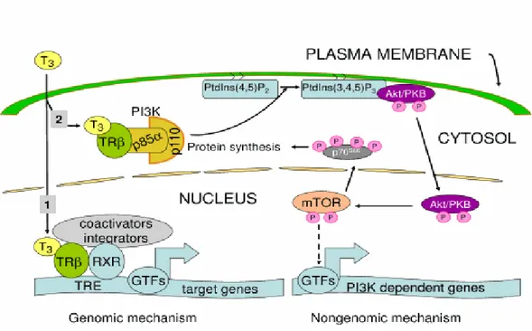
Fig. 2. Genomic and non-genomic action of TH
Recently, Davis and colleagues identified integrin αVβ3 as a plasma membrane
TH-binding site. Previously, they showed that both T4 and T3 activated
mitogen-activated-
protein kinase (MAPK) activity, leading to serine phosphorylation of TRβ
1as
well as to TRβ
1traslocation into the nucleus and co-repressor release. Purified
radiolabeled T4 an
d T3 bind specifically to integrin αVβ3. Moreover, siRNAs against
the integrin αVβ3 subunits block MAPK activation by TH in CV-1 cells. These data
thus provide strong evidence that TH activates the MAPK cascade via TH binding to a
membrane receptor, integr
in αVβ3. This mechanism leads to phosphorylation of nuclear
receptors and can induce angiogenesis and promote cell growth (Oetting, 2007; Davis et
7
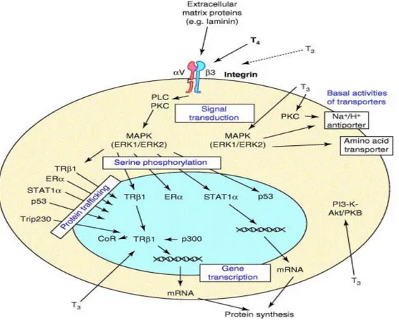
Fig. 3. Summary of membrane-initiated cellular actions of thyroid hormone
Many laboratories have reported tumor cell proliferation in vitro in response to
TH and there are also several reports of anti-apoptotic actions of the hormone on
specific tumor cell lines. Davis et al. have demonstrated that TH, acting at integrin
αVβ3 receptor, non-genomically activates MAPK signal transduction cascade, causing
proliferation of human papillary and follicular thyroid cancer cell line. Moreover they
showed that TH was able to exert an anti-apoptotic effect in both thyroid cancer cell
lines (Davis et al., 2006).
8
In this study we investigated if T3 non-genomic signaling, was able to stimulate
cell proliferation and cell survival in a well-differentiated papillary thyroid cancer cell
line, designed FB2.
9
MATERIALS AND METHODS
Reagents
3,5,3’-Tri-iodothyronine (T3), RDG-peptide, PD-98059 and LY-294002 hydrochloride
were obtained from Sigma-Aldrich (St Louis, MO, USA)
Cell Culture
Human thyroid papillary carcinoma cell line (FB-2) were established and
characterized by Basolo et al. FB-2 cells, derived from a well-differentiated papillary
carcinoma, harbor the RET/PTC1 chimeric oncogene in which the RET kinase domains
is fused to the H4 gene. FB-2 cells only partially retained the differentiated thyroid
phenotype. In fact, the PX-8 gene, which codes for a transcriptional factor required for
thyroid cell differentiation, was expressed, while tyreoglobulin, TSH-receptor and TPO
genes were not. Moreover, FB-2 cells produced high levels of interleukin (6) and
IL-8 (Basolo et al., 2002).
FB-2 cells were cultured in Dulbecco’s Modified Eagle’s medium (DMEM) plus
glutamax, (GIBCO-BRL, Gaithersburg, MD) supplemented with 10% fetal bovine
serum (FBS; Invitrogen) and 1 mg/ml penicillin-streptomycin (P/S). Cells were
maintained at 37°C under humidified conditions of 95% air and 5% CO2.
FB-2 were cultured to 60% confluence and exposed to treatments added only once, at
the beginning of the individual experiments. Prior to treatments, cells were harvested
for 24 hours.
10
Human Follicular Thyroid cancer cells WRO (a gift from Dr Arturi, University of
Magna Grecia, Catanzaro, Italy) were grown in Dulbecco’s Modified Eagle’s medium
(DMEM) plus glutamax containing 10% fetal bovine serum (FBS, Invitrogen) and 1
mg/ml penicillin-streptomycin (P/S).
Proliferation assays
Cell proliferation by (
3H)thymidine incorporation
FB-2 cells were seeded in 6-well plates in a regular growth medium. On the second day,
the cells were incubated in serum free medium (SFM) for 24 hours and then cultured
with treatments. (
3H)thymidine (1 µCi/ml; New England Nuclear, Newton, MA, USA)
was added to the medium for last 6 hours of the second day. After rinsing with PBS, the
cells were washed once with 10% and thrice with 5% trichloroacetic acid. The cells
were lysed by adding 0,1 M NaOH and then incubated for 30 min at 37°C. Thymidine
incorporation was determinate by scintillation counting.
Reverse transcription-PCR (RT-PCR) assay
Cells were grown in 10 cm dishes to 60-70% confluence and exposed to treatments for
24 h in SFM. The total cellular RNA was extracted using TRIZOL reagent (Invitrogen)
as suggested by the manufacturer. The purity and integrity were checked
spettroscopically and by gel electrophoresis before carrying out the analytical
procedures. The evaluation of gene expression was perfomed by the semiquantitative
RT-PCR method. For
TRβ
1,
TRα
1and the internal control gene 36B4, the primers were:
11
CCA TAT CCT CGT CC-3'; TR
α1 forward: 5'-GCC AAA AAA CTG CCC ATG TTC
TCC GAG-3'; TR
α1 reverse: 5'-GGC AGG CCC CGA TCA TGC GGA GGT CAG-3';
TR
β1 forward: 5'-CTC TGT GTA GTG TGT GGT GA-3'; TRβ1 reverse: 5'-TCA TCC
AGC ACC AAA TCT GT-3' to yield respectively the products of 408 bp with 18
cycles, 445bp with 25 cycles and 229bp with 40 cycles.
Immunoblotting
The cells were grown in 10 cm dishes to 70-80% confluence and exposed to treatments
in SFM, as indicated. They were the harvested in cold PBS and resuspended in a lysis
buffer (RIPA-buffer) containing 50 mM Tris-HCl, (pH 7.5), 150 mM NaCl 1% Nonidet
P-40 (NP-40), 0.5% sodium deoxycholate, 1% Sodio-Dodecil-Solfato (SDS) and
inhibitors (0,1mM Na3VO4, 1% phenylmethylsulphonyl fluoride (PMSF), 20mg/ml
aprotinin).
The protein concentration was determinated using Bio-Rad Assay (Bio-Rad
Laboratories). A 50µg portion of protein lysates was used for western blotting (WB),
resolved on a 10% SDS-polycrylamide gel and transferred to a nitrocellulose membrane
(Bio-Rad). Filters were blocked for non-specific reactivity by incubation for 1 h at RT
in 5% non-fat dry milk dissolved in TBST 1X and then incubated for 16 h at 4°C with:
TRβ
1(Santa Cruz Biotechnology), Total Akt (Santa Cruz), Total ERK (Santa Cruz),
phospho Akt 1/2/3-Ser 473 (Santa Cruz), phospho ERK 1/2 (Santa Cruz). As loading,
all membranes were subsequenthly stripped of the first antibody and reprobed with
anti-GAPDH antibody (Santa Cruz), anti-
β-actin antibody (Santa Cruz), anti-Lamin B
antibody (Santa Cruz). The antigen-antibody complex was detected by incubation of the
membranes for 1 h at RT with peroxidase-coupled goat anti-mouse or anti-rabbit IgG
12
and revealed using the enhanced chemiluminescence system (ECL system, Amersham
Pharmacia). The blots were then exposed to Kodak film (Sigma).
Immunoprecipitation
Cells were lysed as previously described; cell lysate (500µg) was incubated for 2 h with
20 µL protein A/G-agarose beads at 4 C and then centrifuged at 12,000 X g for 5min.
The supernatants were then incubated overnight with 10 µl mouse anti-
TRβ
1(Santa
Cruz, 1 µg) and 20 µL of protein A/G. Immunoprecipitates were collected by
centrifugation at 12,000 X g for 10 minutes, followed by washing three times with
HNTG (IP) buffer (50 mm HEPES, pH 7.4; 50 mm NaCl; 0.1% Triton X-100; 10%
glycerol; 1 mm phenylmethylsulfonylfluoride;10 µg/ml leupeptin; 10 µg/ml aprotinin; 2
µg/ml pepstatin). Following the final wash, supernatant was removed. Samples were
resuspended in the Laemmli sample buffer, subjected to SDS-polyacrylammide gel
electrophoresis (10% gel) and then transferred onto a nitrocellulose membrane. The
immunoprecipitated proteins were detected by Western Blot using a rabbit anti-PI3K
p85α (Santa Cruz, 1:500) and mouse anti- TRβ
1.Immunoprecipitation with protein A/G
alone was used as negative control. Membranes were stripped of bound antibodies by
incubation in glycine (0.2 m, pH 2.6) for 30 min at room temperature. Before reprobing
with different primary antibodies, stripped membranes were washed extensively in
TBS-T and placed in blocking buffer (TBS-T containing 5% milk) overnight.
PI3K Kinase assay
Cells were grown in 10 cm dishes to 70-80% confluence and exposed to treatments for
15 minutes in SFM and then lysates with 500µL of lysis buffer (50 mm HEPES, pH 7,5;
150 mmmol/L NaCl, 1,5 mmol/L MgCl2, 1mmol/L EGTA, 10% glycerol, 1% Triton
X-13
100, a mixture of protease inhibitors ((0,1mM Na3VO4, 1% phenylmethylsulphonyl
fluoride (PMSF), 20mg/ml aprotinin)). Cell lysates were centrifuged at 12,000 x g for 5
minutes and 500µg of total protein were incubated overnight with the anti-
p85α
antibody (Santa Cruz Biotechnology) and 500µL of HNTG (immunoprecipitation)
buffer (50 mmol/L HEPES, pH 7,5; 50 mmol/L NaCl, 10% glycerol, Triton X-100, 1%
phenylmethylsulphonyl fluoride, 10mcg/ml leupeptin, 10mcg/ml aprotinin, 2mcg/ml
pepstatin. Immunocomplexes were recovered by incubation with protein A/G-agarose.
The immunoprecipitates were washed once with cold PBS, twice with 0,5 M LiCl, 0,1
M Tris (pH=7,4) and finally with 10mM Tris, 100mM NaCl and 1 mM EDTA. The
presence of PI3K activity in immunoprecipitates was determined by incubating the
beads with reaction buffer containing 10 mm HEPES (pH 7.4), 10 mm MgCl2, 50 µm
ATP, 20 µCi [
γ-
32P] ATP and 10 µg L-
α-phosphatidylinositol-4,5-bis phosphate
(PI-4,5-P2) for 20min at 37 C. The reactions were stopped by adding 100 µl of 1 m HCl.
Phospholipids were extracted with 200 µl CHCl3/methanol. For extraction of lipids,
200 µl chloroform:methanol (1:1, vol/vol) were added to the samples and vortexed for
20 sec. Phase separation was facilitated by centrifugation at 5000 rpm for 2 min in a
tabletop centrifuge. The upperphase was removed, and the lower chloroform phase was
washed once more with clear upper phase. The washed chloroform phase was dried
under a stream of nitrogen gas and redissolved in 30 µl chloroform. The labeled
products of the kinase reaction, the PI phosphates, then were spotted onto
trans-1,2-diaminocyclohexane-N,N,N_,N_-tetraacetic acid–treated silica gel 60 thin-layer
chromatography plates state running solvent used for TLC. Radioactive spots were
visualized by autoradiography.
14
RNA interference (Rnai)
Cells were plated in 10 cm dishes in the regular growth medium the day before
transfection to 60-70% confluence. On the second day, the medium was changed with
SFM without P/S and the cells were transfected with 25bp RNA duplex of validated
RNAi targeted human
TRβ
1mRNA sequence
5’UUGAUGAGCUCCCAUUCCUCGUCUG3’sequence or with a stealth RNAi control
(Invitrogen) to a final concentration of 100nM, using Lipofectamine 2000 (Invitrogen),
as recommended by the manufacturer. After 5 h, the trasfection medium was changed
with SFM in order to avoid Lipofectamine toxicity and the cells were exposed to T3 and
then treated for WB analysis and proliferation assay.
Immunofluorescent microscopy
50% confluent cultures, grown on coverslips, were shifted to SFM for 24 h and then
treated either for 20 minutes with T3 (10
-7M). Cells were then fixed in 4%
paraformaldehyde, permeabilized with 0.2% Triton X-100, washed three times with
PBS, and incubated for 1 h with primary Abs recognizing
TRβ
1(Santa Cruz
Biotechnology). The anti-
TRβ
1monoclonal Ab (mAb) (Santa Cruz) at 2 mg/ml was
used for
TRβ
1staining. Following the incubation with primary Abs, the slides were
washed three times with PBS, and incubated with a secondary Abs, each 1 mg/ml
concentrated. A rhodamine-conjugated donkey anti-mouse IgG (Sigma) was used as a
secondary Ab for
TRβ
1. The cellular localization of
TRβ
1was studied with fluorescence
microscope with 1000 × magnification. The optical sections were taken at the central
plane.
15
Statistical analysis
Statistical analysis was performed using ANOVA followed by Newman-Keuls testing
to determine differences in means. p<0,05 was considered as statistically significant.
16
RESULTS
Thyroid hormone receptor β
1is expressed in FB-2 cells
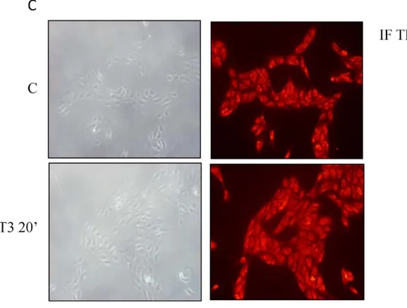
First of all we ascertained the expression of the two major isoforms of TR in
FB-2 cells,
TRα
1and TRβ
1,to demonstrate that the effects of T3 on FB2 cells were
specifically mediated by the binding of thyroid hormone to its own receptor.
As showed in Fig. 1A, by RT-PCR,
TRβ
1mRNA was detected in FB-2 cells and
similar results were observed by Western Blot analysis (Fig. 1B). Follicular thyroid
cancer cells WRO were used as positive control. In contrast, very low levels of
TRα
1mRNA was revealed, while
TRα
1protein was undetectable (data non shown). These
findings indicated that
TRβ
1is the predominant TR isoform in FB-2 cells. Previous
studies reported that
TRβ
1receptor is present at cytoplasmatic and nuclear levels (Davis
et al., 2000; Zhu et al., 1998). Thus, in our cellular context, we performed
immunostaning assay to identify the localization of the thyroid hormone receptor
β
1in
the cell compartment. Our results showed that
TRβ
1receptor was clearly detectable both
at the cytoplasmatic and at nuclear level, confirming its typical localization and that its
expression was not affected by the rapid TH treatment (Fig.1C).
17
Fig. 1. Expression of
TRβ
1in two human thyroid cancer cell line: WRO and FB-2 (A) RT-PCR
analysis showing the expression of mRNA levels of
TRβ
1in WRO and FB-2 cells. 36B4 gene
served as an internal control. (B) Western Blotting analysis using a
TRβ
1-specific antibody.
GAPDH protein served as loading control. (C)
TRβ
1evaluation by fluorescence microscopy in
FB2 cells fixed, permeabilized and stained with anti-
TRβ
1antibody. Cells were treated for 20
18
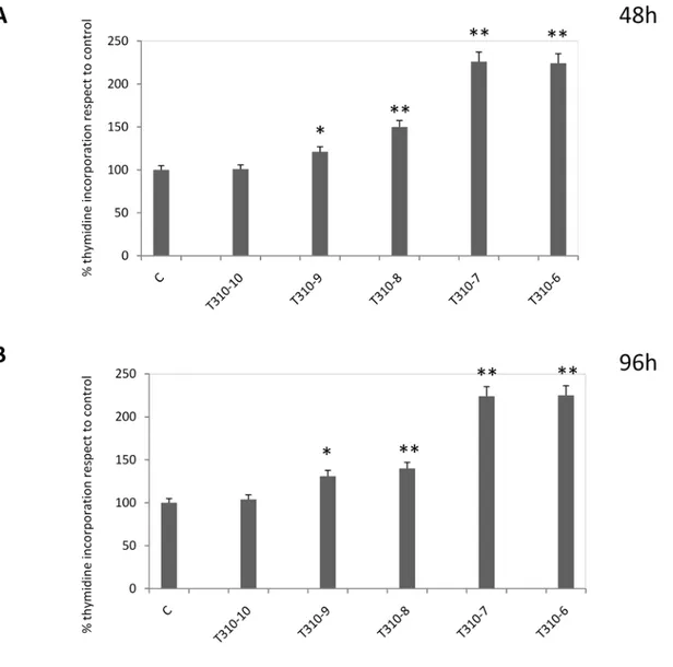
Thyroid hormone T3 induces a proliferative effect in human papillary thyroid
cancer FB-2 cell line
Previous studies have demonstrated the ability of T3 to exert a proliferative
effects in several tumor cells lines (Hsieh et al., 2005;Davis et al., 2006; Lie et al.,
2006; Hall et al., 2008; Poplawski et al., 2008; Lin et al., 2009). Thus, we aimed to
investigate if T3 was able to induce a proliferative effect in a new human thyroid cancer
cell line, designated FB-2, that was derived from a well-differentiated papillary
carcinoma. To this aim, FB-2 cells were treated with increasing doses of T3 for 48
hours. As showed in Fig. 2, T3 induced cell proliferation in a dose-dependent manner.
Notably, the proliferative effect was yet significant using T3 at 1nM concentration, with
higher increment at 100nM; moreover, we observed that the proliferative effects
persisted after 96 hours. These results demonstrate the ability of T3 to stimulate growth
of papillary thyroid cancer cells in vitro.
19
Fig. 2.
Proliferative effect exerted by T3 in FB-2 cells. Cells were cultured in the
presence of increasing doses of T3. Six hours before lysis (
3H)thymidine incorporation was
added. The results represent the means ± SD of three independent experiments, each performed
with triplicate samples and expressed as percentage of growth vs control which was assumed to
be 100%. Statistical significance is shown as * p<0,01; ** p<0,05 vs control.
20
TRβ
1mediates T3 action on FB-2 cells proliferation
Previous studies demonstrated that TR
β
1is essential for the T3 action on the
hCM cell proliferation, survival and size (Verga Falzacappa et al., 2006). To explore
the involvement of thyroid hormone receptor
β
1in the proliferative effect induced by T3
in FB-2 cells, we performed proliferative assay after knoking down
TRβ
1with a specific
siRNA. Our data showed that the cell growth effect exerted by T3 was reduced in the
TRβ
1silenced cells, while no changes was observed after transfection of cells with
scrambled RNA upon identical experimental condition (Fig.3A). These results
demonstrates the crucial role of this receptor in the T3 induced FB-2 cells proliferation.
21
Fig. 3
TRβ
1mediates T3 action on FB-2 cells proliferation. (A) RNA interference experiments
to silence
TRβ
1expression were performed as described in Materials and methods. FB-2 cells
were treated for 48 h with T3 (10
-7M). After 48 hours, the proliferative effect was determined
using (
3H)thymidine incorporation. Data represent the mean ± SD of three independent
experiments performed in triplicate and expressed as percentage of growth vs control which was
assumed to be 100%. *p< 0,05 treated vs control ** <0,05 vs T3-treated in absence of
RNAi
TRβ
1. (B)
TRβ
1protein expression (evaluated by WB) in FB2 cells transfected with RNA
22
TRβ
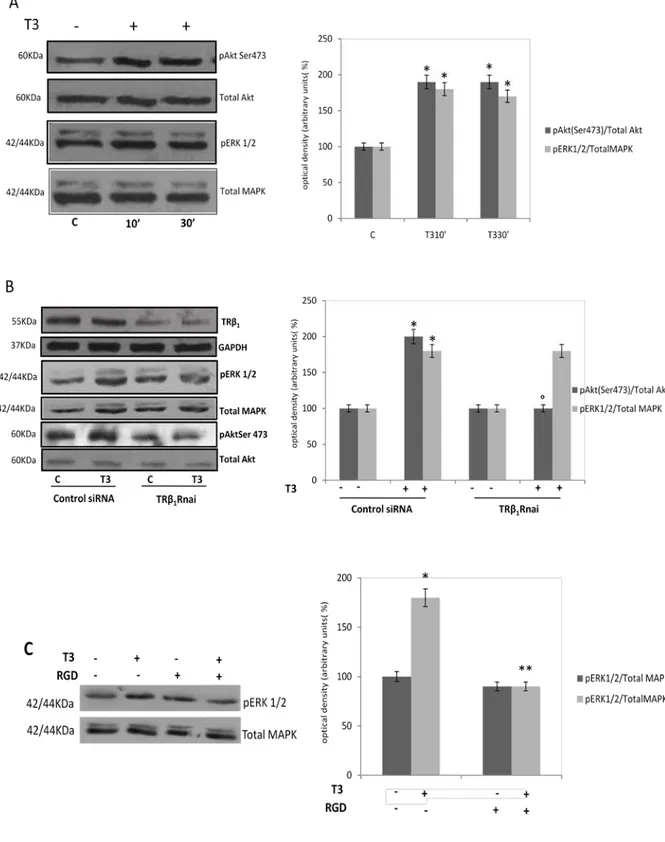
1receptor mediates the rapid activation of AKT induced by T3 in FB2 cells
Considering the proliferative effect induced by T3 in FB-2 cell, we addressed the
contributions of both ERK1/2 and PI3K signal transduction pathways in the action of T3
to specifically assess whether the cell proliferation effects of hormone utilize these
pathways. To this aim we decided to examine an earlier time course of ERK1/2 and Akt
activation by T3 treatment. Cells were cultured in the presence or the absence of the
hormone treatment (T3 10
-7M) for 10 and 30 minutes. As shown in Fig 4A, a significant
increase in Ser 473 phosphorylation of Akt and pERK1/2 was detected as early as 10
minutes after T3 addition and persisted for up 30 minutes.
In the presence of
TRβ
1knocked down, we observed a significant reduction in
the phosphorylation of Akt T3-induced (Fig. 4B). Notably, the activation of MAPK
induced by hormone treatment still persisted in the presence of
TRβ
1siRNAs. This latter
data indicate that T3 membrane signaling, different from those mediated by
TRβ
1,may
be responsible for the maintainement of MAPK stimulation. For instance, it has been
identified on the extracellular domain of integrin
αVβ3 a cell surface receptor for
thyroid hormone which activation induces cellular MAPK signal transduction cascade.
Since thyroid hormone and integrin interaction occurs at or near the Arg-Gly-Asp
(RGD) peptide sequence, we used an RGD peptide to verify if displacement of thyroid
hormone from integrin could block thyroid hormone-MAPK-activation. As shown in
Fig 4C, after 3 hours, the RGD treatment was able to reverse the activation of ERK1/2
by T3 (Fig 4C).
23
Fig. 3
TRβ
1receptor mediates the rapid activation of AKT induced by T3
(A) Before the
treatment, cells were starved for 24 hrs in SFM. Immunoblot of pAkt and pMAPK (ERK1/2)
from FB-2 treated or not (control) with T3 (10
-7M) for 10 and 30 min. The expression of total
24
AKT and total MAPK was analyzed as a control for gel loading. The side panels represent the
means SD of three separate experiments in which band intensities were evaluated in terms of
optical density arbitrary units and expressed as percentages of the control which was assumed to
be 100%. *p<0,005 vs control. (B)
RNA interference experiments to silence TRβ
1were
performed as described in Materials and methods on FB-2 cells exposed or not to T3 (10
-7M)
for 20 min. Western Blot analyses showed a specific band corresponding to the phosphorylated
Akt (Ser 473) phosphorylated ERK1/2 and TRβ
1. The expression of total Akt, total MAPK and
GAPDH were analyzed as controls for gel loading. The side panels represent the means ± SD of
three separate experiments in which band intensities were evaluated in terms of optical density
arbitrary units and expressed as percentages of the control which was assumed to be 100%;
*p<0,01 vs control; ° p<0,05 vs T3-treated cells transfected with control siRNA.
.(C) Cells were
starved for 24 hrs in SFM, pre-incubated with RGD peptide (50nM) for three hours and then
treated for 20 minutes with T3 (10
-7M). Western Blot analyses showed a specific band
corresponding to the phosphorylated ERK1/2. total MAPK was analyzed as a control for gel
loading. The side panels represent the means ± SD of three separate experiments in which band
intensities were evaluated in terms of optical density arbitrary units and expressed as
percentages of the control which was assumed to be 100%. ** p<0,001vs T3-treated cells; *
p<0,005 vs control.
25
TRβ
1complexes with p85
α subunit of PI3K in a ligand independent manner
Recently, some authors (Cao et al., 2005; Storey et al., 2006, Verga Falzacappa
et al., 2009), in different cell lines have shown an interaction
between the subunits p85α
of PI3K and
TRβ
1,similar to that observed for the TR
α
1in endothelial cells (Hiroi et al,
2006). In our cellular system, we evaluated the interaction between cytoplasmatic
TRβ
1and PI3K
p85α subunit.
FB-2 cellular extracts immunoprecipitated with anti-
TRβ
1antibody showed a
constitutive association with the catalytic subunit of PI3K
p85α, that was not influenced
by rapid treatment with T3 (Fig.3).
Fig. 3. Coimmunoprecipitation (IP) experiments . Cells were exposed to T3 (10
-7M) for 20 min.
Immunoprecipitation experiments for
TRβ
1were performed on total extracts. Western blot
analyses for PI3Kp85a and
TRβ
1were performed as described in materials and methods.
Whole-cell lysated (INPUT) were used as input controls. All the data shown are representative
of at least three independent experiments.
26
T3 induces TRβ
1–associated PI3K activity and promotes the nuclear
translocation of activated Akt
To investigate whether T3 is able to affect the PI3K activity, we performed a
PI3K assay on
TRβ
1pulled down samples. As shown in Fig. 5A,
T3
(10
-7
M)
treatment
provoked, after 20 minutes, a significant increase in the kinase activity of the
TRβ
1-associated PI3K. Moreover, to evaluate if the concomitant increase in Akt
phosphorylation was accompanied by an up-regulation in the Akt kinase activity, we
analyzed the activation of the mean target of Akt activity:
glucogen synthetase kinase β
that is phosphorylated by Akt on Ser 9 residues. Interestingly, we found that the
phosphorylation of GSK3β
Ser9was increased by T3 treatment, after 20 minutes. All
these data suggested that in FB-2 cells, the rapid T3 treatment is able to induce PI3K
activity and its downstream effectors. Besides, as shown in Fig 5 C, the rapid thyroid
hormone treatment induces the nuclear translocation of phosphorylated Akt.
27
Fig 4.
T3 induces TRβ
1–associated PI3K activity and Akt activity
. A.
FB-2 cells were incubated in
absence or in presence of T3 100nM for 20 min. Cell lysates were used for PI3K activity. The
autoradiograph presented is representative of experiments that were performed at least three times.
B. FB2 cells were exposed to T3 (10
-7M) for 20 min before lysis. Western blot analyses were
performed as described in Materials and Methods and specific bands corresponding to the
phosphorylated Akt (Ser 473)
and phosphorylated GSK3β
Ser9, were detected. The expression of
total Akt
and GSK3β were analyzed as controls. The side panels represent the means SD of three
separate experiments in which band intensities were evaluated in terms of optical density arbitrary
units and expressed as percentages of the control which was assumed to be 100%. * p<0,01vs
control. C Western Blot analysis on the cytoplasmic and nuclear fractions of protein extracts from
FB-2 cells exposed to T3 treatment for 10 and 30 min were performed and a specific band
corresponding to the phosphorylated Akt (Ser 473) was detected. The expression of Lamin B
(nuclear) and β-actin (cytosol) was analyzed as a control for gel loading and to exclude the
contamination of the cytosol with the nuclear components and vice versa.
28
Activation of PI3-Kinase and MAP-Kinase is involved in thyroid
hormone-induced proliferation in FB2 cells
Finally, we investigated the involvement of PI3K and MAPK pathway in the
proliferative effect T3 induced in FB-2 cells. To this aim we performed a thymidine
incorporation assay using a specific inhibitors of PI3K and MAPK pathway,
respectively LY-294002 and PD-98059, in the presence or absence of T3 treatment.
We found that the combined treatment with LY (5µM) or PD (10 µM),
significantly reduced the proliferation in cells treated for 48 hours with T3 (10
-7M) (Fig.
7), suggesting that in our cellular system, the activation of these pathways is strongly
implicated in the proliferative effect induced by thyroid hormone treatment.
29
Fig. 7 Activation of PI3-Kinase and MAPK is involved in thyroid hormone-induced
proliferation in FB2 cells
.
FB-2 cells were treated for 48 h with T3 (10
-7M), LY-294002
hydrochloride (5µM),
PD (10 µM),
alone or in combination. After 48 hours, (
3H) thymidine
incorporation was determined by scintillation counting. The results represent the means ± SD of
three independent experiments, each performed with triplicate samples and expressed as
percentage of growth vs control which was assumed to be 100%. * p< 0,05 treated vs control;.
** p< 0,05 T3 + Ly or PD treated vs T3 treated.
30
DISCUSSION
Some of the non-genomic actions of TH, involve TRs located outside the
nucleus, since it is known that 10% of TRs are cytoplasmatic in absence of T3
(Baumann et al., 2001), whereas others utilize other proteins that can bind TH, such as
integrin
αVβ3 (Bergh et al., 2005). Non-genomic mechanisms of TRs appear to be
relevant to motility of endothelial cells and to proliferation of several tumor cells;
indeed, many authors reported that TH causes in vitro proliferation of a variety of
cancer cells, including thyroid cancer cells
(H.Y.Tang et. al 2004, M.Cristofanilli et. al
2005 , F.B.Davis et. al 2006; G.B.Hernandez et. al 1999, F.B.Davis et. al 2006, H.Y.Tang et. al
2004; M.L.Hsieh and H.H.Juang 2005; P. Poplawski et al 2008)
.
In the present study we investigated the non-genomic signaling effects induced
by thyroid hormone T3 on a novel thyroid papillary human cancer cell line, designated
FB-2. Our data demonstrated that T3 short exposure is able to activate MAPK/ERK1/2
signaling as well as PI3K/Akt pathway.
Cellular processes, such as proliferation and survival, induced by different
hormones and growth factors are dependent on the activation of PI3K. PI3K is a kinase
that consists of a catalytic subunit of about 110KDa (p110) and a tightly associated
regulatory subunit (p85α, p85β, or p55y). The subunit regulates the association of PI3K
with membrane-associated signaling complexes. Upon activation by membrane
receptors, PI3K phosphorylates phosphatidyl-inositol-4,5 biphosphate (PIP2) to form
phosphatidyl-inositol-3,4,5-triphosphate (PIP3). Through
phosphatidylinositol-dependent kinases, the downstream effectors of PI3K, the serine/threonine Kinase Akt,
is phosphorylated and activated to further phoshorylate downstream protein substrate
31
signaling cascades that affect cellular functions. The activity of PI3K is negatively
regulated by PTEN, a protein phosphatase that dephosphorylates PIP3 to form PIP2
(Eng, 2002).
The activation of PI3K pathway can occur within minutes or hours, and is
related to non-genomic action of specific activators. Recently, it has been observed that
liganded or unliganded TRs bind to the p85α subunit of PI3K and activate the PI3K
signaling pathway, including the phosphorilation of Akt (serine 473), a mammalian
target of rapamycin (mTOR) and its substrate p70
S6K(Hiroi et al., 2006; Verga
Falzacappa et al., 2009). Refetoff et al. showed, in human fibroblast, that T3-treatment
induced a rapid mTOR activation, with detectable phosphorylation as early as 10
minutes and not sensitive to cycloheximide treatment, indicating that this effect of
thyroid hormones uses preexisting proteins. It is not clear why the mechanism of
activation of PI3K signaling by TR differs in the cytoplasm and the plasma membrane,
but may be due to different post-
translational modifications of TRβ
1and p85
α subunit in
the cytoplasm versus the plasma membrane, different receptor/enzyme complex
formation, or cell-specific effects (Moeller et al., 2006; Oetting, 2007).
Recent studies of human cancer specimens by several groups, showed Akt
over-expression and over-activation in primary thyroid cancers (Ringel et al., 2001;
Miyakawa et al., 2003; Motti et el., 2005). In particular, Furuya et al., have reported that
in a mouse model of follicular thyroid carcinoma (TR
β
PV/PV), harbors a knockin mutant
TR
β
1gene, the physical interaction of PV with p85
α subunit results in a constitutive
activation of the PI3K signaling that leads to an increase of thyroid tumor growth and
enhanced cell motility. Moreover, they showed that the inhibition of the PI3K activity
by LY decreased cell proliferation, suggesting that LY treatment, in combination with
32
other specific inhibitors of downstream effectors of PKB, could provide additional
therapeutic effectiveness in treating thyroid cancer.
Tumors of follicular thyroid cells are highly heterogeneous in terms of histology
and response to treatment. Malignant thyroid tumors include (a) well differentiated
carcinomas, which comprise papillary (PTC) and follicular (FTC) carcinomas; (b)
poorly differentiated carcinomas; and (c) undifferentiated carcinomas (Basolo et al,
2002). Well-differentiated PTC is the most frequent type of thyroid cancer and it
accounts for the vast majority of thyroid carcinomas associated with previous exposure
to ionizing radiation. The prognosis of papillary thyroid carcinoma is generally
favorable. However, a number of patients develops recurrences (local or nodal) and
distant metastases, or dies (Mazzaferri et al, 1999). The molecular mechanisms
underlying the initiation and progression of thyroid carcinoma are not full understood,
but it is generally believed that deregulation of cell growth and cell death is involved.
In this study we demonstrated that in FB-2 cells,
TRβ
1receptor was clearly
detectable both at the cytoplasmatic and at nuclear level, confirming its typical
localization and its expression was not affected by the rapid thyroid hormone treatment.
It has been recently reported that thyroid hormone receptor
β
1is involved in the
proliferative effect induced by thyroid hormone T3 in human insulinoma cell line
(Verga Falzacappa et al., 2009).
To explore the involvement of thyroid hormone receptor
β
1in the proliferative
effect induced by T3 in FB-2 cells, we performed proliferative assay using a specific
“knockdown” of the gene encoding TR
β
1., through experiments of RNA interference.
Interestingly, our data showed that the T3 effect on proliferation was completely
abolished in the TR
β
1silenced cells, demonstrating the involvement of this receptor for
33
The non-genomic mechanisms of thyroid hormone action include activation of
the ERK1/2 signal transduction pathway and PI3K signaling. Since in our study we
showed that T3 is able to induce a rapid phosphorylation of Akt and ERK1/2, we
explored the involvement of thyroid hormone receptor
β
1in the activation of these
pathways. Our data demonstrated that the “knockdown” of the gene encoding TR
β
1,
results in a abolishment of AKT activation T3-induced, while the phosphorylation of
MAPK was not affected. It’s well known that the proliferative effect exerts in vitro by
thyroid hormone in some cancer cells, is membrane-initiated at a hormone receptor site
on integrin
αVβ3 through which thyroid hormone non-genomically actives the MAPK
signal transduction cascade (Oetting, 2007; Davis et al., 2007). To verify the role of
integrin receptor in hormone induced activation of MAPK in FB2 cells, we used the
well known RGD peptide that displace thyroid hormone from integrin, blocking
MAPK-activation. Our data evidenced that in the presence of RGD peptide an inhibition
of MAPK activation T3-induced was observed.
It has been evidenced the action of the thyroid hormone T3 on the PI3K/Akt
pathway, showing that T3 can stimulate the phosphatidylinositol 3-kinase at the plasma
membrane (Cao et al., 2005; Furuya et al., 2006; Verga Falzacappa et al., 2006, Lin et
al., 2009).
This activation involves the binding of the TRβ
1and the subunit p85
α of the
PI3K: the said interaction has been proved to be both nuclear and extranuclear (Furuya
et al., 2006). The activation of the PI3K leads to events that include the triggering of the
AKT Kinase and its downstream mTOR. These evidences have suggested that the
action of TRβ
1on the PI3K pathway might drive the increment of cell proliferation and
the suppression of apoptosis.
Our findings showed that in FB2 cells, thyroid hormone receptor
β
1is able to
34
however, nanomolar concentrations of T3, enhances the TR
β
1-associated PI3K activity.
In addition, T3-induced kinase activation can trigger a cascade of events that are
PI3K-dependent, including a rapid phosphorylation of Akt at Ser 473 and of GSK3
β at Ser 9,
one of the main Akt substrates.
In the biological counterpart of the present study, we evidenced the involvement
of PI3K pathway and MAPK signaling in the growth effect induced by T3 in FB-2 cells,
since in the presence of LY or PD the proliferation of FB-2 cells induced by T3
treatment was reversed. All these findings demonstrate how the short exposure to T3
induces tumor cell proliferation concomitantly with anti-apoptotic actions.
If this proliferative action of thyroid hormone in supra-physiological
concentrations is reproduced in the intact organism, it has several implications that are
specific for thyroid cancer. First, the coexistence of thyroid cancer (TC) and
hyperthyroidism in patients is well described, with a reported incidence highly variable,
ranging between 0,2% and 21,0%; moreover, is reported that in toxic thyroid
carcinoma, the papillary hystotype is predominant (Gabriele et al, 2003;Vaiana et al.,
1999; Gulcelik et al., 2006;). Such a wide range of incidence may be due to different
study protocols, diagnosis and follow-up, but the most important factor may be variable
iodine intake of the patients living in different area of iodine supply (Cappelli et al.,
2006; Gulcelik et al., 2006; Pazaitou-Panayiotou et al., 2008; Giles et al., 2008).
However, the nature of “hyperthyroidism-thyroid cancer” relationship is yet
controversial. Experimental data and clinical reports suggest that a patho-physiological
role may be exerted by TSH that not only stimulates normal thyroid cell growth and
function but may promote changes favorable to subsequent tumor development before
that the toxic status suppressed its concentration. Second, it is not clear whether T3 or
T4 influence the growth of normal thyroid or thyroid tumors (Lin et al., 2003);
35
therefore, the conventional use of exogenous thyroid hormone to suppress endogenous
TSH in patients with multinodular goiter or in the thyroid cancer patient who has
undergone thyroidectomy and radioablation, may be carefully considered, in view of the
growth-promotion action of T3 on tumor thyroid cells.
36
BIBLYOGRAFY
F.Basolo, R.Giannini, A.Toniolo, R.Casalone, M.Nikiforova, F.Pacini,
R.Elisei, P.Miccoli, P.Berti, P.Faviana, L.Fiore, Ca.Monaco, G.M.Pierantoni,
M.Fedele, Y.E.Nikiforov, M.Santoro and A.Fusco, (2002) Establishment of a
non-tumorigenic papillary thyroid cell line (FB-2) carrying the RET/PTC1 reattangement,
The Internationa Union Against Cancer, DOI 10.1002/ijc.10116.
S.T.Chen, H.Y.Shieh, J.D.Lin, K.S.S.Chang and K.H.Lin,
(2000)
Overexpression of thyroid hormone receptor b1 is associated with thyrotropin receptor
gene expression and proliferation in a human thyroid carcinoma cell line. Society for
Endocrinology.
M.Cristofanilli, Y.Yamamura, S.W.Kau et al (2005) Thyroid hormone and
breast carcinoma. Primary hypothyroidism is associated with a reduced incidence of
primary breast carcinoma. Cancer, 103:1122-8.
Davis PJ, Shih A., Lin HY., Martino LJ., and Davis FB (2000) Thyroxine
promotes association of mitogen-activated protein kinase and nuclear thryroid hormone
receptor (TR) and causes serine phosphorylation of TR. Journal of Biological Chemistry
275 38032-38039
P.J. Davis, J. L. Leonard, F.B. Davis, (2007) Mechanisms of nongenomic actions
of thyroid hormone, Elsevier Ireland Ltd. 0091-3022.
F.B.Davis, H.Y.Tang, A.Shih, T.Keating, L.Lansing, A.Hercbergs,
R.A.Fenstermaker, A.Mousa, S.A.Mousa, P.J.Davis and H.Y.Lin (2006) Acting via
a Cell Surface Receptor, Thyroid Hormone Is a Growth Factor for Glioma Cells,
American Association for Cancer Research, Cancer Research 66, 7270-7275, July 15
37
P. J. Davis, F. B. Davis and V. Cody, (2005) Membrane receptors mediating
thyroid hormone action. Endocrinology and Metabolism. Nov. Vol 16 N. 9.
J. H. Duncan Bassett, C. B. Harvey, G. R. William, (2003) Mechanisms of
thyroid hormone receptor-special nuclear and extra nuclear actions, Elsevier Ireland
Ltd. 0303-7207.
P. Felig, J. D. Baxter, L. A. Frohman, A .E. Broadus, (1997) Endocrinology and
Metabolism, Terza edizione, McGraw-Hill.
M. Fu, C. Wang, Z. Li, T. Sakamaki and R. G. Pestell (2004) Endocrinology
145 (12): 5439-5447.
A W. Furmanchuk, J I. Averkin, B. Egloff, Ruchti C, Abelin T, Schäppi
W,Korotkevich et al. (1992) Pathomorphological findings in thyroid cancers of
children from the Republic of Belarus: a study of 86 cases occurring between 1986.
Histopathology. Nov;21(5):401-8.
G.B.Hernandez, K.S.Park, A.ace, Q.Zhan and S.y.Cheng (1999) Thyroid
Hormone-Induced Cell Proliferation in GC Cells Is Mediated by Changes in G1
Cyclin/Cyclin-Dependent Kinase Levels and Activity. The Endocrine Society,
Endocrinology Vol. 140, No. 11 5267-5274.
Y. Hiroi, H.H.Kim, H.Ying, F.Furuya, Z.H.Huang, T.Simoncini, K.Noma,
K.Ueki, N.H.Nguyen, T.S.Scanlan, M.A.Moskowitz, S.Y.Cheng, and J.K.Liao,
(2006) Rapid nongenomic actions of thyroid hormone. by The National Academy of
Sciences of the USA.
38
M.L.Hsieh and H.H.Juang, (2005) Cell growth effects of triiodothyronine and
expression of thyroid hormone receptor in prostate carcinoma cells, American Society
of Andrology, Journal of Andrology, Vol. 26, No. 3, May/June.
M.A.Lazar, W.W.Chin, (1990), Nuclear thyroid hormone receptors, The
American Society for Clinical Investigation, Inc. 0021-9738/90/12/1777/06.
H.Y.Lin, F.B.Davis, J.K.Gordinier, L.J.Martino, and P.J.Davis, (1999)
Thyroid hormone induces activation of mitogen-activated protein kinase in cultured
cells. Am J Physiol Cell Physiol 276: C1014-C1024; 0363-6143/99.
H.Y.Lin, H.Y.Tang, A.Shih, T.Keating, G.Cao, P.J.Davis, F.B.Davis (2006)
Thyroid hormone is a MAPK-dependent growth factor for thyroid cancer cells and is
anti-apoptotic. Elsevier Inc.
H.Y. Lin, M. Sun, H. Y. Tang, C. Lin, M.K. Luidens, S A. Mousa, S. Incerpi,
G.L. Drusano, F.B. Davis and P.J. Davis (2009) L-Thyroxine vs
3,5,3’-triiodo-L-thyroxne and cell proliferation: activation of mitogen-activated protein Kinase and
phosphatidylinositol3-kinase. Am J Physiol Cell Phys.May; 295(5):C980-91.
L.C.Moeller, X.Cao, A.M.Dumitrescu, H.Seo, and S.Refetoff, (2006) Thyroid
hormone medited changes in gene expression can be initiated by cytosolic action of the
thyroid hormone receptor
β through the phosphatidylinositol 3-kinase pathway. Nucl
Recept Signal.
A.Oetting, P.M.Yen, (2007) New insights into thyroid hormone action,
Endocrinology and Metabolism.
P. Poplawski, A. Nauman (2008) Thyroid hormone – triiodothyronine- has
contrary effect on proliferation of human proximal tubules cell line (HK2) and renal
cancer cell lines (caki2, Caki-1)- role of E2F4, E2F5 and p107, p130. Thyroid Reserch.
39
H.Y.Tang, H.Y.Lin, J.Zhang, P.J.Davis, F.B.Davis, (2004) Thyroid hormone
causes mitogen-activated protein kinase-dependent phosphorylation of the nuclear
estrogen receptor. Endocrinology 145:3265-72
C. Verga Falzacappa, E. Petrucci, V. Patriarca, S. Michienzi, A. Stgliano, E.
Brunetti, V. Toscano and S. Misiti (2007) Thyroid hormone receptor TR
β1 mediates
Akt activation by T3 in pancreatic
β cells. J. of Molecular Endocrinology
C. Verga Falzacappa, V. Patriarca, B. Bucci, C: Mangialardo, S. Michienzi,
G. Moriggi, A. Stgliano, E. Brunetti, V. Toscano and S. Misiti (2008) The TR
β1 is
essential in mediating T3 action on Akt pathway in Human Pancreatic Insulinoma Cells.
J. of Cellular Biochemestry
Zhu XG., Hanover JA., Hager GL., Cheng SY. (1998) Hormone-induced
translocation of thyroid hormone receptors in living cells visualized using a receptor
green fluorescent protein chimera. Journal of Biological Chemestry 273 27058-27063
40
Scientific Publications Performed during the Program
1) Bonofiglio D, Catalano S, Perri A, Baldini MP, Marsico S, Tagarelli A,
Conforti D, Guido R, Andò S. Beneficial effects of iodized salt prophylaxis on
thyroid volume in an iodine deficient area of Southern Italy. Clin
Endocrinol (Oxf)
. 2008 Sep 23.
2) Tonacchera M, Banco ME, Montanelli L, Di Cosmo C, Agretti P, De Marco G,
Ferrarini E, Ordookhani A, Perri A, Chiovato L, Santini F, Vitti P, Pinchera A.
Genetic analysis of the PAX8 gene in children with congenital hypothyroidism
and dysgenetic or eutopic thyroid glands: identification of a novel sequence
variant.
Clin Endocrinol (Oxf). Jul 2007;67(1):34-4
3) Tonacchera M, Di Cosmo C, De Marco G, Agretti P, Banco M, Perri A,
Gianetti E, Montanelli L, Vitti P, Pinchera A. Identification of TSH receptor
mutations in three families with resistance to TSH. Clin Endocrinol (Oxf). 2007
Nov;67(5):712-8.;
4) Lisi S, Botta R, Pinchera A, Di Cosmo C, Perri A, De Marco G, Menconi F,
Marinò M. Sequencing of the entire coding region of the receptor associated
protein (RAP) in patients with primary hypothyroidism of unknown origin. J
41
Comunications in National and International Conferences
during the Program
1) Workshop “L’Ipotiroidismo Congenito in Italia, Istituto Superiore di Sanità”
(Roma, 4 Luglio 2007)
2) Workshop “La iodoprofilassi in Italia, Istituto Superiore di Sanità” (Roma, 5
Luglio 2007)
3) VI Corso teorico-pratico di citologia e citopatologia della tiroide (ROMA,
UNIVERSITA' CATTOLICA DEL SACRO CUORE - 28/29 NOVEMBRE
2007)
4) Primo Congresso multidisciplinare sull’acne - 19 gennaio 2008 – Pisa
5) XXIX National Congress Italian Society of Pathology (University of Calabria
Rende (CS) Italy, September 10-12 2008)
6) Corso SIAMS Calabria – “Impotenza che fortuna!” (Cosenza, 9 Maggio, 2009):
Relazione dal titolo “Terapia del “Late-onset hypogonadism”
7) 33° Congresso Nazionale Società Italiana di Endocrinologia (Sorrento, 27-30
maggio 2009): POSTER dal titolo “Rapid c-Src mediated effects induced by
triiodothyronine on proliferative and survival signaling in FB-2 papillary
thyroid carcinoma cells”
8) Workshop “L’ipotiroidismo congenito”, Istituto Superiore di Sanità” (Roma, 4
Luglio 2009)
9) Workshop “La iodoprofilassi in Italia”, Istituto Superiore di Sanità” (Roma, 5
Luglio 2009)
42
10) Corso ECM “La iodoprofilassi per la prevenzione del gozzo endemico e delle
patologie ad esso correlato, Università della Calabria (Rende, 28 novembre
2009). Relazione del titolo “Linee guida sulla gestione del paziente con
patologia tiroidea: ruolo del Medico di Medicina Generale, del Pediatra e dello
Specialista”.
Clinical Endocrinology (2007) 67, 34–40 doi: 10.1111/j.1365-2265.2007.02831.x
© 2007 The Authors
34 Journal compilation © 2007 Blackwell Publishing Ltd
O R I G I N A L A R T I C L E
Blackwell Publishing Ltd
Genetic analysis of the
PAX8
gene in children with congenital
hypothyroidism and dysgenetic or eutopic thyroid glands:
identification of a novel sequence variant
Massimo Tonacchera*, Maria Elena Banco*, Lucia Montanelli*, Caterina Di Cosmo*, Patrizia Agretti*, Giuseppina De Marco*, Eleonora Ferrarini*, Arash Ordookhani*, Anna Perri*, Luca Chiovato†, Ferruccio Santini*, Paolo Vitti* and Aldo Pinchera*
*Dipartimento di Endocrinologia e Metabolismo, Centro di Eccellenza AmbiSEN, Università di Pisa, Pisa, Italy, †Cattedra di
Endocrinologia, U.O. di Medicina Interna e Endocrinologia, Università di Pavia, Fondazione S. Maugeri IRCCS, Pavia, Italy
Summary
Objective To analyse the coding region of PAX8 in individuals with congenital (CH) or post neonatal hypothyroidism due to dysgenetic (TD) or eutopic thyroid glands.
Design and patients Forty-three children with CH and TD (13 agenesis, 23 ectopia, and seven hypoplasia), one subject with post neonatal onset of hypothyroidism and thyroid ectopia, 15 children with CH and eutopic thyroid glands and six euthyroid adults with thyroid hemiagenesis were enrolled as cases, along with 120 healthy individuals as controls.
Measurements Exons 2–8 of the PAX8 were directly sequenced. HeLa and HEK293 cells were transfected with PAX8 wild-type (PAX8-WT), mutant PAX8, p300, thyroid transcription factor 1 (TTF-1) and thyroglobulin promoter pGL3 (TG prom-pGL3). Synergism of TTF-1 with PAX8-WT vs. mutant and activity of PAX8 -WT vs. mutant in accompaniment with p300 on TG prom-pGL3 were also assessed. The luminescence produced by PAX8-WT and mutant PAX8 was measured.
Results Among patients and controls only a 15-year-old girl with thyroid ectopia showed a heterozygous transition of cytosine to thymine at position 674 in exon 6, which changed a conserved threo-nine at position 225 to methiothreo-nine (PAX8-T225M). Her father and sister harboured PAX8-T225M without abnormal thyroid pheno-types. PAX8-T225M and PAX8-WT similarly increased luciferase activity and had a similar synergistic effect with TTF-1. At 500 ng p300, however, PAX8-T225M could not significantly increase TG
promoter activity when compared to PAX8-T225M alone, while
PAX8-WT +500 ng p300 induction was significantly higher than
PAX8-WT alone (P < 0·001). Cotransfection of TTF-1 together with PAX8-T225M resulted in rescuing of the lack of synergism with p300.
Conclusions PAX8 mutations in congenital hypothyroidism due to dysgenetic or orthotopic thyroid glands are rare. PAX8-T225M is probably a rare variant.
(Received 2 August 2006; returned for revision 23 August 2006; finally revised 30 November 2006; accepted 17 January 2007)
Introduction
Primary congenital hypothyroidism (CH) is the most common con-genital endocrine disorder, occurring in 1 in 3000 –4000 live births.1 In iodine replete areas, thyroid developmental defects (75–80%) and inborn errors of thyroid hormone biosynthesis (10 –15%) are the most frequent, while transient (5–10%) and central (hypothalamic– pituitary) hypothyroidism (approx. 5%) are the least frequent causes of CH.2,3 Thyroid dysgenesis (TD) presents as agenesis (athyreosis), hypoplasia, ectopia or hemiagenesis of the thyroid gland.4,5 The pathogenesis of TD is largely unknown.6 While most cases are spo-radic, up to 2% of patients with TD have a positive family history of this condition.7 Although there is discordance of TD among monozygotic twins8, mild thyroid abnormalities in first degree relatives of patients with TD,9 the association of parental consan-guinity and TD occurrence in a population with high blood-related marriages10 and a recent report on the mouse model suggestive of a multigenic origin of TD11 suggest that the mechanisms underlying thyroid dysgenesis are complex and heterogeneous. The current data suggest that there is a spectrum ranging from monogenic to multifactorial genetic aetiologies, and environmental and epigenetic modifiers are likely to be contributing factors.12,13
PAX8, a transcription factor of the mammalian Pax protein family, is known to recognize DNA via a highly conserved paired domain.14–16 It is detected in the developing thyroid in early embryonic days at the time of specification and its expression is maintained in thyroid follicular cells during all stages of development.17,18 The PAX8 gene maps to human chromosome 2q12–2q14 and consists of 11 exons.4,19 Initial screening of 145 patients with known TD revealed de novo
monoallelic mutations of PAX8 in two sporadic cases and in a family with three CH-affected members.20 Subsequently, other mutations
Correspondence: Massimo Tonacchera, Dipartimento di Endocrinologia, Università degli Studi di Pisa, Via Paradisa 2, 56124, Cisanello, Pisa, Italy. Tel: +50/995048; Fax: +50/578772; E-mail: [email protected]
PAX8 mutation in congenital hypothyroidism 35
© 2007 The Authors
Journal compilation © 2007 Blackwell Publishing Ltd, Clinical Endocrinology, 67, 34–40
have been discovered.21–25 In the familial cases transmission was autosomal dominant.18 However, in one family there was profound discrepancy between two related individuals bearing the same heterozygous mutation denoting that the phenotype is highly vari-able even with the same mutation.22 Recently, the novel mutation (S48F) located in the PAX8 paired domain has been shown to have a specific defect in transactivation26 due to a loss of co-operation with the general co-activator p300.
In this study we performed mutational analyses of the PAX8 gene in CH-affected individuals (and in one with post neonatal hypo-thyroidism) with dysgenetic and orthotopic thyroid glands.
Subjects, materials and methods
Subjects
The study population comprised 43 children with CH and thyroid dysgenesis, either agenesis, ectopia, or hypoplasia, one subject with post neonatal onset of hypothyroidism and thyroid ectopia (group 1), six young adults with thyroid hemiagenesis without clinical or biochemical hypothyroidism (group 2), 15 children with CH and eutopic thyroid gland of normal size (group 3) and 120 consecutively selected healthy volunteers with no thyroid abnormality (group 4). Except for one subject in group 1, CH was diagnosed in all group 1 and group 3 children during national newborn screening for CH using the primary thyrotropin (TSH) screening test. Levothyroxine (L-T4) replacement treatment, 10 –12 µg/kg/day, was started within the first month of life and L-T4 dosage was adjusted during infancy and childhood according to serum TSH and thyroxine (T4) in follow up sessions (2). Determination of CH type (permanent vs. transient), after 30 days of L-T4 therapy discontinuation, and of permanent CH aetiologies (dysgenesis vs. eutopic thyroid glands) by thyroid scintigraphy using technetium-99 m pertechnetate (99mTC) were performed at ≥ 3 years of age in CH-affected individuals.
Group 1
Forty-three subjects in this group were born at term (≥ 37 weeks’ gestation) after an uneventful pregnancy. Thyroid 99mTc scan was performed during the 6th year of age after 30 days of discontinuation of L-T4 replacement therapy, and subsequent elevation of serum TSH confirmed the permanent nature of CH. Thyroid scintigraphy revealed absent uptake (agenesis confirmed by subsequent ultra-sonography) in 13, ectopia in 23, and hypoplasia in seven individuals. On physical examination no craniofacial, truncal or limb anomalies were found and no structural visceral, in particular cardiac and renal, anomalies were observed upon echocardiography and abdominal ultrasonography.
The last patient in this group, a female subject, had been diagnosed with hypothyroidism at 15 years of age during the evaluation of primary amenorrhoea. At the time of diagnosis she had a serum TSH of 71 (normal: 0·3– 4) mU/l, free T4 (FT4) of 7·7 (normal: 10·3–24·5) pmol/l, and free triiodothyronine (FT3) of 4·6 (3·1–7·5) pmol/l. TPOAb and TgAb were negative. Cervical ultrasonography showed a 10-×-11-mm round mass at the base of the tongue and 99mTc scanning confirmed the presence of ectopic thyroid tissue. Her past medical
record showed that she was born at term after an uncomplicated pregnancy and had normal TSH on neonatal screening testing. It remains unclear when biochemical and clinical hypothyroidism first appeared. Nevertheless, no signs of mental retardation were present at the time of CH diagnosis. At 17 years of age her height was 157 cm (25th percentile for the age- and sex-matched Italian population) and her weight was 39·5 kg.
Group 2
Six adult subjects (mean age of 20·5 years) with clinical and bio-chemical euthyroidism and thyroid hemiagenesis, detected by ultra-sonography, were included in the study. All subjects showed hemiagenesis of the left thyroid lobe.
Group 3
This group comprised 14 children born at ≥ 37 weeks’ gestation after uneventful pregnancies and a premature female offspring of a twin delivery. All had abnormally high TSH at newborn screening and serum TSH and T4 confirmed CH diagnosis during neonatal and infancy periods. Cervical ultrasound showed a bi-lobed, properly located and normal-sized thyroid gland in all of the subjects during the neonatal period.
In six children aged ≥ 3 years old, permanent CH was diagnosed by abnormal serum TSH and FT4 after 30 days of L-T4 discontinu-ation, and orthotopic thyroid glands were also confirmed by 99mTc thyroid scans. In the remaining eight children L-T4 therapy was not stopped because they were < 3 years of age.
The only premature subject, product of a provisionally monozygotic twin delivery according to phenotype similarities, was born at 28 weeks’ gestation after an uneventful pregnancy. CH was diagnosed via a neonatal screening programme and subsequent ultrasonography revealed in-place thyroid gland, which was also confirmed by thyroid
99m
Tc scintigraphy during the neonatal period. L-T4 replacement therapy, 10 µg/kg/day, was started at 19 days of life. Her twin sister had normal TSH at CH screening.
The study was approved by the local ethical committee of our university and informed consent was obtained from all the parents of the children.
Thyroid ultrasound
Ultrasound evaluation was performed by the same examiner (M.T.) using a linear transducer (7·5 MHz) attached to a real time instru-ment (AU 590 Asynchronous Apparatus, Esaote Biomedica, Milan, Italy).
Laboratory evaluation of thyroid function
TSH was assessed by sensitive-TSH IRMA (Delfia, Wallac, Finland). FT4 and FT3 were measured in the laboratory of our department by radioimmunoassay (FT4 RIA, FT3 RIA, Lysophase; Technogenetics, SpA, Milan, Italy). TPOAb and TgAb were measured by passive agglutination (SERODIA-AMC and SERODIA-ATG, Fujirebio, Tokyo, Japan).