PhD COURSE IN BIOCHEMISTRY
XXXIII CYCLE
ENDOCANNABINOIDS MODULATE NEUROGLIAL
PHENOTYPE AND PROTEOTOXIC STRESS
PhD student
MADDALENA GRIECO
Tutor Coordinator
Prof. Maria d’Erme Prof. Stefano Gianni
ACKNOWLEDGMENTS
I would like to thank Prof. Maria d’Erme for guiding me during my PhD and for teaching me that tenacity and critical thinking are the basis of a good researcher.
I would like to thank Prof. Luciana Mosca for supporting and encouraging me along with all the wonderful S30 Lab group.
A special thanks goes to Dr. Anna Maggiore, for being a formidable friend and colleague with whom to share pain and joys of work and life. She was an indispensable support to achieve this goal.
I would like to thank Prof. Patrizia Mancini, Prof. Rita Businaro, Prof. Tiziana Bisogno, Dr. Maria Giovanna De Caris and Dr. Elisa Maggi for their fundamental collaboration to this project.
I would like to thank Prof. Marcel Leist and his lab for the hospitality and the support given to me during the ten months spent in Konstanz.
I would like to thank Prof. Fabien Gosselet and Prof. Filomena Fezza for the careful supervision of my thesis and for their valuable advice.
Furthermore, I would like to thank Prof. Francesco Malatesta for teaching us that the knowledge sharing represents a precious treasure to achieve whatever goals.
I would like also to thank all Professors of the Department of Biochemical Sciences, and all my colleagues for their valuable contribution to my scientific education.
Last but not least, I would like to thank Domenico and my whole family for always believing in me!
SUMMARY
ABSTRACT ... 1
1. INTRODUCTION ... 4
2. ENDOCANNABINOID SYSTEM ... 6
2.1. FAAH enzyme ... 13
2.2. The endocannabinoid system and its therapeutic exploitation .... 16
2.3. URB597: a selective FAAH inhibitor ... 18
2.4. Endocannabinoid system in neurodegenerative disorders ... 19
2.5. Alzheimer disease (AD) and Endocannabinoid system ... 22
Therapeutic approaches ... 25
Modulation of the endocannabinoid system as new therapeutic approach for Alzheimer’s disease... 28
3. AIM OF WORK ... 30
4. MATERIALS AND METHODS ... 32
4.1. Materials and chemicals ... 32
4.2. SPERIMENTAL MODELS ... 33
Microglia ... 33
Astrocytes... 35
LUHMES cells as neuronal model system ... 37
4.3. Preparation of Aβ25–35 and URB597Stock Solution ... 38
4.4. Fatty acid amide hydrolase assay ... 38
4.5. Cell cultures and treatments ... 39
Human microglia conditioned medium ... 43
4.6. Cell viability assays ... 43
MTT assay ... 43
Resazurin ... 43
LDH release ... 44
ATP assay ... 44
4.8. Migration assays ... 46
4.9. Fluorescein isothiocyanate (FITC)-dextran uptake assay ... 46
4.10. Western Blotting ... 47
4.11. Pull down assay for activated Rho GTPases ... 48
4.12. Determination of total glutathione ... 49
4.13. Real-time quantitative PCR analysis ... 50
5. PROJECT I ... 52
Results ... 52
5.1. Aβ25-35 does not affect the activity of the FAAH enzyme ... 52
5.2. A− induces upregulation of the Iba1 microglia marker ... 53
5.3. URB597 does not exhibit cytotoxic effects in BV-2 cells ... 54
5.4. URB597 on BV-2 cells viability ... 56
5.5. URB597 reverts morphological changes induced from Aβ25-35 .... 57
5.6. Effect of URB597 on Cellular Migration ... 60
5.7. URB597 increases phagocytic capacity of microglia ... 62
5.8. URB597 promotes the activation of Rho GTPase family ... 64
5.9. URB597 reduces mRNA expression of IL-1 and TNF- and increases TGF- and IL-10 ... 67
5.10. URB597 modulates iNOS and Arg-1 expression ... 68
DISCUSSION ... 70
6. PROJECT II ... 76
Results ... 76
6.1. mRNA expression of microglia and astrocytes markers after stimulation with LPS and cytokines ... 76
6.2. Cytokines modulations after LPS and cytokines treatment in human neuroglial cells. ... 79
6.3. NF-kB translocation after TNF- treatment in human neuroglial stem cells. ... 81
DISCUSSION ... 83
Results ... 85
7.1. URB597 does not exhibit cytotoxic effects in LUHMES cells ... 87
7.2. Protection of LUHMES against proteasome inhibition by neuroglial cells and endocannabinoid inhibitors... 88
7.3. Co-culture effect on ATF4 proteins levels in presence of a proteasome impairment... 89
7.4. GSH levels modulation in LUHMES cells in presence of proteotoxic stress and the neuroglial role ... 90
7.5. Modulation of the GSH level triggered by URB597 on LUHMES cells treated with MG-132. ... 91
7.6. Modulation of mRNA levels of enzymes involve in the GSH metabolism ... 92
7.7. URB597 influences NRF2 LUHMES cells in presence of proteasome impairment... 93 DISCUSSION ... 95 CONCLUSIONS ... 98 SUPPLEMENTARY DATA ... 99 REFERENCES ... 100 APPENDIX ... 126
1
ABSTRACT
Neuronal survival in neurodegenerative diseases and brain damage is closely related to the cell populations of the environment and in particular to glial cells. Astrocytes, microglia and oligodendrocytes oversee brain homeostasis providing the intrinsic brain defence system. Damage to brain cells triggers a condition generally referred to as reactive gliosis, which includes astrogliosis and activation of microglia. Neuroglia is also thoroughly involved in pathogenesis of many chronic neurological disorders and in neurodegeneration. Endocannabinoids modulating the behaviour of microglia and astrocytes might act as possible targets for therapeutic intervention. Recent studies have indicated that endocannabinoid levels and metabolic enzymes change during the progression of Alzheimer's disease (AD) and that the inhibition of fatty acid amide hydrolase (FAAH), the main catabolic enzyme of anandamide (AEA), has beneficial effects in mice with AD. The aim of this study was to determine whether URB597, a FAAH inhibitor, targets microglia polarization by altering the cytoskeleton reorganization induced by amyloid-β peptide (Aβ) in BV-2 microglial cells. Evaluation of actin cytoskeleton showed that Aβ treatment increased the surface area of BV-2 cells, which acquired a flat and polygonal morphology. Although URB597 did not affect cell morphology only, it partially rescued the control phenotype in BV-2 cells incubated with the combined treatment. Rho family proteins have a critical role in the plasticity of the actin cytoskeleton, influencing morphological changes, migration and phagocytic activity of cells. We observed an increase of Rho protein activation in Aβ samples and a decrease in samples treated with URB597 alone or in combination with Aβ compared to controls, while an increase of Cdc42 protein activation was observed in all samples with respect
2
to control. Aβ induced the migration of BV-2 cells up to 2 h after stimulation. We also found that by reducing Rho protein activity, URB597 was able to reduce the migration rate. URB597 also increased the number of BV-2 cells performing phagocytosis. Taken together, these data suggest that an increase of anandamide (AEA), due to FAAH inhibition, may induce cytoskeleton reorganization, regulating phagocytosis and cell migration processes, and promote microglial polarization towards an anti-inflammatory phenotype.
As most research worldwide has focused on neurons, there is a dearth of protocols to generate glial cells and to produce co-culture systems for biomedical research. The aim of this project has also been the generation of co-culture with neurons, astrocytes and microglia cells and the subsequent characterization of the resulting model, evaluating interspecies differences through the generation of co-cultures with murine microglia. We focused our interest on the repair functions during brain injury and on the interactions between microglia and astrocytes. The protective effect of astrocytes and microglia against neuronal cells in the presence of inflammatory and pro-apoptotic processes was investigated. Human astrocytes and human microglia cells were activated with TNF-, IL-1 and IFN- to evaluate the inflammatory response. The results showed an increase of inflammatory cytokines gene expression such as IL-6 and IL-8 in both cell lines examined. The astrocytes activation by TNF-, or by conditioned medium (CM) of activated microglia cells was confirmed by NF-kB nuclearization. Therefore, the arise of inflammatory process in astrocyte cells is driven not only by TNF- induction, but also by a synergic effect due to microglia activation. Neuroinflammation, oxidative stress, and progressive degeneration of specific brain regions is also driven by proteasomal impairment, promoting protein accumulations. Since LUHMES neurons are quite susceptible cells to
3
proteotoxic stress and amino acid starvation, we investigated whether murine microglia and human astrocytes exerted a protective effect also when the cell lines were treated with URB597. The obtained data demonstrated that the astrocytes through the glutathione (GSH) release, were able to attenuate neuronal proteotoxic stress in LUHMES cells. URB597 contributed to GSH anti-oxidant effects modulating GSH metabolism. The overall data demonstrated that neuroglial cells play a pivotal role on neuronal protection from noxious stimuli.
4
1. INTRODUCTION
Numerous studies indicate that inflammation-mediated neurodegeneration processes and disturbed neuron-microglia interactions may mediate the pathogenesis of several neurodegenerative diseases [1,2]. The two
most important endogenous cells in the Central Nervous System (CNS) that promote inflammation are astrocytes and mononuclear phagocytes, which include microglia and perivascular macrophages. Astrocytes are the most important abundant glial cells in the CNS. They derive from the neuroectoderm and are essential in the synapse formation and function, in ion and neurotransmitter concentrations, contributing to the integrity of the blood– brain-barrier (BBB) and in brain homeostasis and neuronal survival [3]. Astrocytes show an important role in neuroinflammation with the production of anti-inflammatory cytokines, such as transforming growth factor (TGF) able to contrast microglia activation during an inflammatory process [4,5]. Two different phenotypes have been identified in the CNS, A1
and A2. Neuroinflammation stimulate A1 astrocytes to release neurotoxins that induce death of neurons. On the other hand A2 astrocytes promote neuronal survival and tissue repair [6]. This terminology parallels the M1 and M2
macrophage nomenclature, which has also been applied to microglia in the CNS [7]. Microglia are the resting macrophages of the CNS generated by the
embryonic yolk sac. They migrate into the developing neural tube and after they extend to the brain parenchyma [8].Several markers and receptors in
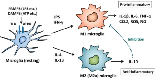
microglia are similar to monocytes and macrophages of peripheral tissue. The M1 phenotype is induced by toxins, lipopolysaccharides (LPS), interferon gamma (IFN-) and pro-inflammatory cytokines. On the contrary M2 phenotype release anti-inflammatory cytokines such as IL-4 and IL-10
5
counteracting neuroinflammation and neuron damages [9]. Under
neuroinflammatory conditions, neurons or astrocytes produce endocannabinoids (eCB) as a means of recruiting microglia [10]. Neuroglial
cells involved in the inflammation process express functional cannabinoid receptors and produce and degrade eCB, suggesting that the endocannabinoid signaling system has a regulatory function in the inflammatory response. Inflammation are on the bases of several diseases as diabetes [11],
cardiovascular diseases and neurodegenerative diseases [12], and eCB exhibit
pleiotropic effect on the complex development of these diseases. Several studies have indicated that eCB levels and metabolic enzymes change during neurodegenerative process and that the inhibition of the major N-Arachidonoylethanolamine (AEA)-hydrolyzing enzyme, fatty acid amide hydrolase (FAAH), has a possible neuroprotective role towards oxidative stress, inflammation and excitotoxicity [13].
6
2. ENDOCANNABINOID SYSTEM
Over the past millennia, Cannabis sativa was considered healers in early civilizations for the therapeutic effects of its psychotropic compounds obtained from desiccated flowers. The most important natural compounds derive from cannabis plant flowers are Δ9-tetrahydrocannabinol (THC) and the non-euphoric cannabidiol (CBD) [14,15]. More recent study suggest the therapeutic effects of these plants [16], but the first cannabis-derived compounds for neurological disorder were approved only in the 20th century,
with the commercialization of nabiximolz (Sativex®) [17]. In animals, the THC and a less degree the CBD, produce similar effects to those of marijuana, such as catalepsy, hypo-locomotion, analgesia and hypothermia in mice and static ataxia in dogs [18,19]. There is no evidence that allows us to associate medical
beneficial effects to THC alone. The CDB shows a safer therapeutic window and it is more amenable to clinical development, even for paediatric populations [20,21]. The first evidence for the existence of a specific binding
site for THC took place in the 1988, when Devane et al. (1988), during experiments using radio-marked CP55940, identified the cannabinoid receptor 1 (CB1), a G protein-coupled receptor (GPCR) that is expressed most abundantly in the brain [22]. Cannabinoid receptor 2 (CB2) was identified only
in the 1990 through homology cloning. It is also a GPCR and is highly expressed in the immune system. CB1 and CB2 receptors exhibit 68% structure homology [23,24]. CB1 is expressed in both presynaptic and
postsynaptic neurons. CB1 on the presynaptic membrane can inhibit voltage-gated Ca2+ channels and vesicular release of GABA or glutamate by a
retrograde modulation [25]. In the postsynaptic neurons, CB1 mediate slow
7
of appetite- controlling peptides in the arcuate nucleus of the hypothalamus
[26]. CB1 is also located in the external membrane of mitochondria, where it
inhibits electron transport and the respiratory chain, thereby affecting brain metabolism and memory formation [27,28]. In astrocytes, CB1 is involved in
the regulation of synaptic plasticity in the hippocampus and in leptin signalling in the hypothalamus [29]. Activation of CB1 also stimulates proliferation of
adult progenitor stem cells and their differentiation into neurons or astrocytes, a role that could be relevant to neurodegenerative disorders [30]. CB2 receptor
is considered an immune-modulator receptor. CB2 is highly expressed in microglia and it shown a close correlation with neurodegenerative pathology like Alzheimer disease (AD), Multiple sclerosis (MS) and Amyotrophic lateral sclerosis (ALS) [31,32]. Some evidence suggests an important role of CB2 in the modulation of inflammatory cytokines. Indeed, CB2 is able to reduce pro-inflammatory cytokines release from microglia cells in AD [33]. Today we know that there are several other receptors related to the endocannabinoid system (ECS) such as metabotropic receptors GPR55, GPR119, GPR18 and transient receptor potential vanilloid receptor (TRPV). TRPV1 receptor is present in GABAergic and glutamatergic terminals and neuronal somata in the hippocampus and cerebellum [34,35]. Some study suggests an involvement of
TRPV1 in the generation of Ca2+ influx and depolarization in spinal or sensory
neurons, but the demonstration is difficult. TRPV1 is implicated in short- term and long- term synaptic plasticity , in the regulation of mood, fear, memory, food intake, visual development and locomotion [36]. TRPV1 is also important
in the reduction of inflammatory cytokines from activated microglia [37].
PPARα and PPARγ are expressed in neurons, astrocytes and microglia in the brain, where they have anti-inflammatory and neuroprotective effects during acute and chronic neuroinflammatory insults, such as brain trauma, ischaemia,
8
AD and MS [38]. PPARα also reduces food intake [39], whereas PPARγ is
involved in neuronal differentiation [40]. The role of GPR55 is not so clear
today. It is able to stimulates excitatory hippocampal neurons [41,42], whereas
the expression in microglia suggests an involvement of this receptor in the inflammation processes [43].
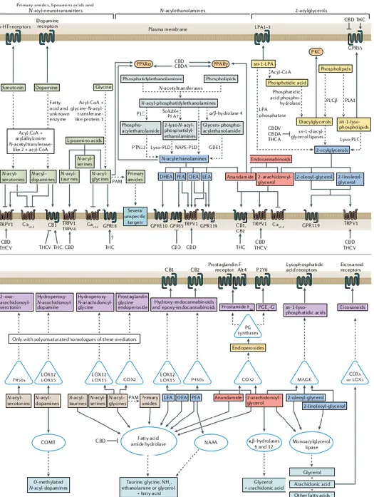
The identification of CB1 and CB2 allowed the sudden characterization of two endogenous ligands that have high affinity for these two receptors, which are the lipids anandamide (ethanolamide of arachidonic acid - AEA) and 2-arachidonoylglycerol (2-AG) [44-47]. AEA can bind both receptor subtypes as a partial agonist [48,49]. 2-AG is also able to activate both receptor subtypes, however, behaving as a full agonist [45]. AEA is able to target also TRPV1 and PPARγ and inhibits Ca2+ channels and transient
receptor potential cation channel subfamily M member 8 (TRPM8) channels, whereas 2-AG activates TRPV1 channels and GABAA receptors [50]. Many
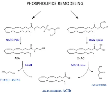
enzymes are involved in the endocannabinoids’ synthesis and degradation. N-acylphosphatidylethanolamine (NAPE)-specific phospholipase d-like hydrolase (NAPE-PLD) catalyses the synthesis of AEA and other N-acylethanolamines, and fatty acid amide hydrolase (FAAH) catalyses the hydrolysis of AEA (and other N-acylethanolamines and fatty acid primary amides). Diacylglycerol lipase α and (DAGLα, DAG) catalyse the biosynthesis of 2-AG and other monoacylglycerols and monoacylglycerol lipase (MAGL) catalyses the hydrolysis of 2-AG (and that of other monoacylglycerols) [51-54] (Fig. 1).
9
Figure 1: Endocannabinoids metabolism
AEA and 2-AG can be metabolized via oxidation by cyclooxygenase 2 (COX-2) and the end products like prostaglandin ethanolamides and prostaglandin glycerol esters, are able to activate other receptors than cannabinoid and prostanoid receptors [55]. AEA and 2-AG can also be
inactivated by other hydrolases, but these enzymes also metabolize other lipids, so targeting them would create other problems [56,57]. Although 2-AG
is an excellent CB1 agonist, the hyperactivation of the receptor leads to its consequent desensitization. Furthermore, chronic administration of MAGL inhibitors produces an effect opposite to that obtained from the CB1 receptor
[58,59]. At the same time 2-AG is a precursor of arachidonic acid and pro-
inflammatory proteinoids [60,61]. Therefore, inhibition of the two main enzymes involved in endocannabinoid synthesis, like NAPE-PLD and DAGL, does not show selectivity and effectively in the reduction of eCB levels, this because there is a compensation from other mediators [62]. Several pathways
10
and enzymes are involved in the synthesis of AEA, 2-AG and other N-acylethanolamines and monoacylglycerols. In nerve cells, eCB are formed by the Ca2+-dependent activation of phospholipases C or D which catalyse the
formation of 2-AG and AEA respectively from membrane phospholipids. The synthesis of endocannabinoids is favoured by all the processes that lead to an increase in [Ca2+] such as, for example, the activation of the phosphoinositide
pathway with synthesis of IP3 and release of Ca2+ from the endoplasmic
reticulum (ER) or opening of the Ca2+ VOC channels following membrane
depolarization. 2-AG and AEA are fat-soluble molecules that diffuse across the cell membrane and interact with cannabinoid receptors on surrounding cells (Fig. 2).
11
12
Retrograde signaling is the principal mode by which endocannabinoids mediate short- and long-term forms of plasticity at both excitatory and inhibitory synapses. However, growing evidence suggests that endocannabinoids can also signal in a non-retrograde manner [63] (Fig. 3)
Figure 3: Endocannabinoid signaling at the synapse (From Castillo P.E. et al. Neuron 2012)
Retrograde activation of the receptor underlies short-term and long-term forms of synaptic plasticity, including depolarization-induced and metabotropic receptor-mediated suppression of excitatory and inhibitory neurotransmission, long- term depression of excitation or inhibition, and long- term potentiation [64]. Retrograde signal of endocannabinoids which regulates
the release of GABA at the level of the inhibitory terminations in some neurons of the hippocampus and cerebellum. This effect is absent after treatment with an endocannabinoid antagonist. The molecular mechanism underlying this response is the Ca2+-dependent activation of the PLC which leads to the
synthesis of 2-AG. 2-AG back scatters in the synaptic space and interacts with CBRs present on the synaptic membrane. CBRs are believed to be associated
13
with a Go protein which through βγ dimer to cause the closure of N-channels of Ca2+ [65] (Fig. 4).
Figure 4:Molecular mechanisms underlying endocannabinoid-mediated short (A)- and
long-term synaptic plasticity (B) (From Castillo P.E. et al. Neuron 2012)
2.1.
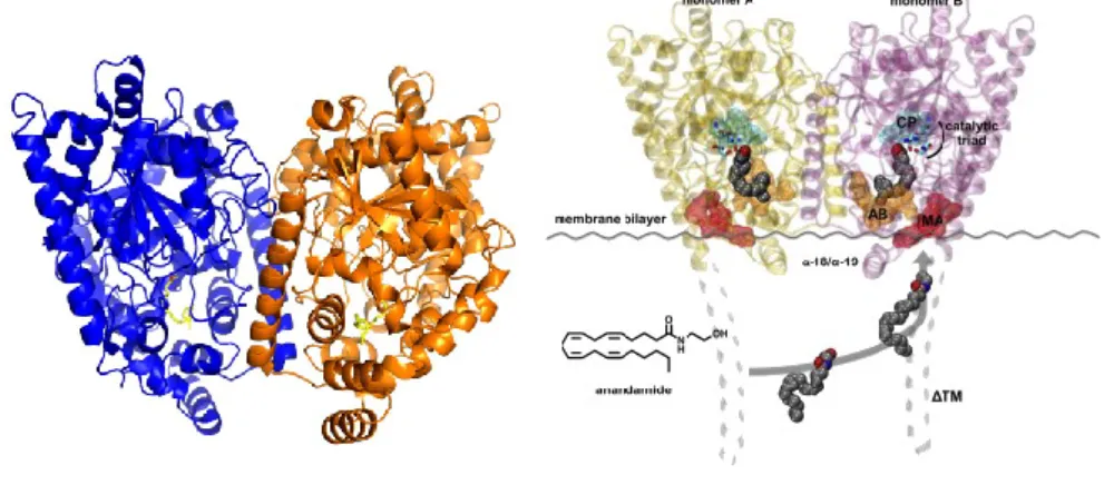
FAAH enzymeThe role of FAAH enzyme was first reported in 1993[66], but a similar enzyme was identified in 1960s [67]. FAAH is a protein of 65kDa and 579 amino acids, purified for the first time in 1996 by Cravatt's group from rat liver membranes [68] (Fig. 5). Subsequently through sequence analysis it was found
that it is a protein belonging to the amidase family. FAAH is the only member characterized in mammals predominantly in microsomal and mitochondrial fractions [69,70]. It is a membrane-bound serine hydrolase, responsible for the
hydrolysis of a family of naturally occurring fatty acid amides, abundantly expressed in the CNS and peripheral tissues, such as kidney, liver, lung, testicle, small intestine and prostate [71]. Enzymes of this family are found in
14
bacteria, archaea and eukaryotes and hydrolyse different substrates represented by a series of physiologically active lipids, including AEA. The human, porcine, mouse, and rat FAAH enzymes are 73% identical at the amino acid level overall and 90% identical in the amidase signature sequence, amino acids 215–257 [72,73].
Figure 5:Fatty acid amide hydrolase (FAAH) (From Palermo et al. Med Chem. 2015)
The reaction consists in the hydrolysis of amide bonds, present in these substrates. This family of enzymes is characterized by a highly conserved consensus sequence rich in Serine and Glycine residues of approximately 50 amino acids. Cluster analysis of 21 amidases revealed a highly conserved central region rich in glycine, serine and alanine residues. This region has been defined as the signature consensus sequence of amidase of 56 amino acids with GGSSGG that is the longest block of strictly conserved residues in different enzymes. In mammalian, consensus sequence corresponds to amino acids 215-257. Other 17 stable positions were identified throughout the sequence. To date, over 45 proteins containing the amidase identification sequence have been identified, including in addition to FAAH, many prokaryotic and fungal
15
enzymes, one avian enzyme and three putative members of C Elegans [74] (Fig.
6).
Figure 6:Amidase signature sequences (FromPatricelli et al. Biochemistry. 1999)
FAAH is able to hydrolyse AEA and, more less, (2-AG), N-oleoylethanolamine, N-palmitoylethanolamine, and N-oleoyltaurine. FAAH degrades AEA through a hydrolysis mechanism that involves an unusual catalytic triad: Ser 241 - Ser 217- Lys 142, which replaces the typical Ser-His-Asp motif of serine hydrolases [66,75] (Fig. 7).
16
Other members of the same FAAH family are soluble enzymes that hydrolyse hydrophilic substrates such as acetamide, malonamide, and glutamine. Compared to these enzymes, two distinguishing features of FAAH are its integration into membranes and its strong affinity for hydrophobic substrates. For this reason, the pharmacological inhibition of FAAH allows for the modulation of endogenous levels of cannabinoids, so it is a promising target for treatment of pain, inflammation and many other diseases [76].
2.2.
The endocannabinoid system and its therapeutic exploitationNew pharmacological approaches for treatment of neurological disorders were identified in the ECS modulation. Many studies suggest how an increase of eCB levels and an enhancement of CB1/CB2 receptors activity can improve neuropathological condition [77-79]. However, an up-regulation of
ECS has been associated with controlling or contributing to pathological effects [80]. Many compounds have been identified to regulate eCB degradation and biosynthesis acting as indirect agonist or indirect antagonist. Drugs that alkylate serine, cysteine and histidine residues or BTNP, an inhibitor of Ca2+-independent PLA2, are able to inhibit the formation of AEA
in cortical neurones [81], while RHC80267 [1,6- bis-(cyclohexyloximino
carbonylamino)-hexane] and tetrahydrolipstatin (THL), have been shown to inhibit DAGL- reducing the production of 2-AG. O-3640 and O-3841 compounds have been developed as inhibitors of 2-AG biosynthesis with hight selectivity for DAGL- but they are not suitable for systemic use in vivo [82].
EMT inhibitors have been identified and they represented a perfect case study to evaluate the influence of ECS in different pathological conditions of both the central and peripheral nervous system. AM404, VDM-11, OMDM-1 and-2, UCM707 and AM1172 represent the most important compounds of this
17
class that includes long chain fatty acid derivatives such as amides or retroamides of arachidonic and oleic acid with amines containing an aromatic constituent [83,84]. Several studies have focused their attention of the
possibility to counteract endocannabinoid inactivation. In particular, serine hydrolase inhibitors have showed therapeutic value in the treatment of several disorders. Belonging to this class we find fluorophosphonates, trifluoromethyl ketones, the a-keto heterocycles OL-135 and a-KH 7, and the carbamates URB597, BMS-1, SA-47 and SA-72 [85]. Another important class of
compounds act on the modulation of CB1/CB2 receptors. The most important are SR141716A, a selective CB1 receptor antagonist, AM251, inverse agonist of the CB1 receptor, AM630, inverse agonist of the CB2 receptor [86,87], AM6545, a neutral antagonist CB1 receptor, URB447 [88,89], a CB1 antagonist/CB2 agonist, and last but not least, WIN55,212-2 [90], a non-selective cannabinoid receptor agonist [91]. In autoimmune encephalomyelitis (EAE) mice SR141716A inhibited the expression of CB1 receptor, whereas increased the expression of CB2 receptor in brains, spinal cords and spleens. At the same time this compound increased IFN‐γ, IL‐17 and inflammatory cytokines such as IL‐1β, IL‐6 and tumor necrosis factor-alfa (TNF‐α) in brains and spinal cords [92]. In hypoxia-ischemia (HI) neonatal rats was observed that
URB447 is able to reduce neurodegeneration after brain injury. A comparable effect was observed with SR141716A, whereas the WIN-55,212-2 reduced the effect of URB447 [93]. WIN-55,212-2 and URB597 have showed
neuroprotective effect in neurological disorders. Su et al. (2017) have demonstrated how these two compounds can improve neuroinflammation in rats with chronic cerebral hypoperfusion (CCH) reducing the number of activated microglia and astrocytes in frontal cortex and hippocampus CA1 region. Moreover, the combined treatment can reduce the release of
pro-18
inflammatory cytokines and contain the translocation of NFkB. Antioxidant and anti-apoptotic properties of these two compounds was observed against the adverse effects of ischemia [94]. eCB are also able to prevent BBB integrity
by attenuation of LPS-induced modifications in the diameter and permeability of vessels and margination of leukocytes. suggesting that ECS could be considered to develop a new strategic approach to protect BBB in different neurodegenerative disorders [95].
2.3.
URB597: a selective FAAH inhibitorMain actor of this work is URB597, also known as KDS-4103. URB597 (cyclohexylcarbamic acid 3’-carbamoylbiphenyl-3-yl ester) is a crystalline white solid with a molecular weight of 338.4 (Fig. 8). The compound has two hydrogen bond donors and five hydrogen bond acceptors
[96,97].
Figure 8: URB597 molecular structure
URB597 has a high degree of membrane permeability, evaluated in CaCo II TC7 cells, with an apparent permeability coefficient of 45.3 x 10⁻⁶
cm/sec [98]. URB597, an aryl ester of archilcarbamic acid, is able to block the hydrolysis enzyme FAAH activity [99,100]. It is a covalent irreversible inhibitor, hydrolysed by the enzyme itself and the enzyme is inhibited by
19
carbamylation of the nucleophilic residue Ser241 [101,102]. Russo et al. (2007)
observed that a repeated oral administration of the URB597 (1–50 mg/kg, once daily, for 4 days) in mouse chronic constriction injury (CCI) model produced antipyralgia and pain relief [103]. Moreover, URB597 increased activity of
serotonergic neurons in the dorsal raphe nucleus and noradrenergic neurons in the nucleus locus ceruleus in C57BL/6 [104]. In the lipopolysaccharide (LPS)
mouse model of inflammatory pain, URB597 attenuated the development of LPS-induced paw edema and reversed LPS-induced hyperalgesia, reducing levels of the proinflammatory cytokines IL-1β and TNF-α [105].
2.4.
Endocannabinoid system in neurodegenerative disordersNumerous studies have well analysed the important role of the ECS in neurodegenerative diseases [13,106]. In animal models was observed a
dysregulation of endocannabinoid mediators, which contribute to disease in different ways depending on the location and timing of their production and on the progression of the disease [50]. eCB play a key role in peripheral and brain immune functionand its agonism is typically associated with a modulation of pro- and anti-inflammatory activities, like the release of inflammatory mediators, including nitric oxide, IL-2 and TNF-, the inhibition of the activation of the cell-mediated immune processes, and the inhibition of proliferation and chemotaxis [107-109].
Animal models of Parkinson disease (PD), pathology characterised by an impairment of dopaminergic system, showed a biphasic dys-regulation of CB1, such as hypoactivity in pre-symptomatic and early PD and hyperactivity at later stages [110]. PET and MRI analysis showed an increased level of CB1
in patients with PD, as well CB2 receptor [111,112]. In 6-OHDA- treated rat
20
MPTP-induced neurotoxic and neuroinflammatory events in mice [113,114].
Another study, showed how an up-regulation of CB2 in LPS-treated rats, is able to reduce expression of inflammatory markers [115]. An impairment of
ECS was demonstrated in patients with PD and in 6-OHDA-treated and reserpine-treated rats, where abnormal eCB levels in cerebrospinal fluid (CSF) were observed. A treatment with levodopa has demonstrated a revert of the pathological condition [116-118]. MAGL inhibitors mediate neuroprotection
through CB2 activation, while FAAH inhibition improve motor behaviour via CB1/CB2 receptors, but no neuroprotective effects in MPTP-treated mice
[119,120].
Huntington disease (HD) is another neurodegenerative disease that shows involvement of the ECS. HD is caused by death of dopaminergic neurons in the globus pallidus, due to expansion of CAG triplet repeats in the gene that encodes the huntingtin protein. This condition leads to progressive locomotor impairment and mental impairments. R6/1, R6/2 and HD94 mice are used like experimental models to study HD. The induction of the pathology through treatments with neurotoxins injected into the globus pallidus has showed a loss of CB1 receptors and the same condition was observed post-mortem in patents with HD [121-123]. In HD, a protection of medium spiny
neuron populations by endocannabinoids might involve a retrograde inhibition of glutamate excitotoxicity at excitatory terminals [124,125]. CB1 and CB2
agonists are considered important targets to prevent motor impairment and loss of medium spiny neurons [126]. A reduction of AEA and 2-AG and in the
striatum of R6/2 mice model was observed [127], while in R6/1 2-AG was
increased and AEA levels were decreased [128]. Battista et al. (2007) found a
21
peripheral lymphocytes of HD patients compared to those of healthy subjects
[129].
Alterations in CB1 and CB2 receptors were reported also in Multiple sclerosis (MS). Experimental autoimmune encephalomyelitis (EAE) or chronic relapsing EAE (CREAE) and Theiler’s Murine Encephalitis Virus-Induced Demyelinating Disease (TMEV-IDD) are clinically relevant murine models of multiple sclerosis (MS). Several studies showed beneficial effects due to the activation of CB1 and CB2 receptors. In CREAE mice, CB1 agonists ameliorated tremor and spasticity [130,131], while in TMEV-IDD mice an improvement of clinical diagnosis via immunomodulatory and anti-inflammatory mechanisms after treatments with CB1 and CB2 agonists was observed [132]. AEA and 2-AG were found increased in brain and spinal cord of EAE and TMEV-IDD mice, but blood tests on patients with MS have showed an increase of endocannabinoids with a consequent reduction of AEA in CSF [133,134].
A down regulation of CB1 receptors was observed in Amyotrophic lateral sclerosis (ALS) experimental models. The causes of this pathology are still unknown, although in 1993 Rosen et al. found that mutations in the gene of Cu/Zn superoxide dismutase (also known as the SOD1 enzyme) were associated with some cases of familial ALS (~ 20%). This enzyme has an antioxidant function, as it reduces the level of superoxide ion (O2−) a toxic free
radical produced during cellular oxidative metabolism capable of altering proteins, membranes and DNA [135]. In SOD1 mice, characterised by an
overexpression of SOD1, was observed an upregulation of CB2 receptors in the spinal cord [136], as well in post-mortem primary motor cortex and spinal
cord sample from patients with ALS [137]. In SOD1 mice, AEA and 2-AG in
22
showed a damping of symptoms, while treatment with MAGL inhibitors have showed also an increased survival [139,140].
2.5.
Alzheimer disease (AD) and Endocannabinoid systemWith the increasing life span of the general population, Alzheimer’s disease (AD), which is an age-related neurodegenerative disorder, is becoming the major worldwide cause of dementia. With a prevalence of 46.8 million people in the world AD is one of the most important global health issues. This number will double by 2040 [141]. AD is a chronic neurodegenerative condition
clinically characterized by progressive memory impairment and cognitive deficits. Other cognitive deficits include changes in attention and problem-solving abilities, impaired judgment, decision-making and orientation. As dementia progresses, language dysfunction and personality changes frequently appear [142]. Mild cognitive impairment (MCI) is a hallmark of AD. For this
reason, MCI profiles may prove to be a promising link to identify AD in its earliest stages and therefore provide an opportunity to apply preventive measures to slow down the disease (Fig. 9).
23
Figure 9. This diagram illustrates the progression from normal cognitive aging to mild cognitive impairment (MCI) and Alzheimer’s disease (AD) as indicated by related sector changes (From Lee et al. Mutat Res. 2015)
AD can be classified into two forms: 1) sporadic AD, which accounts for the majority of cases, with less apparent or no familial aggregation and usually later onset age (> 60 years, SAD) and 2) the rarer, familial early-onset form, with Mendelian inheritance of predominantly early-onset (< 60 years, FAD), in which there are mutations of genes encoding, for example, amyloid beta precursor protein (APP) and presenilin-1 and -2 [143]. The macroscopic
level of patients’ brain shows a cortical diffuse atrophy, a loss of volume and a loss weight of the brain. On a microscopic level it is possible to observe the presence of extracellular senile plaques mainly composed of amyloid-beta peptide (A) and intraneuronal neurofibrillary tangles (NFT) containing hyperphosphorylated tau protein [144,145]. Aβ is derived from Amyloid
Precursor Protein (APP) that is an integral membrane protein expressed in many tissues and concentrated in the synapses of neurons. It is implicated in synapse formation, neural plasticity, antimicrobial activity, iron export and
24
intracellular calcium concentration functions through a kinase-dependent mechanism [146-149]. The gene encoding for APP is located on the long arm of
chromosome 21 [150]. It generates a transcript that can undergo different
processes of alternative splicing leading to the formation of different isoforms of the protein. In the cytoplasm APP cleavage occurs via two distinct ways, non-amyloidogenic and amyloidogenic pathways, through proteolytic processing by three enzymes BACE (β-secretase), α-secretase and γ secretase (Fig. 10). In the non-amyloidogenic pathway APP is hydrolysed by α secretase on the transmembrane segment [151,152], generating a different soluble APP variant (sAPPα) and a shorter C-terminal fragment, C83, which is subsequently hydrolysed by γ secretase producing the peptide P3, a non-toxic fragment of
Aβ [153]. In the amyloidogenic pathway APP is hydrolysed by BACE,
releasing a soluble APP fragment (sAPPβ) and a shorter C-terminal fragment, C99, which is subsequently hydrolysed by γ secretase generating an intracellular peptide called AICD (APP intracellular C-terminal domain) and Aβ. Besides the N-terminal variations due to BACE, Aβ can also have different C-terminal endings depending on the γ-cleavage site (Aβ40 or Aβ42). The most abundant Aβ species produced in the brain and found in the CSF is Aβ1– 40, whereas levels of the more readily aggregating Aβ1–42 generally make up only 5 to 10% [154].
25
Figure 10: APP protein proteolysis (Modified from Heppner et al. Nat Rev Neurosci 2015)
Therapeutic approaches
The molecular basis of the pathogenesis of AD is not yet clear and efficacious drug therapy to counteract the clinical course of the disease still does not exist. The pharmacological approach currently adopted tries to modulate the neurotransmitter imbalances to counteract the neuronal death. Treating AD is the biggest unmet medical need in neurology. The main therapies currently adopted aim to improve symptoms, firsts of all inhibitors of acetylcholinesterase enzyme (AChE) (donepezil, galantamine and rivastigmine) and N-methyl-D-aspartate (NMDA) receptor antagonist (memantine). AChE inhibitors enhance cholinergic neurotransmission by preventing conversion of acetylcholine in choline and acetate, increasing acetylcholine in the inter-synaptic spaces and enhancing post-synaptic activation [155]. There is no significant difference in efficacy between the three
drugs that are well tolerated by patients [156]. This class of drugs promotes a
26
while the therapeutic efficacy on the most acute forms remains in doubt. Memantine, the last drug approved by the FDA, is a non-competitive NMDA antagonist. It is able to reduce the excitatory neurotoxicity effect of glutamate and it showed modest efficacy and safety profile in moderate and severe AD when used as monotherapy [157]. In combination with AChE inhibitors
suggests benefits when compared to a non-combination treatment [158]. Some
evidence suggests that anti-inflammatory drugs are able to delay neurodegeneration. Indeed, numerous mediators of inflammation are often associated with A aggregation and increasing of toxicity [159]. To date, no
drugs used in therapy seems to improve the prognosis of the disease. Actually, the current therapeutic strategies are aimed exclusively at relieving the symptoms and slowing down the course. For this reason, enormous efforts have been made to discover new molecules with the potential to change the course of the disease (Fig. 11).
Figure 11: New therapeutic targets for treatment of Alzheimer's disease (Vaz et al. Eur J Pharmacol 2020)
27
The new targeting of research follows essentially the two pathological features of AD: the amyloid plaques (Aß) and the NFT (tau-protein). Therefore, the future therapies of AD will divide into two main strategies: anti-amyloid therapy and tau therapy. Three approaches have been studied as anti-amyloid therapy: reduction of Aß production, prevention of Aß aggregation and promotion of Aß clearance. To reduce A production BACE1 inhibitors were tested (verubecestat, lanabecestat, atabecestat, umibecestat and elenbecestat). All these drugs showed effective reduction of Aß levels in cerebrospinal CSF of AD patients, but in some cases, a cognitive exacerbated was observed [160,161]. Anti-aggregation agents represent a new strategy in
disease-modifying therapy. Studies are currently focused on two molecules, scyllo-inositol and tramiprosate, but they showed severe adverse effects without a significant difference in cognitive scores [162,163]. One promising concept for preventing and treating AD is based upon stimulating the immune system to remove Aβ from the brain, promoting its clearance [164]. These
therapeutic approaches involve use of specific anti-Aβ antibodies to induce the immune system to develop its own antibodies or direct injection of exogenous antibodies (active and passive immunization) [165]. On the other hand, the
anti-tau therapies include the prevention of anti-tau hyperphosphorylation and aggregation and the promotion of tau clearance (Tideglusib, Lithium). Several protein kinases are involved in tau phosphorylation, such as glycogen synthase kinase 3-beta (GSK-3β). These drugs showed a reduction in CSF levels of p-tau, but at the same time a slowing of cognitive decline [166]. Another
therapeutic approach is the inhibition of tau aggregation constitutes (Methylthioninium chloride) [167,168]. These new approaches represent the
28
Modulation of the endocannabinoid system as new therapeutic approach for Alzheimer’s disease
The ECS can be activated by different stimuli and may induce multiple responses, such as the anti-excitotoxic and antioxidant defense, as well as neuromodulatory and immunomodulatory actions, which help to restore cerebral homeostasis in different pathological conditions [62].
Alterations of ECS were observed in AD. Studies post-mortem on brains of patients with AD have shown the increased of expression of CB1/CB2 receptors in the microglia located close to the amyloid plaques [169]. Furthermore, the expression of FAAH, the enzyme responsible for the degradation of AEA, is increased in the senile plaques [170]. Oxidative stress, inflammation and excitotoxicity are typical feature observed in the pathogenesis of AD and the ECS display a possible neuroprotective role against these effects [171]. Gliosis and degeneration of neurons in the hippocampus due to Aβ aggregation were associated with an increase of 2-AG, suggesting that the ECS might activate in response to neuronal damage, representing a physiological neuroprotective response of the organism [172].
Tg2576 transgenic mice, which overexpress a mutant form of APP, and in APP/PS1 mice, which express the same mutant APP and mutant presenilin 1, did not show an alteration of CB1 levels. An alteration in CB1 levels was only observed in presymptomatic Tg2576 mice [173,174]. CB2 agonists ameliorated
memory and cognitive impairments in Tg2576 mice and in APP/PS1 mice
[175], while amnesia induced by Aß is counteracted by CB1 antagonism [176].
An upregulation of CB2 was observed in microglia of APP/PS1 mice treated with intracerebral injection of Aß, suggesting a protective effect against inflammation by CB2 activation [177]. In AD disease in vitro and in vivo
29
models, CB2 activation reduced levels of pro-inflammatory mediators produced by reactive astrocytes and microglial cells, decreasing Aβ levels
[178,179]. In AD mice models hippocampal levels of 2-AG are increased in the
early stage of disease, while AEA levels resulted decreased in later stage [180].
These observations were confirmed in human AD [181]. In a study on
post-mortem brains from patients with AD, AEA levels in the midfrontal and temporal cortex were reduced, while the expression and activity of FAAH enzyme were found increased in neuritic plaque-associated astrocytes and microglia [170,182].
30
3. AIM OF WORK
Recent reports pointed out on the possible role of the ECS in inflammatory processes of the brain and, specifically, on the one associated to
AD [170,183,184]. Studies in transgenic model of the familiar form of AD
showed a cognitive recovery by a modulation of the ECS [185]. Double
transgenic TASTPM mice, overexpressing the APP and the presenilin-1 genes respectively, exposed to a mild stress procedure, showed a down-modulation of eCB and CB1 levels in brain limbic areas [186]. At the same time a chronic unpredictable stress (CUS) protocol was widely used in presence of the CB1 receptor agonist HU-210, to study the impact of stress exposure in several animal models. These data showed that HU-210 completely blocked the deficits in reversal learning and perseveratory behaviour seen following CUS
[187]. Kofalvi et al. (2016) demonstrated as the levels of AEA were reduced in
the hippocampus of AD mouse genetic model [182]. In the neurodegenerative processes an important role is played by the proteasome. A dysfunction of the proteasome could induce an accumulation of aggregated proteins and oxidative stress, conditions closely related to the increase of the inflammation [188,189].
Therefore, this projects thesis is aimed to:
I. Study the immune-modulatory effects and the cytoskeletal reorganization induced by URB597 in BV-2 cells treated with amyloid- peptide (A)
II. Study the cross-talk between microglia and astrocytes cells in a co-culture system to characterize the inflammatory processes. These experiments were performed at the University of Konstanz (Germany) in Marcel Leist’s laboratory.
31
III. Study the protective effect of the co-culture system of human astrocyte stem cells and murine microglia against proteotoxic stress induced in LUHMES neurons and to analyse the potential neuroprotective effect of URB597 against proteasome dysfunction. These experiments were performed at the University of Konstanz (Germany) in the Marcel Leist’s laboratory.
32
4. MATERIALS AND METHODS
4.1.
Materials and chemicalsURB597 [3-(3-carbamoylphenyl) phenyl] N-cyclohexylcarbamate was from Selleck Chemicals (Selleck Chemicals, Houston, TX, USA). -amyloid peptide (Aβ25-35) fragment was synthesized by conventional solid phase [190].
Tissue culture medium and serum were from Gibco BRL (Life Technologies Inc., Grand Island, NY, USA). 3-(4,5-dimethylthiazol-2-yl)-2,5 diphenyltetrazolium bromide (MTT), 4’, 6-diamidino-2-phenylindole (DAPI) and TRITC-phalloidin were purchased from Sigma-Aldrich (St. Louis, MO).
Bradford Protein Assays was from Bio-Rad (BioRad, Segrate Milano, IT).
Fluorescein isothiocyanate (FITC)-dextran was from Sigma-Aldrich (Sigma-Aldrich, St. Louis, MO). The primary antibody mouse monoclonal IgG -Actine antibody was from Millipore (Merck-Millipore Darmstadt, DE), mouse monoclonal IgG anti-GAPDH was from Abcam (CA, United States), Anti-Rabbit IgG (H+L)-HRP Conjugate and Goat Anti-Mouse IgG (H+L)-HRP Conjugate were from BioRad (BioRad, Segrate Milano, IT), goat anti-rabbit TRITC secondary antibody was from Jackson ImmunoResearch (Jackson ImmunoResearch Europe Ltd, Cambridge House, St. Thomas’ Place, UK) and goat anti-rabbit Alexa Fluor 488 secondary antibodies were from Biotium (Biotium, Inc, Landing Parkway, Fremont, CA, USA). Pull-Down and Detection Kits were from Thermo Scientific Pierce, Rockford (Thermo Scientific Pierce, Rockford, IL). The miRNeasy Micro kit was obtained from QIAGEN (Hilden, Germany), NanoDrop One/One C was from Thermo Fisher Scientific (Waltham, MA, USA). The High-Capacity cDNA Reverse Transcription kit and Power SYBR® Green Master Mix was purchased from
33
Applied Biosystems (Foster City, CA, USA). MG-132 and URB597 [3-(3-carbamoylphenyl) phenyl] N-cyclohexylcarbamate was purchased from Selleckchem Chemicals (Selleck Chemicals, Houston, TX, USA). Dibutyryl-cAMP (Dibutyryl-cAMP), fibronectin, Hoechst bisbenzimide H-33342, resazurin sodium salt, tetracycline, L-cysteine Adenosine triphosphate (ATP), Nicotinamide adenine dinucleotide phosphate (NADPH) and reduced glutathione (GSH) were purchased from Sigma (Steinheim, Germany). Recombinant human fibroblast growth factor (FGF), recombinant human glial cell-derived neurotrophic factor (GDNF) and epidermal growth factor (EGF) were purchased from R&D Systems (Minneapolis, USA). Leukaemia inhibitory factor (LIF) was purchased from Merckmillipore (Germany). Tween-20 and sodium dodecyl sulphate (SDS) were purchased from Roth (Karlsruhe, Germany). All cell culture reagents were purchased from Gibco/Fisher Scientific (Hampton, New Hampshire, USA)unless otherwise specified.
4.2. SPERIMENTAL MODELS
MicrogliaMicroglia are the resting macrophages of the CNS generated by the embryonic yolk sac. They migrate into the developing neural tube and after they extend to the brain parenchyma [8]. Microglia rapidly respond to brain
injury and disease by altering their morphology and phenotype to adopt an activated state [191]. Activation of microglia takes place upon exposure to
different stimuli in the CNS, including trauma to brain or spinal cord, ischemia, infection, air pollutants, neurotoxic agents, dysregulated cellular functions or the accumulation of A [192]. A BBB damage can expose microglia to multiple
34
lipopolysaccharide (LPS) and bacterial DNA, that induce microglial activation triggering surface toll-like receptors (TLRs), with a consequence morphological modification [193,194]. Numerous markers and receptors in
microglia are similar to monocytes and macrophages of peripheral tissue. The M1 phenotype is induced by toxins, LPS, IFN- and pro-inflammatory cytokines. On the contrary M2 phenotype release anti-inflammatory cytokines such as IL-4 and IL-10 to bring down neuroinflammation and neuron damages
[9,195] (Fig. 12).
Figure 12: Microglia activation (Modified by Nakagawa e Chiba, 2014)
More recent studies have shown that microglia are never quiescent, but are in a state of motility, which allows them to probe the environment to identify alterations in cerebral homeostasis [196]. In physiological conditions, microglia use their ramifications as sentinels of the surrounding microenvironment, modulating their length and direction, without significant changes in the
35
cellular soma. In neuropathological conditions or in the presence of infectious agents, microglia undergo a rapid activation, adopting an amoeboid phenotype characterized by a large cell body. Davalos et al. (2005) showed that the induction of brain's damage induces microglia cells to extend their ramifications towards the site of insult [192]. Depending on the source of the
signal, microglia give a specific response, through different receptors and signaling pathways, which include phagocytosis, increased migration, proliferation and release of bioactive molecules [197]. Although, activation of
microglia has a neuroprotective function. Hyperactivation of these cells can cause neurotoxicity through the release of inflammatory cytokines and chemokines [191]. In AD’s patients, the areas of amyloid plaques in the brains are surrounded by activated microglia, which suggests that the cytokines and cytotoxic molecules might be released from microglia [198]. Microglia express neurotransmitter receptors to communicate with neurons [199], to detect damaged neurons and to support them in secreting neurotrophic factors for neuronal regeneration [200]. The function of microglia differs during aging.
This assumption is based on the facts that aged microglia secret more IL-6 and TNF-, they are less responsive to stimulation and they have reduced levels of Glutathione [201].
Astrocytes
Astrocytes are the most important abundant glial cells in the CNS. They derive from the neuroectoderm and are essential in the synapse formation and function, in ion and neurotransmitter concentrations, contributing to the integrity of BBB and in brain homeostasis and neuronal survival [3,202].
Astrocytes are in contact with different brain cells as well as the vasculature, thereby coupling neurons and other brain cells to blood supply through
36
astrocytic end feet. Astrocytes show an important role in neuroinflammation with the production of anti-inflammatory cytokines, such as TGF able to contrast microglia activation during an inflammatory process [4,203]. Two
different phenotypes have been identified in the CNS, A1 and A2. Neuroinflammation stimulates A1 astrocytes to release neurotoxins that induce death of neurons. On the other hand A2 astrocytes promote neuronal survival and tissue repair [6]. Moreover, astrocytes themselves are connected through
gap junctions (connexins), forming highly regulated networks of complex cell interactions [5]. During neurotransmission, the levels of extracellular
potassium increase, which would disturb the depolarization of neurons. Astrocytes release glutamate that binds to receptors at the postsynaptic membrane leading to postsynaptic neuron depolarization. A rapid and efficient removal of remaining glutamate from the synaptic interspace is indispensable for terminating glutamate signaling and preventing excitotoxicity of glutamate. Astrocytes can also store glutamate in vesicles by the expression of vesicular glutamate transporters (vGLUT). As mentioned above, astrocytes play a crucial role also in the formation and maintenance of the BBB, by directly interacting with brain capillary endothelial cells [204,205]. They are one of the
main inductors of the BBB phenotype [206]. Interactions between astrocytes
and capillary endothelial cells during disease may affect the BBB integrity, as for example in PD and AD [207,208]. In AD brain endothelial cell-derived
extracellular vesicles (EVs), are able to transfer A to astrocytes and pericytes and then across the BBB into the brain. These EVs from astrocytes seems to be altered in AD [209].
37
LUHMES cells as neuronal model system
LUHMES (Lund human mesencephalic) cells are a subclone of the originally generated MESC2.10 cell line [210], obtained by preparation of
precursor cells from embryonic ventral mesencephalic tissue and immortalized with a LINX v-myc retroviral vector system [211]. The mesencephalon, or
midbrain, is considered as part of the brainstem and is associated with multiple brain functions, such as vision, motor control, alertness and reward [212]. The
LINX v-myc retroviral vector consists of a tetracycline-controlled transactivator (tTA), a minimal CMV promotor (Tet-O), the oncogene v-myc and a gene that confers a neomycin resistance (neo) In this system, tTA strongly activates transcription of v-myc from Tet-O in the absence of tetracycline (Fig. 13). This allows the cells to continuously proliferate in a medium containing fibroblast growth factors (bFGF). To start the differentiation process, the cells are incubated with medium containing non-toxic concentrations of tetracycline, dibutyryl cyclic adenosine 3’,5’-monophosphate (db-cAMP) and glial cell line-derived neurotrophic factor (GDNF). The role of tetracycline is that to suppress the transcription activation by tTA. In this way the production of v-myc is blocked and the cells readily start to transform into post-mitotic neurons. LUHMES cells are an interesting model system to investigate the processes of neurodegenerative diseases as well as to test compounds interfering with this process in vitro [263-267].
38
Figure 13: Structure of LINX v-myc retroviral vector system (Modified from Hoshimaru 1996 Proc Natl Acad Sci U S A. 1996).
4.3.
Preparation of Aβ25–35 and URB597Stock SolutionAβ25–35 was dissolved in sterile phosphate buffered saline, pH 7.4 (PBS)
at a concentration of 1 mM. To induce the aggregation, the solution was incubated in a sonicator bath on ice for 30 min. After treatment the solution was stored at -20°C until use. URB597 was dissolved in dimethyl sulfoxide (DMSO) at a final concentration of 1 mM.
4.4.
Fatty acid amide hydrolase assayFAAH enzymatic activity was studied by using membranes prepared from BV-2 cells treated with vehicle or A− for 4 h and 24 h, or
pre-treated with 5 μM URB597 for 4 h and incubated with 30 μM Aβ25-35 for 24 h.
Membrane preparations were then incubated with [14C]-AEA (85.0 mCi/mmol,ARC St. Louis, MO, USA) properly diluted with AEA (Cayman
39
Chemicals, Ann Arbor, MI, USA) in 50 mM Tris–HCl, pH = 9, for 30 min at 37°C. [14C]-Ethanolamine produced from [14C]-AEA hydrolysis was measured by scintillation counting of the aqueous phase after the extraction of the incubation mixture with two volumes of CHCl3/CH3OH (1:1, v/v) and the activity was expressed as percentage of the maximum effect observed in absence of treatments.
4.5.
Cell cultures and treatmentsProject I
Mouse microglia cell line (BV-2), kindly provided by Dr. Mangino, Sapienza University of Rome, were seeded in DMEM/F-12 medium containing 5% fetal bovine serum, 4 mM L-glutamine and 1% of penicillin-streptomycin (Gibco BRL Life Technologies Inc., Grand Island, NY, USA), at 37 ◦C in a humidified atmosphere with 5% CO2. Cells were plated at an appropriate density according to each experimental setting and after 24 h treated with Aβ25–35 30 M in presence or absence of URB597 5 M. The cells
were pre-treated with URB597 for 4h before adding the Aβ25–35. Project II and III
LUHMES cells (Lund human mesencephalic cells) were generated as described in detail [213]. Proliferating LUHMES cells were cultivated in
PLO/fibronectin coated T75 flasks (Sarstedt, Germany) in 12 ml proliferation medium composed of advanced DMEM/F12 supplemented with 2 mM glutamine (Sigma-Aldrich, St. Louis, MO), 1x N2 supplement and 40 g/ml FGF. Cells were passaged at a confluence approximately of 80%. Before being detached, the medium was removed and the cells were washed with 10 ml
40
phosphate buffered saline (PBS) (Merck-Millipore Darmstadt, DE). After they were incubated for 3 min at 37°C in 2 ml 0,05% trypsin-ethylenediaminetetraacetic acid (EDTA) solution. Afterwards, 13 ml of advanced DMEM-F12 without supplements were added to the trypsinized cells and centrifuge at 1200 rpm for 5 min. Supernatant was aspired and cells were resuspended in 5 ml of advanced DMEM-F12. Cells number was obtained using a Neubauer chamber. Cells were seeded at a confluence of 40 000 cells/cm2 for two days of proliferation and at a confluence of 15 000 cells/cm2
for three days of proliferation. Cells were cultivated at 37°C in a humidified atmosphere with 5% CO2. To start the differentiation process, proliferating LUHMES were seeded at a confluence of 87 000 cells/cm2 in differentiation
medium (indicated as d0 in the Fig.14). The differentiation medium is composed by advanced DMEM-F12 F12 supplemented with 2 mM glutamine, 1 mM cAMP, 2 ng/ml GDNF and 2,25 M tetracycline. LUHMES cells were differentiated for two days before the seeding in the appropriate plates at a confluence of 145 000 cells/cm2. They were treated on day 6, according to each
experimental setting.
Human astrocytes were differentiated from human stem cells H9 [214]
according to Palm et al. 2015 [215] until they reached the neural stem cell
(NSC) state. At this state, NSC were cultivated for 49 days in NSC-medium consisting of DMEM-F12 supplemented by 0,5x N2, 1x B27 with Vitamin A, 2 mM glutamine, 20 g/ml FGF, 20 g/ml EGF and 1,5l/ml LIF and frozen in liquid nitrogen. For further differentiation towards astrocytes, NSC were thawed and seeded on Matrigel-coated (1:20 dilution in cold DMEM/F-12) plates in astrocyte differentiation medium for at least 35 days until start of experiments. Astrocytes differentiation medium is composed by DMEM/F12, 2 mM L-glutamine, 0.5x N2, 0,5x B27 and 1% FBS. Therefore, astrocytes
41
were detached with Accutase (Corning, USA) for 5 min at 37 °C, added of DMEM/F-12 and centrifuged at 500 x g for 5 min. After supernatant was aspirated, pellet was resuspended in LUHMES differentiation medium and counted using a Neubauer chamber. Astrocytes monoculture or in co-culture with microglia or BV-2, were seeded at a confluence of 30 000 cells/cm2, while
astrocytes in co-culture with LUHMES cells were seeded at a confluence equal to 10% of LUHMES cells, that is 145 000 cells/cm2 as mentioned above. Cells
were cultivated at 37°C in a humidified atmosphere with 5% CO2.
Human monocytes were generated from an iPSC line (STBCi026-A, StemBANCC) that stably express green fluorescent protein (GFP) under a constitutive promotor by lentiviral transfection. iPS cells were differentiated in macrophage progenitors as described in detail [216,217]. iPSC cells were detached with Accutase (Corning, USA) for 5 min at 37°C. Afterwards, mTESR (StemCell Technology, Canada) was added to the cell suspension and centrifugate for 5min at 300 x g. To promote the Embryoid Body (EB) formation, 4*106 cells per well were seeded in an Aggrewell 800 plate
(StemCell Technology, Canada) in mTESR medium containing 10 μM Rock inhibitor Y27632 (Tocris, United Kingdom). The cells were cultured for four days with a 66% medium change every 24 h. Subsequently, to induce the mesoderm formation were added to the medium 50 ng/ml BMP4 (Peprotech, USA), 50 ng/ml VEGF (R&D Systems, USA) and 20 ng/ml SCF (R&D Systems, USA) at 24 h after replanting. After four days, the EBs formed were transferred in cell culture dishes in XVIVO medium (Lonza, Switzerland) supplied with M-CSF (Miltenyi, Germany) and 25 ng/ml IL-34 (Miltenyi, Germany). After seven days the 50% of the medium was renewed and only after 14 days the EBs were reseeded. When the cell culture was confluent and the pre-macrophages was ongoing, the medium was change twice per week.
42
To induce the differentiation of the pre-macrophages in microglia-like cells, the cells were cultivated for seven days in XVIVO medium supplied with 1x GlutaMAXTM (Gibco, USA), 1x Penicillin-Streptomycin (Gibco, USA), 100 ng/ml IL-34 (R&D Systems, USA) and 10 ng/ml GM-CSF (R&D Systems, USA). Microglia monoculture were seeded at a confluence of 30 000 cells/cm2.
The same confluence was used for the co-culture with the astrocytes. Cells were cultivated at 37°C in a humidified atmosphere with 5% CO2.
BV-2 cells, a mouse microglia cell line (kindly provided by E. Blasi, Perugia) [218] were seeded in DMEM/F-12 medium containing 10% fetal bovine serum, 4 mM L-glutamine and 1x Penicillin-Streptomycin (Gibco, USA), at 37 °C in a humidified atmosphere with 5% CO2. The cells were passaged with the same procedure used for LUHMES cells and plated at an appropriate density according to each experimental setting. BV-2 monoculture or in co-culture with astrocytes were seeded at a confluence of 30 000 cells/cm2
while BV-2 in co-culture with LUHMES were seeded at a confluence equal to 10% of LUHMES cells, that is 145 000 cells/cm2 as mentioned above.
Figure 14: Co-culture were generated by mixing LUHMES d2 and astrocytes. The BV-2 cells were added on d5.
43
Human microglia conditioned medium
Conditioned medium (CM) was obtained from human microglia seeded with a confluence of 30 000 cells/cm2 in a 6 wells plate. Cells were treated with
10 ng/ml TNF- and 100ng/ml LPS for 24 h. After the incubation, the medium was centrifugated and cell debris. Then, the CM was transferred to the mono and co-culture astrocytes.
4.6.
Cell viability assaysMTT assay
Cell viability was evaluated with MTT cell metabolic activity assay. Cells were seeded in 96-wells plates at a density of 3000 cells for each well. After treatments, 20 L of a 5 mg/mL solution of MTT in PBS was added to the culture medium at a final concentration of 0,5 mg/mL and cells were incubated at 37°C for 2h. The supernatant was removed from each well and the formazan crystals were solubilised in 100 L of DMSO. The optical density (OD) was measured at 570 nm with a reference at 690 nm using a microplate reader (Thermo Scientific Appliskan Multimode Microplate Reader).
Resazurin
Cells were seeded in a 96 wells plate at an appropriate density according to the experimental setting. Metabolic activity was determined by resazurin assay. Briefly, resazurin solution was added to the cell medium to obtain a final concentration of 10g/ml. After 60 min of incubation at 37°C, the fluorescence signal was measured at an excitation wavelength of 530 nm, using a 590-nm long-pass filter to record the emission. The normalization of
44
the fluorescence values was calculated considering the untreated wells as 100% [219].
LDH release
Cells were seeded in a 96 wells plate at an appropriate density according to the experimental setting. LDH activity was detected separately in the supernatant and in the cell homogenate. The medium was transferred into a separate plate and the cells were lysed in PBS/0,1% Triton X-100 for at least 2h or ON. After that, 20 L of sample, derived from the supernatant and from the lysate, were transferred in a new plate and added of 180 L of reaction buffer containing 100 M NADH and 600 M sodium pyruvate in potassium-phosphate buffer (KPP-buffer, pH 7,4). Absorption at 340 nm was measured at 37 °C in 1 min intervals over a period of 15 min. The slope of the absorption intensity was calculated. The ratio of LDH supernatant/LDH total was calculated using the slopes of supernatant and homogenate. LDH release was expressed in percent [220].
ATP assay
Cells were seeded in a 96 wells plate at an appropriate density according to the experimental setting. For the detection of ATP levels, a commercially available ATP assay reaction mixture CellTiter-Glo (Promega, USA), containing luci-ferin and luciferase, was used. 100 l sample and 50 μl of assay-mix were added to a black 96-well plate. Standards were pre-pared by serial dilutions of ATP disodium salt hydrate (Sigma, Steinheim, Germany) to obtain final concentrations ranging from1000 nM to 7.8 nM. Determination of