PALEOECOLOGY AND FUNCTIONAL MORPHOLOGY OF THE PERMIAN LYTTONIID
BRACHIOPOD PIRGULIA
DANIEL STADTMAUER1 & SUSAN BUTTS2
1Department of Ecology and Evolutionary Biology, Yale University, 165 Prospect Street New Haven, CT, 06520-8106, USA. E-mail: [email protected]
2Yale Peabody Museum, Yale University, Division of Invertebrate Paleontology, 170 Whitney Avenue New Haven, CT, 06520-8118, USA. E-mail: [email protected]
To cite this article: Stadtmauer D. & Butts S. (2019) - Paleoecology and functional morphology of the Permian lyttoniid brachiopod Pirgulia.
Riv. It. Paleontol. Strat., 125(3): 679-688.
Abstract. The lyttoniid brachiopods of the Permian exhibit a unique valve morphology: a branched lobate structure takes the place of the dorsal valve. In one group of lyttoniids, the genus Pirgulia, the ventral valve wraps around to form a cone that fully encloses the lobate structure. This has consequences for the dynamics of water flow and mode of life possible for these heteromorphic brachiopods. Here, we describe the skeletal microstructure and morphology of Pirgulia collected from the Upper Permian Sosio Limestone megablocks of Sicily and housed at the Yale Peabody Museum. We reconstruct the paleoecology of Pirgulia, characterizing it as semi-infaunal in soft sediment. By analogy to Richthofenia, the conical ventral valve and flapping dorsal valve functional morphology could have resisted fouling and assisted feeding in this environment. By comparison with the functional morphology of Pirgulia with other lyttoniids and richthofenids, we propose a revised mode of life for this genus, which involves adap-tation to secondary soft-bottom substrates and support by sediment sticking. Despite constraints to the fundamental brachiopod body plan, modification of the valves in Pirgulia to achieve a conical morphology allowed it to inhabit a paleoecological niche distinct from that of other reef-building lyttoniids.
Received: April 11, 2019; accepted: August 21, 2019
Keywords: Semi-infaunal; brachidium; conical ventral valve; active filtration; pseudopunctae.
I
ntroductIonThe family Lyttoniidae is a group of pro-ductide brachiopods that were common during the Permian. Lyttoniids are remarkable for their aberrant morphology, with a branching lobate structure that serves as the dorsal valve, general-ly leaving much of the ventral valve inner surface exposed. Having gone extinct by the end of the Permian, they have no close modern relatives, nor are there many adequate analogues for their mor-phology among extant fauna (Rudwick & Cowen 1967). As such, much effort has been directed into reconstructing the functional morphology of these unique brachiopods.
Lyttoniid shell shapes vary from cup or scoop-shaped (Leptodus, Oldhamina, and
Poikilo-sakos) to subconical (Eolyttonia and Choanodus) or
fully conical (Pirgulia). Based on the presence of
a cicatrix of attachment, most are thought to have cemented to hard substrates such as reefs, rocky bottoms, or clasts, including skeletal debris. Re-constructions for the angle of attachment range from completely vertical to completely horizontal. Scoop-shaped lyttoniid shells are often found at-tached to one another and based on the orientation of multiple-shell assemblages, these taxa have been reconstructed as cemented in a vertical orientation (Seilacher 2013) or at a semi-reclining angle result-ing from deflected shell growth away from the ce-mentation disc formed early in development (Stehli 1956).
The Yale Peabody Museum (YPM) holds a small collection of brachiopods, including lytto-niids, from the Upper Permian Sosio Limestone of Sicily, collected by G. Bonafede of Palermo
and given to Charles Schuchert in 1936. They were collected from within the Sosio megablocks that crop out three km from the Palazzo Adriano (“Pie-tra de Salomone” megablock of Flügel et al. 1991; 37.660653°, 13.382482°, uncertainty = 1.3 km ra-dius). The assemblage also includes ammonoids, fusulinides, crinoid fragments, sponges, corals, and other brachiopods (orthide, orthotetide, productide, rhynchonellide, spiriferide, spiriferinide, and ter-ebratulide species). The lyttoniids had been previ-ously catalogued as Leptodus fasciculata and Leptodus princeps. However, based on the conical wrapping of
the ventral valve and similarity with those described by Verna et al. (2010) from Tunisia, we place these in the genus Pirgulia sp. (species undetermined). Pirgulia
also occurs in the Lower Permian of Thailand (Yan-agida 1967) and the Upper Permian (Guadalupian) of Tunisia (Verna et al 2010, which also contains a comprehensive taxonomic history of the genus).
Lyttoniid brachiopods have a unique mor-phology characterized by a lobate dorsal valve with rib-like processes, resembling a ptycholophe sup-port, which correspond with ridges, or septa, in the ventral valve. Pirgulia is morphologically like other
lyttoniids in being inequivalve with a lobate dorsal valve. Whereas most lyttoniids have two valves par-allel or subparpar-allel to the commissural plane, Pirgulia
has lateral margins which wrap around the dorsal valve and converge dorsally to form a conical ventral valve, like the trail of some productides (Proboscidella,
Muir-Wood & Cooper 1960). It bears morphological similarity, and therefore possibly functional similar-ity, to richthofenioids in its conical shell, although it differs in the orientation of the dorsal valve relative to the ventral valve (parallel in lyttonioids and per-pendicular in richthofenioids). The goal of this study was to use various techniques to examine the func-tional morphology of the modified valves to infer the lifestyle of the Pirgulia. Serial sections were used
to examine shell structure and internal morphology and to infer the relationship to substrate and feeding methods.
B
Iologyandgrowthofthe loBate structureand lophophoreDue to the strong similarity with the outline of the dorsal valve, virtually all reconstructions propose that the lobate structure supported a ptycholophous
lophophore (Williams 1953; Stehli 1956; Rudwick & Cowen 1967). The dorsal valve structure consists of two submedian primary lobes that branch out peri-odically into secondary lobes that are equally spaced from one another (Rudwick & Cowen 1967). A sub-marginal groove present on the ventral side of the lobate structure, termed a flange by Watson (1917) or vallum by Cooper and Grant (1974: 385), has been proposed as the point where the lophophore would have attached (Stehli 1956). In this orienta-tion, cilia occupying the gaps between lobes could have filtered water passing or pushed through. The large number of lobes to this theoretical lophophore at adult size would outnumber known extant ex-amples, but is otherwise like modern ptycholophes (Williams 1953).
On the other hand, the homology of the lo-bate structure with standard brachiopod anatomy is controversial. The interpretation that is perhaps most parsimonious, and most consistent with the structure’s function, is that it is a modified dorsal valve. Under this interpretation, abnormal structure of the valve may be explained by proposing a de-rived method by which the dorsal valve could have been deposited to achieve this shape. Based on the study of ontogenetic series of lyttoniids and appar-ent teratologies, Rudwick (1968) devised an ontoge-netic growth model where the dorsal valve develops by controlled accretion and periodic branching. Pat-terns of growth lines suggest that the lobes of the dorsal valve grew independently by accretion on the distal edges, increasing in length but not in width, and expanded over ontogeny by addition of sec-ondary lobes but not by enlargement (Rudwick & Cowen 1967; Rudwick 1968). That is, juvenile lytto-niids had fewer lobes of approximately the adult size rather than small multilobed structures (Rudwick & Cowen 1967).
However, an alternative interpretation of the lyttoniid lobate dorsal valve is that it is actually an internal skeleton-like structure of spicular calcite homologous to a brachidium (lophophore support), extending from a reduced true dorsal valve that ar-ticulates posteriorly at the hinge (Termier & Termi-er 1949). Williams (1953) proposed that the lobate dorsal valve structure was entirely encased in mantle tissue, with cirri extending from a ptycholophous lo-phophore mirroring the lobes. Observation of the microstructure of the dorsal valve in thin section revealed an internal pseudopunctate secondary layer
and no primary layer, except in the posterior region in the false interarea. This triangular structure at the posterior was identified by Williams (1953) as a vestigial remnant of the true dorsal valve, based on the restriction of the putative primary layer to this area, and the presence of remnants of a cardinal process and denticular sockets. While the lyttoniids appear to be functionally bivalved, that interpreta-tion makes them anatomically univalved.
A similar case has been reported in Falafer, a
small cooperinid brachiopod that is preserved with a corresponding calcified ptycolophous brachidium. Grant (1972) used Falafer to argue by analogy that
the lyttoniid lobate structure is also a brachidium. Secondary lateral lobes that support a ptycholophe could have evolved from a form with two primary lobes that supported a schizolophe (Williams 1953; Grant 1972), thereby paralleling the development of a ptycholophe from a simpler schizolophe (Rud-wick 1962).
Methods
All specimens used for this study are housed in the Yale Pea-body Museum (New Haven, CT, USA). Specimens studied are Pirgulia from Sosio, Italy (locality described above). Comparative material in-cluded the lyttoniids Leptodus and Collemataria and the richthofeniid Richthofenia from the Permian of West Texas, from the Guadalupian (Word Fm.) and Leonardian (Bone Spring Fm.) and Oldhamina from the Salt Range of Pakistan. Specimens from the Permian of West Texas are silicified. All comparative material was free from matrix, so microfossil and lithological analyses were not possible. All mate-rial is available for study. Macrophotography was performed with a Canon EOS 60D camera with a 60 mm EF-S macro lens. Z-stacking was performed using the Cognisys StackShot rail on a Kaiser copy stand with incandescent light and stacked using Helicon Focus ver-sion 6.7.1 Pro.
Computerized Tomography (CT)
Two specimens (YPM IP 238672 and YPM IP 238678) from the Sosio Limestone, Palermo, Italy, were CT scanned (Harvard Uni-versity, Cambridge, MA). Due to the similarity in mineral composi-tion of the shell and the infill matrix, the visual contrast was insuffi-cient to reveal the microstructure and disposition of the two valves.
Serial Sectioning
Standard unpolished serial sections were prepared both parallel (YPM IP 238672 and YPM IP 238678) and perpendicular (YPM IP 238677) to plane of symmetry by Vancouver GeoTech Labs, Richmond, British Columbia, Canada. Serial thin sections were viewed using a Leica M205 C Fusion Optics Stereo Microscope with a Leica DMC4500 camera using Leica Application Suite X Version 3.0.7.19082.
Abbreviations
YPM, Yale Peabody Museum; Fm., formation.
r
esultsVentral valve morphology
The ventral valve of Pirgulia forms a rugate
cone which completely encloses and obscures the dorsal valve from the exterior (Fig. 1A). The dorsal
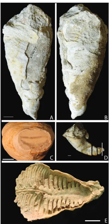
Fig. 1 - Pirgulia sp. from the Yale Peabody Museum Schuchert Col-lection A) YPM IP 238675, dorsal view, scale bar is 1 mm. B) YPM IP 238675, ventral view, showing partially exfoli-ated ventral valve bearing lobate structure, scale bar from 1A. C) cross section of YPM IPS 002295 with dorsal valve indicated with arrow, and D) specimen YPM IPS 002295 indicating the location of the cross section in Fig. 1C. E) YPM IP 036782, Leptodus sp., showing the morphology of a typical lyttoniid dorsal valve.
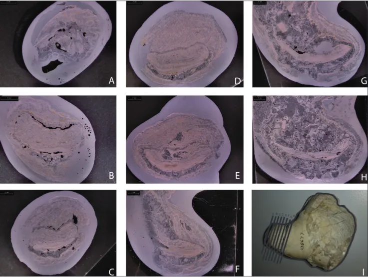
extension of the ventral valve to create a conical shell shape matches reports in Sicilian lyttoniids (Rudwick & Cowen 1967). Specimens in which part of the ventral valve is exfoliated show the lobate pattern indicative of the septa (Fig. 1B). In cross section, the dorsal valve has a concave flexure and is nested in the ventral valve (Fig. 1C; Fig. 1D shows the location of the cross section). In serial sections of YPM IP 238677 (Pirgulia sp.), cut approximately
perpendicular to the direction of growth (Fig. 2), it is possible to see the position of the dorsal valve relative to the ventral valve. The shell exhibits irreg-ular torsion around the longitudinal axis, which sug-gests that the growth direction shifted throughout growth of the organism. We find no evidence of a cicatrix for attachment, even on specimens where the hinge is well-preserved, and no specimens are found cemented to other Pirgulia (although
speci-mens were not observed in situ). The lack of an at-tachment surface (and lack of rhizoid spines, which
can accompany the attachment surfaces), differen-tiates Pirgulia from the other lyttoniidines and the
richthofenioids, their most morphologically similar relatives.
Dorsal valve morphology and apposition
Thin sections reveal lobes in the dorsal valve that are gently arched toward corresponding callused shell on the ventral valve, forming the septal appa-ratus. Although a disarticulated dorsal valve was not available in any of the Pirgulia specimens at YPM,
it would likely have a similar disposition the dorsal valve in Leptodus (Fig. 1E), shown here with dorsal
valve overlying a broad slightly flexed ventral valve. As viewed in thin section, the callused ridges of Pir-gulia correspond to septa in the ventral valve which
interdigitate with the lobes of the dorsal valve. The gaps between the lobes of the dorsal valve are relatively consistent in size, as evidenced by im-prints of the ventral valve septa on some exfoliated Fig. 2 - Serial sections of Pirgulia sp., YPM IP 238677, showing the orientation of the dorsal valve relative to ventral valve, shell microstructure,
specimens. The two primary lobes are fused at the posterior end. The edges of the dorsal valve lobes are marked by a submarginal groove on the ventral side, which may have supported the lophophore (Ste-hli 1956). The fusion of the submarginal grooves of the two adjacent primary lobes in specimens where the primary lobes are close together forms a median incision (Rudwick 1968).
Shell structure
Pirgulia specimens were sectioned and
exam-ined directly by light microscopy, and thin sections were prepared to examine shell microstructure. The dorsal valve exhibits pseudopunctae (Fig. 1C), that give the surface a pustular texture made up of small rosettes (Williams et al. 2000: 310). Connected to the pseudopunctae appear to be taleolae, rods of calcite permeated by networks of organic material (Williams & Brunton 1993). More pseudopunctae appear to emerge from the dorsal side of the dorsal valve than from its ventral side. The layers accrete and form two convex primary lobes, the sides of which meet in the median of the ventral side of the valve to form the median incision. Primary shell is observed in the posterior region of the dorsal valve; the shell is otherwise secondary.
d
IscussIonShell structure
One aim of studying the valve microstruc-ture was to generate new evidence using different methods and taxa (the conical Pirgulia) to address the
assertion made by Williams (1953) that the lack of a primary shell layer on the dorsal lobate structure implies that it was fully internalized. The laminar secondary shell layer that makes up the majority of both the ventral valve and the dorsal lobate structure was readily evident and resembled the texture from Williams’ (1953) figures. Primary shell on the ex-ternal surface of the ventral valve is expected to be thin, only around 10-100 µm (Williams 1997: 268). The fact that it was not confidently identified may have resulted from its failure to be preserved in the specimens studied, which is commonplace.
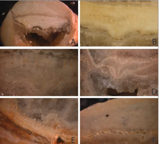
Pseudopunctae are evident under direct ob-servation of sections by light microscopy (Fig. 3). The pseudopunctae appear to be the calcitic taleo-lae characteristic of the aberrant productides, and
which are thought to have evolved independently in multiple strophomenate groups (Williams & Brun-ton 1993; Williams 1997: 305-309). Pseudopuncta-tion is a trait in strophomenate brachiopod shells and therefore its presence is not surprising, but there is a possibility that the distribution of pseudopunct-ae may infer properties of the mantle-mediated shell deposition. One proposed function of pseudopunc-tae is as a holdfast for mantle tissue (Williams 1997: 310-311). This would imply that surfaces directly in contact with mantle would have more pseudopunct-ae penetrating the surface. Although both valves ex-hibit pseudopunctation throughout (Fig. 3A), pseu-dopunctae in contact with the surface of a valve are concentrated on the dorsal side of the dorsal valve (Fig. 3B-D) and the inner surface of the ventral valve (Fig. 3E-F). Curiously, this is different from what would be expected if the lobate structure is a normal valve, in which case mantle tissue would be expected on the ventral side but not the dorsal side. However, the apparent asymmetry in the distribution of pseu-dopunctae also does not fit perfectly with the theory by Williams (1953) of complete mantle tissue enclo-sure of the dorsal valve, as pseudopunctae would be expected to serve as holdfasts on the ventral side of the structure as well. It is possible that the presence of a lophophore on the ventral side of the lobate structure affects the growth of mantle tissue, or if cells in the submarginal groove are indeed involved in lophophore attachment, this may limit mantle growth. In the posterior shell, the dorsal and ventral valves appear similar in structure and we confirm the presence of taleolar pseudopunctae, consistent with the theory that at least the posterior dorsal structure is a normal brachiopod valve. However, more ante-rior sections also exhibited similar typical productide secondary layer microstructure, making it difficult to support theories that the rest of the lobate structure is a modified brachidium or internal calcified skele-ton. Based on our findings in YPM specimens, we cannot reject the hypothesis that the lobate structure is merely a modified valve rather than a novel inter-nal skeleton. Going forward, better resolution from sections at more locations along the antero-poste-rior axis will be helpful in testing this provocative theory more rigorously.
Musculature
Lyttoniids have very little in the way of iden-tifiable sites of attachment for diductor muscles,
which led Williams (1953) to suggest that lyttoniid musculature was atrophied and weak. Later work used phylogenetic inference and the potential iden-tification of muscle scars on lyttoniids to support the presence of developed muscle (Stehli 1956; Rudwick & Cowen 1967). Stehli (1956) argued that reduced but still functional musculature was neces-sary due to the extreme shallowness of the shell that is characteristic of oldhaminids, because without active movement, their gape would have been insuf-ficient for passive filtration. This formed the basis of the active “flapping valve” model for feeding by rhythmic movement of the dorsal valve (Fig. 4A) (Rudwick & Cowen 1967; Cowen 1975), for which the need for well-developed musculature and ener-getic cost were primary points of criticism (Grant 1972). If musculature was indeed developed, Stehli (1956) argued that its presence is incompatible with the dorsal valve being a mantle-enclosed structure, although Grant (1972) pointed out that muscles attaching posteriorly could have moved the lobate structure even if it was fully enclosed in tissue and not homologous to a true valve.
Alternatively, Seilacher (2013) reconciled the apparent absence or atrophy of diductor muscula-ture with the need for active motion by proposing
that the role of a diductor muscle was accomplished by a ligament between the dorsal and inner ventral valves. This ligament would have stored energy upon ventral valve closure for a quick reopening and made active filtration possible whereas slow (smooth) diductor muscles were limiting (Seilacher & Gishlick 2014: 214). The ligament, in this model, was proposed to be made of an extension of man-tle tissue from the dorsal side of the dorsal valve, therefore linking it with the Williams (1953) mod-el and providing a mechanism for active filtration with a tissue-enclosed valve. The observation that many Leptodus are preserved with shells in an open
state and at a consistently small angle was given as support for this hypothesis (Seilacher and Gishlick 2014: 214); however, other fossil evidence for this theory is poor. Muscle presence and strength are considerations in assessing theories of active filter feeding, but were not observable based on the pres-ervation of our specimens.
Paleoecology and functional morphology
The lifestyle of Pirgulia was inferred from
morphology, microstructure, and by analogy to other organisms. Based on attachment scars, most lyttoniids are thought to have been attached by
ce-Fig. 3 - Pseudopunctae in Pirgulia sp., YPM IP S-2295, show-ing dorsal and ventral valve structure (A), dorsal valve (B-D), and ventral valve (E-F) from cut and pol-ished specimens. Within these three groups, images are ordered from posterior to anterior slices. Dorsal side is upward in all images. Red arrows mark the places where pseudopunctae (cal-citic taleolae) protrude from the surface of a valve. The dorsal valve exhibits growth lines and a median incision made up of the meeting of the two primary lobes (C-E). Scale, width of image is A) est. 6 mm (specimen frag-ment was prepared as serial longitudinal thin sections), B) 5 mm, C) 3.5 mm, D) 8 mm, E) 6.5 mm, and F) 6 mm.
mentation to hard substrates such as reefs, rocky bottoms, or invertebrate skeletal debris (Stehli 1956; Seilacher 2013); the Pirgulia examined in this study
exhibited no cicatrix, apical spines, or other signs of attachment. The absence of signs of a fixed sessile lifestyle, deviating from both the richthofenioids and from other lyttoniidines, led us to investigate types of secondary soft-bottom dwellers (Seilacher 2005) as a potential lifestyle for Pirgulia. Secondary
soft-bottom dwellers lack functional pedicles/pedi-cle openings and have functional morphologies that point to resting, passive stabilization, or anchor-ing on soft substrates. Three modes of secondary soft-bottom lifestyles are recliners, passive implant-ers, and sediment stickers (Seilacher & Gishlick 2014). Thin sections of Pirgulia did not reveal any
shell deflections, thickening or ballasting of the ven-tral valve (as in Seilacher 2005), nor keel (Seilacher et al. 2008) which would support a theory of sec-ondary soft bottom reclining. Passive implanters also bear thickening of the shell in the umbonal re-gion (Seilacher & Gishlick 2014: 206). Despite the aberrant morphology of Pirgulia not producing the
typical umbonal region of a typical biconvex bra-chiopod, there is still no shell thickening in the area that would have been embedded in the sediment in the manner of a passive implanter. In Pirgulia, the
shell is relatively uniform in thickness, externally rugate, and exhibits torsion throughout the growth of the organism. These characters are suggestive of an organism which inhabited low-energy environ-ments characterized by deposition of mud or fine sediment, in which the organism grew upwards to keep up with sedimentation, a lifestyle facilitated by the presence of a conical shell (“sediment sticking” of Seilacher & Gishlick 2014: 187). The conical shells described by Seilacher and Gishlick (2014) are achieved by expansion of interarea and umbon-al region (for example, on an othothetide), rather than a true conical shell, but the conical ventral valve is better adapted to this lifestyle, since there is less possibility of fouling via burial of the gape. Al-though apical attachment to the substrate may have occurred in the juvenile to support initial growth, the thinness of the Pirgulia ventral valve suggests
that the surrounding sediment was likely the pri-mary support system for the organism. Rudwick and Cowen (1967) suggested that the conical forms would have laid flat on top of substrate, but the po-tential for such an orientation to quickly bury an
organism with this shape, and the irregular torsion of some Pirgulia specimens, suggest otherwise. The
rugosity of the ventral valve would have provid-ed additional purchase in accumulating sprovid-ediments during growth. Torsion of the conical shell would have slightly reoriented the aperture relative to sed-imentation, but those deflections were insufficient to preclude functioning of the dorsal valve.
For lyttoniids like Leptodus, reconstructions
for the angle of attachment range from complete-ly vertical to completecomplete-ly horizontal. Attachment to substrate in most lyttoniids is often established by root lamellae of the ventral valve and through ce-mentation at the hinge or along the ventral valve. Seilacher (2013) reconstructed the scoop-shaped lyttoniids as building reefs of individuals that stick out of a hard substrate in subparallel arrays. Alter-natively, Rudwick and Cowen (1967) agreed that vertical attachment to hard surfaces was feasible for juvenile Lyttonia “Form B” (mentioned as Pirgulia
therein and reclassified as Pirgulia per Verna et al.,
2010), but argued that due to their lack of spines, adults would have been secondarily free-lying. In-terpretation of assemblages of multiple individuals and of different ontogenetic stages in situ, when preserved in life position, is one of the best meth-ods for determining these different reconstructions. Several functional explanations for the unique morphology of lyttoniids have been proposed. Rudwick (1961) first used the aberrant morpholo-gy of Prorichthofenia (now classified as Richthofenia)
to exemplify the paradigm method for reconstruct-ing function, in which he proposed a flappreconstruct-ing valve functional morphology. Later, Rudwick and Cowen (1967) applied this method to the aberrant lytto-niids as well and suggested a similar flapping valve mechanism. Rudwick and Cowen (1967) proposed that the unique dorsal valve of lyttoniids aided in active filter feeding by rhythmic movement. The conical ventral valve divided the shell into two chambers. Flapping both passed water through the lophophore as it moved between the chambers and created negative and positive pressure moving cur-rent in and out of the aperture of the cone. One potential difficulty with this model is the tendency for recirculation of already filtered water within the end of the cone. Active filtration was supposed-ly necessary due to the shallow angle of opening (Stehli 1956; Rudwick & Cowen 1967; Seilacher & Gishlick 2014: 284).
In contrast to the flapping valve model, in which filter feeding occurs as water passes between the dorsal valve lobes as it opens and closes, Wil-liams (1953: 284) and Grant (1972) proposed that filter feeding in the scooplike or subconical lytto-niids was achieved simply by water passing through troughs formed by the septal apparatus of the vtral valve. In this passive filtration model, water en-tered the distal ends of the ventral valve second-ary troughs, passed by lophophore cilia protruding from the lobes, and left via the two primary troughs of the median incision. This more closely approx-imates the function of a normal ptycholophe, and differs from Rudwick and Cowen’s model (1967), where active closure of the dorsal valve pushes wa-ter through the narrow gaps between the lobes and food is filtered by cilia reaching between the lobes (Fig. 4B). To support this model, Williams (1953: 284) suggested that the septal apparatus on the ven-tral valve served to prop up the dorsal valve from below to ensure a steady flow of water through the troughs. However, we find instead that the septal ridges commonly fall between, rather than directly under, the dorsal valve lobes (e.g. Fig. 1E), resulting in a tighter closure that may have precluded the flow of water in this manner.
It has also been suggested that the lyttoniids housed photosymbionts, analogous the modern gi-ant clam Tridacna supporting photosynthetic
Zoox-anthellae (Cowen 1970). The theoretical necessities of photosymbiosis can be analyzed through com-parison with a model paradigm (sensu Rudwick 1961), but such a trait generally leaves little trace in the fossil record. However, photosymbiosis and active filtration are not necessarily exclusive, for ex-ample, the mixotrophic lifestyle of Gigantoproductus,
determined through biogeochemical signatures of symbionts (Angiolini et al. 2019).
Our findings for Pirgulia valve morphology
are consistent with the flapping valve functional morphology proposed for richthofenioids (Rud-wick 1961) and modified for lyttoniids (Rud(Rud-wick and Cowen 1967). The flapping valve behavior is most effective when the ventral valve completely encloses the dorsal valve, because water is pushed through the lobes on both adduction and diduction. The dynamics of water flow to the lophophore via flapping are potentially complicated by the fact that
Pirgulia exhibits complete ventral valve enclosure,
and has the conical form of a richthofeniid
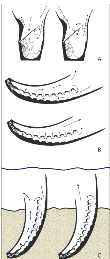
com-A
B
C
Fig. 4 - Water flow models in lyttoniid and richthofeniid morphologiesA) Sicularia (Richthofenia sicula) with inflow phase with dorsal valve opening (top) and filtering phase with dorsal valve clos-ing bottom. Modified from Fig. 17 of Rudwick and Cowen (1967). B) Water flow model for with inflow phase with dor-sal valve opening (top) and filtering phase with dordor-sal valve closing (bottom). Modified from Fig. 23 from Rudwick and Cowen (1967) C) Re-interpretation of flow regime of reclin-ing semi-infaunal Pirgulia based on this analysis of functional morphology. Blue arrows indicate direction of water flow (smaller blue arrows indicate flow near the lophophore), red arrows indicate direction of valve movement.
bined with the dorsal valve orientation of a lytto-niid. However, this morphology also makes active contraction of the dorsal valve even more valuable not only for feeding but also as a way to remove sediment (Fig. 4C).
We infer that the conical shape of Pirgulia
is derived with respect to other lyttoniids like Lep-todus and Poikilosakos that were open scoops. The
fully conical ventral valve is not widespread among productides, occurring in Pirgulia and Richthofenia,
although several subconical or cup-shaped forms exist, as well as innovations like apron trails. That
said, there may be a general trend within lyttoniids towards wrapping around of the ventral valve, as subconical morphology appears to have evolved multiple times in both the Lyttoniinae (e.g. Eolytto-nia, Keyserlingia, and Loxophragmus) as well as in their
sister clade the Poikilosakinae (Pseudoleptodus and Choanodus) (Williams et al. 2000: 635-637). Wrapping
around of the ventral valve may have functioned as a protective dorsal covering within this clade of uniquely exposed brachiopods. It also could have adjusted the circulation of water within the valve to enable the flapping valve as a feeding mechanism. If Pirgulia indeed lived stuck in soft sediment, the
combination of lophophore-driven water flushing and dorsal valve flapping with the conical ventral valve morphology would have played an important role in preventing fouling and could have been used to flush sediment from the corpus. Understanding the morphological evolution of the highly modified lineage Pirgulia helps contextualize wider lyttoniid
evolution and convergent patterns in other biomin-eralizing species.
Pirgulia in context of lyttoniid evolution A substantial difference between the mor-phology of the scoop-shaped (Leptodus; Rudwick
& Cowen 1967) and conical (Pirgulia; Williams et
al. 2000: 634-635) morphologies is the complete enclosure of the dorsal valve by the ventral valve. Of the specimens in this study, all of the conical forms identified were collected from the Sosio me-gablocks of Sicily, but others have reported both scoop-shaped and conical forms in multiple locali-ties including West Texas (Williams et al. 2000: 634-635); thus, both forms were widespread and coex-isted.
Potential adaptive hypotheses for the evolu-tion of conical ventral valve morphology from
pla-nar-convex “scoops” or vice versa are numerous. Polarity of this trait between Leptodus and Pirgulia
is not absolute, but the conical morphology is not widespread among lyttoniids, occurring only in
Pirgulia, with Pseudoleptodus, Chaoella, and Choanodus
having subconical ventral valves that wrap around partially (Williams et al. 2000: 635-637). Poikilosakos,
the oldest known lyttoniid, is also a small and scoop-shaped form. If the conical shell is indeed derived, it may have arisen for protection from predation, as a secondary reacquisition of protective dorsal cov-ering after this feature was lost from the ancestral brachiopod body plan by modification of the dor-sal valve into the lobate structure. Notably, if the superposition of valves in Leptodus leaves exposed
mantle tissue on the ventral valve (as in Williams 1953), the conical ventral valve resolves this issue. In the flapping valve model of Rudwick and Cowen (1967), the conical extension of the ventral valve functions to create a semi-closed chamber wall for the dorsal valve to push water against to direct flow between the lobes. There was presumably a benefit to losing this protective function in the first place, such as better access to current for filter feeding or even access to light to host photosymbiotic organ-isms (Cowen 1970). The conical and scoop-shaped lyttoniids may therefore represent different niches or modes of life possible with this modified dorsal valve morphology, and there need not be a single reconstruction for mode of life for all lyttoniids.
The heteromorphic morphology of the lo-bate dorsal valves of lyttoniid brachiopods has been a topic of debate and speculation for more than 100 years (e.g. Watson 1917). Numerous theo-ries for their functional morphology exist (summa-rized in Williams et al. 2000: 619-630), and the pur-pose of this study was to study the Yale specimens through the lens of these theories in order to test their plausibility. Initial microstructural analysis of the dorsal and ventral lyttoniid valve structures has identified pseudopunctate lamellar secondary shell layers in both dorsal and ventral valves and shown structural similarity of the lobate structure to the ventral valve. On the other hand, the presence of pseudopunctae emerging on the dorsal side of this structure suggest that it may have had mantle tissue on this side, unlike a normal valve. Ongoing analy-sis of these and other microstructural data should allow for the reassessment of the hypothesis by Williams (1953) of the lobate structure being an
in-ternal skeleton and the dorsal valve being reduced to a posterior support. The conical shell is condu-cive to flow of water and prevention of fouling via lophophore-mediated flow and flapping of valves, although whether that was accomplished by the action of muscles or a functioning ligament is still unclear. By comparing the functional morphology of Pirgulia (aberrant amongst the aberrant
brachi-opods) with other lyttonioids and richthofenioids, we are able to provide a revised mode of life for members of this genus which involves adaptation to secondary soft-bottom substrates and support by sediment sticking. This peculiar group of brachio-pods has the potential to become a study case in fundamental evolutionary issues such as homology and the processes by which drastic morphological innovation can occur.
Acknowledgments: This research was conducted initially as an
independent term project (by D.S.) for the Yale College course Inver-tebrate Paleontology, which benefited from constructive comments from Professor Derek E. G. Briggs, Department of Geology and Geophysics and Yale Peabody Museum, Yale University. The authors appreciate thoughtful reviews from Lucia Angiolini, David A.T. Harper, and Francisco Sour-Tovar.
RefeRences
Angiolini L., Crippa G., Azmy K., Capitani G., Confalonieri G., Della Porta G., Griesshaber E., Harper D.A.T., Leng M.J., Nolan L., Orlandi M., Posenato R., Schmahl W.W., Banks V.J. & Stephenson M.H. (2019) - The giants of the phylum Brachiopoda: a matter of diet? Palaeontology, early view: https://doi.org/10.1111/pala.12433.
Cooper G.A. & Grant R.E. (1974) - Permian brachiopods of West Texas, II. Smithsonian Contrib. Paleobiol., 15: 233-793. Cowen R. (1970) - Analogies between the recent bivalve Tri-dacna and the fossil brachiopods Lyttoniacea and Rich-thofeniacea. Palaeogeogr., Palaeoclimatol., Palaeoecol., 8: 329-344.
Cowen R. (1975) - ‘Flapping valves’ in brachiopods. Lethaia, 8: 23-29.
Flügel E., Di Stefano P. & Senowbari-Daryan B. (1991) - Mi-crofacies and depositional structure of allochthonous carbonate base-of-slope deposits: The Late Permian Pietra di Salomone megablock, Sosio Valley (Western Sicily). Facies, 25(1): 147-186.
Grant R.E. (1972) - The lophophore and feeding mechanism of the Productidina (Brachiopoda). J. Paleontol., 46(2): 213-248.
Muir-Wood H. & Cooper G.A. (1960) - Morphology, classifi-cation, and life habits of the Productoidea (Brachiopo-da). Geol. Soc. America Mem., 81: 1-447.
Rudwick M.J.S. (1961) - The feeding mechanism of the Permi-an brachiopod Prorichthofenia. Palaeontology, 3(4): 450-471. Rudwick M.J.S. (1962) - Filter-feeding mechanisms in some
brachiopods from New Zealand. Zool. J. Linnean Soc., 44 (300): 592-615.
Rudwick M.J.S. (1968) - Some analytic methods in the study of ontogeny in fossils with accretionary skeletons. Memoir (The Paleontological Society), 2: 35-49.
Rudwick M.J.S. (1970) - Living and fossil brachiopods. Hutchinson University Library, London, 199 pp. Rudwick M.J.S. & Cowen R. (1967) - The functional
mor-phology of some aberrant Strophomenide brachiopods from the Permian of Sicily. Boll. Soc. Paleontol. It., 6(2): 113-176.
Seilacher A. (2005) - Secondary soft-bottom dwellers: conver-gent responses to an evolutionary “mistake”. In: Briggs D.E.G. (Ed.) - Evolving form and function: fossils and development: 257-271. Peabody Museum of Natural History, New Haven, Connecticut.
Seilacher A. (2013) - Patterns of macroevolution through the Phanerozoic. Palaeontology, 56(6): 1273-1283.
Seilacher A. & Gishlick A.D. (2014) - Morphodynamics. CRC Press, Boca Raton, Florida, 531 pp.
Seilacher A., Olivero E.B., Butts S.H. & Jäger M. (2008) - Soft-bottom tube worms: from irregular to programmed shell growth. Lethaia, 41(4): 349-365.
Stehli F.G. (1956) - Notes on oldhaminid brachiopods. J. Pale-ontol., 30(2): 305-313.
Termier H. & Termier G. (1949) - Sur la classification des bra-chiopodes. Bull. Soc. Hist. Nat. Afrique Nord, 40: 51-63. Verna V., Angiolini L., Chaouachi C., Soussi M., Henderson
C., Davydov V., Nicora A. & Bougdar M. (2010) - Gua-dalupian brachiopods from Djebel Tebaga de Medenine, south Tunisia. Riv. It. Paleontol. Strat., 116(3): 309-349. Watson D.M.S. (1917) - Poikilosakos, a remarkable new genus
of brachiopods from the Upper Coal-measures of Tex-as. Geol. Mag. (Decade VI), 4(5): 212-219.
Williams A. (1953) - The morphology and classification of the oldhaminid brachiopods. Washington Acad. Sci. J., 9: 279-287.
Williams A. & Brunton C.H.C. (1993) - Role of shell structure in the classification of the orthotetidine brachiopods. Palaeontology, 36: 931-966.
Williams A. (1997) - Shell structure. In: Williams A., Brunton C.H.C., Carlson S.J. et al. (2000) - Treatise on Inverte-brate Palaeontology. Part H, Brachiopoda (Revised), Volume 1: 267-320. Geological Society of America and University of Kansas.
Williams A., Harper D.A.T. & Grant R.E. (2000) - Lyttonii-dina. In Williams A., Brunton C.H.C., Carlson S.J. et al. (2000) - Treatise on Invertebrate Palaeontology. Part H, Brachiopoda (Revised), Volume 3: 619-643. Geological Society of America and University of Kansas.
Yanagida J. (1967) - Early Permian brachiopods from north-central Thailand. Geol. Paleontol. Southeast Asia, 3: 46-97.