UNIVERSITY OF SASSARI
PhD School in Biomedical Sciences
Director: Professor Andrea Fausto Piana
Curriculum: Neuroscience
CYCLE XXXI
Facial motor system:
voluntary and emotional control
Supervisor:
PhD Candidate:
2
Summary ... 4
Introduction ... 6
Research question 1: Corpus callosum or brainstem: which is most involved in coordination of facial muscles? ... 12
Introduction... 13
Methods ... 16
EMG ... 16
TMS ... 16
Experimental design ... 17
Experiment 1. Interhemispheric inhibition between primary motor cortices innervating the DAO, FDI and UT muscles. ... 17
Experiment 2. Investigation of a possible direct activation of the DAO by the conditioning stimulus alone and by IHI at the shortest ISIs ... 18
Experiment 3. Recruitment curve response of the DAO muscle in resting and active conditions. ... 18
Statistical Analysis ... 18
Results ... 19
Experiment 1. Interhemispheric inhibition between primary motor cortices innervating the DAO, FDI and UT muscles. ... 19
Experiment 2. Investigation of a possible direct activation of the DAO by the conditioning stimulus alone and by IHI at the shortest ISIs ... 21
Experiment 3. Recruitment curve response of the DAO muscle in resting and active conditions. ... 22
Discussion ... 23
Research question: How cerebellum influence facial primary motor cortex? ... 28
Introduction... 29 Methods ... 32 EMG ... 32 TMS ... 33 Statistical Analysis ... 34 Results ... 34 Discussion ... 35
3
Introduction... 39
Methods ... 42
EMG ... 42
TMS ... 43
Short-latency intracortical inhibition and intracortical facilitation in fM1 ... 43
M1 connectivity with SMA and dPM. ... 43
Neuronavigation ... 44
Facial expressions’ stimulation ... 44
Experimental design ... 44
Experiment 1. Influence exerted by emotional stimuli on primary motor cortex innervating facial and hand muscles. ... 44
Experiment 2. Influence of emotional stimuli exerted on DAO fM1, dPM and SMA. ... 45
Statistical analysis... 45
Results ... 47
Experiment 1. Influence exerted by emotional stimuli on primary motor cortex innervating facial and hand muscles. ... 47
Experiment 2. Influence of emotional stimuli exerted on DAO fM1, dPM and SMA. ... 48
Discussion ... 50
Conclusion ... 53
4
Summary
Face expressions have a crucial role in human nonverbal behaviour and can be either voluntarily or emotionally controlled. The variety of functions of facial muscles, is reflected in their anatomical and histological characteristics. Face muscles present a peculiar coordination, in fact also during voluntary movements, facial muscles’ groups are always recruited together to produce a facial posture, but how and at which level they are coordinated and controlled by the emotional system is still unknown. For long time was impossible to study the motor control of facial muscle in humans. Only recently several studies have demonstrated that it is possible to probe it using transcranial magnetic stimulation (TMS).
This work thesis attempted to investigate: 1) the interhemispheric connections between facial motor cortices (fM1);2) the cerebellar-fM1 connectivity; and 3) the influence of emotional stimuli over fM1 and pre-motor cortices, using TMS protocols.
Results provided the first demonstration that : 1) the corpus callosum plays a minor role in the coordination of bilateral face movements; 2) A clear cerebellar-fM1 connectivity operates in the facial motor system; 3) fM1 is directly connected with the emotional system and specifically modulated by stimuli with positive connotation.
5
“A man's face as a rule says more, and more interesting things,
than his mouth, for it is a compendium of everything his mouth
will ever say, in that it is the monogram of all this man's thoughts
6
Introduction
The important role of the human facial expression has been reflected in art, philosophy and science since antiquity. Facial muscles contribute significantly to human behaviour in a wide range of functions such as, for example, feeding, speech production and communication of affective states (Cattaneo and Pavesi, 2014). For these reasons, these muscles are peculiar and different from any other muscle of the body, in fact facial movements can be categorized into involuntary movements that are controlled by cortical commands, and reflex movements, which involve mainly brainstem generators (Muri, 2016).
The facial muscle system consists of a flat web of muscular fascicles, embedded in a variable matrix of connective tissue and packed into a small two-dimensional matrix under the facial skin; differently from the upper limb district where a single muscle is characterized by its own bony insertions, the joint upon which it acts, and its individual fascia of connective tissue (D’Andrea and Barbaix, 2006).
The motor innervation of facial muscles belongs to the facial nerve while the somatosensory information is pertinent to the trigeminal nerve. In humans, the facial nucleus is the largest motor nucleus in the brainstem and is located in the caudal portion of the ventrolateral pontine tegmentum. All motoneurons, in this nucleus, innervate mimetic facial muscles. Animal studies demonstrated that neurons innervating the same facial muscles are arranged together in longitudinal columns, which are oriented craniocaudally (Morecraft et al., 2001). Holstege and Colleagues (2002) suggested that the dorsal subgroup of the facial nucleus contains motoneurons innervating the muscles around the eye while the medial subgroup innervates the muscles of the ear. Contrarily, the lateral subgroup, that in humans is the largest, innervates the muscles of the mouth; particularly its dorsal portion innervates the muscles of the upper mouth, whereas its ventral portion innervates the muscles of the lower mouth (Holstege et al., 2002).
7
An animal study demonstrated that the facial nucleus receives at least 5 cortical projections: one from the primary motor cortex (M1), one from ventral lateral premotor cortex (LPMCv), one from the supplementary motor area (SMA or M2), one from rostral cingulate motor cortex (M3) and one from caudal area of the anterior midcingulate (M4) (Morecraft et al., 2001; Cattaneo and Pavesi, 2014; Muri, 2016). It is well known that face representation in M1 (fM1), for humans and monkeys, is located anterior to the most lateral segment of the central sulcus. M1 projects to all subdivisions of the facial nucleus (Kuypers, 1958b; Jenny and Saper, 1987; Morecraft et al., 2001), with an important portion of the projections innervating the contralateral lower facial musculature (Morecraft et al., 2001). M1 also sends corticofacial fibres bilaterally to all musculo-topical subdivisions of the facial nucleus, as shown by Jenny and Saper (1987) and by Morecraft et al. (2001).
Contrariwise, the LPMCv is localized anterior to the facial representation of M1 and it projects mainly to in the contralateral lateral subnucleus, innervating the lower facial muscles (Morecraft et al., 2001). Monkey studies demonstrated that the M2 is located in the superior frontal lobule of the medial surface, anterior to the arm representation of M2 and caudal to a region known as the pre-supplementary motor area (Luppino and Rizolatti, 2000) while M3 is located in the rostral midcingulate motor cortex (Morecraft et al., 2001). Both M2 and M3 project bilaterally to the medial part of the facial nucleus, innervating the upper face. A human study demonstrated that direct electric stimulation of the rostral portion of the SMA elicits complex patterns of facial movements (Fried et al., 1991), with both contralateral and ipsilateral movements. Finally, the M4 is localized in the caudal cingulate motor cortex (Picard and Strick, 1996). M4 projections end specifically within the dorsolateral part of the lateral subnucleus, which contains motoneurons innervating the upper, but not the lower and lip muscles (Morecraft et al., 2001).
In humans, anatomy and physiology of the facial motor system have not been studied in details and the comparison with monkey or other animal models is not appropriate
8
because the functions that subtend facial muscles in humans and animals are completely different.
The study of cortical-facial motor projection has become possible using non-invasive transcranial magnetic stimulation (TMS). TMS of the facial motor cortex is a much more complex procedure than TMS on hand muscle or lower limb motor cortex for two reasons: one is the location of face representation in M1 (fM1) which is located anterior to the most lateral segment of the central sulcus; the other one depends on the multiple response of facial muscles to TMS, of which only one is due to activation of the primary motor cortex, while the others can be acknowledged as reflexes or responses to the peripheral stimulation of the facial nerve. Accurate examination of the latency of the TMS-evoked responses can help in distinguishing that produced by each of the 3 mechanisms. One of the elements that help identifying cortical responses are that they are elicited preferentially 8–10 cm lateral to the vertex (Rödel et al., 1999), are modulated by voluntary activity and have an onset latency roughly comprised between 9 and 13 ms. Moreover previous studies, demonstrated that even with high intensity stimulation it could be not possible evoked a response from facial muscle in some subjects, in fact facial muscles present a higher motor threshold than upper limb muscles (Cattaneo and Pavesi, 2014; Pilurzi et al., 2013; Paradiso et al., 2005). Furthermore some authors report a crosstalk phenomenon during the surface registration in perioral muscle (Cruccu et al., 1990a; Paradiso et al., 2005).
In spite of the large amount of technical difficulties to record motor evoked potentials (MEP) in facial muscles, it has been recently observed an increasing interest in the understanding of the voluntary control of facial muscles (Pilurzi et al., 2013; Paradiso et al., 2005; Kobayashi et al., 2001). All these studies suggested that the pattern of cortical representation seems to be dependent on which muscular group is recorded and both, perioral and orbicularis oris muscles, seem more represented than orbicularis oculi or frontalis muscles (Cruccu et al., 1990b; 1997; Liscic´ and Zidar, 1998; Sohn et al., 2004). All facial muscles seem to have a bilateral representation, predominantly contralateral, as demonstrated for different muscle groups such as lip
9
depressors (Meyer et al., 1994; Rödel et al., 1999), muscles active in pursing of lips (Triggs et al., 2005; Yildiz et al., 2004), for the buccinator muscle (Urban et al., 1997; 2001a), the nasalis muscle (Dubach et al., 2004; Fischer et al., 2005) and the depressor anguli oris (DAO) (Pilurzi et al., 2013). Pilurzi and Colleagues (2013) investigated systematically intracortical circuitry in the facial motor cortex, with specific regard to the DAO muscle. The authors demonstrated the presence of short-latency intracortical inhibition (SICI) and facilitation (ICF) in this muscle and demonstrated that these phenomena are present in both the ipsilateral and the contralateral motor cortex, with a contralateral predominance (Pilurzi et al., 2013).
Since facial muscles are also involved in the emotional expressiveness, they are controlled not only by the primary motor cortex, but also by different internal and external inputs.
In humans, facial emotional displays are a mainstay of nonverbal communication. These are highly stereotyped facial postures that are thought to be, at least in part, archetypal (Ekman, 1993) and hard-wired in the human motor systems owing to their evolutionary advantage (Darwin, 1872). Facial expressions are part of stereotyped physiological responses to peculiar affective states involving both the autonomic and the somatic systems and are controlled by the so-called “emotional motor system” (Holstege, 1992; Holstege et al., 1996). Ekman (1993) described that only a limited number of elementary emotional states are associated with stereotyped facial expressions, namely fear, disgust, joy, sadness, anger and surprise (Ekman, 1993; Fridlund et al., 1984). In humans, the most common and reliable information on the affective innervation of facial motoneurons comes from the study of focal lesions that produce the infrequent symptom of isolated emotional facial palsy. Previous studies described these conditions following lesion of the contralateral thalamus and anterior striato-capsular region and frontal lobes (Bogousslavsky et al., 1988; Hopf et al., 1992; Michel et al., 2008; Ross and Mathiesen, 1998; Trosch et al., 1990). Moreover, studies in epileptic patients suggested a direct role of the amygdala in producing contralateral facial emotional postures (Jacob et al., 2003). It is interesting to note that isolated
10
emotional palsy can occur for very distal sites of lesion, i.e. in the ipsilateral pons and medulla (Cerrato et al., 2003; Hopf et al., 2000; Khurana et al., 2002). Also the much more frequent condition of voluntary facial palsy, with sparing of emotional movements, can occur after brainstem lesions (Bouras et al., 2007; Topper et al., 1995; Trepel et al., 1996; Urban et al., 1998; 2001b). These findings suggested a dissociation between emotional and voluntary facial movements at the level of the brainstem, with a possible reciprocal independence of these two systems up to the facial nucleus, which is the final common pathway. The behavioural counterpart of these physiological findings is that the voluntary motor system cannot access at any level a genuine emotional motor pattern. In other words, this is the precise reason why it is not possible to produce by volition a genuine emotional expression. Conversely, the two systems are known to undergo reciprocal, non-movement-specific, influences. As in the rest of the motor system, the pyramidal system exerts a tonic inhibition on involuntary motor patterns, which becomes evident in the release phenomena which occurs after pyramidal lesions. This is the case of old observations of the enhancement of emotional movements on the paretic lower facial quadrant in some cases of unilateral voluntary facial palsy (Monrad-Krohn, 1924; 1939; 1958).
However facial muscles are rarely contracted in asymmetrical way. In fact, also during voluntary movements, facial muscles’ groups are always recruited together to produce a facial posture. These synergies are very stable and stereotyped (Cattneo and Pavesi, 2014). Previous studies carried out in heathy subjects using electromyography coherence between two bilateral muscles provided some valuable insights into the cortical control of voluntary facial movements. It was shown that side–to-side coherence between homologous muscles is constantly present in voluntary movements, no matter what task is being performed, indicating that the two sides of the face tend to be represented together and act as a single functional unit (Wohlert, 1996; Wohlert and Goffman, 1994).
On the other hand, in the coordination of movements also the cerebellum play an important role, in fact studies carried out in patients with cerebellar diseases clearly
11
showed a vast array of impairments, where abnormal motor control and coordination (ataxia) are one of the cardinal features (Celnik, 2015). Previous studies demonstrated that it could be possible to study the connection between cerebellum and motor cortex using non-invasive TMS in intact human subjects (Ugawa et al., 1991; 1995; Pinto and Chen, 2001; Werhahn et al., 1996). Indeed Ugawa and Colleagues (1995) demonstrated that stimulation over the cerebellum resulted in modulation of the responses to a second stimulus delivered over the primary motor cortex (M1). They showed that delivering TMS pulse over the contralateral cerebellar hemisphere few milliseconds before a second stimuli over M1, resulted in an inhibition of MEPs. This phenomenon was called cerebellar inhibition (CBI) and was attributed to the activation of Purkinje cells which inhibited a facilitatory pathway resulting in the temporary inhibition of M1 (Ugawa et al., 1995; Celnik, 2015).
In spite of the large amount of work that investigated the cortical control in facial muscles, how and at which level they are coordinated is still unknown. This study proposed to investigate, using double-coil TMS protocols : 1) the possible connections between fM1 of the two sides by exploring the interhemispheric inhibition (IHI) circuit, and 2) the possible role of the cerebellum in facial motor control, by studying the CBI in fM1 Finally, the influence of emotional stimuli on motor control of facial muscles was also investigated, to give a contribution to the understanding of connections between motor and emotional systems.
12
“Things are connected by invisible bonds: you can’t pluck a flower
without disturbing a star. ” (Galileo Galilei)
Research question 1: Corpus callosum or brainstem: which is
most involved in coordination of facial muscles?
13
Introduction
In daily life, many tasks have a bimanual feature, such as dressing yourself, tying shoelaces, carrying objects, eating or typing an email and playing a musical instrument. Therefore the ability to perform precisely coordinated movements using both hands is an important aspect of several human abilities (Takeuchi et al., 2012; Wahl and Zieman, 2008). It is well known that in order to do bimanual movements it is not important only the activation of particular regions such as the supplementary motor area (SMA) and the lateral premotor cortex (Sadato et al., 1997; Toyokura et al., 1999), but also the functional link between the premotor and sensorimotor areas of both hemispheres. Previous studies in patients suggested that interhemispheric connections of premotor and sensorimotor commands plays a major role in bimanual activities (Geffenet al., 1994; Jeeves et al., 1988; Leonard et al., 1988; Preilowski, 1972; Sperry, 1968).
The corpus callosum is the biggest white matter structure in the brain that connects homologous and non-homologous cortical areas of the two cerebral hemispheres. Several studies demonstrated that it plays a crucial role in the transfer of sensory, cognitive and motor information (Perez and Cohen, 2009). In the field of motor control, many works have demonstrated that modulations of interhemispheric interactions are engaged in the execution of movements involving both body sides. Moreover, modulations of transcallosal fibres contribute also to learn bimanual skills. Ferbert and co-worker (1992) described for the first time powerful interhemispheric interactions between M1 of the two sides in intact human subjects using double-pulse TMS. This study investigated the effect of a supra-threshold conditioning stimulus over one M1 on the size of a test motor evoked potential (MEP) elicited by stimulation of the opposite M1. The authors found an inhibition of the test MEP at conditioning–test interstimulus intervals (ISI) between 6 and 15 ms (Figure 1). This phenomenon was termed short inter-hemispheric inhibition (IHI) and was supposed to be due to
14
activation by the conditioning pulse of transcallosal outputs to the test hemisphere since the effects were absent in patients with no corpus callosum (Meyer et al., 1995).
Figure 1. Interhemispheric inhibition between the hand area of the primary motor cortex investigated with the twin coil method.
A test coil stimulates the left motor hand area to evoke a MEP in the right FDI muscle (top trace on left ‘‘control”). A conditioning stimulus is applied at different interstimulus intervals before the test stimulus. When the interval is 10-20 ms (right panel) a clear inhibition was detected. The graph (bottom left) shows the mean time course of the inter-hemispheric effect in healthy subjects. (Rothwell, 2011 modified).
IHI, founded by Febert et al. links homologous distal hand representations of M1 (Di Lazzaro et al., 1999). Differently, the strength of this linkage between other homologous body part representations in humans, such as proximal and axial muscles, has not been clarified in detail. A previous study investigated IHI between two proximal arm muscle pairs (triceps muscle) and compared their interhemispheric control to that of the first dorsal interossei muscle (FDI); it was detected that the IHI observed bilaterally in the triceps muscle pair was less than that found in the FDI
15
muscle pair (Harris-Love et al., 2007). Moreover, another study carried out in proximal scapulo-thoracic muscles observed a small degree of short latency IHI using low conditioning stimulus intensities (Matthews et al., 2013). These findings are in line with an animal study observing that the control of proximal muscles is less affected by callosal section presumably because of the bilateral connections of the motor cortex to proximal muscles via cortico-reticulospinal pathways (Brinkman and Kuypers, 1972). On the other hand interhemispheric interactions between fM1 of the two sides is still controversial. Indeed a previous study, using neuroimaging techniques, failed to identify callosal motor fibres connecting fM1s (Wahl et al., 2007). This observation is in contrast with previous anatomical tracer studies, which demonstrated that fM1, as defined by intracortical microstimulation, is connected with its homolog in the other hemisphere through callosal fibers, at least in the owl monkey (Gould et al., 1986) and in the macaque monkey (Rouiller et al., 1994).
The facial motor system has several unique features. Facial muscles undergo two types of controls: one is voluntary and other one is emotional (Muri, 2016; Cattaneo and Pavesi, 2014). Furthermore previous TMS studies, carried out on fM1 innervating lower facial muscles showed a bilateral and asymmetrical corticolbulbar projection with the ipsilateral projection being weaker than the contralateral. In this study the authors described an ipsilateral MEP (iMEP) which was hypothesized as not being mediated by the transcallosal pathway (Pilurzi et al., 2013).
In spite of a large amount of human studies demonstrating an interhemispheric interaction between M1s innervating the upper limbs and axial muscles there is an absence of studies that investigated how fM1s are connected one to each other. Therefore aim of this study was to investigate whether short latency IHI can be evoked in fM1, using as a model the DAO muscle and, for comparison, the FDI as a model for the upper limb and the upper trapezius muscle (UT) as a model for axial and peri-axial muscles, in physiological conditions.
16
Methods
Experiments were conducted in ten healthy volunteers (4 females and 6 males; mean age 30.00 ± 1.17 years), all right handed according to the Oldfield inventory scale (Oldfield, 1971). All subjects gave their informed written consent to participate in the study, which was approved by the local ethical committee and conducted in accordance with the declaration of Helsinki. None of the subjects had history or current signs/symptoms of neurological diseases. Subjects sat in a comfortable chair and were asked to stay relaxed but alert during the experiments.
EMG
EMG was recorded bilaterally, in different experimental sessions, from the DAO, the FDI and from UT muscles, using 9 mm diameter Ag-AgCl surface electrodes. For the DAO EMG recordings, the active electrode was placed at the midpoint between the angle of the mouth and the lower border of the mandible, the reference electrode over the mandible border, 1 cm below the active electrode and the ground electrode over the right forehead (Pilurzi et al, 2013). For the FDI EMG recordings, the active electrode was placed over the muscle belly, the reference electrode at the second finger metacarpo-phalangeal joint and the ground electrode over the forearm (Farbert et al., 1992; Rossini et al., 2014). For the UT EMG recording, the active and reference electrode were placed 3 cm apart over UT with a distance of 3 cm between each other’s and the ground on the sternum (Matthews et al., 2013). Unrectified EMG signals were recorded (D360 amplifier, Digitimer Ltd, Welwyn Garden City, UK), amplified (x1000), filtered (bandpass 3-3000 Hz), sampled (5 kHz per channel; window frame length: 250 ms) using a 1401 power analog-to-digital converter (Cambridge Electronic Design, Cambridge, UK) and Signal 6 software on a computer and stored for off-line analysis.
TMS
TMS was performed using a figure-of-eight shaped coil with external loop diameter of 7 cm connected to a Magstim 200 stimulator (Magstim Co., Whitland, and Dyfed, UK).
17
The optimal stimulation site, for the contralateral DAO, FDI or UT was carefully searched and then marked with a soft tip pen over the scalp, to maintain the same coil position throughout the experiments. For the DAO muscles the handle of the coil pointed posteriorly and laterally, at approximately 30-45 deg to the interhemispheric line (Pilurzi et al., 2013; Kujirai et al., 2006); for both FDI and UT the coil pointed backwards and laterally at 45° away from the midline. The resting motor threshold (RMT) was taken as the lowest TMS intensity that elicited, in the relaxed muscle, MEPs of 0.05 mV in at least 5 out of 10 consecutive trials and was expressed in percentage of the maximum stimulator output (MSO) (Groppa et al., 2012; Rossini et al., 2014). Active motor threshold (AMT) was established as the minimum stimulus intensity able to evoke MEPs >200 μV peak-to-peak amplitude in at least five out of ten consecutive trials during isometric contraction of the tested muscle at 10% of maximum voluntary isometric contraction (MVIC) (Rossini et al., 2014). The intensity of the test stimulus (TS) for TMS was 120% of RMT.
Experimental design
The design of the study comprised a main experiment (experiment 1) and two control experiments (experiment 2 and 3) which took place one week apart from the main experiment.
Experiment 1. Interhemispheric inhibition between primary motor cortices innervating the DAO, FDI and UT muscles.
In all ten subjects, the IHI was tested in the M1 representation of the DAO, FDI and UT. IHI was tested using 7-cm double coils and delivering a conditioning stimulus (CS) to the M1 of one side before the administration of a test stimulus to the contralateral M1, using a conditioning stimulus intensity between 90-130% of RMT. The experiment was divided up into three blocks: IHI in DAO, IHI in FDI and IHI in UT muscles. In each block, TS alone and 4, 6, 8, 10, 12 ms ISIs were tested. The three blocks and all states (TS alone and ISIs) were randomized in each subject. Ten unconditioned MEPs and 10 conditioned responses for each ISI were recorded.
18
Experiment 2. Investigation of a possible direct activation of the DAO by the conditioning stimulus alone and by IHI at the shortest ISIs
To exclude a possible stimulation of the ipsilateral DAO by the CS alone and during the paired-pulse TMS at the shortest ISIs, the effects of the CS alone and of paired pulse TMS at 1, 2 and 4 ms ISIs were investigated in 6 out 10 subjects (4 females and 2 males; mean age 31.5± 0.38 years) in the DAO muscles using CS intensities between 110% and 130% of RMT. Ten unconditioned MEPs and 10 conditioned responses for each ISI were recorded in a random order.
Experiment 3. Recruitment curve response of the DAO muscle in resting and active conditions.
To compare the effect of muscle contraction on the DAO MEP, in 5 out of 10 subjects (3 females and 2 males; mean age 31.60 ± 0.42 years), the recruitment curve (RC) was constructed plotting peak-to-peak amplitudes of mean MEPs, recorded from both the active (active RC) and resting (rest RC) contralateral DAO, induced by a single-pulse TMS delivered to the contralateral fM1 at intensities from 90 to 130% of RMT and AMT. Three blocks rest-RC, active-RC with intensity of 90-130% of RMT (RMT-RC) and active-RC with intensity of 90-130% of AMT (AMT-RC) with ten stimuli for each intensity were collected. For the active-RC the subject was required to keep a constant contraction of the DAO at a level of at least 10% of MICV. The results were compared with those obtained in experiment 2.
Statistical Analysis
Statistical analysis was performed with SPSS 20 software (SPSS Inc, Chicago, IL, USA). Student’s paired t-test, repeated measures analysis of variance (ANOVA) and planned post hoc t-test with Bonferroni correction for multiple comparison were used. Compound symmetry was evaluated with the Mauchly’s test and the Greenhouse-Geisser correction was used when required. Significance was set for p value <0.05. Unless otherwise stated, values are expressed as means ± standard error of the mean
19
(SEM). In all experiments latency and amplitude of conditioned and unconditioned MEPs were analysed.
Experiment 1 and 2: A three-way repeated measure ANOVA with ISIs (Experiment 1: TS, 4, 6, 8, 10 and 12; Experiment 2: CS, TS, 1, 2, 4), INTENSITY of CS (Experiment 1: 90-130% RMT; Experiment 2: 110-90-130% RMT) and SIDE (left or right muscle) as within subject factors was used. In case the analysis detected a non-significant SIDE effect, left and right responses were pooled together as a single distribution. In that case a two-way ANOVA with a ISIs and INTENSITY as a within factors was performed.
Experiment 3: To compare MEPs obtained in the RC with those obtained in experiment 2, a two-way repeated measure ANOVA with INTENSITY (110-130% RMT or AMT, according to the resting or active condition) and TYPE OF MEP (TS, CS, conditioned-MEP at 1, 2, 4 ms ISIs, rest-RC, active RMT-RC and active-AMT-RC) as a within subject factors was used.
Results
Experiment 1. Interhemispheric inhibition between primary motor cortices innervating the DAO, FDI and UT muscles.
No significant effect of SIDE for all muscles (DAO: F1,7=0.007 p=0.937, FDI: F1,7= 0.323
p= 0.590, UT: F1,7= 0.020 p=0.901) was detected, thus right and left MEPs were pooled
together. In the relaxed DAO, the mean RMT was 51.34 ± 3.73% MSO. No clear IHI was detected, a significant facilitation was rather found at 4ms ISI (Figure 2). Indeed ANOVA on DAO’s MEP amplitude showed a non-significant main effect of INTENSITY (F5,13 =1.021, p=0.378) but a significant effect of ISI (F5,13 =4.756, p=0.013) and a
significant interaction among factors (F5,13 =2.945, p=0.011). Post Hoc analysis showed
the conditioned MEP was bigger than the test MEP at 4 ms with 110% (p=0.007), 120% (p=0.04) and 130 (p=0.005) of RMT intensities.
20
In the FDI muscle, the mean RMT was 40.54 ± 2.12% of MSO. A clear IHI at ISIs of 8 and 10 ms with high intensity (120 and 130% of RMT) was detected (Figure 2). ANOVA showed a non-significant effect of INTENSITY (F5,13 =1.391, p=0.258) but a significant
main effect of ISI (F5,13 =8.232, p<0.001) and a significant interaction among the factors
(F5,13 =1.990, p=0.051). Bonferroni test showed a clear inhibition at 8-10 ms for an
intensity of 120% RMT (p=0.026 and p=0.011 respectively) and only at 8 ms ISI with 130% RMT Intensity (p=0.005).
In the resting state, the high threshold for UT muscle allowed to complete the experiment in only in 6 subjects. The mean RMT was 52.85 ± 2.58% MSO. A clear inhibition was detected at an ISI of 8 ms with an intensity of 130% RMT (Figure 2). Statistical analysis showed a non-significant effect of INTENSITY (F5,13 =1.353, p=0.270)
but a significant effect of ISI (F5, 13 =4.664, p=0.020) and interaction among the factors
(F5,13 =1.990, p=0.051).
Figure 2. IHI in the UT (light blue) and FDI (dark blue) muscle was clearly detected as expected at 8-10 ms ISIs. By contrast, no IHI was detected in the DAO (violet), in which a significant facilitation was instead observed at 4 ms ISI. The left panel shows MEP amplitude expressed as a percentage of the unconditioned MEP induced by the TS alone at 130% RMT of CS. The graphs reports means + SEM. *p < 0.05. The right panel shows averaged test MEPs obtained from single and paired pulse TMS
21
(upper traces) and conditioned MEPS obtained with paired pulse TMS (lower trace) in the DAO (ISI 4 ms), in the FDI and UT muscles (ISI 8 ms), in a representative subject.
Experiment 2. Investigation of a possible direct activation of the DAO by the conditioning stimulus alone and by IHI at the shortest ISIs
The participants in this experiment had a mean RMT of 52.5 ± 3.60% of MSO, which was not statistically different from that detected in experiment 1 (p=0.40).
Within subject ANOVA showed a significant effect on MEP amplitude of the INTENSITY (F2,9=10.836, p=0.001) and ISI (F2,9=26.964, p<0.001) but a non-significant interaction
among factors (F2,9=1.523, p=0.212). Post-Hoc analysis showed that the test MEP was
not different from the response induced by the CS alone (p=0.9) but both MEPs were smaller than the conditioned MEP at ISIs of 1, 2 and 4 ms (all p<0.01) (Figure 3).
Figure 3. Investigation of a possible direct activation of the DAO by the conditioning stimulus alone and by IHI protocol at the shortest ISIs: amplitude. The graph shows MEP amplitudes elicited by the test stimulus on the contralateral fM1 (MEP), by the conditioning stimulus alone delivered on
22
the ipsilateral fM1 (CS) and by paired TS-CS stimuli at ISIs of 1, 2 and 4 ms) at different CS intensities (110% RMT, 120% RMT, 130% RMT). The graphs report means + SEM. *p < 0.05. The conditioned MEP was bigger than both test and CS- induced MEPs.
The mean latency of the conditioned MEP at 1, 2 and 4 ms ISIs was significantly shorter than that of the test MEP and of the MEP induced by the CS alone (Figure 4). ANOVA detected a significant effect of ISI (F2,9=43.515, p<0.001) but a non-significant effect of
INTENSITY (F2,9= 1.707, p=0.201) nor interaction among the factors (F2,9= 0.656,
p=0.614).
Figure 4. Investigation of a possible direct activation of the DAO by the conditioning stimulus alone and by IHI at the shortest ISIs: latency. The latency of the MEP obtained from single and paired pulse TMS (1, 2, and 4 ms ISIs) with different CS intensities is represented. The graphs report mean + SEM; *p<0.005. The conditioned MEP was faster than the unconditioned MEP in any condition.
Experiment 3. Recruitment curve response of the DAO muscle in resting and active conditions.
Statistical analysis of MEP amplitude revealed a significant effect of INTENSITY (F6,5=53.924, p<0.001) TYPE OF MEP (F6,5=16.443, p<0.001) and a significant interaction
23
among factors (F6,5=16.291, p<0.001). ANOVA of latency showed a significant effect of
TYPE OF MEP (F6,5=21.993, p<0.001) but a non-significant effect of INTENSITY
(F6,5=0.931, p=0.409) or interaction among factors (F6,5=1.150, p=0.347).
Bonferroni post Hoc test showed that the amplitude and latency of conditioned MEPs were different from that of test MEP and of the MEP induced by CS alone, but not from the MEP obtained in active RMT-RC and AMT-RC with intensity of 120-130% RMT and AMT, respectively.
Discussion
The main finding of the present study is that the IHI does not operate in the M1 innervating the lower facial muscles, differently from what confirmed in the hand and axial muscle districts. In consideration of the well-known role played by interhemispheric inhibitory connections in the coordination of bilateral movements, it can be hypothesized that the callosum seems not to be involved in the coordination of bilateral movements of facial muscles, as acknowledged in hand muscles. It seems rather attributable to brainstem circuits.
It is well known that facial muscles rarely contract asymmetrically, especially during voluntary movements. Groups of facial muscles are always recruited together to produce a facial posture (Cattneo and Pavesi, 2014) indicating the two sides of the face tend to be represented together and act as a single functional unit (Wohlert, 1996; Wohlert and Goffman, 1994).
Voluntary movements are generated from cortical motor strips and projects to the facial nucleus through the corticobulbar projections. Corticobulbar pathways to the facial nucleus are of two types: direct and indirect (Brodal, 1981; Noback and Demarest, 1975). The direct fibres form synapses on the facial nucleus and topographically map the motor strip face representation to the face representation in the facial nerve nucleus. The indirect pathway projects first to interneurons in the
24
brain stem reticular formation and, secondly, these neurons deliver the impulses to the facial nerve nucleus (Rinn, 1984).
For long time it was established that cell body groups that innervate the lower half of the face (i.e., the ventrolateral and lateral groups) emanate exclusively from the contralateral cortex while the upper face had bilateral projections (Cattaneo and Pavesi, 2014; Muri, 2016). There is some debate over the presence of bilateral cortical projections to the facial muscles, in fact several studies carried out in healthy subject using TMS found different results. These studies have evaluated the motor cortical projections to a range of facial muscles, including frontalis (Cruccu et al., 1990), orbicularis oculi (Benecke et al., 1988; Cruccu et al., 1997; Liscić and Zidar, 1998; Kobayashi et al., 2001; Sohn et al., 2004; Paradiso et al., 2005), nasalis (Rösler et al., 1989; Dubach et al., 2004), orbicularis oris (Cruccu et al., 1990; 1997; Liscić and Zidar, 1998; Rösler et al., 1989; Sohn et al., 2004; Yildìz et al., 2004; 2007; Triggs et al., 2005), mentalis (Benecke et al., 1988; Cruccu et al., 1990; Werhahn et al., 1995; Kobayashi et al., 2001) and depressor anguli oris muscles (Rösler et al., 1989; Meyer et al., 1994; Rimpiläinen et al., 1992; Paradiso et al., 2005; Pilurzi et al., 2013). Some of these found no ipsilateral response in lower facial muscles (Cruccu et al., 1990; Kobayashi et al., 2001; Paradiso et al., 2005), while others suggested that the cortical projections to lower facial muscles are bilateral, although with a contralateral predominance (Benecke et al., 1988; Meyer et al., 1989; 1994; Werhahn et al., 1995; Urban et al., 1997; Liscić and Zidar, 1998; Rödel et al., 2000; Yildiz et al., 2004; 2007; Triggs et al., 2005, Pilurzi et al., 2013). Pilurzi and colleagues (2013) showed that the ipsilateral response had a 1–2 ms longer onset latency and a higher threshold than the contralateral response. The same difference of latency, roughly up to 2.0–2.5 ms between ipsi- and contralateral responses has been reported in several upper and lower facial muscles by many authors (Benecke et al., 1988; Cruccu et al., 1990; Liscić and Zidar, 1998; Triggs et al., 2005). Since this latency difference was shorter than that necessary to send activation of transcallosal inputs to the opposite hemisphere, which involves a longer conduction delay of 5–10 ms (Meyer et al., 1995), they suggested
25
that an involvement of the corpus callosum in the activation of ipsilateral facial muscles could be ruled out (Pilurzi et al., 2013).
The results of the present study seems to support this speculation. In fact the main experiment failed to detect a clear IHI in DAO muscles although in the same subjects a clear IHI was found for hand and axial muscles. A facilitation was instead observed in DAO at short intervals. This facilitation is not comparable to that already described as interhemispheric facilitation in hand muscle, since in the hand this phenomenon was obtained only with subthreshold conditioning stimuli (Hanajima et al., 2001), differently from our protocol where the facilitation was observed only when suprathreshold conditioning stimuli were used. Since Pilurzi et al. (2013) demonstrated that fM1 sends bilateral projections to DAO muscles, experiment 2 was planned to investigate whether the facilitation observed was attributable to the conditioning stimulation of the ipsilateral cortex. The results showed that the conditioned MEPs at short intervals were different from the test MEP and from the MEP induced by the CS alone, thus allowing to exclude the possibility of an excitatory influence exerted by the ipsilateral stimulation.
One possibility to explain the facilitation observed at the shortest intervals could be an activation of the corticalbulbar tract, which, in active conditions, is engaged more than the trancallosal fibers (Rinn, 1989). The results of experiment 3, support this hypothesis since DAO MEPs recorded in active state were not different as for amplitude and latency from the conditioned MEPs recorded at rest.
In line with our findings, a previous study using a combined functional magnetic resonance imaging/diffusion tensor imaging fiber-tracking procedure, failed, to track lip callosal motor fibres (Wahl et al., 2007). This result is in contrast with a previous animal study demonstrating that the M1 face area, as defined by intracortical microsimulation, is connected with its homolog in the other hemisphere through callosal fibers, at least in the owl monkey (Gould et al., 1986) and in the macaque monkey (Rouiller et al., 1994). Therefore the authors suggested that the failure to track
26
lip callosal motor fibres in Whal’s study was more likely due to technical reasons (Basser et al., 2000; Wiegell et al., 2000; Behrens et al., 2007). On the other hand, Guandalini and Collegues (1990) demonstrated that the transection of the corpus callosum had no effect on ipsilateral facial responses to intracortical stimulation in the cat. This result lead to hypothesize that the functional significance of the overlapping contralateral and ipsilateral representations within fM1, as well as within motor areas devoted to jaw (Clark and Luschei, 1974), tongue (Gould et al., 1986) and tail (Tigges et al., 1979) muscles may be viewed in the light of the functional requirement for simultaneous activation of bilateral pairs of muscles acting close to the midline (Guandalini et al., 1990; Huang et al., 1988). In addition, fM1 can act on ipsilateral muscles through its ipsilateral efferent zones and can promote bilaterally synergistic movements by its bilateral efferent areas through pathways which do not include callosal connections with other areas in the opposite hemisphere.
In this framework, the results of the present study are in line with the above reported animal studies (Guandalini et al., 1990) and might better explain the absence of callosal motor fibres found it in neuroimaging studies in healthy subjects (Wahl et al., 2007 ).
The difference between previous studies carried out in macaque monkey (Rouiller et al., 1994) and owl monkey (Gould et al., 1986) and the results of the present study could be explained by evolution’s reasons. Facial muscles are involved in the emotional expressiveness and their motor control in humans has changed differently from other animals, to allow an evolutionary advantage which occurred in humans to allow social behaviour (Darwin, 1872). In this line, Sherwood et al. (2005) studied the evolution of the brainstem orofacial motor system in 47 species of primates. They found that hominids present significantly larger volumes of the facial nucleus than predicted and they suggested that the phylogenetic specialization of the facial nucleus may be related to the variation in facial muscle differentiation and to increased descending inputs from neocortical areas. It might be possible that in humans compared to
27
primates the transcallosal pathway has lost a importance for the face muscle in order to allow expressiveness.
28
“Well, the way of paradoxes is the way of truth. To test reality we
must see it on the tightrope. When the verities become acrobats,
we can judge them. ” (Oscar Wilde)
Research question: How cerebellum influence facial primary
motor cortex?
29
Introduction
It is well known that the cerebellum, the cerebral cortex and other sub-cortical structures play an important role in adaptive learning (Manto et al., 2012; Medina and Lisberger, 2008; Yanagihara and Kondo, 1996), and in the initiation of voluntary movement (Heiney et al.,2014; Ito, 2006; Manto et al., 2012). Several studies demonstrated that the cerebellum exerts an inhibitory tone on the motor cortex, known as cerebellar brain inhibition (CBI), and its change may act as an important indicator of motor learning (Jayaram et al., 2011).
The cerebellum’s connections with the cerebral cortex ride through two different pathways: inputs arrive to the cerebellum via the cortico-ponto-cerebellar pathway, while outputs are sent via the cerebello-thalamo-cortical system (Fernandez et al., 2018). The afferent pathway to the cerebellum arrives from the cortex via the pontine nuclei and mossy fibres. From here, Golgi and basket cell interneurons form inhibitory synapses with Purkinje cells, while climbing fibres originating from the inferior olive, and axons of granule cells, which relay mossy fibres from the brainstem and spinal cord, form excitatory synapses with Purkinje cells in the cerebro-cerebellum. Granule cells synapses are also located along parallel fibres in the molecular layer of the cerebellar cortex, which form excitatory synapses onto Purkinje cells, basket cell, and stellate cell dendrites (Huang et al., 2006; Ito, 2001). Purkinje cells represent the main output from the cerebellar cortex and are instrumental in the formation of CBI through inhibitory connections with neurons in the vestibular and cerebellar nuclei (Ito, 2006). From the cerebellar nuclei, the signals are sent to the cortex via the thalamus from where they project to a number of areas, including the motor cortex (Dum and Strick, 2003; Kelly and Strick, 2003; Purves et al., 2001; Ramnani, 2006). Inhibition of cortical output occurs as a result of Purkinje cells activity, while a reduction in activity allows excitatory signals to pass from the cerebellar nuclei to the cortex (Ishikawa et al., 2014).
30
Previous studies demonstrated a reduction of CBI following a period of adaptive motor learning (Baarbé et al., 2014; Jayaram et al., 2011; Schlerf et al., 2012a; 2015; Spampinato and Celnik, 2017), at the onset of movement (Kassavetis et al., 2011) and in patients with movement deficits such as Parkinson’s disease, schizophrenia, supra nuclear palsy and cerebellar ataxia (Carrillo et al., 2013; Daskalakis et al., 2005; Ni et al., 2010; Shirota et al., 2010). Thus, CBI is an important marker of both degree and capacity of cerebellar-mediated motor learning.
The CBI was first studied, in intact humans, by transmastoid electrical stimulation (Ugawa et al., 1991a) and later by magnetic stimulation (Ugawa et al., 1995b). Ugawa and colleagues (1995) demonstrated that stimulation over the cerebellum resulted in modulation of the responses to a second stimulus delivered over the M1. Specifically they showed that delivering a TMS pulse (conditioning stimuli, CS) over the contralateral cerebellar hemisphere, 5 to 7 ms prior to a second stimulus over M1 (test stimuli, TS) resulted in inhibition of the TS (Ugawa et al., 1995; Werhahn et al., 1996; Celik, 2015).
Figure 1. Stimulation of the Cerebellum with TMS: the protocol to measure CBI. A test coil stimulates the left motor
31
hand area to evoke a MEP in the right FDI muscle (bottom trace on left ‘‘Test MEP”). A conditioning stimulus is applied over the right cerebellum, 3 cm inferior and lateral to the inion. When the interstimulus interval is 5–7 ms, a clear inhibition of the Test MEP was detected (MEPs following the conditioned test stimulus lower right (Schlerf et al., 2012 modified).
This protocol is likely to involve the activation of Purkinje cells in the cerebellar cortex, and this results in suppression of excitatory output from cerebellar nuclei and, subsequently, suppression of excitatory signals passing from the ventrolateral nucleus of the thalamus to neurons in cortical layers I, III, V and VI (Daskalakis et al., 2004; Na et al., 1997; Shinoda et al., 1993). Neurons in these layers mediate cortical output via connections with pyramidal tract neurons, interneurons and cortico fugal neurons (Daskalakis et al., 2004; Na et al., 1997; Shinoda et al., 1993), leading to the suppression of MEP response seen in CBI.
Recently, a review paper showed that CBI was mostly investigated in first dorsal interosseous muscle (Fernandez et al., 2018) while it was rarely studied in other muscles such as abductor pollicis brevis (Kassavetis et al., 2011; Panyakaew et al., 2016; Popa et al., 2010), extensor digitorium communis and flexor digitoum superficialis of the forearm (Fisher et al., 2009), flexor carpi radialis and extensor carpi radialis of the forearm (Panyakaew et al., 2016) and tibialis anterior (Jayaram et al., 2011; Spampinato et al., 2017). However, cerebellar lower-limb representation is smaller than upper limb representation, which makes it difficult to target (Mottolese et al., 2013). This, and the higher stimulus intensities required for a response may make assessing CBI on lower limb muscles impractical in some circumstances.
In the facial muscles, the linkage between cerebellum and motor cortex for their control is different, because it has been shown that lesions in the red nucleus are implicated in the mechanisms and pathways through which the cerebellar control over facial nucleus motoneurons is realized (Fanardjian and Manvelyan, 1984). This suggests
32
that cerebellar output from the cerebellum to the thalamus may be relayed by the brainstem. Both the poor knowledge on the cerebellar control over the facial muscles and the technical difficulties in studying the facial system might be at the base of the absence of works that investigated CBI in facial muscles. In particular, it has not been clarified if there is a direct connection between the cerebellum and fM1, like it happens in the hand M1, or in which way their coordination involve the cerebellum. Therefore aim of this work was investigated CBI protocol in the DAO, using TMS protocols.
Methods
Thirteen healthy volunteers (9 females and 4 males; mean age 28.77 ± 1.11 years), all right handed according to the Oldfield inventory scale (Oldfield, 1971), participated in the study. An informed written consent was obtained from all subjects before the study and the experimental procedure was approved by the local ethical committee and conducted in accordance with the Helsinki Declaration. None of the participants had history and/or current signs/symptoms of neurological and/or psychiatric diseases. Recordings were carried out in a quiet room and subjects sat in a comfortable chair and were asked to stay relaxed but alert during the experiments.
EMG
EMG was recorded from the right DAO using 9 mm diameter Ag-AgCl surface electrodes. The active electrode was placed at the midpoint between the angle of the mouth and the lower border of the mandible, the reference electrode over the mandible border, 1 cm below the active electrode and the ground electrode over the right forehead (Pilurzi et al., 2013). Unrectified EMG signals were recorded (D360 amplifier, Digitimer Ltd, Welwyn Garden City, UK), amplified (x1000), filtered (bandpass 3-3000 Hz), sampled (5 kHz per channel; window frame length: 250 ms) using a 1401 power analog-to-digital converter (Cambridge Electronic Design, Cambridge, UK) and Signal 6 software on a computer and stored for off-line analysis.
33
TMS
TMS was performed using a figure-of-eight coil with external loop diameter of 7 cm connected to a Magstim 200 stimulator connected in a Bistim module (Magstim Co., Whitland, Dyfed, UK). The optimal stimulation site was carefully searched and then marked with a soft tip pen over the scalp, to maintain the same coil position throughout the experiments. The handle of the coil pointed posteriorly and laterally, at approximately 30-45 deg to the interhemispheric line (Pilurzi et al., 2013; Kujirai et al., 2006). The resting motor threshold (RMT) was taken as the lowest TMS intensity that elicited, in the relaxed muscle, MEPs of 0.05 mV in at least 5 out of 10 consecutive trials (Groppa et al., 2012; Rossini et al., 2014). The intensity of the test stimulus (TS) was the intensity sufficient to evoke a 0.3 mV MEP in the relaxed DAO. Level of cerebellar–M1 connectivity or CBI, in the right DAO muscle, was measured using a standard paired-pulse TMS paradigm (Ugawa et al., 1995; Werhahn et al., 1996; Pinto and Chen, 2001; Daskalakis et al., 2004). Cerebellar stimulation, namely conditioning stimulus (CS), was delivered by double cone coil with external loop diameter of 7 cm connected to a Magstim 200 stimulator connected in a Bistim module (Magstim Co., Whitland, Dyfed, UK). A CS intensity of 60% of maximum stimulator output (MSO) was used, to avoid potential artefacts caused by antidromic stimulation of the pyramidal tract itself (Fisher et al. 2009). Brainstem threshold was first assessed. The experiment was divided up into two blocks: central CBI and lateral CBI. In the central CBI the CS was delivered at the inion while in the lateral CBI the CS was delivered 3 cm lateral to the inion and contralaterally to M1. In order to control for the effect of CS intensity, the same experiment was performed with CS stimulus intensity of 50% and 70% MSO. In each block TS alone and 5, 10 and 12 ms ISIs were tested randomly in each subject. Twelve unconditioned MEPs and 12 conditioned responses for each ISI were recorded. Amplitudes of conditioned and unconditioned MEPs were measured. CBI was expressed as the average MEP amplitude evoked by the cerebellar-conditioned stimulation relative to the average MEP amplitude evoked by the unconditioned TMS pulses over fM1.
34
Statistical Analysis
Statistical analysis was performed with SPSS 20 software (SPSS Inc, Chicago, IL, USA). Student’s paired t-test, repeated measures analysis of variance (ANOVA) and planned post hoc t-test with Bonferroni correction for multiple comparison were used. Compound symmetry was evaluated with the Mauchly’s test and the Greenhouse-Geisser correction was used when required. Significance was set for p value ≤0.05. Unless otherwise stated, values are expressed as means ± standard error of the mean (SEM).
In order to investigated the presence of CBI in DAO muscle, a two-way repeated measure ANOVA on MEP’s amplitude with ISIs (TS, 5, 10 and 12), and SIDE (central or lateral CS) as within subject factors was used. To investigate the effect of intensity of CS, a three-way repeated measure ANOVA of the amplitude of the conditioned MEP (expressed as conditioned amplitude/unconditioned amplitude) with ISIs (5, 10 and 12), SIDE (central or lateral CS) and INTENSITY (50%, 60%, 70% of MSO) as within subject factors was performed.
Results
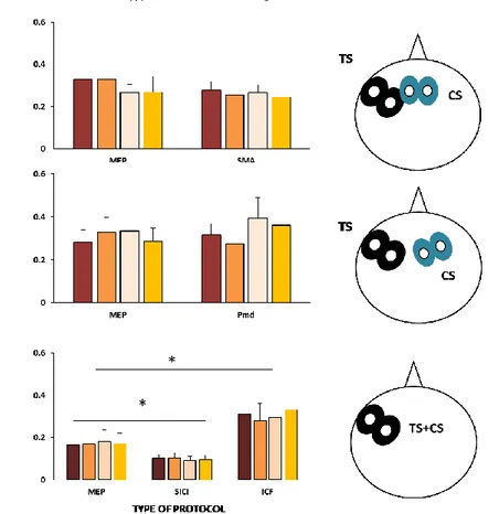
In the relaxed DAO, the mean RMT was 52.62 ± 2.93% MSO and the TS was 68.85 ±4.33 % MSO. A clear CBI was detected with an ISI of 5 ms without any difference between the two positions of CS (Figure 2). Indeed ANOVA on DAO’s MEP amplitude showed a significant main effect of ISI (F1,12 =3.225, p=0.040) but a non-significant effect of SIDE
(F1,12=0.158, p=0.702) or interaction among factors (F1,12 =0.483, p=0.593). Post Hoc
analysis showed that the conditioned MEP was smaller than TS at 5 ms ISI (p=0.05). Statistical analysis of the CBI ratio showed a non-significant effect of SIDE (F1,8=0.342,
p=0.575), INTENSITY (F1,8=1.329, p=0.292) ISI (F1,8 =1.465, p=0.261) and no interaction
35
showed that the CBI obtained with the lateral position of CS was stronger than the central one only for 70% CS intensity (p=0.027).
Figure 2: Cerebellar–fM1 connectivity assessed in the cortical representation of the DAO at rest.
Mean data obtained from 13 subjects at rest are reported. Paired TMS of the left fM1 and of the cerebellum (conditioning stimulus at 60% RMT) in both central and lateral positions, induced a significant inhibition (cerebellar-cortical inhibition, CBI) of conditioned DAO’s MEP at the interstimulus interval (ISI) of 5 ms. Ordinate indicates MEP amplitude; abscissa the ISIs. *p < 0.05. Error bars represent means ± SEM.
Discussion
Overall, the present study showed, for the first time, a clear cerebellar–M1 connectivity in the facial motor cortex at 5 ms ISI, which is the same ISI necessary to evidence the cerebellar connection with hand M1 (Ugawa et al., 1995; Werhahn et al., 1996; Pinto and Chen, 2001; Daskalakis et al., 2004). All these works suggested that CBI
36
seems to involve the activation of Purkinje cells in the cerebellar cortex and this results in a suppression of excitatory signals passing from the ventrolateral nucleus of the thalamus to neurons in cortical layers I, III, V and VI (Daskalakis et al., 2004; Na et al., 1997; Shinoda et al., 1993). These neurons mediate cortical outputs to lower motoneurons via connections with pyramidal tract neurons, interneurons and cortico fugal neurons (Daskalakis et al., 2004; Na et al., 1997; Shinoda et al., 1993). The CBI results in the inhibition of MEP responses to M1 stimulation. Since CBI in the facial muscles was detected at the same ISI used in hand studies it might be hypnotized that the same pathways may subtend the cerebellar connection with fM1. In hand muscles the cerebellar conditioning stimulus was found to be more efficacious when the coil was positioned 3 cm lateral to the inion on the line joining the external auditory meatus, contralateral to the TS in the hand M1 (Fernandez et al., 2018). In particular, the vertical coordinate is at the same level of the inion, or 1 cm below to access cerebellar grey matter and cerebellar hand representations in Lobules V and VIII. By contrast, the present study did not show a clear difference between the two different sites of cerebellar stimulation (central versus lateral). This difference, may be explained in terms of both focality of the double-cone coil stimulation and anatomical representation of hand and facial muscles over the cerebellar cortex. The double-cone coil effectively stimulates the cerebellum at relatively low stimulus intensities. However, it has less focality than other types of coil (Hardwick et al., 2014; Fernandez et al., 2018). In fact a previous study, that performed tissue depth analyses, showed that the double-cone coil is specifically designed to achieve greater depth of stimulation thus allowing to probe deep lying cerebellum and motor representations. On the other hand, this increased depth of stimulation is likely to occur at the expense of focality (Deng et al., 2013).
Animal studies demonstrated that the spinocerebellum comprises the vermis and intermediate parts of the hemispheres and receives somatosensory and proprioceptive inputs from the spinal cord. It has projections, through the fastigial nucleus, to cortical and brainstem regions that give rise to the medial descending systems controlling
37
proximal muscles of the body, face and limb muscles (Thach, 1980). The adjacent intermediate parts of the hemispheres also receive somatosensory input from the limbs and project to the interpositus nucleus, which provides inputs to the lateral corticospinal and rubrospinal systems and controls the more distal muscles of the limbs and digits. By contrast, the cerebrocerebellum outputs involve connections with the cerebral cortex (Thach, 1980). The output is transmitted through the dentate nucleus, which projects to motor, premotor, and prefrontal cortices, via the motor nuclei of the thalamus (Asanuma et al., 1983). Furthermore, there is a general agreement that the dentate nucleus contains at least one rostrocaudally oriented body map, with the leg represented rostrally in the nucleus, face caudally, and the arm at intermediate levels (Hoover and Strick, 1999). According to these anatomical data, hand representation over the cerebellar cortex is more lateral than face, which may explain why the optimal site of stimulation for the hand is lateral. Face representation is more medial than hand, but when using a non-focal coil, it can be efficaciously stimulated if positioned both centrally and laterally.
Although the present study is the first documenting in healthy subjects the presence of the CBI in face muscles, some limitations have to be acknowledged such as its exploratory nature in a small sample size, with multiple statistical test performed. However, once data are confirmed in a proper sample size, data could provide interesting insights into the understanding of the cerebellar control over facial muscles and the role of this control in learning of adaptive movements. In addition, these data may provide an useful tool for further investigations on a possible involvement of the cerebellum in movement disorders such as dystonia.
38
“Two faces, which resemble each other, make us laugh, when
together, by their resemblance, though neither of them by itself
makes us laugh. ” (Blaise Pascal)
Research question 3: Emotional stimuli do influence facial
motor control?
39
Introduction
In humans the ability to rapidly attend, recognize and react to facial expressions is crucial for survival and social communication (Blair, 2003; 2004).
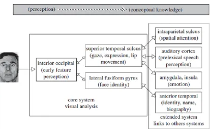
Neurobiological models of face perception propose that face selective areas in the inferior occipital gyrus represent facial information before their analysis in downstream areas in the fusiform gyrus and superior temporal sulcus (Haxby et al., 2000; Calder and Young, 2005). In one of these model, Haxby and colleagues (2000) suggested that face perception involves the coordinated participation of multiple neural regions, broadly delineated into the ‘core’ and ‘extended’ systems (Figure 1). The core system itself consists of two processing streams, one leading from inferior occipital cortex to inferotemporal cortex, in which relatively invariant aspects of faces are represented, and another route, leading from inferior occipital cortex to superior temporal cortex, in which changeable aspects of faces resulting from movement of the facial features are represented. This model finds support from different kind of work such as neuroimaging and neuropsychological studies in patients (Rossion et al., 2003; Winston et al., 2003; Steeves et al., 2006; Engell and Haxby, 2007).
The extended system consists in regions that are also parts of neural systems for other cognitive functions. These regions such as the amygdala and the intraparietal sulcus, may have some capacity for visual analysis of faces (Haxby et al., 2000).
40
Figure 1. The face processing model of Haxby et al. 2000 (Atkinson and Adolphs, 2011 modified).
On the other hand many studies demonstrated that showing face expressions triggers a cascade of central and peripheral physiological processes associated with action preparedness (Dalgleish, 2004; Frijda, 1986; Vuilleumier and Pourtois, 2007). This aspect highlights the importance to consider the neural substrates that link emotions and motor control in the human brain. Anatomical, neuroimaging and neurophysiological evidences support the existence of anatomo-functional links from the limbic association cortex to premotor/motor areas, via the cingulate and prefrontal cortical regions.
Interestingly, human studies using TMS demonstrated an increase in the corticospinal motor tract (CST) excitability to emotional stimuli (Hortensius et al., 2016; Schutter et al., 2008; Baumgartner et al., 2007; Hajcak et al., 2007; Oliveri et al., 2003) with selective increases in CST excitability to fearful facial expressions (Schutter et al., 2008). Moreover, several studies found evidence for increased CST excitability in response to simultaneously presented emotional pictures and congruent music (Baumgartner et al., 2007) or to pleasant and unpleasant scenes (Hajcak et al., 2007). Oliveri and colleagues (2003) provided evidence that the supplementary motor cortex (SMA) may act as a neural gateway in conveying emotion-related information from the
41
limbic system to M1 for initiating action programs. All of these works demonstrated a direct connections between emotion and motor control systems. However all these studies, used the hand motor system as a general model, since this system is easier to study through TMS.
Facial muscles have been demonstrated to be involved in the emotional expressiveness, thus some authors suggested that their control receives different type of inputs including the emotional ones. Several studies demonstrated that it is possible to study the motor control of facial muscle with TMS (Cruccu et al., 1990a; Paradiso et al., 2005; Pilurzi et al., 2013; Cattaneo and Pavesi, 2014). It is well known that the facial muscular system is bilaterally represented with a predominant representation for the contralateral side. This was demonstrated for different muscle groups such as lip depressors (Meyer et al., 1994; Rödel et al., 1999), muscles active in pursing of lips (Triggs et al., 2005; Yildiz et al., 2004), the buccinator muscle (Urban et al., 1997; 2001a), the nasalis muscle (Dubach et al., 2004; Fischer et al., 2005) and the depressor anguli oris (DAO) (Pilurzi et al., 2013). Furthermore, the pattern of cortical representation seems to be dependent on which muscular group is recorded and both, perioral and orbicularis oris muscles, seem more represented than orbicularis oculi or frontalis muscles (Cruccu et al., 1990b; 1997; Liscic´ and Zidar, 1998; Sohn et al., 2004). With specific regard to the DAO muscle, Pilurzi and Colleagues (2013), investigated systematically intracortical circuitry of this perioral muscle in the facial motor cortex. They demonstrated, for the first time, the presence of short-latency intracortical inhibition (SICI) and facilitation (ICF) in both the ipsilateral and the contralateral motor representations of the DAO, with a smaller SICI in the ipsilateral than contralateral DAO (Pilurzi et al., 2013).
In spite of the large amount of studies that demonstrated a link between limbic system and motor control in the hand motor cortex there is an absence of studies that investigated the same effect in the facial muscles. In order to understand relations between emotional stimuli represented by different facial expressions and the activity