PET/CT with
18
F
–choline: Physiological whole bio-distribution in male
and female subjects and diagnostic pitfalls on 1000 prostate
cancer patients
☆
,
☆☆
,
★
18
F
–choline PET/CT bio-distribution and pitfalls. A southern Italian experience
Ferdinando Calabria
a,⁎
, Agostino Chiaravalloti
b, Carmelo Cicciò
c, Vincenzo Gangemi
d, Domenico Gullà
a,
Federico Rocca
a, Gianpasquale Gallo
e, Giuseppe Lucio Cascini
d, Orazio Schillaci
f,ga
Neuroimaging PET/MRI Research Unit, Institute of Molecular Bioimaging and Physiology, Italian National Research Council, IBFM-CNR, Catanzaro, Italy
bDepartment of Diagnostic and Molecular Imaging, Interventional Radiology and Radiotherapy, University Hospital“Tor Vergata”, Rome, Italy c
Department of Diagnostic Imaging, Sacro Cuore Hospital“Don Calabria”, Verona, Italy
d
Department of Diagnostic Imaging, Nuclear Medicine Unit, Magna Graecia University, Catanzaro, Italy
e
Department of Medical and Surgical Sciences, Clinical Surgery and Endoscopy Unit, University "Magna Graecia" Medical School, Viale Europa, Catanzaro, Italy
f
Department of Biomedicine and Prevention, University“Tor Vergata”, Rome, Italy
g
IRCCS Neuromed, Pozzilli (IS), Italy
a b s t r a c t
a r t i c l e i n f o
Article history:
Received 14 September 2016 Received in revised form 7 March 2017 Accepted 10 April 2017
Keywords:
18
F–choline
Diagnostic pitfalls, PET/CT Bio-distribution Physiological variants Female patients Radiolabeled choline
Introduction: The11C/18F–choline is a PET/CT radiopharmaceutical useful in detecting tumors with high lipogenesis. 11C/18F–choline uptake can occur in physiological conditions or tumors. The knowledge of its bio-distribution is
es-sential to recognize physiologic variants or diagnostic pitfalls. Moreover, few information are available on the bio-distribution of this tracer in female patients. Our aim was to discuss some documented18F–choline PET/CT pitfalls
in prostate cancer patients. Our secondary aim was to describe the18F
–choline bio-distribution in the female body. Methods: We collected diagnostic pitfalls in three PET centers examining 1000 prostate cancer by18F–choline PET/
CT. All pitfalls were ensured by follow-up, imaging and/or histology. We also performed whole body18F–choline
PET/CT in 5 female patients.
Results: 169/1000 (16.9%) patients showed pitfalls not owing to prostate cancer. Thesefindings were due to inflam-mation, benign tumors while, in 1% of examined patients, a concomitant neoplasm was found. In the female body, the breast showed low physiological uptake.
Conclusions: The accurate knowledge of18F–choline PET/CT bio-distribution and diagnostic pitfalls is essential.
Cor-relative imaging and histological exam are often necessary to depict pitfalls. In women, the uptake in the breast is due to the physiological gradient of18F
–choline uptake in the exocrine glands.
Advances in knowledge: Our results confirm the possibility of18F–choline uptake in several diseases other than
pros-tate cancer. However, our experience was acquired on a large population and shows that a conspicuous amount of
18F–choline diagnostic pitfalls are easily recognizable and attributable to inflammation. A new advance in knowledge
is the minimal difference in terms of physiological tracer bio-distribution between male and female patients. Implications for patient care: The knowledge of the physiological bio-distribution and of the potential pitfalls linked of a tracer could help physicians to choose the best diagnostic and therapeutic approaches for a better patient quality of life. © 2017 Elsevier Inc. All rights reserved.
1. Introduction
The11C/18F–choline has become a useful positron emission
tomog-raphy/computed tomography (PET/CT) radiopharmaceutical, for its ca-pability to be enhanced in neoplastic lesions with low rate of glucose metabolism, as for prostate cancer (PC) cells. The choline is a marker of lipogenesis, being the precursor of phosphatidylcholine, an important element of the cell membrane. The biosynthesis of the cell membrane is particularly increased in PC, inducing high uptake of the tracer[1].
☆ Reprints to: Ferdinando Calabria (MD), Istituto di Bioimaging Molecolare e Fisiologia, IBFM CNR, Viale Europa, 88100, Germaneto, Catanzaro, Italy.
☆☆ Disclosure of funding: The authors declare that they did not received funding for this work by any of the following organizations: National Institutes of Health (NIH), Wellcome Trust, Howard Hughes Medical Institute (HHMI),or others.
★ Conflict of interest: the authors declare that they have no conflict of interest to declare. ⁎ Corresponding author at: Neuroimaging PET/MRI Research Unit, Institute ofMolecular Bioimaging and Physiology, National Research Council, IBFM CNR, Viale Europa, Germaneto, Catanzaro, Italy. Tel.: +39 328 333 70 36.
E-mail address:[email protected](F. Calabria).
http://dx.doi.org/10.1016/j.nucmedbio.2017.04.004
0969-8051/© 2017 Elsevier Inc. All rights reserved.
Contents lists available atScienceDirect
Nuclear Medicine and Biology
j o u r n a l h o m e p a g e :w w w . e l s e v i e r . c o m / l o c a t e / n u c m e d b i oSeveral studies, mostly developed in western Europe and USA, have been depicted PET/CT with radiolabeled choline as a useful technique in the management of PC patients[2–4], especially in relation to absolute PSA and PSA kinetics value at the time of the scan[5].
Unfortunately, an intrinsic property of this tracer is the tendency to be enhanced in benign conditions and malignant diseases other than PC, due to the high rate of cell membrane synthesis, inducing a rise of intra-cellular lipogenesis. Similarly to the18F–FDG, the variation of the
tracer uptake can be due to physiological and inflammatory effects or to neoplastic processes[6].
Various groups of authors have been described the potential false pos-itive cases, physiological variants and diagnostic pitfalls linked to the var-iability of the bio-distribution of the radiolabeled choline, as a series
[7–12]or in several reports of a case[13–16]. All these experiences prob-ably contributed to develop new clinical applications for radiolabeled choline PET/CT; some recently published papers investigate the possible role of this tracer in imaging brain tumors (BT)[17,18], parathyroid ade-nomas [19] and urothelial carcinoma [20]. In fact, the uptake of
radiolabeled choline can provide important information about the exten-sion and growth of tumors[21]or inflammation[22].
All the experiences expressed above helped to reach a satisfactory knowledge on the“in vivo” bio-distribution of the various kinds of radiolabeled choline, which is essential for nuclear physicians.
On a theoretical basis, still needs to be addressed is the possibility of radiolabeled choline uptake in other diseases, in order to improve the accuracy and the expertise of nuclear physicians in thisfield. From this point of view, sharing different clinical experiences of various groups of researchers could bring an added value on the overall skill on this topic.
It is noteworthy to introduce that, despite few differences linked to the faster urinary excretion of 18F–ethylcholine in comparison to 18F–
methylcholine and slightly different acquisition protocols for11C–choline PET/CT, for the shorter half-decay of11C[1,23], all these kinds of tracer
fol-low the same metabolic pathway, being all markers of lipogenesis. Unfor-tunately, being the experience with radiolabeled choline PET/CT largely linked to studies developed on PC patients, few information are available
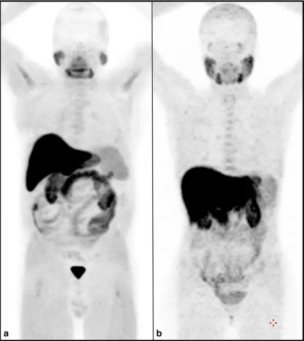
Fig. 1. Maximum intensity projection of two whole body18F–choline PET/CT scans respectively performed in a man (a) and in a woman (b). In both patients a 18F–choline dose of 330 MBq
was administered, without anesthesia and the length of the scan was 3 min per bed position (7 beds). No pathologic foci of uptake are evident in both images. Therefore, thisfigure can be representative of the physiological bio-distribution of radiolabeled choline in the male and female body.
on its“in vivo” bio-distribution in female patients. In fact, this radiophar-maceutical was administered in female patients only in few papers inves-tigating parathyroid adenomas[24]or BT[25]. As a matter of fact, in these last cited papers, the acquisition protocols were developed with segmen-tal scans of brain and/or neck, not including the rest of the female body. For the best of our knowledge, no data are available on the whole bio-distribution of radiolabeled choline in the female body.
For the reasons expressed above, in the last years we collected a large series diagnostic pitfalls in three PET centers performing PET/CT with18F–choline for imaging PC patients. The aim of our study was to
show our results in a multicenter acquired experience, trying to explain the possible meaning of thesefindings. Moreover, our secondary aim was to describe the“in vivo” bio-distribution of the tracer in the female and male body, with the help of semi-quantitative analysis of maximum standardized uptake value (SUVmax) in the target organs[26]. 2. Materials and methods
We registered 169 cases of diagnostic pitfalls on a population of 1000 male patients undergoing whole body18F–choline PET/CT during
the staging or restaging of PC.
The term“diagnostic pitfall” was intended as an area of potentially pathologic tracer uptake, higher than surrounding background, not re-lated to the disease under study.
All 1000 patients were included from three different centers: 1) University Hospital“Tor Vergata” in Rome, Italy (n = 300); 2) IRCCS INM Neuromed, Pozzilli (IS), Italy (n = 410);
3) University Hospital“Magna Græcia”, Catanzaro, Italy (n = 290). All PET centers contributed to the study joining the diagnostic pit-falls registered during the routinely clinical practice.
All these three centers used18F–methylcholine (18F–choline) to
per-form PET/CT scans. Examined patients fasted 6 h before18F–choline
in-travenous administration; they were also asked, in the week before the exam, to avoid foods containing high levels of choline[7,27]. Patients re-ceived 300–400 MBq of 18F–choline with an intravenous injection
and hydrated per os (500 ml) to reduce pooling of the radiotracer in the kidneys.
In all the three centers PET/CT scans were acquired using a Discovery ST scanner (Discovery ST16 GE Medical Systems, Tennessee, USA). This system combines a high-speed ultra 16-detector row (912 detectors per row) CT unit and a PET scanner. With 10,080 bismuth germanate crys-tals in 24 rings.
A low amperage CT scan was performed for attenuation correction of PET images (80 mA, 140 kV,field of view about 420–500 mm; CT slice thickness 3.75). Just after CT, whole-body PET images were acquired 45 min after18F–choline administration, 5–7 bed positions, 4 min per bed, from upper thighs to vertex; images were reconstructed using a stan-dard iterative algorithm, Ordered Subsets Expectation Maximization (OSEM). All patients gave their written informed consent for the exam.
The18F–choline PET/CT data set was evaluated by 6 nuclear
physi-cians on a dedicated workstation (Advantage-Windows 4.4, GE; General Electric Medical System, Tennessee, USA). All clinical, histological, ra-diological, nuclear medical information, available within the 3–6 months months before examination, were consulted to verify suspected18F–
cholinefindings.
In some cases, magnetic resonance imaging (MRI), CT with or with-out contrast agents and/or histological exam were performed to verify some uncertainfindings.
Table 1
Mean values of registered SUVmax in various organs in 5 female patients submitted to whole body18F–choline PET/CT.
Organs Mean SUVmax SD RANGE
Liver 12.8 5.5 8.9–16.7 Pancreas 6.8 4.5 3.6–10.0 Lacrimal glands 1.4 1.2 0.5–2.3 Salivary glands 3.4 2.7 1.5–5.3 Spleen 3.1 1.9 1.8–4.5 Bone marrow 1.5 1.2 0.6–2.3 Kidneys 7.6 4.2 4.6–10.5 Breast 0.8 0.5 0.4–1.1 Uterus 1.2 0.7 0.7–1.7 Table 2
Mean values of registered SUVmax in various organs in 30 male patients submitted to whole body18F–choline PET/CT.
Organs Mean SUVmax SD RANGE
Liver 11.4 4.6 8.1–14.6 Pancreas 7.8 5.4 3.9–11.6 Lacrimal glands 1.2 1 0.5–1.9 Salivary glands 3.4 2.4 1.7–5.1 Spleen 4.3 3 2.2–6.4 Bone marrow 1.7 1.4 0.7–2.7 Kidneys 7.9 5.3 4.1–11.6 Testicles 1.9 0.4 1.6–2.4 Table 3
The table summarizes the documented 169 cases of18F–choline PET/CT pitfalls on a per
tis-sues analysis.
Table 4
The table summarizes the documented18
F–choline PET/CT pitfalls according to their etiology.
Finally, in the Department of Nuclear Medicine of the University Hospital“Magna Græcia”, a18F–choline whole body PET/CT was
per-formed in 5 female patients, examined for suspected BT relapse, beyond the brain PET/CT. Patients were administered 300–400 MBq of18
F –cho-line intravenously. The acquisition protocol was the same described above for male patients.
Therefore, in these 5 female patients, the SUVmax in liver, uterus, breast, pancreas, salivary and lacrimal glands, spleen, bone marrow and kidneys, was recorded, in order to compare these data to those ob-tained in 30 male patients submitted to whole body PET/CT with18F–
choline for PC and a scan negative for secondary localizations.SUVmax was measured by placing spherical regions of interest (ROIs) on the cited organs in the maximum intensity projection (MIP).
All patients gave their written consent to the study. The study was approved by our local ethic committees.
3. Results
3.1. Physiological distribution of18F–choline in female and male patients
The most relevant physiological tracer uptake of18F–choline, in both
male and female patients, was documented in the liver and pancreas; moderate-to-high uptake was observed in the spleen and in salivary and lachrymal glands. Less intense uptake was registered in bone mar-row. Other further sites of mild uptake were the small and large intes-tines, probably due to the peristalsis. Testicles presented mild uptake. Due to the urinary excretion of the tracer, high rate of tracer uptake was documentedin kidneys and bladder. The uptake of18F–choline in
the brain was usually negligible, except in the choroid plexus and pineal gland, occasionally visualized in PET imaging. No significant differences in18F–choline physiological bio-distribution were recorded at the visual
analysis of PET data (Fig. 1) and with the semi-quantitative analysis with SUVmax in the whole body, between female (Table 1) and male (Table 2) patients.
In the examined women, low uptake was detected in the breast, with a mean SUVmax of 0.8 (range 0.4–1.1), and in the uterus, with a mean SUVmax of 1.2 (range 0.7–1.7). Ovaries were not detectable in the CT component of the exam, being performed without contrast agent administration. No differences in terms of bio-distribution were recorded between male and female populations.
3.2. General overview of diagnostic pitfalls in 1000 male patients Among the 1000 examined male patients with PC, we collected 169 cases (16.9%) of abnormal sites of uptake of18F–choline, dubious or
un-expected, not related to PC.
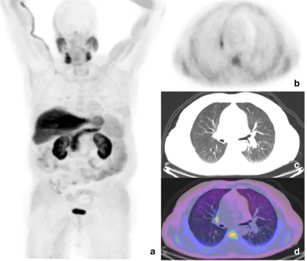
Globally, on a per-organ analysis, the abnormal sites of uptake were observed in lymph nodes (67/169 patients; 40% of overallfindings), in the adrenal glands (23/169, 13%), lungs (15/169, 9%); thyroid (15/69, 9%); brain (14/169, 8%); colon (11/169, 6%); bones (6/169, 3.5%), skin (4/169, 2.5%); thymus (3/169,b2%); liver (2/169, ≈1%); esophagus (1/169,b1%), bladder (1/169, b1%) and prostate (1/169, b1%) (Table 3).
Considering the diagnostic results, pitfalls were due to inflammation in 80/169 patients (47%), to non-specific uptake in 46/169 patients (27%), to benign tumors in 26/169 patients (15%), to malignant tumors
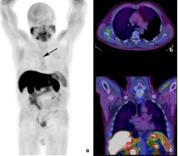
Fig. 2. Maximum intensity projection of the whole body18F–choline PET (a) shows single area of focal uptake in the mediastinum, corresponding to a 6 mm wide lymph node in the
Barety's space, in corresponding axial (b) and coronal (c) PET/CT views. SUVmax was 3.6 and thefinding was considered as inflammatory due to the low PSA serum level and referred persistent cough since one month before the exam.
in 10/169 patients (6%), to a condition of hypermetabolism in 7/169 pa-tients (4%) (Table 4).
The inflammatory findings were localized in lymph nodes (n = 64), bones (n = 5), skin (n = 4), thyroid (n = 4),lungs (n = 2) and esoph-agus (n = 1).
Nonspecific uptake was considered as a condition of high radiolabeled choline uptake without clinical evidence, laboratory tests and correlative imaging positive for benign or malignant disease. This feature was documented in adrenal glands (n = 21), lungs (n = 12 sol-itary pulmonary nodes), thyroid (n = 8) and pleura (n = 5).
Benign tumors were represented by meningiomas (n = 12), colon adenomas (n = 9), thymomas (n = 3), a papilloma of the choroid plex-us (n = 1) and a neuroendocrine lung tumor (n = 1).
Malignant tumors were lymphomas (n = 2), colon cancers (n = 2), a lymphadenopathy due to bladder cancer relapse (n = 1), a glioma (n = 1), a case of multiple myeloma (n = 1), a pleural mesothelioma (n = 1), a case of bladder cancer (n = 1) and a primary squamous cell carcinoma of the prostate (n = 1).
A condition of hypermetabolism was supposed when clinical and laboratory data confirmed the18
F–choline findings of uptake in condi-tions of hyperthyroidism (n = 3), adrenal adenomas (n = 2) and hepat-ic focal nodular hyperplasia (n = 2).
The mean SUVmax registered in inflammatory findings, nonspecific uptake cases, benign tumors, malignant tumors and in conditions of hy-permetabolism was respectively 2.9, 2.5, 4.5, 8.9 and 4.2 while the SUVmax documented in the vascular lesion was 11. Differences be-tween these groups were not statistically significant.
All the citedfindings are described below more accurately, following the order ofTable 3.
3.3. Lymph node uptake
A large amount of patients (67/169; 40% of overall documented pit-falls) showed low/mild uptake of18F–choline in lymph-nodes. In the most part of these cases the uptake was confined in a single lymph-node, generally localized in axillary regions, neck, mediastinum or in-guinal regions. Rarely thisfinding was recorded in the abdominal district.
In 64 cases the uptake was considered as inflammatory with the help of anamnestic data and CT features. Among these patients, in particular, in a 67-year-old patient in restaging of PC, 1 year after radical prostatec-tomy (PSA 0.36 at the time of the scan), a single area of focal, mild up-take was detected in a 6 mm lymph node, with recognizable hilum, of the Barety's space. Registered SUVmax was 3.6 (Fig. 2). The patient re-ferred persistent cough since one month before the exam.
Conversely, in two cases a lymphoma was documented. In thefirst case a low grade lymphoma was detected in multiple supra- and infra-diaphragmatic lymph-adenopaties[7]. In the second case, we examined for restaging PC a patient previously submitted to radiotherapy on the prostate for curative intent (PSA 1.8 at the time of the scan). Intense
18F–choline uptake was observed in three small lymph nodes without
recognizable hilum (diameter≈1 cm) in the left inguinal region. More-over, the patient presented splenomegaly at the CT component of the exam, with low18F–choline uptake (SUVmax 2.8). Being these findings
not congruent with the diagnosis of secondary localizations of PC a histo-logical specimen was obtained in the left inguinal region. Histohisto-logical diagnosiswas positive for non-Hodgkin lymphoma (Fig. 3).
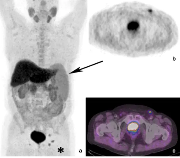
Finally, in a patient in restaging of PC after radiotherapy (PSA 1.1 ng/ml), a focal area of intense uptake (SUVmax 8.3) was detected
Fig. 3. PET/CT maximum intensity projection (a) shows splenomegaly (black arrow) and intense18F–choline uptake in the left inguinal region, with a focal area evident in axial PET detail
in correspondence of a 2.3 cm wide lymph node in the pre-sacral region (Fig. 4). The patient had also diagnosis of bladder cancer and wassubmitted to chemiotherapy two years before the scan. Considering also the relatively low PSA serum level, thisfinding was considered as pelvic lymph nodal relapse of bladder cancer.
3.4. Adrenal glands
In 23/169 patients mild uptake of the tracer was documented in ad-renal glands (13%). Generally, the uptake was mono-lateral. In 21 cases the uptake was not related to meaningful morphological abnormalities, thisfinding was therefore considered as exclusively functional, non-specific. In two cases the uptake was in correspondence of a nodular hypodense area in right (n = 1) or in left adrenal gland (n = 1): thesefindings were considered suspicious for adrenal adenomas[9]. 3.5. Lungs
The pulmonary district was the third most important district for prevalence of unexpectedfindings of18F–choline uptake (15/169; 9%).
Twelve patients showed focal uptake in correspondence of a solitary pulmonary node, generally with a low SUVmax (range: 1.5–2.6). In most of these cases, the suspicion of benign lung nodule was done,
standing to the clinical history or the comparison with previous imaging
[7]. In one case a neuro-endocrine tumor of the lung was diagnosed in a mass in the right lung with SUVmax 4.5 and diameter 10 cm[9].Finally, in two cases the diagnosis of sarcoidosis was done: in particular, in a 58-year-old patient in restaging of PC 3 years after radical prostatectomy (PSA 2.6 ng/ml), a focal area of uptake was detected in the right pulmo-nary hilar region (SUVmax 4.1) in correspondence of a 1 cm wide lymph node (Fig. 5). Thisfinding was suspicious for secondary localization of PC but anamnestic data of the patients were also suggestive for chronic respiratory disease (cough, fatigue, and shortness of breath since 6 years before the exam). Thus, a suspicion of sarcoidosis was done, accordingly with the pulmonologist. Histological sampling from the mediastinal lymph nodes, by endobronchial ultrasound-guided transbronchial needle aspiration, confirmed non-caseating granulomas with epitheli-oid cells.
3.6. Thyroid
The thyroid was interested by uptake of18F–choline in 15 patients
(9% of overall amount of pitfalls). In 8 cases laboratory data and/or cor-relative imaging did not show abnormalities and thisfinding was con-sidered as non-specific. In 4 patients laboratory data were suggestive for a condition of thyroiditis[7]. In the remaining 3 patients, laboratory
Fig. 4. PET/CT maximum intensity projection (a) of a patient in biochemical relapse of PC after radiotherapy (PSA 1.1 ng/ml), previously also submitted to cystectomy and ileo-cutaneous ureterostomy for bladder carcinoma. The axial PET view (b) shows diffuse uptake in the pre-sacral region, corresponding to a 2.3 cm wide lymph node in CT (c) and PET/CT (d) views, indicative of lymph nodal pelvic bladder cancer relapse.
data allowed to diagnose a condition of hyperthyroidism, due to the concomitant rise of thyroid hormones and reduction of TSH serum levels.
3.7. Brain
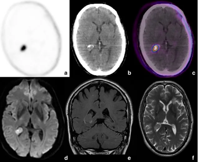
Abnormal uptake of18F–choline was documented in the brain in 14/
169 cases (8%). In 12 patients the uptake was linked to a meningioma (Fig. 6); in one patient a single area of18F–choline uptake in the brain
was related to a glioma, as confirmed by MRI[7].Finally, in apatient ex-amined for restaging PC, 1 year after radiotherapy for curative intent (PSA 1.6 ng/ml at the time of the scan), a focal area of uptake (SUVmax 4.5) was observed in the occipital horn of the right cerebral ventricle, as-sociated with hyperdense area in the CT. MRI diagnosed a papilloma of the choroid plexus (Fig. 7).
3.8. Colon
In 11 patients (6%) abnormal focal uptake of18F–choline was
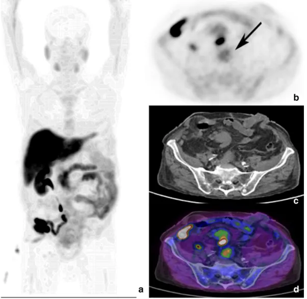
docu-mented in the colon. Among these, in9 patients the colon endoscopy allowed to diagnose colic adenomatosis. In two cases the histological sam-ple, performed by endoscopy, diagnosed colon carcinoma. In particular, in a 74-year-old patient in restaging of PC, focal uptake was detected in a thickening of the superior tract of the sigma, due to colon cancer (Fig. 8). 3.9. Bones
In 6/169 patients (3.5%) we observed abnormal tracer uptake in the skeleton. In particular, in 3 patients the uptake was detectable in the
maxillary sinus, extended to mucosal thickenings respectively of the right (n = 1)[9]and left (n = 2) maxillary sinus.All patients were then evaluated with CT of the cranial bones within 10 days. The CT scan showed soft tissue in the right maxillary sinus without erosion of the osseous cortex. All the imagingfindings were considered suggestive for maxillary sinusitis. Similarly, in one patient presenting18F–choline
uptake in the right middle ear and in the ipsilateral mastoidcells, a right otomastoiditis was documented. In one patient a multiple myeloma was diagnosed due to multiple18F–choline avid, lytic bony lesions. In this
pa-tient, examined for restaging PC for low rise in PSA serum level (0.9 ng/mL), 2 years after radical prostatectomy, the diagnosis was al-ready obtained from elevatedγ-globulin levels and bone marrow biopsy. Finally, the last patient showed was examined due to abone scan suspicious for a PC metastasis in the right femoral neck: the PET/CT scan confirmed a single area of focal uptake in this site, withSUVmax 6 and without morphological abnormalities in the corresponding CT[9]. The PSA serum level was too low to support the diagnosis of bone me-tastasis (0.2 ng/ml). Moreover, the patient reported a severe post-traumatic pain in that region. The biopsy of the right femoral neck was performed and diagnosed reactivefibrous cells without malignancy. 3.10. Pleura
In the pleural district we encountered 6 pitfalls (3.5%): in 5 of them a solitary area of uptake was observed in correspondence of a single pleu-ral thickening in the thoracic wall. In all these cases the anamnesis, follow-up and/or correlative imaging allowed to consider the uptake as non-specific. Unfortunately, in these patients histological exam was not available.
Fig. 5. PET/CT maximum intensity projection (a) and axial PET view (b) show a focal area of18F–choline uptake in the right pulmonary hilar region (SUVmax 4.1), corresponding to a 1 cm
In the last patient, examined for restaging PC and rise of PSA serum level (1 ng/ml at the time of the scan) one year after surgical interven-tion of radical prostatectomy, we observed multiple areas of intense
18
F–choline uptake in several pleural thickenings in the right lung, with some areas of confluence producing consolidation as detectable in the CT component of the exam. Considering the radiologic criteria and the high SUVmax documented in the lesions (range 7.2–16) a CT-guided pleural needle biopsy was performed. Histological exam diag-nosed pleural mesothelioma (Fig. 9).
3.11. Skin
We registered 4 cases (2.5%) of a single area of focal uptake in the skin. In all cases the uptake was localized in correspondence of a sub-centimetric nodule in the skin of the neck (n = 2), of the right axilla (n = 1) and right inguinal region (n = 1). In all these patients the an-amnesis or the direct clinical investigation allowed to diagnose a com-mon cutaneous furuncle.
3.12. Thymus
In three patients we documented abnormal tracer uptake in the thy-mus. Considering the age-related thymic involution in all adult patients,
we further examined thisfinding with follow-up or histological exam. In all cases a thymoma was diagnosed. In particular, in a patient exam-ined for staging PC (PSA 6 ng/ml), we observed prominent18F–choline
uptake in the upper mediastinum (SUVmax 7), corresponding to a well-defined,hypodense area of 2.8 cm. The patient did not suffer any symp-toms related to a“mediastinal mass”. Subsequently, histopathological analysis, after a CT-guided biopsy, diagnosed epithelial thymoma. No lymph node or bone metastases from PC were detected on the exam (Fig. 10).
3.13. Liver
In 2 patients we observed a single hepatic area of focal uptake. Con-sidering the clinical suspicion of hepatic secondary lesions, in both case histological CT-guided sampling allowed to diagnose focal nodular hy-perplasia (FNH). Particularly, in a 67-year-old patient a focal area of
18F–choline uptake was documented in the 4th hepatic segment
(SUVmax 7), corresponding to a non-homogeneous hypodense area in the CT. The subsequently performed MRI of the upper abdomen did not help to reach the correct diagnosis, being dubious for the differential diagnosis between secondary localization of PC and FNH (Fig. 11). Only the histological exam allowed to obtain the correct diagnosis.
Fig. 6. Maximum intensity projection (a) and axial PET view (b) show focal18F–choline uptake in right, parasagittal frontal lobe, corresponding to hyperdense area with calcifications in
Fig. 8. Sagittal (a) and axial (b) PET/CT views show focal tracer uptake in the superior sigmoidal tract, in correspondence of a thickening in related sagittal (c) and axial (d) views. The colonoscopy (e, f) displayed a pedunculated malignant lesion, as confirmed by subsequent histological exam.
Fig. 7. Axial PET (a), CT (b) and PET/CT (c) views show focal18F–choline uptake a hyperdense area in the occipital horn of the right cerebral ventricle. MRI diagnosed a papilloma of the
3.14. Esophagus
A 72-year-old patient was referred for18F–choline PET-CT staging
of PC (PSA serum levels of 11 ng/ml; Gleason score = 8). Only an area of focal and intense uptake in the distal tract ofesophagus was docu-mented. Contrast enhanced CT did not show suspected abnormal tissue in correspondence of the site of uptake. Thisfinding was highly sugges-tive for oesophagitis, according to clinical symptoms referred by the pa-tient (dyspepsia and dysphagia), as subsequently confirmed by gastroscopy[7].
3.15. Bladder
A patient was submitted to the exambecause of suspicion of bone metastases in a bone scan2 years after radical prostatectomy (PSA serum level, 1.5 ng/ml at the time of the scan). No bone metastases were detected at PET/CT; nevertheless, a focal area of intense uptake (SUVmax 18.1) was detected in a thickness of the trigonal area of the bladder higher than the surrounding physiological high rate of radioac-tive urine in the bladder. A contrast enhanced CT of the pelvis showed a lesion of the trigone with irregular margins and high contrast enhance-ment. Histological sample diagnosed a bladder carcinoma.
3.16. Prostate
A patient was examined with18F–choline PET/CT to stage PC histo-logically proven (PSA 7.7 ng/ml). The exam showed intense tracer up-take in the prostate gland, with remarkable increase in size, non-homogeneous density and air bubbles in the prostatic tract of the ure-thra. Furthermore, an important quote of air-fluid level was observed in the bladder, by evaluating the CT component of the exam.
Finally, some areas of18F–choline uptake were observed in several
mediastinal lymphadenopathies (Fig. 12). The patient referred to suffer of pelvic pain and haematuria from three months. Considering that these data were suggestive for a particularly aggressive kind of neo-plasm, not completely in agreement with the biological behavior of PC and with the PSA serum level, a new biopsy, done in accordance with urologists, allowed to diagnose a primitive squamous cells cancer of the prostate.
4. Discussion
Being a marker of lipogenesis and cell membrane synthesis, the11C/ 18F–choline is a tracer with a clear role in the management of PC
pa-tients, superior to18F–FDG in different clinical settings[28,29].
Numer-ous studies deepen the knowledge of the diagnostic pitfalls inclinical
Fig. 9. Maximum intensity projection (a) shows multiple areas of pathologic18F–choline uptake in the right lung, corresponding to several pleural thickenings in axial (b) and coronal
practice with the18F–FDG[6,30–32]: all these experiences allowed to
reach a satisfactory knowledge on the physiological bio-distribution of this tracer, its physiologic variants and the potential diagnostic errors linked to its intrinsic properties of a glucose-analogue, non-tumor spe-cific marker. On the other hand, few data are actually available on this topic on the radiolabeled choline. Nevertheless, the radiolabeled choline is becoming one of the most useful PET tracers, beyond18F–FDG.
Concerning the physiological bio-distribution of18F–choline in the
whole body, our data (Tables 1 and 2) show that the18F–choline
ex-hibits a high uptake gradient in the exocrine and endocrine glands: in liver is registered the highest uptake while pancreas, salivary and lach-rymal glands, normally enhance18F–choline. It is known that hepatic
and pancreatic cells present an endodermal lineages, also common to salivary glands and lachrymal glands[33,34]: as reported in two studies comparing11C–choline and the two fluorinated kinds of choline[1,23],
all these tracers present an high uptake gradient in liver, being this or-gans the main site of phospholipids metabolism and catabolism in the human body.The uptake in the other exocrine glands can be due to an alternative excretion, being pancreas and salivary and lachrymal glands associated by a common embryologic origin[33].
Other sites of physiological uptake are bone marrow and spleen: this could be due to a slight gradient of choline enhancement in the reticu-loendothelial system.
The kidneys and bladder are usually visualized due to the urinary ex-cretion of the tracer. Interestingly, we did not observe significant differ-ences in the bio-distribution in the whole body between female and male patients (Fig. 1). Testicles show mild uptake and breast show low-mild uptake, due to their exocrine function, that in the breast is normally absent. The uptake in the uterus is negligible. Nevertheless, our data on the bio-distribution of the tracer in female body need to be considered preliminary, considering the poor examined population. The main feature that our results in 1000 patients examined for PC, shows the high probability to detect18F
–choline uptake in inflammato-ry conditions (47%, 80 cases on 169 pitfalls), in particular in reactive lymph nodes. In our series, a large number of documented pitfalls were linked to inflammation in lymph nodes, lung nodes and skin. The uptake of18F–choline in inflammatory conditions could be due to
the activation of cellular types as in the monocyte-to-macrophage dif-ferentiation, expressing a high rate of lipogenesis and cell membrane synthesis[35]. However, the recognition of the most part of these false positive cases is easily reachable with an accurate collection of the an-amnesis. Nevertheless, among all the cases of lymph node uptake, we documented one case of pelvic lymph nodal relapse of bladder cancer and two lymphomas. Both these cases were not congruent with a diag-nosis of lymph nodal metastases of PC, due to the anamnestic data and their peculiar anatomic localizations, as in a case of low grade
Fig. 10. Maximum intensity projection (a) and axial PET view (b) show18F–choline uptake in the upper mediastinum, corresponding to a well-defined, hypodense, 2.8 cm wide area in
lymphoma with supra and infra-diaphragmatic localizations. Only few papers have been already described the possibility of18F–choline
up-take in non-Hodgkin and Hodgkin lymphomas[16,36]and we must consider the incidental detection of a lymphoma as a rare but not un-usual condition.
The molecular pathway of lipogenesis is also at the basis of the18F–
choline uptake documented in benign tumors: thymomas, a neuroen-docrine tumor of the lung, colic adenomas, meningiomas, representing the 15% of all registered pitfalls (25/169,Table 3). Also in these cases, a clinical approach, aware of the clinical history of the patients, allowed to recognize thesefindings as non-related with PC. In particular, is well known that some mediastinal diseases can show18F/11C–choline
up-take, as already reported for thymomas[13,37]and mediastinal in flam-matory lymph nodes[38]while the uptake in thoracic granulomatous diseases as sarcoidosis, anthracosis and tuberculosis is also known the possibility of uptake in thoracic granulomatous diseases as sarcoidosis
[39], anthracosis[40]and tuberculosis[41]: in these cases, a deepen clinical anamnesis was sufficient to reach the final diagnosis. On the other hand, caution is needed when observing tracer uptake in numer-ous pleural thickenings as in the case of malignant mesothelioma we re-ported (Fig. 10). In this last case the accurate knowledge of the CT component of the exam can help in correctly diagnosing a concomitant malignant tumor other than PC.
Regarding the liver, we documented a case of focal nodular hyper-plasia. In the liver, we observed a single case of focal nodular hyperpla-sia; in fact, it is known that some benign hepatic lesions as focal nodular hyperplasia and hepatocellular adenoma can show radiolabeled choline uptake, with different uptake rate[42]. Interestingly, in our case, the up-take in the focal nodular hyperplasia was a clearly visible respect to the
surrounding background of the liver, which is commonly the site with the highest rate of radiolabeled choline uptake.
As known, the uptake in the brain is usually negligible with all the kinds of radiolabeled choline[1,23]. As already reported by our group
[7]and by the experience of Mertens et al.[43], only the pineal gland and the choroid plexus can occasionally show a minimal rate of physio-logical uptake, not associated with morphophysio-logical lesions. In our series, we described a case of18F–choline uptake linked to a benign tumor in
the choroid plexus (Fig. 8): in this asymptomatic case a hyperdense area was detectable on the CT but only correlative MRI allowed to reach the correct diagnosis. The possibility of uptake in benign and ma-lignant tumors of the brain was described in several papers[44–46]: all these studies support the usefulness of radiolabeled choline PET/CT in the management of brain tumors, due to the low rate of physiological uptake in the normal white and gray matter that allows the recognition of tumors expressing high synthesis of cell membrane. In our series,we documented 1 papilloma of the choroid plexus, 1 glioma and 11 menin-giomas. Therefore, 1% of examined patients for PC (11/1000) presented a18F–choline avid meningioma at the whole body scan. Interestingly,
these data are similar to the experience of Fallanca et al.[11], which re-ported the possibility to incidentally detect meningiomas in patients undergoing a whole body11C–choline PET/CT for PC. The inclusion of
the brain in the whole body scan of PC patients examined with radiolabeled choline PET/CT should be recommended in order to ensure the possibility to detect malignant tumors or benign lesions of the brain and also the verify the possibility of PC metastases in the cranial teca or in the brain that can occur in a significant minority of PC patients[47,48]. In our opinion, the most peculiarfinding we encountered was the squamous cell carcinoma of the prostate: probably, for the best of our
Fig. 11. In this patient, a focal area of18F–choline uptake was documented in the 4th hepatic segment, as evident in maximum intensity projection (a, arrow) and axial PET (b) and PET/CT
knowledge, this is thefirst case described in literature as18F-choline
avid[49]. Also in this case, the high rate of tracer uptake was probably due to a faster synthesis of cell membrane expressed by this cancer[50]. Globally, the uptake of radiolabeled choline in malignant tumors could be due to the highestsynthesis of cell membrane induced by the faster mitosis of cellular types expressing a high rate of replication, as known for malignant tumors, hepato-cellular[51] and bronchiolo-alveolar carcinoma[21].
Limit of our study was the lack of an adequate follow-up for all de-scribed cased, in particular for those case we classified as non-specific uptake or inflammatory. Furthermore, we did not provide the
histological diagnosis for all registered pitfalls. Anyway, we muststate that the recognition of all cited false positivefindings, physiological var-iants and diagnostic pitfalls was of the utmost importance in order to choose the best therapeutic approach when a malignant condition was diagnosed.
In the last decade, several PET radiopharmaceuticals have been pro-posed forthe management of PC patients[28], also taking into account the documented low specificity of radiolabeled choline. Recently, the use of radiolabeled choline PET/CT has significantly reduced its use in the clinical practice due to the marketing of new agents, like68
Ga-Prostate Specific Membrane Antigen (PSMA)[52]. In particular, many
Fig. 12. Maximum intensity projection (a) of a patient with primitive squamous cell cancer of the prostate. High tracer uptake is more evident in axial PET views in the pelvis (a, black arrow) and the mediastinum (b, curved arrow), corresponding to respectively the prostate and to metastatic mediastinal lymph node in related axial CT (b, c) and PET/CT views. Only the repeated histological sample allowed to reach thefinal diagnosis.
Table 5
The table summarizes some papers developed on patients, suggesting the potential applications of radiolabeled choline,68
Ga-PSMA and18F–Fluciclovine PET/CT in different clinical
settings.
Authors Journal Year Tracer Clinical setting Nr. of patients Trigger PSA Conclusion
Cimitan et al.[60] Eur J Nucl Med Mol Imaging 2006 18F–choline
Biochemical relapse 100 4 ng/ml Useful
Calabria et al.[2] Nucl Med Commun 2013 18F–choline
Staging 45 18 ng/ml Limited role
Evangelista et al.[59] Abdom Imaging 2015 18F–choline
Local recurrence 1031 N2 ng/ml Limited role
Chiaravalloti et al.[61] Eur J Nucl Med Mol Imaging 2016 18F–choline Local recurrence 79 ≤2 ng/ml Limited role
Marzola et al.[5] Clin Nucl Med 2013 18F–choline Distant metastases and
biochemical relapse
331 N0.2 ng/ml Useful
Eschmann et al.[3] Nuklearmedizin 2007 11C–choline
Staging 42 / Useful only in secondary
localization detection.
Castellucci et al.[4] J Nucl Med 2014 11C–choline
Restaging 605 N0.2 ng/ml Useful
Freitag et al.[52] Eur J Nucl Med Mol Imaging 2016 68Ga-PSMA Restaging 26 / Useful
Giesel et al.[53] Eur J Nucl Med Mol Imaging 2016 68Ga-PSMA Primary tumor location
(PET/CT and PET/MRI)
10 / Useful
Eiber et al.[54] Eur Urol 2016 68
Ga-PSMA Primary tumor
location (PET/MRI)
66 / Useful
Verburg et al.[55] Eur J Nucl Med Mol Imaging 2016 68
Ga-PSMA Biochemical relapse 155 / Useful
Odewole et al.[62] Eur J Nucl Med Mol Imaging 2016 18F–fluciclovine
Biochemical relapse 53 / Useful
Nanni et al.[63] Clin Nucl Med 2015 18F–fluciclovine
11C–choline
Biochemical relapse 50 / 18F–fluciclovine more
papers documented a good diagnostic performance of68Ga-PSMA PET/ CT and PET/MRI in primary tumor detection and localization[53,54]and in the detection of secondary lymph nodes and bone metastases[52], with a diagnostic accuracy superior or slightly higher than radiolabeled choline PET/CT(Table 5).
Moreover, the detection rate of68Ga-PSMA PET/CT it is also related
to the PSA and PSA kinetics at the time of the scan[55]. On a theoretical basis,68Ga-PSMA is more specific than radiolabeled choline due to its
peculiar biological affinity for PC cells; anyway, similarly to radiolabeled choline, it has also been already demonstrated the possibility to detect
68Ga-PSMA uptake in conditions other than PC[56,57]. On the other
hand, radiolabeled choline PET/CT shows a very low specificity in the detection of primary PC[58]and local recurrence[59]while68
Ga-PSMA is more useful in thesefields due to its specific membrane antigen ligand[54]. However, radiolabeled choline PET/CT remains the most cost-saving technique.
Standing to these considerations, we can hypothesize that radiolabeled choline and68Ga-PSMA will play complementary roles in
different clinical settings of PC.
As future trend, PET/CT with radiolabeled choline should be per-formed in patients during the restaging of PC with advanced biochemi-cal relapse (in example with PSA higher than 2 ng/ml, supporting the possibility of distant secondary metastases rather than local recurrence)
[60,61]. On the other hand,68Ga-PSMA PET/CT is becoming useful
dur-ing the stagdur-ing, for tumor detection or durdur-ing the early biochemical re-lapse after surgery or radiotherapy[55]: on this last topic, it has recently demonstrated that, in 32 patients with suspicion of local relapse of pros-tate cancer after curative therapy (surgery and/or radiotherapy) and a negative18F–choline PET/CT scan,68Ga-PSMA PET/CT detected local
re-currence in 43.8% (14/32) of the choline negative patients.[62]This study suggests that68Ga-PSMA is more able than radiolabeled choline
in detecting early recurrence of prostate cancer.
However, it is necessary to underline that a conspicuous amount of papers on the diagnostic accuracy of radiolabeled choline PET/CT is ac-tually available with large examined populations, while the interesting results regarding68Ga-PSMA PET/CT are obtained on limited
popula-tions. Moreover, in the panorama of PC imaging, a new tracer, the18F
– fluciclovine[63], is showing encouraging results in terms of diagnostic accuracy in the management of PC patients, even in comparison with
11C
–choline[64](Table 5).
Globally, it is also mandatory to consider the large amount of studies on the diagnostic accuracy of radiolabeled choline PET/CT, which allowed to reach a satisfactory knowledge of the physiologic distribu-tion of this tracer and on the physio-pathologic pathways at the basis of its uptake in several, different conditions as benign lesions, in flamma-tion and malignant tumors that can occur in clinical practice[1,7–16]. Beyond the few cited reports of a case on68Ga-PSMA[56,57], diagnostic
pitfalls linked to the abnormal uptake of 68Ga-PSMA and 18F–
fluciclovine still need to be addressed on a large population with studies focused on this topic.
Finally, we must also consider a well-known favorable quality imag-ing for radiolabeled choline, while preliminary available data suggest a poor quality of imaging due to the entrapment of18F
–fluciclovine in muscular structures, suggesting the necessity of an early imaging win-dow with this tracer, in order to provide the best visual results[65]. On the other hand,68Ga present high emission positron energy, which could lead a low quality offinal imaging[66].
Therefore, despite the potentialgood diagnostic accuracy of68
Ga-PSMA PET/CT in detecting recurrent PC, especially for low PSA serum levels after primary treatment with curative intent, a head to head com-parison with radiolabeled choline PET/CT is still missing[67]. The rou-tine use of68Ga-PSMA in clinical practice is also limited by the
registration in several different countries. Considering also this feature, the radiolabeled choline PET/CT still needs to be considered the method of choice in the diagnostic panorama of PC imaging. Therefore, the accu-rate knowledgeof its biodistribution and the expertise in the
management of diagnostic pitfalls linked to the increasing growing rate and augmented uptake in cells due to the up-regulation of choline kinase[68], appear to be absolutely necessary.
In conclusion, the interpreting nuclear medicine physician must be aware of the physiological biodistribution of the tracer and about the unusualfindings of uptake, in order to avoid misdiagnosis of benign conditions or malignancy, as well as missing out on actual pathology. The clinical history and the exact anatomical evaluation provided by CT are of the utmost importance, in clinical practice. When needed, cor-relative imaging with MRI is also important. Histological sample is nec-essary to reach thefinal diagnosis.
References
[1]Calabria F, Gallo G, Schillaci O, Cascini GL. Bio-distribution, imaging protocols and di-agnostic accuracy of PET with tracers of lipogenesis in imaging prostate cancer: a comparison between 11C-choline, 18F-Fluoroethylcholine and 18F-Methylcholine. Curr Pharm Des 2015;21(32):4738–47.
[2]Calabria F, Chiaravalloti A, Tavolozza M, Ragano-Caracciolo C, Schillaci O. Evaluation of extraprostatic disease in the staging of prostate cancer by F-18 choline PET/CT: can PSA and PSA density help in patient selection? Nucl Med Commun 2013; 34(8):733–40.
[3]Eschmann SM, Pfannenberg AC, Rieger A, Aschoff P, Müller M, Paulsen F, et al. Com-parison of 11C-choline-PET/CT and whole body-MRI for staging of prostate cancer. Nuklearmedizin 2007;46(5):161–8.
[4]Castellucci P, Ceci F, Graziani T, Schiavina R, Brunocilla E, Mazzarotto R, et al. Early biochemical relapse after radical prostatectomy: which prostate cancer patients may benefit from a restaging 11C-choline PET/CT scan before salvage radiation ther-apy? J Nucl Med 2014;55(9):1424–9.
[5]Marzola MC, Chondrogiannis S, Ferretti A, Grassetto G, Rampin L, Massaro A, et al. Role of 18F-choline PET/CT in biochemically relapsed prostate cancer after radical prostatectomy: correlation with trigger PSA, PSA velocity, PSA doubling time, and metastatic distribution. Clin Nucl Med 2013;2013(38):1.
[6]Corrigan AJ, Schleyer PJ, Cook GJ. Pitfalls and artifacts in the use of PET/CT in oncol-ogy imaging. Semin Nucl Med 2015;45(6):481–99.
[7]Schillaci O, Calabria F, Tavolozza M, Cicciò C, Carlani M, Caracciolo CR, et al. 18F-choline PET/CT physiological distribution and pitfalls in image interpretation: experience in 80 patients with prostate cancer. Nucl Med Commun 2010; 31(1):39–45.
[8]García Vicente AM, Núñez García A, Soriano Castrejón AM, Jiménez Londoño GA, Cordero García JM, Palomar Muñoz A. Pitfalls with 18F-choline PET/CT in patients with prostate cancer. Rev Esp Med Nucl Imagen Mol 2013;32(1):37–9.
[9]Calabria F, Chiaravalloti A, Schillaci O. (18)F-choline PET/CT pitfalls in image inter-pretation: an update on 300 examined patients with prostate cancer. Clin Nucl Med 2014;39(2):122–30.
[10]Beheshti M, Haroon A, Bomanji JB, Langsteger W. Fluorocholine PET/computed to-mography: physiologic uptake, benignfindings, and pitfalls. PET Clin 2014;9(3): 299–306.
[11]Fallanca F, Giovacchini G, Picchio M, Bettinardi V, Messa C, Fazio F. Incidental detec-tion by [11C]choline PET/CT of meningiomas in prostate cancer patients. Q J Nucl Med Mol Imaging 2009;53(4):417–21.
[12]Hodolic M, Huchet V, Balogova S, Michaud L, Kerrou K, Nataf V, et al. Incidental up-take of (18)F-fluorocholine (FCH) in the head or in the neck of patients with pros-tate cancer. Radiol Oncol 2014;48(3):228–34.
[13]Calabria F, D'Auria S, Sannino P, Schillaci O. A case of thymoma detected by 18F–cho-line positron emission tomography/computed tomography. Eur J Nucl Med Mol Im-aging 2011;38(3):602.
[14]Calabria F, Calabria E, Chiaravalloti A, Barbarisi M, Schillaci O. A case of intracranial meningioma detected by18F-choline PET/CT and examined by PET/MRI fusion
imag-ing. Rev Esp Med Nucl Imagen Mol 2014;33(5):306–7.
[15]Cascini GL, Restuccia A, De Vincenti T, Manti F, Calabria F. A vascular lesion mimick-ing a primitive brain tumour in a patient examined by (18)F-choline PET/CT and MRI. Rev Esp Med Nucl Imagen Mol 2015;34(5):335–6.
[16]Garzon JG, Bassa P, Moragas M, Soler M, Riera E. Incidental diagnosis of diffuse large B-cell lymphoma by 11C-choline PET/CT in a patient with biochemical recurrence of prostate cancer. Clin Nucl Med 2014;39(8):742–3.
[17]Li W, Ma L, Wang X, Sun J, Wang S, Hu X. (11)C-choline PET/CT tumor recurrence detection and survival prediction in post-treatment patients with high-grade glio-mas. Tumour Biol 2014;35(12):12353–60.
[18]Takenaka S, Asano Y, Shinoda J, Nomura Y, Yonezawa S, Miwa K, et al. Comparison of (11)C-methionine, (11)C-choline, and (18)F-fluorodeoxyglucose-PET for distinguishing glioma recurrence from radiation necrosis. Neurol Med Chir (Tokyo) 2014;54(4):280–9.
[19]van Raalte DH, Vlot MC, Zwijnenburg A, ten Kate RW. F18-choline PET/CT: a novel tool to localize parathyroid adenoma? Clin Endocrinol 2015;82(6):910–2.
[20]Sassa N, Kato K, Abe S, Iwano S, Ito S, Ikeda M, et al. Evaluation of 11C-choline PET/CT for primary diagnosis and staging of urothelial carcinoma of the upper urinary tract: a pilot study. Eur J Nucl Med Mol Imaging 2014;41(12):2232–341.
[21]Balogova S, Huchet V, Kerrou K, Nataf V, Gutman F, Antoine M, et al. Detection of bronchioloalveolar cancer by means of PET/CT and 18F-fluorocholine, and compar-ison with 18F-fluorodeoxyglucose. Nucl Med Commun 2010;31(5):389–97.
[22]Liang X, Tang G, Wang H, Hu K, Tang X, Nie D, et al. Comparative uptake of18
F–FEN-DPAZn2,18F–FECH,18F–fluoride, and18F–FDG in fibrosarcoma and aseptic
inflam-mation. Appl Radiat Isot 2014;90:158–64 (no issue).
[23]Haroon A, Zanoni L, Celli M, Zakavi R, Beheshti M, Langsteger W, et al. Multicenter study evaluating extraprostatic uptake of 11C-choline, 18F-methylcholine, and 18F-ethylcholine in male patients: physiological distribution, statistical differences, imaging pearls, and normal variants. Nucl Med Commun 2015;36(11):1065–75.
[24]Michaud L, Balogova S, Burgess A, Ohnona J, Huchet V, Kerrou K, et al. A pilot com-parison of 18F-fluorocholine PET/CT, ultrasonography and 123I/99mTc-sestaMIBI dual-phase dual-isotope scintigraphy in the preoperative localization of hyperfunc-tioning parathyroid glands in primary or secondary hyperparathyroidism: influence of thyroid anomalies. Medicine (Baltimore) 2015;94(41):e1701.
[25]Wyss MT, Spaeth N, Biollaz G, Pahnke J, Alessi P, Trachsel E, et al. Uptake of 18F-Fluorocholine, 18F-FET, and 18F-FDG in C6 gliomas and correlation with 131I-SIP(L19), a marker of angiogenesis. J Nucl Med 2007;48(4):608–14.
[26]Wetter A, Lipponer C, Nensa F, Heusch P, Rübben H, Schlosser TW, et al. Quantitative evaluation of bone metastases from prostate cancer with simultaneous [18F] choline PET/MRI: combined SUV and ADC analysis. Ann Nucl Med 2014;28(5):405–10.
[27]Calabria F, Rubello D, Schillaci O. The optimal timing to perform 18F/11C–choline PET/CT in patients with suspicion of relapse of prostate cancer: Trigger PSA versus PSA velocity and PSA doubling time, 29(4); 2014 423–30.
[28]Jadvar H. Prostate cancer: PET with 18F-FDG, 18F- or acetate, and 18F- or 11C-choline. J Nucl Med 2011;52(1):81–9.
[29]How Kit N, Dugué AE, Sevin E, Allouache N, Lesaunier F, Joly F, et al. Pairwise compar-ison of 18F-FDG and 18F-FCH PET/CT in prostate cancer patients with rising PSA and known or suspected second malignancy. Nucl Med Commun 2016;37(4):348–55.
[30]Hernandez Pampaloni M, Facchetti L, Nardo L. Pitfalls in [18F]FDG PET imaging in gynecological malignancies. Q J Nucl Med Mol Imaging 2016;60(2):124–38.
[31]Even-Sapir Einat. Imaging the normal and abnormal anatomy of the female pel-vis using (18)F FDG-PET/CT, including pitfalls and artifacts. PET Clin 2010;5(4): 425–34.
[32]Laffon E, de Clermont H, Lamare F, Marthan R. Estimating the amount of FDG uptake in physiological tissues. Nucl Med Biol 2014;41(9):717–20.
[33]Hisatomi Y, Okumura K, Nakamura K, Matsumoto S, Satoh A, Nagano K, et al. Flow cytometric isolation of endodermal progenitors from mouse salivary gland differen-tiate into hepatic and pancreatic lineages. Hepatology 2004;39(3):667–75.
[34]Okumura K, Nakamura K, Hisatomi Y, Nagano K, Tanaka Y, Terada K, et al. Salivary gland progenitor cells induced by duct ligation differentiate into hepatic and pancre-atic lineages. Hepatology 2003;38(1):104–13.
[35]Zhu L, Yuan C, Ma Y, Ding X, Zhu G, Zhu Q. Anti-inflammatory activities of phospho-lipase C inhibitor U73122: inhibition of monocyte-to-macrophage transformation and LPS-induced pro-inflammatory cytokine expression. Int Immunopharmacol 2015;29(2):622-327.
[36]Goineau A, Colombié M, Rousseau C, Sadot-Lebouvier S, Supiot S. Incidental detec-tion of a Hodgkin lymphoma on choline PET/CT and comparison with 18F-FDG in a patient with prostate cancer. Clin Nucl Med 2015;40(8):670–1.
[37]Fallanca F, Picchio M, Spinapolice EG, Ugolini C, Proietti A, Messa C. Imaging of a thymoma incidentally detected by C-11 choline PET/CT. Clin Nucl Med 2011;36(2):134–5.
[38]Rietbergen DD, van der Hiel B, Vogel W, Stokkel MP. Mediastinal lymph node uptake in patients with prostate carcinoma on F18-choline PET/CT. Nucl Med Commun 2011;32(12):1143–7.
[39]Takesh M, Haberkorn U, Strauss LG, Roumia S, Dimitrakopoulou-Strauss A. Inciden-tal detection and monitoring of spontaneous recovery of sarcoidosis via fluorine-18-fluoroethyl-choline positron emission tomography/computed tomography. Hell J Nucl Med 2012;15(1):63–5.
[40]Pinaquy JB, Fernandez P, Pasticier G, Parrens M, De Clermont H. Anthracosis mimick-ing mediastinal lymph node metastases with 18F-FCholine in high-risk prostate can-cer. Clin Nucl Med 2015;40(4):235–54.
[41]Hara T, Kosaka N, Suzuki T, Kudo K, Niino H. Uptake rates of 18F-fluorodeoxyglucose and 11C-choline in lung cancer and pulmonary tuberculosis: a positron emission to-mography study. Chest 2003;124(3):893–901.
[42]van den Esschert JW, Bieze M, Beuers UH, van Gulik TM, Bennink RJ. Differentiation of hepatocellular adenoma and focal nodular hyperplasia using 18F-fluorocholine PET/CT. Eur J Nucl Med Mol Imaging 2011;38(3):436–40.
[43]Mertens K, Ham H, Deblaere K, Kalala JP, Van den Broecke C, Slaets D, et al. Distribu-tion patterns of 18F-labelledfluoromethylcholine in normal structures and tumors of the head: a PET/MRI evaluation. Clin Nucl Med 2012;37(8):196–203.
[44]Utriainen M, Komu M, Vuorinen V, Lehikoinen P, Sonninen P, Kurki T, et al. Evalua-tion of brain tumor metabolism with [11C]choline PET and 1H-MRS. J Neuro-Oncol 2003;62(3):329–38.
[45]Fraioli F, Shankar A, Hargrave D, Hyare H, Gaze MN, Groves AM, et al. 18F-fluoroethylcholine (18F-Cho) PET/MRI functional parameters in pediatric astrocytic brain tumors. Clin Nucl Med 2015;40(1):e40–5.
[46]Lam WW, Ng DC, Wong WY, Ong SC, Yu SW, See SJ. Promising role of [18F] fluorocholine PET/CT vs [18F] fluorodeoxyglucose PET/CT in primary brain tumors-early experience. Clin Neurol Neurosurg 2011;113(2):156–61.
[47]Messina M, Ricci F, Spina B, Boccardo F. Single skull metastasis 15 years after primary treatment of prostate cancer and with undetectable PSA levels: a case report and re-view of the literature. Tumori 2013;99(5):220–4.
[48]Tremont-Lukats IW, Bobustuc G, Lagos GK, Lolas K, Kyritsis AP, Puduvalli VK. Brain metastasis from prostate carcinoma: the M. D. Anderson cancer center experience. Cancer 2003;15(98):363–8.
[49]Li J, Wang Zhe. The pathology of unusual subtypes of prostate cancer. Chin J Cancer Res 2016;28(1):130–43.
[50]Bertagna F, Bertoli M, Treglia G, Manenti S, Salemme M, Giubbini R. Incidental 11C-choline PET/CT uptake due to esophageal carcinoma in a patient studied for prostate cancer. Clin Nucl Med 2014;39(10):e442–4.
[51]Castilla-Lièvre MA, Franco D, Gervais P, Kuhnast B, Agostini H, Marthey L, et al. Diag-nostic value of combining (11)C-choline and (18)F-FDG PET/CT in hepatocellular carcinoma. Eur J Nucl Med Mol Imaging 2016;2016(43):5.
[52]Freitag MT, Radtke JP, Hadaschik BA, Kopp-Schneider A, Eder M, Kopka K, et al. Comparison of hybrid (68)Ga-PSMA PET/MRI and (68)Ga-PSMA PET/CT in the evaluation of lymph node and bone metastases of prostate cancer. Eur J Nucl Med Mol Imaging 2016;2016:70–83.
[53]Giesel FL, Sterzing F, Schlemmer HP, Holland-Letz T, Mier W, Rius M, et al. Intra-individual comparison of 68Ga-PSMA-11-PET/CT and multi-parametric MR for im-aging of primary prostate cancer. Eur J Nucl Med Mol Imim-aging 2016;43:1400–6.
[54]Eiber M, Weirich G, Holzapfel K, Souvatzoglou M, Haller B, Rauscher I, et al. Simulta-neousbsupN68b/supNGa-PSMA HBED-CC PET/MRI improves the localization of pri-mary prostate cancer. Eur Urol 2016 [Epub ahead of print].
[55]Verburg FA, Pfister D, Heidenreich A, Vogg A, Drude NI, Vöö S, et al. Extent of disease in recurrent prostate cancer determined by [(68)Ga]PSMA-HBED-CC PET/CT in rela-tion to PSA levels, PSA doubling time and Gleason score. Eur J Nucl Med Mol Imaging 2016;43:397–403.
[56]Krohn T, Verburg FA, Pufe T, Neuhuber W, Vogg A, Heinzel A, et al. [(68)Ga]PSMA-HBED uptake mimicking lymph node metastasis in coeliac ganglia: an important pit-fall in clinical practice. Eur J Nucl Med Mol Imaging 2015;42:210–4.
[57]Huang YT, Fong W, Thomas P. Rectal Carcinoma on 68Ga-PSMA PET/CT. Clin Nucl Med 2016;41:167–8.
[58]Schwarzenböck S, Souvatzoglou M, Krause BJ. Choline PET and PET/CT in primary di-agnosis and staging of prostate cancer. Theranostics 2012;2:318–30.
[59]Evangelista L, Cimitan M, Hodolič M, Baseric T, Fettich J, Borsatti E. The ability of 18F-choline PET/CT to identify local recurrence of prostate cancer. Abdom Imaging 2015;40:3230–7.
[60]Cimitan M, Bortolus R, Morassut S, Canzonieri V, Garbeglio A, Baresic T, et al. [18F]fluorocholine PET/CT imaging for the detection of recurrent prostate cancer at PSA relapse: experience in 100 consecutive patients. Eur J Nucl Med Mol Imaging 2006;33:1387–98.
[61]Chiaravalloti A, Di Biagio D, Tavolozza M, Calabria F, Schillaci O. PET/CT with 18F-choline after radical prostatectomy in patients with PSA≤2 ng/ml. Can PSA velocity and PSA doubling time help in patient selection? Eur J Nucl Med Mol Imaging 2016;43:1418–24.
[62]Bluemel C, Krebs M, Polat B, Linke F, Eiber M, Samnick S, et al. 68Ga-PSMA-PET/CT in patients with biochemical prostate cancer recurrence and negative 18F-choline-PET/ CT. Clin Nucl Med 2016;41:515–21.
[63]Odewole OA, Tade FI, Nieh PT, Savir-Baruch B, Jani AB, Master VA, et al. Recurrent prostate cancer detection with anti-3-[(18)F]FACBC PET/CT: comparison with CT. Eur J Nucl Med Mol Imaging 2016;43:1773–83.
[64]Nanni C, Schiavina R, Brunocilla E, Boschi S, Borghesi M, Zanoni L, et al. 18F-Fluciclovine PET/CT for the detection of prostate cancer relapse: acomparison to 11C-choline PET/CT. Clin Nucl Med 2015;40:e386–91.
[65]Sörensen J, Owenius R, Lax M, Johansson S. Regional distribution and kinetics of [18F]fluciclovine (anti-[18F]FACBC), a tracer of amino acid transport, in subjects with primary prostate cancer. Eur J Nucl Med Mol Imaging 2013;40:394–402.
[66]Lütje S, Blex S, Gomez B, Schaarschmidt BM, Umutlu L, Forsting M, et al. Optimiza-tion of acquisiOptimiza-tion time of 68Ga-PSMA-ligand PET/MRI in patients with local and metastatic prostate cancer. PLoS One 2016;18(11):e0164392.
[67]Zattoni F, Guttilla A, Evangelista L. Can (68)GA-PSMA or radiolabeled choline PET/CT guide salvage lymph node dissection in recurrent prostate cancer? Eur J Nucl Med Mol Imaging 2016;43:1407–9.
[68]Cuccurullo V, Di Stasio GD, Evangelista L, Castoria G, Mansi L. Biochemical and path-ophysiological premises to positron emission tomography with choline radiotracers. J Cell Physiol 2017;232:270–5.