1 A
AllmmaaMMaatteerrSSttuuddiioorruumm––UUnniivveerrssiittààddiiBBoollooggnnaa
DOTTORATO DI RICERCA IN:
BIOLOGIA CELLULARE E MOLECOLARE Ciclo XXVI
Settore Concorsuale di afferenza: 05/E2 BIOLOGIA MOLECOLARE Settore Scientifico disciplinare: BIO/11 BIOLOGIA MOLECOLARE
TITOLO TESI:
Identification and characterization of novel tumor-associated proteins as potential tumor markers for diagnosis and therapy
Presentata da: RENATA MARIA GRIFANTINI
Coordinatore Dottorato Relatore
Prof. Vincenzo Scarlato Prof. Vincenzo Scarlato
2 INDEX
PAGE
ABSTRACT 5
1. INTRODUCTION 6
1.1 Cancer, the disease 6
1.2 Tumor Markers 10
1.3 MAbs and their use in cancer therapy 12
1.3.1 Mechanisms of action of therapeutic mAbs 17
1.4 Externautics approach for tumor marker discovery 22
1.5 EXN11 and EXN6, two novel tumor-associated proteins 25
2. AIM OF THE PROJECT 25
3. RESULTS 26
3.1 EXN6 26
3.1.1. Confirmation of EXN6 detection in human cancers 26 3.1.2. The IHC-reactive anti-EXN6 polyclonal antibody specifically
recognizes its target protein 27
3.1.3. EXN6 is endogenously expressed and surface exposed in breast
and ovary cancer cell lines 29
3.1.4. EXN6 is involved in cell proliferation and invasiveness 30 3.1.5. MAbs towards EXN6 specifically recognized the
protein on the surface of breast and ovary cancer cells 32 3.1.6. Two anti-EXN6 mAbs are efficiently internalized by breast and 34 ovary cancer cells
3.1.7. The two anti-EXN6 mAbs promote cell killing in an indirect
ADC assay. 35
3.1.8. Anti-EXN6 mAb with ADCC activity 36
3.1.9. The anti-EXN6 mAbs show marginal reactivity on normal human 38 tissues.
3.2. EXN11 39
3.2.1. Confirmation of EXN11 detection in human cancers 39 3.2.2. EXN11 has a marginal endogenous expression under in vitro
3
standard growth of tumor cell lines 40
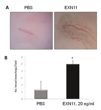
3.2.3. EXN11 expression is induced by hypoxia in cancer cells 41 3.2.4. EXN11 is associated with exosomes 41 3.2.5. EXN11 exerts pro-angiogenic activities on differentiated endothelial
cells in vitro 44
3.2.6. The human endothelial progenitor cells respond differently to the
stimulation induced by EXN11 47
3.2.7. EXN11 promotes vascularization in the mouse matrigel sponge
assay 48
4. DISCUSSION AND FUTURE PERSPECTIVES 50
5. MATERIALS AND METHODS 55
6. ACKNOWLEDGEMENTS 64
4 ABSTRACT
This study deals with the discovery and characterization of EXN6 and EXN11 as novel tumor-associated proteins and promising therapeutic targets for cancer at high morbidity and mortality. These proteins were discovered in the context of an immuno-histochemistry (IHC) screening of a collection of approximately 1600 murine polyclonal antibodies towards membrane/secreted human proteins on tissue microarrays (TMA) containing cancerous and normal sample specimens of breast, colon, lung, ovary and prostate (5 patients per cancer). Both proteins were over-expressed in more cancer types, with concomitant negligible expression in corresponding normal samples.
An expanded IHC analysis on 50 samples per cancer confirmed that EXN6 is mainly present at abnormal levels in breast and ovary cancers (40 and 35%) while it is less frequently found in colon and lung cancer (less than 10%). Concerning EXN11, it is mainly detected in colon cancer (40%). Most importantly, EXN11 is expressed in hepatic metastasis from colon cancer, suggesting that this protein could be important for tumor progression and dissemination.
A molecular and biological characterization of the two proteins was undertaken to understand whether these proteins could be exploited as molecular targets for therapeutic interventions. Concerning EXN6, results showed that it is endogenously expressed and surface exposed in different breast and ovary cancers cell lines, as confirmed by gene silencing. Knock down of EXN6 expression significantly affects relevant cancer processes in vitro, such as cell invasiveness and proliferation, thus providing the first evidence that this protein could be a potential therapeutic target. Five highly specific monoclonal antibodies (mAbs) towards EXN6 were generated, able to bind the surface of EXN6 positive cells, as judged by FACS and confocal microscopy. Interestingly these antibodies are efficiently internalized by cancer cells, a property that makes them amenable for the generation of antibody-drug-conjugates (ADC). In agreement with this, both antibodies are capable to drive a toxin-conjugated secondary antibody into cancer cells and induce cell lysis. Moreover, one of them also shows ADCC activity. Thanks to these encouraging results, efficacy studies are ongoing to test the ability of the anti-EXN6 mAbs to prevent tumor formation or progression in mouse cancer models, either as naked antibodies or as ADCs.
Concerning EXN11, an expression profile analysis in human cancer epithelial, stromal and endothelial cells showed that the protein is normally endogenously expressed at very low level
5 while it is specifically up-regulated by hypoxia. Interestingly, this protein is secreted and partially associated with the exosomial fraction, suggesting that it could be released in the systemic circulation of oncologic patients and it may act in an endocrine way. Moreover, we showed that EXN11 exerts pro-angiogenetic activities on human differentiated endothelial cells by stimulating their motility, invasiveness and capability to form capillary-like networks, whereas it does not stimulate endothelial progenitor cells. Finally, EXN11 promotes vascularization in vivo in the mouse matrigel sponge assay. Experiments are ongoing to understand the role of EXN11 in cancer angiogenesis.
Overall, this study highlights the relevance of EXN6 and EXN11 as potential cancer markers and molecular targets for novel therapies. The identification of the molecular ligands and the cellular pathways in which they are involved, currently under investigation, would significantly facilitate the design of specific drugs. MAbs offer interesting therapeutic opportunities for both proteins. Concerning EXN6, this study already contributed to this aspect by providing novel anti-EXN6 mAbs with therapeutic potential.
6 1. INTRODUCTION
1.1 Cancer, the disease
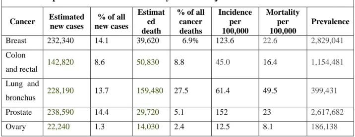
Cancer represents the second and third cause of death in the developed countries and worldwide, respectively. Cardiovascular and infectious diseases are the other two major killer diseases. However, in the near future, cancer is expected to become the leading cause of death. The first reason is that deaths from cardiovascular and infectious diseases are declining, thanks to the scientific achievements in the understanding of the pathogenic mechanisms and risk factors associated with these diseases, and to the overall improvement of sanitary conditions. The second reason is due to the genetic mutations that occur every day, because of metabolic reactions and of environmentally induced cellular damages, which accumulate because of the extension of human life span. The National Cancer Institute of the United States of America (NCI) recently estimated that a 2013 newborn has a 40% possibility to get a cancer during his/her life. In the last decade, malignancies affecting breast, colon, ovary, prostate and lung cancers had the highest incidence and mortality, and are therefore considered as the “five killers”. Prostate, breast and lung cancers have been the tumors at the highest incidence, followed by colorectal and ovary cancers. Table 1 reports the epidemiological data of the five tumors in 18 SEER geographic areas of the United States for the first six months of 2013 (http://www.cancer.gov/statistics/).
Table I – Expected Incidence and mortality rates of major tumors in the USA in 2013
Cancer Estimated new cases % of all new cases Estimat ed death % of all cancer deaths Incidence per 100,000 Mortality per 100,000 Prevalence Breast 232,340 14.1 39,620 6.9% 123.6 22.6 2,829,041 Colon and rectal 142,820 8.6 50,830 8.8 45.0 16.4 1,154,481 Lung and bronchus 228,190 13.7 159,480 27.5 61.4 49.5 399,431 Prostate 238,590 14.4 29,720 5.1 152 23 2,617,682 Ovary 22,240 1.3 14,030 2.4 12.5 8.1 186,138
Despite cancer is still as a devastating disease, cancer-associated deaths are constantly declining since 1995 (Figure 1), thanks to the significant progress in the understanding of tumor biology, improved and early diagnosis, and the identification of appropriate therapies.
7 Figure 1 – Cancer death rates in the United States from 1975 to 2010 (NCI:http://seer.cancer.gov/csr/1975_2010/)
This favorable trend will probably continue and improve in the next years. In an optimistic view of the future of cancer prevention and treatment, cancer will be a sort of “chronic disease” that will allow oncologic patients to live with a good quality of life with for several years, similarly to what now occurs with diabetes, hypertension, and other once deadly pathologies.
The fight against cancer is based on the combined action of: i) prevention, ii) early diagnosis, iii) accurate surgery, and iv) optimized and personalized therapy (1)
- Prevention – A number of environmental factors and personal behaviors are risk factors for cancer, with higher relevance for specific pathologies such as smoking and lung cancer, ultraviolet light exposure and melanoma, excessive meat assumption and colon-rectal cancer. Moreover, different microorganisms are responsible for aggressive forms of cancer, such as HBV and HCV for hepatocarcinomas, HPV for cervical cancers, and Helocobacter pylori whose chronic infection can lead to stomach cancer. Thus, a proper surveillance of healthcare and correction of wrong behaviors aimed at reducing the exposure and prolonged contact with these tumorigenic agents should have a profound effect on cancer control.
- Early and accurate diagnosis – Most cancer deaths are the consequence of the cancer progression and disseminated metastases deriving from primary tumors that are generally refractory to the current therapies. Therefore, the success of a cancer treatment strongly
8 depends upon the ability to diagnose and treat cancer before the metastasis process has started. Moreover, an important aspect is the characterization of the cancer aggressiveness and metastatic potential that has an impact in the clinicians’ decision on the most appropriate treatments. In fact, it is now well established that tumors fall into three classes (Http://cancer.gov/cancertopics): i) low invasive and metastatic cancers which remain in such a state during the life time of the patients; ii) highly aggressive cancers with a high metastatic potential that, in most cases have already disseminated by the time of diagnosis; iii) cancers of intermediate grade that have the potential to disseminate. Presently, a common way of thinking is that only the last group of cancers is really worthy of treatment. Indeed, death rates associated to indolent low invasive tumors is low. In addition, it has been shown that surgery may even provoke dormant cancers to become clinically apparent, or even aggressive. Concerning the highly aggressive cancers, they are generally refractory to the available therapies, whose toxicity sometimes overcome their palliative curative effects. Finally, the treatment of tumors with intermediate aggressiveness have a high probability to lead to a long-term, even relapse-free response. However, at present, the ability of differentiating tumors is still rudimental and all classes are treated similarly, with limited success and high socio-sanitary economical costs. Cancer markers are tumor-associated molecules that enable oncologists to distinguish different type of markers, characterize them for their severity, and monitor their progression. Moreover, markers could allow to discriminate cancers that are likely to respond to specific therapy, thus allowing a more tailored treatment of oncologic patients. Despite the incredible effort of academic and industrial research centers in the cancer markers field, due to the heterogeneity and complexity of cancer, the number of clinically relevant markers is still limited.
Surgery – Surgery is still the first medical intervention to fight cancer and will remain a key therapeutic solution in the years to come. Surgical practice of primary tumors is considered effective in reducing the risk of metastatic relapse. For instance, the five year overall survival of colorectal cancer patients that receive a surgery at early stages of the diseases is 95%. An effective surgery is accompanied to an early and accurate diagnosis and avails of high-resolution imaging and endoscopy systems. Moreover, surgery is becoming less invasive and more selective, to the great benefit of patients in terms of time of recovery and quality of life.
9 1. Optimized and targeted therapies - Radiotherapy and chemotherapy based on alkylating agents, platinum-based drugs and anti-metabolites (such as analogs of DNA bases) have been the first therapeutic strategies still widely used in clinical oncology in association to surgery. These strategies are active against rapidly dividing cells, a characteristics share by all tumors. However, they can be very toxic, poorly tolerated by patients and often leading to secondary tumors (both radiation and anti-cancer drugs are carcinogens themselves). Moreover, they can be ineffective, since often cancer can become resistant to most conventional therapies. For these reasons, cancer therapy is seeking for more selective and personalized approaches, targeting components, which are specifically or abnormally present in cancer cells of oncologic patients. In the last decades, the molecular characterization of cancer has led to the discovery of components that can be selectively targeted by cancer drugs, such as (i) oncogenic proteins responsible for uncontrolled signaling of cell cycle and proliferation, (ii) mutated tumor suppression proteins, and (iii) cancer-specific surface-associated proteins. A plethora of novel targeted therapies are under clinical validations, whose individual components can be categorized in three major types: 1) small molecules, specifically selected to block aberrant signaling proteins or biological pathways activated in cancer due to the absence of properly functioning tumor suppression genes, 2) passive immunotherapy with mAbs able to bind cancer-specific surface proteins and capable of killing cancer cells, and 3) active immunotherapy in which tumor markers are exploited as antigens and delivered to patients with proper adjuvant formulation able to break the immune-tolerance and elicit cytotoxic immune responses.
Overall, targeted cancer therapies give doctors a better way to tailor cancer treatment, especially when a target is present in some but not all tumors of a particular type, as is the case for HER-2-positive breast cancers. Ideally, treatments could be personalized based on the unique set of molecular targets produced by the patient’s tumor. Targeted cancer therapies also hold the promise of being more selective for cancer cells than normal cells, thus harming fewer normal cells, reducing side effects, and improving quality of life. Nevertheless, targeted therapies have some limitations. Chief among these is the potential for cells to develop resistance to them. In some patients who have developed resistance to imatinib, for example, a mutation in the BCR-ABL gene has arisen that changes the shape of the protein so that it no longer binds this drug as well. In most cases, another targeted therapy that could overcome this resistance is not available. It is for this reason that
10 targeted therapies may work best in combination, either with other targeted therapies or with more traditional therapies.
Overall, the discovery highly selective tumor-specific markers will enable an early and accurate diagnosis and offer new perspective for the development of novel targeted drugs, among which mAbs are emerging therapeutic opportunities.
1.2 Tumor Markers
Ideal tumor markers are molecules able to accurately distinguish a cancerous state, discriminate different cancer types and become targets for selective therapy capable of specifically recognizing and destroying tumor cells without damaging the surrounding normal tissues. Tumor marker can be categorized in four major classes based on their clinical use.
1- Diagnostic markers are used for the early detection of cancer. Moreover, their aberrant expression level can be used to monitor cancer progression in patients subjected to a given therapeutic regimen. For instance, a marker decrease or return to a normal level could be associated to a relapse-free state or indicate that cancer is responding to therapy. Of particular relevance are markers released in biological fluids of cancer patients and can be detected with non-invasive immunological assays.
2- Prognostics markers are indicative of the severity and likely outcome of the disease at time of diagnosis. The prediction of poor or favorable prognosis helps the clinician in the decision making process and improve the patients’ management.
3- Predictive markers are helpful to predict the patients’ response to drug therapies and allow clinicians to select the most appropriate therapeutic regimen while avoiding ineffective treatments.
4- Therapeutic markers are molecules, frequently receptors, oncogenes or key components of molecular pathways that can be used to develop tumor-specific ligands able to block their action, such as small molecules and mAbs.
Table 2 reports the protein markers currently used in clinic and their application (Http://cancer.gov/cancertopics/factsheet/detection/tumor-markers)
11 Table 2. Clinical cancer markers currently in use in clinic
TUMOR MARKER CANCER TYPES TISSUE APPLICATION
ALK gene Lung cancer Tumor To help determine
treatment and prognosis AFP(alpha-fetoprotein) Liver cancer and germ cell
tumors Blood
Diagnostic and prognostic B2M (Beta-2-microglobulin)
Multiple myeloma, chronic lymphocytic leukemia, and some
lymphomas Blood and urine Diagnostic and prognostic Beta-Hcg (Beta-human chorionic gonadotropin)
Choriocarcinoma and testicular cancer
Blood and urine
Diagnostic, prognostic and predictive BCR-ABL Chronic myeloid leukemia Blood and/or
bone marrow
Diagnostic and prognostic BRAF mutation V600E Melanoma and colon cancer Tumor Therapeutic
CA15-3/CA27.29 Breast Blood Predictive
CA19-9
Pancreatic cancer, gallbladder cancer, bile duct cancer, and
gastric cancer
Blood Predictive
CA-125 Ovary tumor Blood Diagnostic, predictive
and therapeutic Calcitonin Medullary thyroid cancer Blood Diagnostic, prognostic
and predictive CEA Colon and breast cancer Blood Diagnostic, prognostic
and predictive Chromogranin A (CgA) Neuroendocrine tumors Blood Diagnostic, prognostic
and predictive Chromosome 3, 7, 17and
9p21 Bladder cancer Urine Prognostic
Cytokeratin fragments 21-1 Lung cancer Blood Prognostic
EGFR mutation analysis Non small cells lung cancer Tumor Therapeutic and prognostic Estrogen receptor
(ER)/progesterone receptor (PR) (ER/PR)
Breast cancer Tumor Therapeutic
HE4 Ovary tumor Blood Predictive
HER2/neu Breast cancer, gastric cancer, and
esophageal cancer Tumor Therapeutic Immunoglobulins Multiple myeloma and
Waldenström macroglobulinemia
Blood and urine
Diagnostic, prognostic and predictive KRAS mutations Colorectal cancer and non-small
cell lung cancer Tumor
Therapeutic and prognostic Nuclear Matrix protein 22 Bladder cancer Blood Prognostic
Prostate specific antigen
(PSA) Prostate cancer Blood
Diagnostic, prognostic and predictive
Thyroglobulin Thyroid cancer Tumor Prognostic
21 Gene signature Breast cancer Tumor Prognostic
70 Gene signature Breast cancer Tumor Prognostic
Currently, despite the importance of the cancer markers that account for a considerable area of oncology research, only a few are recognized as valid and used in the clinic, as they do not have
12 sufficient specificity and sensitivity to distinguish cancer cells and allow an accurate and timely diagnosis. For example, the "Carcinoembryonic Antigen" (CEA) and the "Prostate Specific Antigen" (PSA) , are proteins detectable in the sera of patients and routinely used in the clinic to predict the presence of the tumor or to monitor the patient's response to treatments for colon or prostate cancers, respectively. Concerning CEA, this marker has the advantage of being dosed accurately and in a reproducible way, with minimal costs, and for these characteristics it has been used in the past for the screening of colorectal tumors (2). Subsequent studies have shown that CEA is inadequate for this purpose, being expressed in a variety of extra-intestinal tumors , such as cancers of the lung , ovary and bladder. Similarly other markers used for the diagnosis of tumors of the colon, such as the "Tumor - Associated Glycoprotein -72" (TAG -72 ) , a high molecular weight glycoprotein expressed in a variety of tumors , and the " Carbohydrate Antigen " CA19 -9 were inadequate diagnostic tools (2,3). Regarding the PSA, this protein is organ specific rather than tumor-specific. Although this marker has been of great help to facilitate the detection of prostate cancer, it also presents limited specificity as its serological levels may be altered in inflammatory conditions, benign prostatic hyperplasia, and trauma. Recent studies have identified new molecular forms of PSA , such as free PSA ( fPSA ) and PSA derivatives, which could be more appropriate to discriminate prostate cancer from benign neoplastic states (4). Another marker sometimes used for screening of women with high risk of ovarian cancer is the "Cancer Antigen 125" (CA- 125). Even in this case, changes in the levels of serological CA- 125 are not sufficiently specific and sensitive for population screening. CA- 125 is mainly used to monitor response to cancer treatment and check for recurrence of ovarian cancer (3). In addition to the markers mentioned, in the last 30 years, other proteins, hormones and enzymes have been used, but their level increases even in benign conditions and during pregnancy.
Despite thousands of researchers around the world are engaged in the study of cancer, today many areas of biology and physiology of tumors are unexplored and there is a pressing need to identify new markers suitable for clinical use.
1.3 MAbs and their use in cancer therapy
Immunotherapy represents the most attractive opportunity for the treatment of cancer and it is pushing the clinical research from more than a century. In particular, mAb therapy is emerging as a powerful solution for the treatment of cancer. The popularity of mAbs stemmed from the advent of hybridoma technology in 1975 and progressed through the development of chimeric, humanized, and human antibodies (5). In fact, before mAbs can be used in humans, they are
13 “humanized” by genetic engineering by replacing as much as possible of the animal portion of the molecule with human portions to prevent the human immune system from recognizing the mAb as “foreign” and destroying it before it has a chance to interact with and inactivate its target. Being directed against tumor-specific or tumor-associated antigens, mAbs can selectively eliminate tumor cells while maintaining an acceptable toxicity profile. Moreover, an emerging immune-therapeutic strategy exploits mAbs that target immune cells with the goal of breaking local tolerance and stimulating the patient’s anti-tumor immune response.
mAbs can be used as naked molecules or conjugated with cell payloads (radioisotopes, drugs or toxins) to direct kill tumor cells or to activate pro-drugs specifically within the tumors. These antibody-drug conjugates (ADC) can deliver a toxic load selectively to the tumor site while normal tissues are generally spared. ADC are of particular interest in that their therapeutic efficacy is stronger than that of naked antibodies. The concept that mAbs could be used as vehicles for the selective delivery of cytotoxic agents to tumors has been around almost since the beginning. However, until very recently, the idea has eluded successful implementation, probably for three reasons: (i) the use of antibodies against targets that were not sufficiently restricted to tumor cells, (ii) the use of drugs with insufficient potency or (in the case of bacterial or plant toxins) that were immunogenic, and (iii) the linker chemistry used to attach drugs to antibodies was not optimized. The most important property of antibodies to be used for the generation of ADC is their specificity for cancer cells, and the ability to be efficiently internalized by them so as to deliver the toxic compound in the intracellular compartment. In order to minimize toxicity, conjugates are usually engineered based on molecules with a short serum half-life (e.g. the use of IgG3 or IgG4 isotypes). Concerning the linker chemistry issue, this aspect has so far required an enormous empirical effort (6,7). For instance, a linker that is too labile allows a too rapid dissociation of the drug from the antibody causing high drug level in the blood and its exposure of normal tissues. Conversely, stable linkers require complete proteolytic digestion of the ADC to release the cytotoxic drug as the active metabolite (8). Similarly, the choice of drug required a great deal of trial and effort. So far, the most successful drugs are microtubule antagonists.
Currently, thirteen antibodies are approved for use in oncology with different therapeutic indications (9,10), described below and grouped on the basis of their major mechanism of action (http://www.cancer.gov/cancertopics/factsheet/Therapy/targeted).
14 - MAbs blocking specific enzymes and growth factor receptors involved in cancer cell proliferation.
1. Trastuzumab (Herceptin®) is approved to treat certain types of breast cancer as well as some types of gastric or gastroesophageal junction adenocarcinoma. The therapy is a mAb that binds to the human epidermal growth factor receptor 2 (HER-2). HER-2, a receptor with tyrosine kinase activity, is expressed at high levels in approximately 15-20% of breast cancers and also some other types of cancer. The mechanism by which trastuzumab acts is not completely understood, but one likely possibility is that it prevents HER-2 from sending growth-promoting signals (Figure 2). Trastuzumab may have other effects as well, such as inducing the immune system to attack cells that express high levels of HER-2.
2. Pertuzumab (Perjeta™) is approved to be used in combination with trastuzumab and docetaxel to treat metastatic breast cancer that expresses HER-2 and has not been treated with chemotherapy or a HER-2-directed therapy. Pertuzumab is a mAb that binds to HER-2 at a region distinct from trastuzumab. This region allows HER-2 to interact with other receptors, such as Her3, to send growth-promoting signals. The drug likely prevents HER-2 from sending growth signals and induces the immune system to attack HER-2-expressing cells (Figure 2).
3. Cetuximab (Erbitux®) is a mAb that is approved to treat some patients with squamous cell carcinoma of the head and neck or colorectal cancer. The drug binds to the external portion of EGFR, thereby preventing the receptor from being activated by growth signals, which may inhibit signal transduction and lead to antiproliferative effects.
4. Panitumumab (Vectibix®) is approved to treat some patients with metastatic colon cancer. This mAb attaches to EGFR and prevents it from sending growth signals.
Other targeted therapies block the growth of blood vessels to tumors (angiogenesis). To grow beyond a certain size, tumors must obtain a blood supply to get the oxygen and nutrients needed for continued growth. Treatments that interfere with angiogenesis may block tumor growth.
5. Bevacizumab (Avastin®) is a mAb that is approved for the treatment of glioblastoma. The therapy is also approved to treat some patients with non-small cell lung cancer, metastatic colorectal cancer, and metastatic kidney cancer. Bevacizumab binds to VEGF and prevents it from interacting with receptors on endothelial cells, blocking a step that is necessary for the initiation of new blood vessel growth.
15 - MAbs working by helping the immune system to destroy cancer cells.
6. Rituximab (Rituxan®) is a mAb that is approved to treat certain types of B-cell non-Hodgkin lymphoma and, when combined with other drugs, to treat chronic lymphocytic leukemia (CLL). The therapy recognizes a molecule called CD20 that is found on B cells. When rituximab binds to these cells, it triggers an immune response that results in their destruction. Rituximab may also induce apoptosis.
7. Alemtuzumab (Campath®) is approved to treat patients with B-cell CLL. The therapy is a mAb directed against CD52, a protein found on the surface of normal and malignant B and T cells and many other cells of the immune system. Binding of alemtuzumab to CD52 triggers an immune response that destroys the cells.
8. Ofatumumab (Arzerra®) is approved for the treatment of some patients with CLL that does not respond to treatment with fludarabine and alemtuzumab. This mAb is directed against the B-cell CD20 cell surface antigen.
9. Ipilimumab (Yervoy™) is approved to treat patients with unresectable or metastatic melanoma. This mAb is directed against cytotoxic T-lymphocyte-associated antigen-4 (CTLA-4), which is expressed on the surface of activated T cells as part of a “checkpoint” to prevent a runaway immune response. By inhibiting CTLA-4, ipilimumab stimulates the immune system to attack melanoma cells.
- MAbs formulated as ADCs able to deliver the toxic drug into the cancer cells
10. Tositumomab and 131I-tositumomab (Bexxar®) is approved to treat certain types of B-cell non-Hodgkin lymphoma. The therapy is a mixture of mAbs that recognize the CD20 molecule. Some of the antibodies in the mixture are linked to a radioactive substance called iodine-131. The 131I-tositumomab component delivers radioactive energy to CD20-expressing B cells specifically, reducing collateral damage to normal cells. In addition, the binding of tositumomab to the CD20-expressing B cells triggers the immune system to destroy these cells.
11. Ibritumomab tiuxetan (Zevalin®) is approved to treat some patients with B-cell non-Hodgkin lymphoma. The therapy is a mAb directed against CD20 that is linked to a molecule that can bind radioisotopes such as indium-111 or yttrium-90. The radiolabeled forms of Zevalin deliver a high dose of radioactivity to cells that express CD20.
16 12. Brentuximab vedotin (Adcetris®) is approved for the treatment of systemic anaplastic large cell lymphoma and Hodgkin lymphoma that has not responded to prior chemotherapy or autologous stem cell transplantation. This agent consists of a mAb directed against a molecule called CD30, which is found on some lymphoma cells, linked to a drug called monomethyl auristatin E (MMAE). The antibody part of the agent binds to and is internalized by CD30-expressing tumor cells. Once inside the cell, the MMAE is released, where it induces cell cycle arrest and apoptosis.
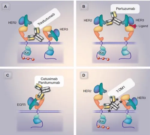
13. Ado-trastuzumab emtansine (Kadcyla®), is approved for the treatment of Her2-positive metastatic cancer who have received prior treatment with Herceptin and Taxane chemotherapy. It consists in Trastuzumab, directed against HER-2, conjugated to the a maytansine derivative DM1 (Figure 2)
Figure 2. Specificity of and mechanism of action of therapeutic targeting HER2 and EGFR. The figure represents the epitope recognition of Trastuzumab (A), Pertuxumab (B), Cetuximab or Panitumumab (C), and the ADC ado-trastuzumab conjugated to DM1 (D). In the Her2 signaling, Her2 phosphorylates the Her3 cytoplasmic domain and this activates the phosphatidylinositol-3-phosphate kinase cascade. Conversely, Her3 activation by its binding ligans (e.g. can allosterically activate Her2 kinase). The anti-Her2 antibodies target the physical and functional interaction of anti-Her2 with Her3. Pertuzumab block the heterodimerization of Her2 and Her3. In the case of Ado-trastuzumab conjugated to DM1, the cytoxic effect is potentiated by the DM1 drug (modified from Mark X, Science 341, 1192, 2013)
17 Despite the encouraging clinical data, available mAbs are still insufficient to cope with the complexity of tumor biology and ultimately to provide satisfactory therapeutic interventions. Indeed, several mechanisms of resistance are emerging that limit the antibody therapy efficacy, including activation of alternative signaling pathways, genetic mutations and expression of immune inhibitory molecules. Altogether, these evidences prompt the search and development of new molecular targets and mAbs to improve future therapies and tumor patient health care. Moreover, the clinical success of antibody therapy is dependent on a thorough comprehension of their effects of on tumor cells and their mechanism of action. In this scenario, highly sensitive technologies are needed to understand the local physiological conditions present at a cellular level, the tumor microenvironment and the regulatory networks that might influence the behavior of molecular targets and their response to mAb therapy. Moreover, as with other treatment modalities, mAb-based immunotherapy is far from perfect and requires additional study to optimize clinical response and overcome therapeutic resistance.
1.3.1 Mechanisms of action of therapeutic mAbs
The mechanism by which naked therapeutic mAbs exert their anti-tumor effect can be grouped the following categories: 1) perturbation of tumor cell signaling, 2) activation of complement dependent cytotoxicity (CDC), 3) antibody dependent cellular cytotoxicity (ADCC), 4) induction of adaptive immunity (10).
Perturbation of tumor cell signaling. Antibodies of this class target soluble mediators (e.g cytokines) or membrane bound receptors to inhibit their ability to bind their cognate ligands and regulate signaling, and can act as agonists or antagonists. For examples, antagonistic antibodies target the epidermal growth factor receptor (EGFR), or other growth factors over-expressed in cancer, and inhibit their ability to mediate mitogenic signaling (11,12) . Other type of antibodies can inhibit immune suppressing receptors, e.g. CTLA-4, or enhance antigen presentation on APCs through the activation of receptors such as CD40 (13,14). Antibodies designed for signal perturbation frequently belong to the IgG2 and IgG4 subclasses, as they do not activate CDC or ADC and have fewer immune related adverse events due to non specific immune activation (15).
Complement Dependent Cytotoxicity (CDC). The human complement is a complex proteolytic cascade comprised of over thirty proteins involved in different innate immunity responses, such as acting to lysis of foreign cells through the assembly of the membrane attack complex (MAC),
18 stimulate inflammatory processes through anaphylatoxins, and remove opsonized targets (16,17). When two or more antibodies bind to a cell, the classical complement pathway is activated through the binding of the C1 complex, a serine protease consisting of C1q, C1r and C1s, to the antibody’s Fc domains. This activates a proteolytic cascade that leads to the formation of the MAC and the release of potent anaphylatoxins and opsonins resulting in cell lysis and phagocytosis (16,18). The ability of human IgG to activate CDC varies depending on the isotype.
IgG3 followed by IgG1 are the most effective isotypes for stimulating the classic complement
cascade: both isotypes bind to C1q leading to formation of C3b on the surface of antibody-coated tumor cells near the site of complement activation. IgG2 antibodies are less efficient in activating
the complement cascade, whereas IgG4 is unable to do so (19).
CDC is an important contributor to the anti-tumor activity of many therapeutic antibodies, such as Rituximab and Ofatumumab. As already said, Rituximab is a type I chimeric antibody targeting CD20 approved for treatment of many B cell malignancies and is a potent activator of CDC. It inhibits internalization and shedding of CD20 and shifts CD20 onto lipid rafts, increasing the likelihood of complement activation through the assembly of rituximab bound receptors (20). The connection of its therapeutic efficacy with CDC activity has been demonstrated in several studies. In one of them, using in vivo lymphoma model it was found that loss of C1q abrogated the protective effects of rituximab therapy (21). Data from clinical studies showed that polymorphisms in the C1qA gene in patients with follicular lymphoma are associated with response to rituximab therapy (22). However, some undesired effect of rituximab seems to due to its CDC activity. One small clinical study found that, following rituximab therapy, patients had high circulating levels of circulating complement components with associated severe toxicity (23,24).
Ofatumumab is another type-I anti-CD20 antibody that binds to a distinct epitope of CD20 and induces greater CDC com-pared to rituximab(25). The higher efficacy of Ofatumumab could be ascribed to the fact that it binds C1q with greater avidity than rituximab and efficiently kills rituximab-resistant large B-cell lymphoma cell lines. Moreover it is able to lyse cell lines expressing low levels of CD20, which are not efficiently killed by rituximab (26). Clinical trials data showed that ofatumumab has high response rates in patients with refractory chronic lymphocytic leukemia (CLL) (27).
Antibody Dependent Cell-Mediated Cytotoxicity (ADCC). ADCC is activated by the interaction of the Fc domain with FcγRs on effector immune cells,. The stimulation of immune-receptor
19 tyrosine-based activation motifs and immune-receptor tyrosine-based inhibitory motifs results in activating or inhibitory signals through FcγRs, respectively. There are three activating FcγRs: FcγRI (CD64), which binds to monomeric IgG and tends to be occupied by plasma IgG, the low-affinity FcγRIIA (CD32A), and FcγRIIIA (CD16A) which bind IgG aggregates or immunocomplexes, and one inhibitory receptor, FcγRIIB (CD32B) (28,29).
Natural killer (NK) cells, which predominantly express FcγRIIIA, are the main effector cells of ADCC, although macrophages and granulocytes cells have been shown to mediate ADCC to a lesser extent (28). These effector cells, through the involvement of FcγRs, recognize antibodies bound to cancer cell and cause their direct lysis through release of granzymes and perforin which culminates in antibody-dependent cell-mediated cytotoxicity (ADCC). Upon crosslinking of FcγRIIIa by the immune complex, the immunoreceptor tyrosine-based activation domain is phosphorylated by the SRC family tyrosine kinase LYN, thereby inducing the formation of a signaling complex (29). This signaling complex results in the activation of phospholipase C, which hydrolyzes phosphatidylinositol-3,4-bisphosphate [PtdIns(4,5)P2] into diacylglycerol (DAG) and inosititol-1,4,5-triphosphate [Ins(1,4,5)P3], thereby inducing a number of signaling events including calcium influx from the endoplasmic reticulum and the opening of calcium-release-activated calcium channels (29,30). The increased intracellular calcium induces the serine/ threonine phosphatase calcineurin to dephosphorylate the nuclear factor of activated T cells (NFAT). Dephosphorylation of cytoplasmic NFAT exposes a nuclear localization sequence that causes translocation of this transcription factor to the nucleus where it induces the expression of a number of genes involved in the ADCC pathway (29,31). These effects are dependent on a number of factors including the density of the antigen on the cell surface and the isotype of the antibody. Of the drugable human IgG isotypes, IgG1 is reported to be the best at inducing effector function and is selected as the isotype for the antibody in indications such as the treatment of cancer where cell killing may be part of the mechanism of action. In cases where cell killing is not wanted, IgG2 or IgG4 are the isotypes of choice since they have limited ability to activate effector function,
Among others, two common mAbs with ADCC activity are trastuzumab and rituximab, which require functional activating FcγRs to exert their activity (32). Recent studies highlighted two determinants that could influence the clinical efficacy of therapeutic antibodies with ADCC activity. The first determinant is the balance between expression of activating and inhibitory FcγRs. In support of this notion, it has been shown that animals lacking expression of FcγRIIB
20 displayed a greater anti-tumor response when treated with therapeutic antibodies (32). The second determinant is the level of macrophages which can act as an effector for ADCC. High levels of macrophages are normally considered as prognostic factor for poor survival. However, two independent clinical studies have shown that follicular lymphoma patients with high levels of tumor associated macrophages have an improved response to rituximab (33), which might be ascribed to an increase in ADCC.
Induction of Adaptive Immunity. Numerous pre-clinical studies support the notion that tumor targeted antibodies can elicit adaptive immune responses, and a growing body of clinical evidence suggests that this mechanism may contribute to the clinical efficacy of antibodies. The mechanism by which this can happen is multiple and involves CDC, ADCC, or antibody dependent cell-mediated phagocytosis (ADCP). Tumor cell fragments and tumor antigens released during these processes can be taken up by professional APCs, such as DCs, to initiate tumor directed adaptive immunity. In addition, antibodies can trigger adaptive immunity by acting as an opsonin and triggering Fc dependent phagocytosis of tumor cells by APCs. Tumor antigens are processed by DCs through the endocytic pathway and presented on MHC II to prime CD4+ T cells. In addition, DCs are capable of presenting engulfed tumor antigens on MHC I molecules and elicit tumor-specific CD8+ cytotoxic T-cells (CTLs). Upon activation, CTLs can directly kill tumor cells that present the cognate peptide on MHC I, or further differentiate into tumor specific memory T cells (34).
Preclinical studies showed that DCs loaded with ovarian and melanoma cells coated with antibodies were able to elicit tumor specific CTLs able to kill primary ovarian and melanoma cells (35). A recent study showed that colon cancer cell lines coated with cetuximab, were able to induce tumor specific CTLs from autologous human DCs (36). Overall, the elicitation of adaptive immunity could potentially sustain anti-tumor immune responses in patients.
Immunotherapy approaches to activate the immune systems.
A number of molecular engineering approaches are undertaken to enhance the antibody capacity to activate immune effector mechanisms and/or to overcome the suppressive tumor microenvironment. One approach is based on the generation of non-fucosylated antibodies, In fact, it has been shown that highly fucosylated Fc significantly impair immune activation. Advanced clinical studies with non-fucosylated antibodies are currently ongoing with promising results (37). Another approach is that of bispecific tri-functional antibodies (so called triomabs) which have
21 two distinct Fab regions capable of binding two distinct epitomes, which can simultaneously bind tumor cells and immune cells while maintaining the capacity to mediate Fc dependent effector functions. To this class belongs Catumaxomab, a tri-omab that targets the tumor antigen EpCAM and the T-cell stimulatory receptor CD3, which allows for direct stimulation of CTLs in the tumor microenvironment (38). However, the complexity and the very high cost associated with this technology are preventing its widespread development.
Finally, a promising cancer immunotherapy strategy is to target the effector cells that largely contribute to the immune suppressive tumor microenvironment. As already said, Ipilimumab inhibits the potent immune suppressor molecule CTLA- 4 that is expressed by Tregs and consequently, the capacity of Tregs to inhibit the anti-tumor immune response is diminished, resulting in increased levels of CTLs, CD4+ T cells, and APCs (14,39). Following the success of ipilimumab, other mAbs that are emerging from clinical studies are those that antagonize the interaction of programmed death–1 (PD-1), another negative regulator of T cells with its ligands PD-L1 and PD-L2 (40). The PD-1/PD-L1 axis serves as another front-line mechanism of immune suppression in tumor, whose role is to prevent the unrestrained activation of T cells that have been previously activated and probably to contribute to maintain peripheral tolerance. During antigen presentation by dendritic cells, PD1 can act as a checkpoint inhibitor which can sent a negative signal by its binding to either PD-L1 or the closely related (and dendritic cell–specific) negative regulatory ligand PD-L2.. The addition of mAbs to either PD-1 or PD-L1 blocks their interaction, thereby rescuing T cell cytotoxic activity. Often, this results in rapid and substantial tumor shrinkage coupled with long-term responses (40). Two antibodies against PD-1 (nivolumab and lambrolizumab) and one to PD-L1 (MPDL3280A) are the most advanced in the clinic studies, and show less serious adverse effect than Ipilimumab.
Antibodies that block immune checkpoints and immune suppression in the tumor bed have so far produced long-term, durable patient responses rarely seen with other therapeutics and, as such, their development is expected in increase in the coming years.
Overall, in the last decade mAb therapy has revolutionized the treatment of cancer and it will continue to dominate in the years to come, in conjunction with other treatment chemotherapy. Presently, the best hope to maximize the efficacy of antibody therapy is represented by rational combinations of mAbs targeting tumor-specific antigens, either as naked or as ADCs, with antibodies targeting the immunosuppressive tumor microenvironment. The use of these antibody
22 combinations is expected to significantly lower the amount of cytotoxic chemotherapy that is still the main pillar of most systemic oncology treatments.
1.4 Externautics approach for tumor marker discovery
This PhD project has been conducted at Externautics, a Company located in Siena at the incubator Toscana Life Sciences (TLS). Externautics is a research company whose objectives consist in the discovery and pre-clinical validation of novel cancer markers and therapeutic targets, and the generation of mAbs for the diagnosis and treatment of prevalent human cancers. Briefly, the Externautics’ approach for marker discovery and development of mAbs exploits the availability of an in-house generated collection of about 2600 murine polyclonal antibodies (pAbs) directed against secreted and membrane-associated human recombinant proteins only marginally characterized in the scientific literature (41). The library of pAbs is used screen Tissue Microarrays (TMAs) carrying clinical samples from pedigreed patients affected by breast, colon, lung, prostate and ovary tumors, generated at the European Oncology Institute (Milan, Italy). The concomitant presence on the TMAs of both cancerous and normal tissues from the same patients allows to discriminate the pAbs able to selectively cancer samples, thus providing a first-hand indication that their target proteins are over-expressed in tumor. After their initial discovery, the novel potential cancer markers are extensively validated using molecular and cellular biology experiments aimed at identifying whether they could be exploited as diagnostic markers or therapeutic targets, including analysis of antibody specificity, gene expression profiling in tumor cell lines, marker cell localization and biological characterization. Finally, murine mAbs (mAbs) are generated against the markers by immunizing animals with the recombinant protein and/or selected domain of the protein. Afterwards, the mAbs are further characterized so as to select the most promising candidates to be brought to the pre-industrial development phase, ready to enter the clinical development phase.
The strategy workflow is expected to generate tumor markers and mAbs with potential applications in the cancer diagnostics, small molecules and antibody therapeutics, and vaccine fields.
23 Figure 3 – Experimental approach used by Externautics for tumor marker discovery and characterization.
The screening of the pAb collection on TMAs carrying clinical specimens from 5 patients per each tumor has currently allowed the identification of 89 novel tumor-associated proteins differentially expressed in one or more tumors. These proteins are distributed in all the five tested tumors with approximately one fifth selectively over-expressed in only one tumor, one third in two tumors and the remaining part in three or four tumor types (Figure 4A). Most of these proteins show several annotated variants, resulting from alternative splicing events, predicted to be found in different cellular compartments. Bioinformatic analysis predicts that approximately 30% of the 89 proteins have at least a variant associated to the plasma membrane, 50% have a secreted isoform and the remaining proteins are associated to the internal cell membranes, such as those belonging to the Golgi apparatus and mitochondrion (Figure 4B).
24 Figure 4 – Representation of the 89 novel markers discovered by Externautics. A) Distribution of IHC positivity of the 89 markers in the five tumor types. B) Predicted cell localization of the markers.
The 89 proteins are currently under characterization. An expanded IHC analysis so far conducted for 27 proteins on 50 patients per tumor showed that 19 of them are over-expressed in one or more cancers with frequencies ranging from 20% to 96% (9 pending patents: PCT/EP2010/000503, PCT/EP2010/000502; PCT/EP2010/066147, PCT/EP2010/066144, PCT/EP2010/066146, PCT/EP2010/066154, PCT/EP2010/066134, EP10161559.9).
25 1.5 EXN11 and EXN6, two novel tumor-associated proteins
The present study focuses on EXN11 and EXN6, two novel markers identified in the primary TMA screening. EXN6 is a single-pass type II membrane protein with a carbohydrate-binding domain. Apart from sequence annotation, EXN6 is almost unknown and no data are available on its expression, biological function and association to cancer. As far EXN11 is concerned, this protein shows homology the angiopoietin-like (ANGPTL) proteins, a family of secreted proteins with structural similarity to members of the angiopoietin (ANG) family, with which they share a coiled-coil domain at the N-terminus and a fibrinogen-like domain at the C-terminus. Most members of this family are emerging as central players of different cell processes and human diseases. Several ANGPTL proteins potently modulate angiogenesis (42-45), though for some of them the effect remains controversial. Similarly, their involvement in tumor and tumor-angiogenesis has been only partially elucidated. ANGPTLs are also involved in other processes independently from angiogenesis, such as augmentation of energy expenditure, induction of inflammation, and metabolism (44-47), as well as in associated diseases, such as metabolic and cardiovascular diseases (48). Concerning the EXN11, its role in angiogenesis and expression in human cancer has not been reported yet.
2. AIM OF THE PROJECT
Scope of the experimental work of this PhD thesis was twofold. The first objective of the project was the confirmation and characterization of EXN6 and EXN11 as novel tumor-associated proteins that could be exploited as diagnostic, prognostic, predictive or therapeutic biomarkers. The second objective was to develop specific mAbs able to specifically detect the markers and to interfere with tumor-related processes in vitro and/or in vivo.
1) Marker confirmation and characterization. The activity consisted in confirming the positivity of the cancer-reactive antibodies towards EXN11 and EXN6 in an expanded number of patients, using TMA representing tissues from 50 patients per each tumor. This confirmatory analysis allows to obtain a preliminary statistical analysis on the frequency with which these proteins are over-expressed in the tumors in which they were originally discovered. Afterwards, a marker characterization study was undertaken, aimed at defining their most likely use of EXN6 and EXN11 as diagnostic and/or therapeutic tools. It consisted in different molecular and cell biology analyses that allow to assess: i) the marker expression in human cell lines, either endogenous or in response to specific environmental stimuli; ii) the markerlocalization on commercially available
26 tumor cell lines; iii) the marker biological role, by investigating the perturbation of major cell processes relevant for tumor development caused by the alteration of marker expression.
3) Generation and validation of mAbs. The activity intended to develop marker-specific mAbs, with priority for the surface-associated protein EXN6. It consisted in generating panel of EXN6-specific mAbs and confirming the mAb ability to detect the target protein in cancer cells using different immunoassays, such as Western blot (WB), flow cytometry, confocal microscopy and immuno-histochemistry. The expected achievement was the availability of mAbs able to detect EXN6 on the surface of cancer cells and, possibly, to interfere with cellular processes relevant for cancer growth.
3. RESULTS
The Results section separately describes the major scientific achievements related to EXN6 and EXN11.
3.1 EXN6
3.1.1Confirmation of EXN6 detection in human cancers
The screening of the antibody collection on TMA carrying tumor and matched normal samples of 5 patients per each tumor provided the first experimental evidence that EXN6 is over-expressed in breast, lung, colon and ovary tumors. We then confirm the altered expression of EXN6 in larger set of samples. To this regard, tumor-specific TMA were generated at IEO carrying clinical specimens of breast, colon, ovary and lung cancer (representing 50 patients/tumor) and analyzed with the anti-EXN6 pAb. EXN6 was mainly detected in breast (40% positivity) and ovary cancer samples (35%). In colon and lung cancer specimen samples it was detected with lower frequency (less than 10%, respectively). In these cancers the immuno-histochemical staining mainly accumulated at the plasma membrane, leading us to hypothesize that the protein is surface exposed (Figure 5).
We then investigated the presence of EXN6 in fresh bioptic specimens of breast cancer by WB. Since surgical biopsies are very inhomogeneous and contain epithelium, stroma, and connective tissues in variable amount, this analysis does not allow a marker quantitative measurement, but it was meant to assess the presence of EXN6 species in cancer, compared to normal samples. Total protein extracts were prepared from four biopsies of breast cancer and matched adjacent normal
27 samples, loaded on PAGE-SDS (25 g/lane) and subjected to immunoblot. As shown in Figure 5, EXN6 bands (ranging from 55 to 65 kDa) were detected in 3 of the 4 samples, which were absent or only marginally visible in normal samples.
Figure 5. EXN6 detection in clinical sample specimen by IHC. A) IHC images of positive cancer specimens and corresponding normal samples stained with the anti-EXN6 pAb. B) Graph reporting the EXN6 detection frequency in clinical specimens from 50 patients/cancer. C)WB analysis of cancer (K) and normal (N) breast specimens stained with the anti-EXN6 pAb. Actin was used as sample normalization control.
3.1.2. The IHC-reactive anti-EXN6 polyclonal antibody specifically recognizes its target protein
The first experimental step necessary to start EXN6 validation and characterization was to assure the specificity of the anti-EXN6 pAb showing reactivity on cancer tissues. This was done by analyzing its ability to recognize EXN6 expressed in mammalian cells by WB and confocal
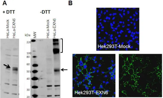
28 microscopy. The EX66 coding sequence was cloned in vector pcDNA3.1D and the resulting plasmid was used to transiently transfect HeLa and/or Hek293T human cell lines. After 24 hours, total cell extracts were prepared with and without DTT, separated by SDS-PAGE (25 micrograms/lane, corresponding to approximately 1x10⁵ cells) and subjected to immunoblot with the anti-EXN6 pAb. As controls, total extracts of EXN6-transfected cells were run in parallel. Under reducing condition the anti-EXN6 pAb was able to detect a EXN6 band (approximately 50 kDa) over-expressed by HeLa cells upon transfection. When WB was conducted under not reducing condition, other more intense high molecular weight bands ranging from 90 to 200 kDa were detected, likely corresponding to EXN6 dimers and multimers (Figure 6A).
For confocal microscopy analysis, EXN6 transfected cells were fixed with para-formaldehyde and incubated with the anti-EXN6 pAb, before or after treatment with BRJ96, a detergent used to permeabilize the plasma membrane. The anti-EXN6 pAb was able to bind EXN6 transfected cells without cell permeabilization (Figure 6B) indicating that, in agreement with its prediction, EXN6 is surface exposed.
Figure 6 –Specificity of the IHC reactive anti-EXN6 polyclonal antibody. Cells were transfected with a EXN6-coding plasmid and the empty vector (mock) and subjected to WB (A) and confocal microscopy (B). For WB, total extracts from EXN6 and Mock transfected HeLa cells were prepared with or without DTT and used for immunoblot with the anti-EXN6 pAb. The EXN6 monomer and multimer bands are marked by arrow and bracket, respectively. For confocal microscopy, trasnfected HEK293T cells were fixed, incubated with the anti-EXN6 pAb, followed by incubation with an Alexa 488-conjugated secondary antibody. DAPI was used to visualize nuclei.
29 3.1.3 EXN6 is endogenously expressed and surface exposed in breast and ovary cancer cell lines
Confocal microscopy analysis of EXN6 transfected cells confirmed that EXN6 is surface exposed. We then investigated EXN6 endogenous expression and localization in cell lines derived from breast, ovary and colon cancer cell lines by flow cytometry, using a surface staining setting. Among 15 cell lines globally analyzed, the breast cell lines SKBr3, MCF7 and T47D and the ovary cell line Ovcar 3 showed FACS positivity (Figure 7A). WB analysis under not reducing condition confirmed the presence of EXN6 in these cells (Figure 7B). Moreover, the specificity of the EXN6 detected band was confirmed by gene silencing. SKBr3 cells transfected with EXN6 specific siRNAs o with an irrelevant siRNA (scrambled siRNA) and at 72 hours later EXN6 loss of expression was assesses by WB. As shown in Figure 7B, treatment with EXN6 siRNA resulted in the reduction of a band of approximately 90 kDa band, likely corresponding to EXN6 dimer. EXN6 transcription was significantly reduced at 48h after siRNA treatment, as judged by q-RT-PCR (not shown).
Overall, the results confirmed that EXN6 is endogenously expressed in a subset of ovary and breast cancer cells, in which it is surface exposed. This evidence suggests that EXN6 is a potential drug target in breast and ovary cancers.
30 Figure 7. EXN6 expression and localization in cancer cell lines. A) FACS analysis. The table reports the panel of cell lines tested by surface staining using an anti-EXN6 pAb. The four FACS positive cell lines are represented in the graphs (Bleu, EXN6-pAb; red,unrelated negative antibody control). B) WB analysis of SKBR3. Cells were treated with EXN6-specific or scrambled siRNAs and 72 hours later total extracts were prepared with/without DTT for immunoblot, probed with the anti-EXN6 pAb. A major band of approximately 90 kDa was detected in cell total extracts, only under not reducing conditions. This band showed a weaker intensity in EXN6 silenced cells.
3.1.4. EXN6 is involved in cell proliferation and invasiveness
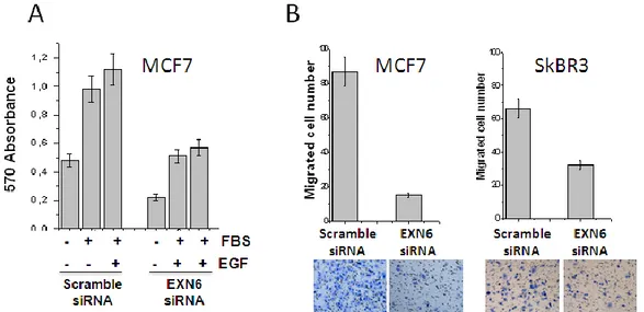
Ideal therapeutic targets are key player in signaling pathways frequently activated or altered in cancer. This type of proteins enables the generation of drugs able to inhibit their expression and function, ultimately leading to the arrest of proliferation, the induction of apoptosis, or alteration of cell motility properties. To obtain a preliminary indication of EXN6 relevance in tumor development we investigated the phenotypic effect of EXN6 knock-down in the viability,
31 proliferation and migration of breast cell lines. In these experiments, EXN6 silencing was verified by q-RT-PCR (not shown). MCF7 cells transfected with EXN6 siRNA or scramble siRNA were incubate for 72 either without FBS, for viability analysis, or in the presence of 2,5% FBS, with and without 10 ng/ml of EGF, to estimate proliferation. Cell viability and proliferation was assessed by the MTT assay, a colorimetric assay that allows to monitor the enzymatic conversion of a tetrazolium salt into a purple-colored formazan product in viable cells, by spectrophotometric measurement of the adsorbance at 570 nm. As shown in Figure 8A, EXN6 silencing caused a 2-fold decrease in cell viability and cell proliferation.
Cell migration / invasiveness was assessed in the Boyden-Matrigel-Assay. This assay employs a trans-well chamber in which the upper and lower compartments are separated by a microporous membrane. Cells are seeded on the upper compartment and are stimulated to migrate the lower compartment under a chemo-attractant stimulus, crossing the porous membrane. When used to measure invasiveness, cells must degrade a 3D extracellular matrix (Matrigel) before crossing the membrane. After 16h, the number of cells that have migrated to the lower side of the membrane is counted. In this assay, MCF7 and SKBR3 cells treated with EXN6-specific siRNAs for 72 hour showed a significant decrease of the invasive phenotype, as compared to control cells (Figure 8B). The effect was more pronounced in MCF7 (5 fold) than in SKBR3 cells (2 fold).
Figure 8– EXN6 is involved in cell proliferation and invasiveness. MCF7 and/or SK-BR-3 cells were transfected with EXN6-specific or scrambled siRNA. After 72 hours cells cell viability/proliferation and invasiveness was assessed. A) MTT assay. MCF7 cells were exposed to 2.5% FBS, 2.5% FBS+10 ng/ml EGF or serum-starved and cell viability/proliferation was measured by the MTT and optical absorbance reading. B) Boyden assay. MCF7 and SK-BR-3 were seeded on the upper chamber of a Matrigel-coated 96-well plates and the number of migrated cells was evaluated after Diff-Quick staining by counting cells in six randomly chosen fields. Images below the graphs show the visual counting.
32 3.1.5. Anti-EXN6 mAbs specifically recognize the protein on the surface of breast and ovary cancer cells.
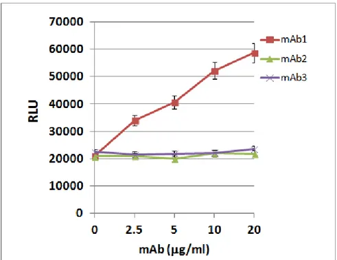
To achieve the proof of concept that EXN6 could be a promising target for mAb therapy, mAbs were generated towards recombinant EXN6 using the standard hybridoma technology based on the fusion of splenocytes from immunized BALB/c mice with Ag8 myeloma cells (mAb production carried out at The Fourth Military University, X’ian, China). After screening by ELISA on the recombinant proteins, approximately 20 EXN6-specific mAbs were obtained. Afterwards, these mAbs were further screened by WB and FACS on EXN6 transfected HeLa/Hek293T cells. In total, five anti-EXN6 mAbs (labelled from 1 to 5) specifically recognized EXN6 transfected cells both by WB and FACS (Figure 9A). The five EXN6 mAbs were also tested for their ability to detect EXN6 on the surface of breast and ovary cancer cell lines (see Figure 7). As shown in Figure 9B, the five mAbs were able to bind the surface of MCF7, T47D, SKBr3 and Ovcar3 cell line (Figure 9B). The mAb specificity was confirmed in the EXN6-positive cells by treating them with different concentration of EXN6 siRNA (ranging from 10 to 50nM) and monitoring the disappearance of surface binding. EXN6-siRNA treated cells showed a significant loss of surface staining, as opposed to cells treated with a scrambled siRNA (see exemplary data for anti-EXN6 mAb1 and mAb2 in SKBR3, Figure 9C). WB analysis on EXN6 silenced cells further confirmed the mAb specificity (data not shown).
33 Figure 9 – The anti- EXN6 mAbs bind the surface of breast and ovary cell lines. A) FACS analysis of EXN6 transfected HEK293T cells with the five anti-EXN6 mAbs (red peak, EXN6 mAbs, Blue peak, irrelevant isotype control); B) FACS analysis on the EXN6-positive breast and ovary cell lines (blue peak, EXN6 mAbs; red peak irrelevant isotype control). C) FACS analysis in EXN6 knocked down SKBR3 cells. Cells were transiently transfected with different
34 concentrations of EXN6-specific sirRNA and 72 hours later the residual mAb surface binding was measured by flow cytometry as compared to cells treated with an irrelevant siRNA.
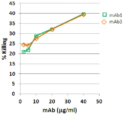
3.1.6 Two anti-EXN6 mAbs are efficiently internalized by breast and ovary cancer cells Antibody-drug conjugates (ADC) are powerful tools to increase the anti-tumor activity of naked antibodies. As already said, the most important property of antibodies suitable for ADC development is the ability to be internalized by cancer cells shortly after their surface binding on cancer cells, so as to efficiently drive their drug-conjugates into the cells. To assess whether the anti-EXN6 mAbs have this important property, internalization experiments have been carried out by FACS and confocal microscopy, in breast and ovary EXN6-positive cell lines. Cells were incubated for 30’on ice with the five anti-EXN6 mAbs (10g/ml) to allow surface binding, subsequently washed to remove the excess of unbound mAbs and then shifted to 37°C for up to 2 hours to permit the internalization process to occur. As negative control, cells were also incubated with a mAb toward CD81, a multipass plasma membrane protein that is not internalized. As controls, samples were kept on ice and analyzed in parallel. For FACS analysis, cells were then incubated with a fluorescently-labeled secondary antibody, and the disappearance of the anti-EXN6 mAbs from the cell surface was monitored as function of time. Two of the 5 anti-anti-EXN6 mAbs (mAb1 and 3) quickly disappeared from the cell surface upon temperature shift (after 30’ approximately 50% of surface bound antibodies disappeared from the cell surface) (Figure 10A). Conversely, the other 3 mAbs remained associated on the cell surface under the observation period (mAb2 example, in Figure 10A). To confirm that the loss of mAb surface binding visible upon temperature shift was effectively due the mAb internalization, confocal microscopy was also carried out to monitor the formation of intracellular immuno-complexes. In this experiment, after 1h incubation at 37°C, cells were methanol-permeabilized and incubated with a fluorescently labeled secondary antibody. As shown in Figure 10B, upon temperature shift the surface bound anti-EXN6 mAb 1 and mAb3 were not detectable on the plasma membrane and accumulated in the intracellular milieu, visible as fluorescent spots, confirming that they were efficiently internalized (mAb3 example, Figure 10B).