DEPARTMENT OF BIOLOGICAL AND ECOLOGICAL SCIENCES (DEB)
XVI PhD COURSE IN GENETICS AND CELLULAR BIOLOGY
BIO/11
Proteomics and metabolomics in transfusion medicine: new technologies to
improve red blood cell storage
PhD student Tutor
Valeria Pallotta Dott.ssa Sara Rinalducci
Coordinator Prof. Giorgio Prantera
1
Index
Aim of the thesis
………..………... 3Chapter 1. Introduction
1.1 Structure, function and metabolism of erythrocytes………. 1.2 Antioxidant defense system of Red Blood Cells (RBCs)………. 1.3 Red cell storage under standard blood bank conditions: historic evolution………..
1.3.1 Blood collection and processing………. 1.3.2 Additive solutions……… 1.4 RBCs and storage lesions: “how long is too long?”………. 1.5 Alternative methods in red cell storage……… 1.5.1 Cryopreservation of erythrocyte concentrates………... 1.5.2 Formulation of rejuvenation solutions………. 1.6 Proteomics and its application in transfusion medicine………... References………...….. 4 7 10 12 13 14 17 18 20 24 26
Chapter 2. A better RBC storage rather than a longer one
2.1 Monitoring of red blood cells during processing for cryopreservation: from fresh blood to to thaw-washing………. 2.1.1 Materials and methods ……….. 2.1.2 Results and discussions………. 2.1.3 Conclusion………. 2.2 Red blood cell storage with vitamin C and N-acetylcysteine prevents oxidative
stress-related lesions: a metabolomics overview………. 2.2.1 Materials and methods……….. 2.2.2 Results and discussion……….
34 34 37 44 46 46 50
2
2.2.3 Conclusion………. References………..
64 65
Chapter 3. A new method to evaluate Red Blood Cell storage quality
3.1 Native protein complexes in the cytoplasm of red blood cells……… 3.1.1 Materials and methods……….. 3.1.2 Results……… 3.1.3 Discussion……….. 3.1.4 Conclusion………. References……… 71 71 75 99 106 107
3
Aim of the thesis
Despite decades of significant technological improvements, red blood cells can be stored under standard blood bank conditions for a limited span of life because RBC undergo a series of biochemical and physical changes, the so-called “storage lesions” that are related to the promotion of apoptosis-like phenomena. These anomalies compromise red cell survival upon transfusion and in turn transfusion efficacy in the recipient.
In the first section, this PhD thesis would evaluate new technologies to improve red cell storage quality, in term of safety and efficacy. To this end it was performed a biochemical and mass spectrometry-based analysis of:
(i) high-glycerol frozen RBCs (i.e., stored with a final concentration of 40% glycerol at -80°C for about 10 years);
(ii) RBCs stored under standard blood banking with the supplementation of antioxidants (N-acetyl-L-cysteine and ascorbic acid).
In the second section, the thesis would describe an innovative method to evaluate red cell storage quality through an extensive investigation of RBC cytosolic multi-protein complexes. In the light of this, it was performed a proteomic analysis in order to identify multimeric protein complexes that play a critical role in the maintenance of red blood cell functionality and survival in vivo. Future investigations should expand the existing knowledge and determine whether and how these complexes might influence red cell ageing in vivo and in vitro, other than the insurgence of specific pathologies.
4
Chapter 1
Introduction
1.1 Structure, function and metabolism of erythrocytes
Blood is a connective tissue that is mainly involved in the transport of many substances such as nutrients, oxygen and metabolic waste products around the body. It also protects against deseases and helps to regulate pH and water balance.
In vertebrates, blood consists of two main components: plasma, which is a clear extracellular fluid, and cells, which are also called “corpuscles” or “formed elements”. These last comprise erythrocytes or red blood cells (RBCs), leukocytes or white blood cells and thrombocytes or platelets.
Erythrocytes are the most abundant blood cells. By volume they constitute about 45% of whole blood in men and about 40% in women. This percentage is called Hematocrit (Hct).
Human erythrocytes are anucleated, disk-shaped and biconcave cells (7,5µm diameter x 2µm thickness) (Figure 1). This shape allows optimal flexibility while traveling through microcapillaries and permits maximal surface area for oxygen and carbon dioxide exchange [Barasa and Slijper, 2013]. Their red color is due to the spectral properties of the hemic iron ions in hemoglobin molecules (Hb). This metalloprotein, firstly described from Perutz in 1960 [Perutz, 1960], is the most abundant cytoplasmic protein in human erythrocytes (about 95% of total proteins). Each cell contains approximately 270 million of these biomolecules, each of which carry four heme goups. Thanks to heme groups of Hb erythrocytes can temporarily bind oxygen molecules (O2) in the
lungs and release them throughout the body. These cells also carry carbon dioxide (CO2) back from the tissues.
RBCs originate from hematopoietic stem cells (HSCs) residing in the bone marrow, which are capable of self-renewal via asymmetric cell division resulting in one identical HSC and one differentiating cell for each HSC. This differentiating cell matures during erythropoiesis through a series of proliferation and differentiation steps into a reticulocyte and eventually into an RBC [Barasa and Slijper, 2013]. By maturation, cells become
5
gradually specialized for their function. In the early stage organelles are present in erythrocytes, so cells can synthesize proteins such as haemoglobin. As the proteins accumulate, the number of organelles slowly diminishes. Complete differentiation of maturing erythrocytes needs of reorganization of membrane skeletal protein network and the loss of nuclei.
In healthy individuals, cells live in blood circulation for about 120 days. At the end of their lifespan they become senescent and are removed from circulation. The process of distruption of erythrocytes is called hemocatheresis and occurs mainly in the spleen, liver and bone marrow.
A balance between production and destruction keeps the erythrocyte number constant.
Since the cells lose intracellular organelles, they are uncapable of protein and lipid synthesis and oxidative phosphorylation [Bossi and Giardina, 1996]. This means that they can not replace damaged lipids or proteins and also produce ATP in oxidative phosphorylation. However, energy is required for the maintenance of osmotic balance through ATP-dependent pumps, of enzymes and membrane lipid protection from oxidative stress, and maintenance of Hb iron in reduced state (hemoglobin oxidation). So energy is needed for RBC integrity and deformability.
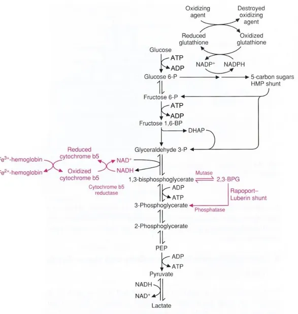
Erythrocytes must depend on two key metabolic pathways for production of high-energy compounds, as summarized in Figure 2. These are glycolysis or Embden-Meyerhoff pathway and hexose monophosphate shunt or pentose phosphate pathway (PPP).
Glycolysis generates 90-95% of energy needed by RBCs. In this pathway, glucose, i.e. the main red cell energy source, is metabolized with the production of 2 mol of ATP (adenosine triphosphate), NADH (nicotinamide adenine dinucleotide reduced form) and lactate as end products per mol of glucose [Bossi and Giardina, 1996]. ATP is the major high-energy phosphate nucleotide that powers the cation pump and NADH is a cofactor in the methemoglobin reductase reaction, which maintains heme iron in the reduced state to avoid the formation of methemoglobin.
Another end product of glycolysis is 2,3-disphosphoglycerate (2,3-DPG), necessary for the regulation of hemoglobin affinity to oxygen [Barasa and Slijper, 2013].
The final yield of ATP and 2,3-DPG varies depending on the activity of the Rapoport-Luebering shunt that allows the RBCs to regulate oxygen transport during conditions of hypoxia or acid-base imbalance.
Although to a lesser extent the PPP contributes to the energy status of the cell with the production of 2 mol of NADPH per mol of glucose entering the cycle [Bossi and Giardina, 1996]. In this pathway, glucose-6-phosphate undergoes oxidation followed by a series of reactions to yield fructose-6-glucose-6-phosphate and glyceraldehyde-3-phosphate, which are intermediates in the glycolytic pathway. The most important product is reduced nicotinamide-adenine dinucleotide phosphate (NADPH) which is required in the glutathione metabolism pathway to reduce oxidized glutathione (GSSG) in GSH by glutathione reductase. Reduced glutathione (GSH) is a tripeptide (glutamyl-cysteinyl-glycine) that reduces protein
6
sulfhydryl group. It is also involved in detoxifying the cellular peroxides [Barasa and Slijper, 2013] (see next paragraph).
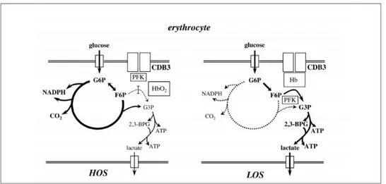
The modulation of erythrocyte metabolism is driven by the oxygenation state of the cell (Figure 3).
During the high oxygenation state (HOS), the risk of oxidative stress should address erythrocyte metabolism toward the activation of those metabolic pathways that may lead to an increase of cell protection, namely the pentose phosphate shunt [Bossi and Giardina, 1996]. During the low oxygenation state (LOS), glucose is addressed towards glycolysis in order to increase ATP and 2,3-DPG production.
Figure 2. Overview of vital pathways in the red blood cells. In the Embden–Meyerhoff pathway glucose is anaerobically
metabolized to pyruvate. Part of this pathway is the Rapoport–Luebering shunt, which is unique for the RBCs. For energy ATP is needed, which is generated through the Embden–Meyerhoff pathway. This pathway also provides reducing power (NADH) to keep hemoglobin in reduced state. NAD+ is produced by the pyruvate metabolism pathway. Other reducing power by NADPH originates from the pentose phosphate pathway, and is essential for keeping high glutathione (GSH) levels [Barasa and Slijper, 2013].
7
Castagnola et al. (2010) have reported the hypothesis that the glycolytic flux is modulated via a competition between glycolytic enzymes and deoxy-Hb for band 3, a transmembrane protein that accounts for about 25% of the total RBC membrane proteins. Band 3 is characterized by three distinct functional domains: the membrane spanning domain, which catalyzes the exchange of anions (mainly Cl- and HCO3-) across the
membrane; the short C-terminal cytoplasmatic domain that binds carbonic anhydrase II; and N-terminal cytoplasmatic domain (CDB3) that binds a variety of proteins by anchoring the RBC membrane to the underlying cytoskeleton via ankyrin and protein 4.2.
In HOS erythrocytes, the inibition of glycolytic enzymes, due to their binding to CDB3, should result in a reduced glucose flux through glycolysis. As a consequence, since glucose consumption is constant, more glucose is metabolized by the PPP (Figure 3, left). Conversely, in the LOS state the displacement of glycolitic enzymes from CDB3, induced by the binding of deoxy-Hb, results in an increased glucose flux throughout glycolysis (Figure 3, right) [Castagnola et al., 2010].
Figure 3. Simplified scheme representing the modulation of erythrocyte metabolism by the O2 transition of Hb and its competition
with glycolytic enzymes (mainly phosphofructokinase, PFK) for the cytoplasmic domain of band 3 (CDB3) [Castagnola et al., 2010].
1.2 Antioxidant defense system of Red Blood Cells (RBCs)
Reactive oxygen species (ROS) are formed and degraded by all aerobic organisms, leading to either physiological concentrations required for normal cell function, or excessive quantities, the state called oxidative stress. As the term ROS implies, intracellular production of those oxygen
8
intermediates threatens the integrity of various biomolecules including proteins, lipids and DNA [Nordberg and Arnèr, 2001].
To counteract oxidative stress, erythrocytes have a self-sustaining activity of antioxidant defense enzymes including superoxide dismutase (SOD), catalase (CAT), glutathione peroxidase (GPx), glutathione reductase (GR) and peroxiredoxins (Prxs), in addition to low-molecular-weight antioxidants, such as glutathione (GSH) and vitamin E and C [Silva et al., 2013].
In eukaryotic cells, superoxide (O2
•-) can be metabolized to hydrogen peroxide (H2O2) by the
cytosolic 32-kDa dimeric Cu/Zn-SOD [Nordberg and Arnèr, 2001]. In this reaction two molecules of superoxide form hydrogen peroxide and molecular oxygen:
2 O2•- + 2 H+ H2O2 + O2
SOD is considered to be one of the major enzymes that protect cells from ROS.
Catalase, however, is a tetrameric heme-containing enzyme that catalyzes the dismutation of hydrogen peroxide to water and molecular oxygen:
2 H2O2 O2 + 2 H2O
H2O2 is not a radical but it is still a harmful byproduct of many normal metabolic processes.
Indeed it can be rapidly converted into hydroxyl radical (•OH) by Fenton reaction, in presence of Cu+ and Fe2+:
H2O2 + Cu+/Fe2+ •OH + OH- + Cu2+/Fe3+
Thus CAT rapidly catalyzes the decomposition of a potential toxic compound into less-reactive molecules.
Glutathione peroxidase is a family of multiple isozymes involved in scavenging oxy- radicals [Lin et al., 2002]. The main reaction they catalyze is the reduction of H2O2 using glutathione
(GSH) as substrate:
2 GSH + H2O2 GSSG + 2 H2O
9
GSSG + NADPH + H+ 2 GSH + NADP+
Therefore GR is a key enzyme in the glutathione pathway for reduced glutathione (GSH) regeneration. This system is critical for the protection of hemoglobin and other proteins against peroxide damage in mammals [Hou et al., 2004].
GPx can also reduce other peroxides (for example lipid peroxides in cell membranes) to alcohols:
ROOH + 2 GSH ROH + GSSG + H2O
Peroxiredoxins [Chae et al., 1994a; Chae et al., 1994b] are a ubiquitous family of antioxidant enzymes that can be divided into three classes: typical 2-Cys Prxs; atypical 2-Cys Prxs; 1-Cys Prxs. All proteins share the same basic catalytic mechanism in which an active-site cysteine (the peroxidatic cysteine) is oxidized to a sulfenic acid by the peroxide substrate. The recycling of the sulfenic acid back to a thiol is what distinguishes the three enzyme classes [Wood et al., 2003]. Prxs exert their role in the cell through their peroxidatic activity so that hydrogen peroxide, peroxynitrite and a wide range of hydroperoxides (ROOH) are reduced and detoxified [Hofmann et al., 2002; Jacobson et al., 1999; Poole and Ellis, 1996; Bryk et al., 2000; Peshenko and Shichi, 2001]:
ROOH + 2 e- ROH + H2O
The typical 2-Cys Prxs are the largest class of enzymes. They act as obligate homodimers containing two identical active site [Hofmann et al., 2002]. This class comprises PrxII, a most abundant protein in red cell.
A large number of low molecular weight compounds are considered to be antioxidants of biological importance, including ascorbic acid (vitamin C) and -tocopherol (vitamin E).
Ascorbic acid is a water-soluble vitamin. Its antioxidant power depends on the high reducing potential of its carbon-carbon double bond (Figure 4) which readily donates one or two hydrogens and electrons to a variety of oxidants, including oxygen free radicals, peroxides, and superoxide.
Ascorbate acts protecting red cell membrane. However it does not directly affect membrane lipid peroxidation but it may perform this function indirectly by reducing the tocopheroxyl free radical
10
in the lipid bilayer. Thus intracellular ascorbate can regenerate -tocopherol in the erythrocyte membrane and in turn -tocopherol protects the cell membrane from lipid peroxidation.
-tocopherol (-TOH) is a lipid-soluble vitamin present in biological membranes (Figure 4). It acts as chain-breaking antioxidant thanks to hydroxyl group by which it reacts with unpaired electrons and can reduce [Nordberg and Arnèr, 2001]. The role of vitamin E is to protect polyunsaturated fatty acids in membranes against lipid peroxidation.
Figura 4. Left: structure of vitamin C (ascorbic acid); right: structure of vitamin E (-tocopherol).
1.3 Red cell storage under standard blood bank conditions: historic evolution
The development of effective red blood cell biopreservation techniques that mantain ex vivo RBC viability and function represents the foundation of modern blood banking. The ability to preserve the integrity of RBCs outside their native environment for extended periods has not only separated blood donors and recipients in space and time, but also has made it possible for blood banks to provide safe, high-quality blood products in an efficient and effective manner [Scott et al., 2005].
The major force driving the field of red cell biopreservation is the enormous clinical need for RBC product. RBC transfusions are necessary for patients with low oxygen-carrying capacity due to traumatic/surgical hemorrhage, decreased bone marrow production (aplastic anemias), defective hemoglobin (hemaglobinopathies and thalassemia), and decrease RBC survival (hemolytic anemias).
Worldwide more than 80 million units of red cells are transfused each year, and in highly developed countries, about 1% of the population receives a transfusion annually. Red cells are the most commonly transfused blood component.
11
Blood storage begane in 1913 when Lee and Vincent demonstrate that citrate could prevent the coagulation of human blood [Stansbury et al., 2005]. Later Agote (1914) showed that citrate anticoagulation could facilitate human transfusion and in 1915 Lewisohn determined the critical amount of citrate required to anticoagulate a donor’s blood without causing hypocalcemia in the recipient [Lewison, 1915]. In 1916 Rous and Turner firstly described the 4-week storage of rabbit red cells in citrate and glucose.
The introduction of citrate anticoagulant and glucose, used to support cellular metabolism, allowed the donor and recipient to be separated in time and in space respectively. This physical separation of donor and recipient is the basis of blood banking.
In 1917 Robertson worked out safe ways to store and transport blood, and transfused human blood stored for 26 days in the Rous-Turner citrate-glucose solutions [Rous and Turner, 1916; Robertson, 1918].
In subsequent decades, plastic blood bags were introduced and later, when the diethylhexyl phthalate (DHEP) was coupled with PVC classic bags, it has been shown a fourfold reduction of haemolysis and the time of storage increased twice. Nowadays it is known that DHEP enters the RBC membrane where it limits membrane loss by microvesiculation.
Around 1950 Gibson added phosphate to acid citrate glucose solution in order to offset the loss of phosphate from red cells [Walter and Murphy, 1952; Gibson et al., 1957]. The subsequent addition of adenine could help storage by cell shape, ATP and viability restoration, extending the storage time to 5 weeks (citrate, phosphate, dextrose, adenine: CPDA-1).
The first RBC additive solution, saline-adenine-glucose (SAG) was developed by European researchers in the late 1970's [Högman CF et al., 1978]. Soon after, in 1981, the same researchers added mannitol to help protect the RBC membrane and reduce hemolysis [Högman CF et al., 1981]. The addition of mannitol extended the storage time to 42 days, a period that is currently used today. The modified SAG formulation was named SAGM and to this day SAGM is the most widely used RBC additive solution (see next paragraph).
The exact formulation of SAG-M varies, so the term is usually restricted to the standard European formulation while U.S. varieties are designated by additive solution (AS) and a number as in AS-1 and AS-5 [Hess, 2010].
The subsequent development of filtration-leukoreduction has improved recovery and reduced hemolysis further, beacause it has been demonstrated that white blood cells break down in the cold and release proteases and lipases that cause damage to the RBCs during storage. So nowadays the practice of leukoreduction, by buffy coat removal or leukofiltration, became a fundamental strategy to increase RBC recovery and reduce haemolysis.
12 1.3.1 Blood collection and processing
Red cell concentrates are obtained starting from whole blood through a centrifugation that allows to separate erythrocytes from leuko-platelet layer (buffy coat) and plasma (Figure 5, left).
So a cellular separator instrument equipped with an optical sensor is used to physically separate blood components into different satellite bags. This multi bag system is composed by a “mother bag” containing CPD solution for the collection of whole blood (about 450 cc) which is connected with a first bag with SAG-M intended for red cell and a second one for the plasma. This procedure is called “top and bottom” collection.
After this step RBCs are ready for leukofiltration using filters that can remove 99,99% of White Blood Cells (Figure 5, right). So red cell concentrates are immediately ready for the use or to be stored for up to 42 days at 4°C.
Figure 5. Left) Blood components after centrifugation. Right) Leukofiltration of RBCs.
1.3.2 Additive solutions
Nowadays SAGM (saline-adenine-glucose-mannitol) is the most widely used RBC additive solution. This is a modified SAG formulation (saline-adenine-glucose) developed by European researchers around 1981. Several countries apply a shorter 5 weeks shelf-life to their SAGM-RBC components despite the fact that SAGM is approved for 6-weeks storage. In Table 1 a list of RBC additive solutions currently in routine use around the world is reported. SAGM has not been licensed by the Food and Drug Administration (FDA), so it is
13
not used in the USA, but only in Europe, UK, Australia, Canada and New Zealand. Other additive solutions, which are all essentially variations of SAG/SAGM, have been developed and commericalised in other countries, including AS-1, AS-3, AS-5, MAP and PAGGSM [Heaton et al., 1984; Simon et al., 1987; Cicha et al., 2000; Walker et al., 1990]. Specifically AS-1 and AS-5 are used in USA, AS-3 both in USA and Canada, MAP and PAGGSM only in Japan and in Germany respectively.
No new RBC additive solutions have been licensed for use for over 20 years.
Licensed RBC additive solutions
NaCl 150 154 70 150 85 72 NaHCO3 - - - - - - Na2HPO4 - - - - - 16 NaH2PO4 - - 23 - 6 8 Citric acid - - 2 - 1 - Na-citrate - - 23 - 5 - Adenine 1.25 2 2 2.2 1.5 1.4 Guanosine - - - - - 1.4 Dextrose (glucose) 45 111 55 45 40 47 Mannitol 30 41 - 45.5 80 55 pH 5.7 5.5 5.8 5.5 5.7 5.7 Anti-coagulant CPD CPD CP2D CPD ACD CPD FDA Licensed No Yes Yes Yes No No Countries used Europe USA USA USA Japan Germany
UK Canada Australia
Canada New Zealand
Table 1 RBC additive solutions currently used around the world [Sparrow, 2012]. Constituents (mM) SAGM AS-1
Adsol Baxter AS-3 Nutricel Pall Medical AS-5 Optisol Terumo MAP PAGGSM MacoPharma
14
All of the currently licensed RBC additive solution have an acidic pH (~5.6-5.8), which is well below the normal physiological pH of 7.3 of venous blood. Acidic additive solutions (and anticoagulants) are used simply because at physiological and alkaline pH, glucose caramelises during heat-sterilization. Erythrocytes have sufficient buffering capacity to adjust the pH closer to physiological levels during the first week of storage. However the buffering capacity of RBCs is soon exhausted due to the generation of lactate (glycolytic pathway) [Sparrow, 2012].
Consequently the extracellular and intracellular pH of RBCs progressively becomes more acidic during storage, reaching a pH of ~6.5 at the end of storage [van der Meer et al., 2011]. An acidic intracellular environment alters the activity of certain enzymes and biochemical pathways. During storage the intracellular concentration of adenosine-5’-triphosphate (ATP) and 2,3-diphosphoglycerate (2,3-DPG) decline [van der Meer et al., 2011]. Over the past 15-20 years, research into the development of new additive solutions has focussed on ways to maintain higher the intracellular levels of ATP and 2,3-DPG during storage of red cells [Hess, 2006]. A significant driver for this research came from the military, who wanted to store RBCs for longer than 6 weeks. However longer storage time is not a key interest of civilian blood service and the medical community, whose desire is to support new technologies, including new RBC additive solutions that offer further improvement to the quality and efficacy of RBC components.
1.4 RBCs and storage lesions: “how long is too long?”
For several decades RBC components have been prepared as concentrates suspended in nutrient additive solution, which preserves and extends the shelf-life of the RBC component, allowing up to 6-7 weeks of refrigerated storage [Hess JR, 2006]. Nevertheless, during storage RBCs undergo a complex and progressive accumulation of physicochemical changes, collectively referred to as the RBC storage lesion [Högman CF and Meryman HT, 1999; Hess, 2010]. These time-dependent changes include metabolic, enzymatic, oxidative and physiologic lesions that reflect the deterioration of RBCs during conventional blood bank storage (Table 2).
Metabolic changes have been the best studied [Hess, 2010]. As previously reported, metabolism of RBCs is centered on glycolysis, the only source of energy for red cells. Protons produced by this process increase the acidity of cells and storage solutions as well. Acidosis inhibits in turn
15
the glycolysis pathway, in fact low pH have adverse effect on activity of hexose kinase (HK) and phosphofructokinase (PFK), so that less ATP and NADH is produced as storage progresses. The inhibition of HK leads to a poor production of NADPH and glutathione, since it is reduced the amount of glucose 6-phosphate going through the exose-monophosphate shunt.
Moreover, at any storage pH less than 7.2, the breakdown of 2,3-DPG is favored, and this in turn leads to an initial burst of ATP production.
As a result of these activities, red cell ATP concentrations initially rise as 2,3-DPG is broken down, stay above their initial concentrations for 2–3 weeks, and then decline steadily. The pH typically declines from 7.0 to 6.5, and the rate of glucose consumption decreases by more than 50%. As it was mentioned before, the slowing of glycolysis also leads to reduced production of NADH leading to reduced activity of methaemoglobin reductase, and thus the methaemoglobin concentration of stored red cells increases over 6 weeks of storage, typically from about 1–2%. Low temperature profoundly reduces the activity of the major membrane sodium-for- potassium pump, which is ATP-dependent. As a result, the potassium that slowly leaks out of RBCs is not returned, and the concentration of extracellular potassium in the suspending fluid in the red cell bag typically rises at a rate of about 1 mEq/L/day [Hess, 2010].
Metabolic 1. Acidosis
2. Lower ATP, DPG
3. Lower glutathione, NADH, NADPH Enzymatic (Reduced with leukoreduction)
1. Loss of surface glycands 2. Lysolipids
3. Protein damage Oxidative
1. Damage to proteins e.g. Band III 2. Decoration of proteins
a. Benign – glycation of Hb b. Inflammatory – e-lysines (AGEs) 3. Oxidized lipids – micro-vesicles 4. Lysolipids – TRALI
Physiologic
1. Shape change 2. Membrane loss 3. Apoptosis
16
Enzymatic lesions were related to the presence of white blood cell (WBC) during blood storage. As previously reported, leukoreduction step has also been shown to improve red cell storage. Removal of metabolically active WBC from RBC concentrates minimizes glucose consumption, waste product accumulation and damage from leukocyte enzymes, resulting in a significant decrease in hemolysis during hypotermic storage [Högman, 1998].
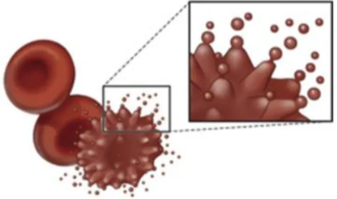
Physical changes include membrane loss and the associated changes in RBC shape and rheology. During storage red cells evolve from smooth biconcave discs to subtly bumpy discs (echinocytes) to grossly bumpy spheres called spheroechinocytes (Figure 6) [Hess, 2010]. It has been demonstrated that this process is associated with decreased pH and ATP concentration and increased intracellular concentration of calcium. The return toward the normal red cell shape occurs in parallel with increasing ATP concentration, regeneration of 2,3-DPG and the restoration of normal sodium, potassium and calcium gradients. However, beyond the early spheroechinocyte stage, red cells lose their membrane through a blebbing mechanism involving cytoskeleton remodeling and membrane asymmetry disruption, with the exposure of phosphatidylserine at the outlet of the plasma membrane. Thus, small corpuscles of less than 1
m, named microvesicles, bud from the tips of erythrocyte spines (Figure 7). This process is irreversible, since the cells have no mechanisms to replace lost membrane. At the end of the storage, the densest cells have lost all extra membrane and have become rigid spheres [Hess, 2010].
Last but not least, oxidative damage to lipids and proteins also contributes to red cell injury during storage [Sharifi et al., 2000]. RBCs are continuously exposed to oxygen and are also rich in polyunsaturated lipids, target of free radicals, and iron (hemoglobin molecules), a powerful catalyzer of free radical through Fenton reaction. Red cells are normally supplied with superoxide dismutase and methemoglobin reductase, so that the dangerous products are quickly removed.
17
Figure 6.Evolution of erythrocyte morphology during blood bank storage.
Figure 7. Blebbing mechanism of red blood cells
.
However, as red cell storage progresses and the flux through the glycolytic pathway is slowed, NADH concentration falls and methemoglobin concentration rises [Dumaswala et al., 2000]. This molecule is less stable than ferrous hemoglobin, increasing the chance that the heme will come free from the globin heme pocket and the iron will then also come out of the heme. Under these circumstances, the chanches that superoxide radicals and water will meet in the presence of a free iron or heme to undergo Fenton transformation into hydroxyl radical increase and hydroxyl radical readily damages lipids and proteins [Hess, 2010].
1.5 Alternative methods in red cell storage
While increased research effort is being directed to better understand the effects of storage on RBCs and the potential impact on transfusion outcomes [Glynn, 2010], slower progress is being made in finding ways to deter the detrimental effects of the RBC storage lesion.
Current research suggest that the RBC hypothermic storage lesion still significantly influences the efficacy of transfusion as it is responsible for the association of blood transfusion with an increased lenght stay in the hospital [Martin et al., 1994], impaired tissue oxygen use [Marik and Sibbald, 1993], proinflammatory and immunomodulatory effects [Blajchman, 2002], increased infections [Leal-Noval et al., 2003], multiple organ system failure [Zallen et al., 1999], and ultimately, increased morbidity and mortality [Vamvakas and Taswell, 1994; Whyte, 1998; Purdy et al., 1997; Ho et al., 2003].
18
The currently accepted biological marker of RBC viability, which is minimal 24-hour post-transfusional survival of 75% od RBCs, does not reflect the clinical effects of transfusion [Ho et al., 2003]. In addition, in vivo assessment of RBC viability by measurement of RBC recovery using radioactive labeling with chromium 51 has many disadvantages, including practical limits and source of error [Hess and Greenwalt, 2002].
Improvement of current red cell hypothermic storage would have an enormous effect on RBC availability, safety and quality.
For the last two decades, the focus of red blood cell biopreservation research has been on lengthening the RBC hypothermic storage period over 42 days by modification of additive solution, blood collection protocol and devices. As a result, solutions allowing 7-week hypothermic storage, such as ErythroSol, MAP and PAGGS-S/M, have been developed for clinical use [Högman, 1999; Walker et al., 1990]. In addition, several rejuvenation solutions have been proposed, of which Rejuvesol is currently the only solution approved by the Food and Drug Administration (FDA).
According to Valeri [2002], for optimum survival and function, liquid preserved RBCs should be hypothermically stored in the currenlty licensed additive solution for no more than 2 weeks. The continuously increasing need for a safe, high-.quality RBC product will ensure future advances in red blood cell biopreservation.
1.5.1 Cryopreservation of erythrocyte concentrates
Cryopreservation is the process of preserving the biological structure and/or function of living systems by freezing to and storage at ultralow temperature. This method uses the beneficial effect of decreased temperature to suppress molecular motion and arrest metabolic and biochemical reactions.
Cryopreservation is the only current technology that maintains ex-vivo biologic functions and provides long-term RBC storage.
The idea of storing RBC at ultralow subzero temperatures has been around for at least 150 years [Rowe, 1994]. Sinche 1950s, cryopreservation has been used in transfusion medicine for long-term storage of RBCs from donors with rare or unusual phenotypes [Rowe, 1994; Valeri and Ragno, 2006] and for military deployment [Kakaiya et al., 2008; Lelkens et al., 2006]. Moreover stockpiling of frozen red cells can be beneficial in emergency or clinical situations, where demand exceeds the supply of RBCs.
19
At the moment, the shelf life of high-glycerol frozen RBCs (i.e., stored with a final concentration of 40% glycerol at -80°C) has been approved for up to 10 years [Kakaiya et al., 2008].
Glycerol is used as cryoprotective agent, in order to protect red cells from low-temperature injury. This molecule is an attractive cryoprotectant for RBCs because it is relatively non-toxic at high concentrations and readily permeates the cell at 37°C. Over the years, two different protocols have been utilized for cryostorage of red cells in presence of glycerol. These differ in glycerol concentration (either 15-20% or 40% w/v), in the cooling rate (rapid or slow) and in the storage temperature (-196°C or -80°C). [Rowe et al., 1968; Meryman and Hornblower, 1972].
Although glycerol has slow toxicity, a post-thaw deglycerolization washing procedure is necessary to avoid hemolytic transfusion reactions and renal failure after infusion [Bechdolt et al., 1986; Cregan et al., 1991; Klein and Anstree, 2005]. The removal of glycerol is achieved by washing RBC units in a continuous flow centrifuge. This procedure results in a loss of about 15% of the cells [Valeri, 2004]. Thawed/deglycerolized units are expected to meet the minimum standards for transfusion (hemolysis below the 0.8% threshold in Europe and <1% in the USA, and in-vivo recovery at 24 h post transfusion >75%).
Henkelman and colleagues [2010] have recently reported that RBC processing steps have the largest effect on cryostored RBC quality, while storage duration itself has minimal effects on red cells. In order to standardize cell processing steps, Haemonetics Corporation (Braintree, Mass) have developed an automated closed system (ACP 215) for the glycerolization and deglycerolization of RBC units. The system uses sterile connectors, inline filters and a disposable polycarbonate bowl with an external seal that allows sequential processing of 2 RBC units (Figure 8).
A body of evidence has been accumulated which indicates that cryostored RBCs apparently do not show any classical “storage lesion”, in contrast to that observed in red cells stored at 1-6°C [Hess et al., 2010].
On the other hand, it has been reported that thawed RBCs are more fragile than fresh units, as they display higher osmotic fragility [Henkelman et al., 2010]. In addition, intra-cellular calcium content has been shown to increase in the presence of glycerol and upon freeze/thawing of RBCs, probably due to the blockade Ca2+ pumps or activation of nonspecific cation channels [Kofanova et a., 2008]. Freeze-thawing and deglycerolization of red blood cells have been thus suggested to compromise ion permeability of the plasma membrane [Kofanova et a., 2008].
20
Figure 8. ACP 215, Haemonetics Corporation (Braintree, Mass).
The current direction of RBC cryopreservation research involves the development of novel methods to eliminate common problems associated with glycerol-preserved erythrocytes. These research areas involve ice-free cryostorage or vitrification, the use of extracellular cryoprotectant agents and the use of intracellular sugar as cryoprotectant. The first method is used in order to bypass ice formation and the detrimental effects of this step. The second alternative approach is cryopreservation with cryoprotective extracellular macromolecules that are biodegradable and well tolerated by the patients (such as some sugars and polymers); because these molecules do not penetrate the RBC membrane, the osmotic fragility and other problems associated with the use of glycerol would be avoided. The last approach involves the use of low concentrations of intracellular sugars such as trehalose and sucrose. This is an emerging area of interest thanks to the properties of these molecules that can protect critical biological structures during freezing and thawing through the formation of a stable glassy matrix.
1.5.2 Formulation of rejuvenation solutions
Red blood cell storage under blood bank conditions is associated with a sequence of biochemical, metabolic and mechanical changes leading to the progressive loss of cell viability.
In particular, the storage-dependent accumulation of reactive oxygen species (ROS) is mainly related to the progressive loss of metabolic modulation [D’Alessandro et al., 2012;Messana et
21
al., 2000], resulting in the impairment of antioxidant defences [Jozwik et al., 1997]. Indeed, in humans, each day about 3% of the body’s hemoglobin undergoes spontaneous autoxidation to methemoglobin because of the high concentrations of iron in red cells. As prevoiusly reported, Fenton reaction ultimately promotes the accumulation of superoxyl and hydroxyl radicals, the latter being considered as the primary mechanism of injury in stored RBCs [Jozwik et al., 1997].
In this view, preventing the accumulation of oxidative stress has been regarded as a viable strategy to improve the quality of stored RBCs. Over the years, several approaches have been proposed to achieve this aim:
(i) Supplementation of metal chelators, such as deferoxamine, diethylenetriaminepentaacetic acid or ethylenediaminetetraacetic acid, which bind iron released from ageing erythrocytes [Knight et al., 1992];
(ii) Oxygen depletion [Yoshida and Shevkoplyas, 2010], to remove the main substrate of ROS-generating reactions;
(iii) Supplementation of anti-oxidants in the additive solutions, using compounds containing thiol groups and vitamins (expecially C in the form of ascorbate/dehydroascorbate and vitamin E) [Knight et al., 1993; Dawson et al., 1981; Dawson et al., 1980; Magnusardottir and Skuladottir, 2006; Ma et al., 2002].
The last strategy has attracted a great deal of interest during recent decades, expecially in the light of the central role of glutathione (GSH) in intracellular antioxidant system. Indeed, glutathione is the substrate of three key RBC antioxidant enzymes: glutathione peroxidase, glutathione reductase and glutathione S-transferase. Glutathione peroxidase catalyses the decomposition of H2O2 and the reduction of lipid hydroperoxides to stable hydroxy-acids.
Therefore, ROS are not generated in glutathione reactions, as is the case for superoxide dismutase (SOD)-catalysed reactions, from which it can be inferred that glutathione-related enzymatic mechanisms are primarly committed to protection against the auto-oxidation of erythrocytes [Raftos et al., 2010].
Oxidized glutathione is constantly reduced by glutathione reductase through its utilization of NADPH, which is mainly generated via the pentose phosphate pathway (PPP). However, it has been recently confirmed that the progressive loss of metabolic modulation over storage proportionally impairs the capacity of RBC to replensih NADPH reservoirs via the PPP, expecially after the second week of storage [Gevi et al., 2012; Messana et al., 2000].
22
- to preserve GSH levels by promoting de novo synthesis of this tripeptide by fuelling a key limiting substrate precursor, L-cysteine, in the form of N-acetyl-L-cysteine (NAC) [Whillier et al., 2009];
- to reduce GSSG back to GSH, via reactions involving dehydroascorbate/ascorbate (vitamin C) [May, 1998; Rizvi et al., 2009].
N-acetyl-L-cysteine molecule is an acetylated cysteine residue, as illustrated in Figure 9. It has long been used therapeutically for the teatment of acetaminophen (paracetamol) overdose, acting as a precursor for the substrate (L-cysteine) in synthesis of hepatic glutathione. Other therapeutic uses of this drug have also emerged, including alleviation of clinical symptoms of cystic fibrosis, nephropathy and thrombosis [Rushworth and Megson, 2013].
NAC has a double biological effect. It can act as a direct antioxidant thanks to the thiolic group –SH (glutathione-independent way) or perform a glutathione-dependent antioxidant activity through the supply of L-cysteine (Cys). Indeed, GSH is synthesized through the conjugation of Cys with L-glutamate (glutamate-cysteine ligase) and subsequently addition of L-glycine thanks to GSH synthase (Figure 10).
The glutathione-dependent antioxidant activity is performed through different mechanisms (Figure 11). It has been reported that N-acetylcysteine can be extracellularly deacetylated and that Cys is taken into cells via amino acid transporters. Moreover intact NAC can penetrate the cell membrane prior to hydrolysis to Cys in the intracellular environment. Whitin red cell, Cys residue partecipates in GSH biosynthesis, as previously reported.
In its reduced form, GSH have a wide variety of functions, from antioxidant protection to protein thiolation and drug detoxification, often supported by specific enzymes (Figue 11). Glutathione is a critical intracellular antioxidant that helps to limit the impact of oxidative stress and to protect vital cellular components (lipids and proteins) against harmful peroxidation.
23
Figure 10. N-acetyl-L-cysteine: a cysteine pro-drug.
While thiol compound like GSH are known to protect directly against oxidative stress, ascorbate helps to preserve -tocopherol from oxidation, a compound that is found in lipoproteins and in the RBC membrane [May et al., 1998].
Uptake of ascorbic acid by erythrocytes is very slow and occurs by simple diffusion [Horning et al., 1971; Hughes et al., 1968; Wagner et al., 1987; Okamura, 1979]. However, dehydroascorbic acid (DHA), the two-electron oxidized form of ascorbate, is taken up by human red cells by facilitated diffusion on the glucose transporter (GLUT1). Once it has entered the cell, DHA is rapidly converted to ascorbate.
The mechanism by which erythrocytes reduce intracellular DHA to ascorbate was initially considered to be NADH-dependent [Orringer et al., 1979]. More recent studies have suggested that it is primarly GSH-dependent [Rose et al., 1993] (Figure 11).
Vitamin C may perform its antioxidant function indirectly by reducing the tocopheroxyl free radical (E•) in the lipid bilayer [Packer et al., 1979; Leung et al., 1981].
The ability of erythrocytes to recycle ascorbate, coupled with the ability of ascorbate to protect vitamin E in the cell membrane, provides a powerful mechanism for preventing lipid peroxidative damage.
24
Figure 11 . Impact of NAC on synthesis and utilization pathways for glutathione (GSH).
1.6 Proteomics and its application in transfusion medicine
Despite the mapping of human genome has allowed the identification of over 30,000 genes, continuous efforts have been made to associate the data acquired with DNA functions. So, new tools of investigation and also new disciplines of study have been developed in order to explore the potential applications of genome-related information. These new fields of study are: (i) genomics, the comprehensive analysis of DNA structure and function; (ii) transcriptomics, the study of the mRNA pool found within a cell and correlated to gene expression; (iii) proteomics, the study of the structure, function and location of all proteins expressed in a biological system. The latter is the field of the “proteome”. The term proteome was coined by Wilkins and colleagues in 1996 to indicate the “PROTeins expressed by a genOME” [Wilkins et al., 1996], that are dynamic and changing based on the type and functional state of a cell.
25
Proteomics exploits several technologies such as two-dimensional polyacrylamide gel electrophoresis (IEF/SDS-PAGE) and other non-gel based separation techniques, such as
High-Performance Liquid Chromatography (HPLC) coupled with mass spectrometry.
This discipline has been used for the analysis of blood components since the mid-1970s and at the moment it seems to be the most promising tool in transfusion medicine, for global quality assessment of the production process of blood components and blood derivates.
In particular, proteomics was successfully employed to study RBCs. The first proteomic study of RBCs dates back to 1981 and was conducted by Rosemblum to analyze red cell membrane proteins in normal, adults, neonates and patients with erythrocyte membrane disorders [Rosemblum, 1981]. A lot of studies were successfully performed on RBCs, such as those regarding the analysis of the changes that red cells undergo during storage, also known as storage lesions.
The proteomic approoach is a valuable way to do a global screening of storage-related changes in order to evaluate protective factors against oxidative stress and also to improve the quality of the shelf-life of RBC units.
26 References
Barasa B, Slijper M. Challenges for red blood cell biomarker discovery through proteomics. Biochim Biophys Acta. (2013) doi:pii: S1570-9639(13)00360-9.
Bechdolt S, Schroeder LK, Samia C, Schmidt PJ. In vivo hemolysis of deglycerolized red blood cells. Arch Pathol Lab Med. 1986;110:344-5.
Blajchman MA. Immunomodulation and blood transfusion. Am J Ther 2002;9:389-395.
Bossi D, Giardina B. Red cell physiology. Mol Aspects Med. (1996);17(2):117-28.
Bryk R, Griffin P, Nathan C. Peroxynitrite reductase activity of bacterial peroxiredoxins. Nature. (2000);407(6801):211-5.
Castagnola M, Messana I, Sanna MT, Giardina B. Oxygen-linked modulation of erythrocyte metabolism: state of the art. Blood Transfus. (2010);8 Suppl 3:s53-8. doi: 10.2450/2010.009S. Review.
Chae HZ, Robison K, Poole LB, Church G, Storz G, Rhee SG. Cloning and sequencing of specific antioxidant from mammalian brain: alkyl hydroperoxide reductase and thiol-specific antioxidant define a large family of antioxidant enzymes. Proc Natl Acad Sci. USA (1994);91:7017– 21.
Chae HZ, Chung SJ, Rhee SG. Thioredoxin-dependent peroxide reductase from yeast. J Biol Chem. (1994);269:27670-8.
Cicha J, Suzuki Y, Tateishi N et al. Gamma-ray-irradiated red blood cells stored in mannitol-adenine.-phosphate medium: rheological evaluaton and susceptibility to oxidative stress. Vos Sang 2000;79:75-82.
Cregan P, Donegan E, Gotelli G. Hemolytic trabsfusion reaction following transfusion of frozen and washed autologous red cells. Transfusion 1991;31:172-5.
27
Dawson RB, Hershey RT, Myers CS, Eaton JW. Blood preservation XLIV. 2,3-DPG maintenance by dehydroascorbate better than D-ascorbic acid. Transfusion 1980;20:321-3.
Dawson RB, Hershey RT, Myers CS, Miller RM. Blood presevration 35. Red cell 2,3-DPG and ATP mantained by DHA-ascorbate-phosphate. Transfusion 1981;21:219-23.
D’Alessandro A, D’Amici, Vaglio S, Zolla L. Time-course investigation of SAGM-stored erythrocyte concentrates: from metabolism to proteomics. Haematologica 2012;97:107-15.
Dumaswala UJ, Wilson MJ, Wu YL, Wykle J, Zhuo L, Douglass LM et al. Glutathione loading prevents free radical injury in red blood cells after storage. Free Radic Res. 2000; 33:517-29.
Gevi F, D’Alessandro A, Rinlducci S, Zolla L. Akterations of red blood cell metabolome during cold liquid storage of erythrocyte concentrates in CPD-SAGM. J Proteomics. 2012;76:10-27.
Gibson TG, Rees SB, McManus TJ, Scheitlin II WA. A citrate phosphate dextrose solution for the preservation of human blood. Am J Clin Pathol. (1957);28:569-78.
Glynn SA. The red cell storage lesion: a method to the madness. Transfusion 2010; 50:1164-9.
Heaton A, Miripol J, Aster R et al. Use of Adsol® preservation solution for prolonged storage of low viscosity AS-1 red blood cells. Br J Haematol 1984;57:467-78.
Henkelman S, Lagerberg JW, Graaff R et al. The effects of cryopreservation on red blood cell rheologic properties. Transfusion 2010;50(11):2393-2401.
Hess JR. An update on solutions for red cell storage. Vox Sang 2006; 91: 13-9.
Hess JR. Red cell storage. J Proteomics (2010);73:368-73.
28
Hess JR, Greenwalt TJ. Storage of red blood cells: new approaches. Transfus Med Rev. (2002);16:283–95.
Ho J, Sibbald WJ, Chin-Yee IH. Effects of storage on efficacy of red cell transfusion: When is it not safe? Crit Care Med.2003;31:S687-S697.
Hofmann B, Hecht HJ, Flohé L. Peroxiredoxins. Biol Chem. (2002);383:347-64.
Högman CF, Hedland K, Zetterstroem H. Clinical usefulness of red cells preserved in protein-poor mediums. N Engl J Med. (1978);299:1377-82.
Högman CF, Hedlund K, Sahleström Y. Red cell preservation in protein-poor media. III. Protection against in vitro hemolysis. Vox Sang. 1981; 41: 274-81.
Högman CF. Preparation and preservation of red cells. Vox Sang. 1998;74:177-178.
Högman CF. Liquid-stored red blood cells for transfusion:a status report. Vox Sang. 1999;76:67-77.
Högman CF, Meryman HT. Storage parameters affecting red blood cell survival and function after transfusion. Transfus Med Rev 1999; 13: 275-96.
Horning D, Weber F, Wiss O. Uptake and release of [1-14C]ascorbic acid and
[1-14
C]dehydroascorbic acid by erythrocytes of guinea pigs. Clin Chim Acta 1971;31:25-35.
Hou WC, Liang HJ, Wang CC, Liu DZ. Detection of glutathione reductase after electrophoresis on native or sodium dodecyl sulfate polyacrylamide gels. Electrophoresis. (2004);25(17):2926-31.
Huges R and Maton SC. The passage of vitamin C across the erythrocyte membrane. Brit J Haematol. 1968;14:247-53.
29
Jacobson FS, Morgan RW, Christman MF, Ames BN. An alkyl hydroperoxide reductase from Salmonella typhimurium involved in the defense of DNA against oxidative damage. Purification and properties. J Biol Chem. (1989);264:1488–96.
Jòzwik M, Jòzwik M, Jòzwik M et al. Antioxidant defence of red blood cells and plasma in stored human blood. Clin Chim Acta 1997;267:129-42.
Kakaiya R, Aronson CA, Julleis J. Whole blood collection and component processing. AABB technical manual. 16th ed. Bethesda (MD):AABB Press;2008. P.201-11.
Klein HG, Anstee D. Mollison’s blood transfusion in clinical medicine. 11th ed. Massachussets:Blackwell Publishing 2005.
Knight A, Voorhees RP, Martin L. The effect of metal chelators on lipid peroxidation in stored erythrocytes. Ann Clin Lab Sci 1992;22:207-13.
Knight A, Blaylock RC, Searles DA. The effect of vitamins C and E on lipid peroxidation in stored erythrocytes. Ann Clin Lab Sci. 1993;23:51-6.
Kofanova OA, Zemlyanskikh NG, Ivanova L et al. Changes in the intracellular Ca2+ content in human red blood cells in the presence of glycerol. Bioelectrochemistry 2008;73(2):151-154.
Leal-Noval SR, Jara-Lopez I, Garcia-Garmndia JL et al. Influence of erythrocyte concentrate storage time on post-surgical morbidity in cardiac surgery patients. Anesthesiology 2003;98:815-822.
Lelkens CC, Koning JG, de Korte D, Floot IB, Noorman F. Experieces with frozen blood products in the Netherlands military. Transfus Apher Sci. 2006;34:289-98.
Leung HW, Vang MJ, Mavis RD. The cooperative interaction between vitamin E and vitamin C in suppression of peroxidation of membrane phospholipids. Biochim Biophys Acta 1981;664:266-72.
30
Lewison R. A new and greatly simplified method of blood transfusion. Med Rec. (1915);87:141-2.
Lin CL, Chen HJ, Hou WC. Activity staining of glutathione peroxidase after electrophoresis on native and sodium dodecyl sulfate polyacrylamide gels. Electrophoresis. (2002);23(4):513-6.
Ma EP, Liu XZ, Han Y et al. New tactics of human red blood cells stored at 4 degrees C-protective effect of antioxidant solution on red blood cells damage. Zhongguo Shi Yan Xue Ye Xue Za Zhi 2002;10:153-5. Abstract.
Magnusardottir AR, Skuladottir GV. Effects of storage time and added antioxidant on fatty acid composition of red blood cells at -20 degrees C. Lipids 2006;41:401-4.
Marik PE, Sibbald WJ. Effect of stored-blood transfusion on oxygen delivery in patients with sepsis. JAMA 1993;269:3024-3029.
Martin CM, Sibbald WJ, Lu X et al. Age of transfused red blood cells is associated with ICU lenght of stay. Clin Invest Med 1994; 17:B21.
May JM. Ascorbate function and metabolism in the human erythrocyte. Front Biosci. 1998;3:d1-10.
May JM, Qu ZC, Mendiratta S. Protection and recycling of alpha-tocopherol in human erythrocytes by intracellular ascorbic acid. Arch Biochem Biophys. 1998;349:281-9.
Meryman HT, Hornblowr M. A method for freezing and washing red blood cells using a high glycerol concentration. Transfusion 1972;12:145-156.
Messana I, Ferroni L, Misiti F et al. Blood bank conditions and RBCs: the progressive loss of metabolic modulation. Transfusion 2000;40:353-60.
Nordberg J, Arnér ES. Reactive oxygen species, antioxidants, and the mammalian thioredoxin system. Free Radic Biol Med. (2001);31(11):1287-312. Review.
31
Okamura M. Uptake of L-ascorbic acid and L-dehydroascorbic acid by human erythrocytes and HeLa cells. J Nutr Sci Vitaminol. 1979;25:269-79.
Orringer EP and Roer ME. An ascorbate-mediated transmemebrane-reducing system of the human erythrocyte. J Clin Invest. 1979;63:53-8.
Packer JE, Slater TF, Willson RL. Direct observation of a free radical interaction between vitamin E and vitamin C. Nature 1979;278:737-8.
Purdy FR, Tweeddale MG, Marrick PM. Association of mortality with age of blood transfused in septic ICU patients.Can J Anaesth.1997;44:1256-1261.
Raftos JE, Whilliert S, Kuchel PW. Glutathione synthesis and turnover in human erythrocyte: alignment of a model based on detailed enzyme kinetics with experimental data. J Biol Chem. 2010;285:23557-67.
Rizvi SI, Pandey KB, Jha R, Maurya PK. Ascorbate recycling by erythrocytes during aging in humans. Rejuvenation Res 2009;12:3-6.
Robertson OH. Transfusion with preserved red blood cells. Br Med J. (1918);1:691-5.
Rose RC and Bode AM. Biology of free radical scavangers: an evaluation of ascorbate. FASEB J 1993;7:1135-42.
Rosenblum BB. Two-dimensional gel electrophoresis of erythrocyte membrane proteins. Prog Clin Biol Res. 1981;56:251-68.
Rous P., Turner JR. The preservation of living red blood cells in vitro. J Exp Med. (1916);23:219-48.
Rowe AW. Cryopreservation of red blood cells. Vox Sang. 1994;67:201-6.
Rowe AW, Eyster E, Kellner A. Liquid nitrogen preservation of red blood cells for transfusion: a low glycero- rapid freeze procedure. Cryobiology 1968;5:119-128.
32
Rushworth GF, Megson IL. Existing and potential therapeutic uses for N-acetylcysteine: the need for conversion to intracellular glutathione for antioxidant benefits. Pharmacol Ther. 2014;141(2):150-9.
Scott KL, Lecak J, Acker JP. Biopreservation of red blood cells: past, present, and future. Transfus Med Rev. (2005);19(2):127-42. Review.
Sharifi S, Dzik WH, Sadrazadeh SM. Human plasma and tirilazad mesylate protect stored human erythrocytes against the oxidative damage of gamma-irradiation. Transfus Med 2000; 10:125-30.
Silva DG, Belini Junior E, de Almeida EA, Bonini-Domingos CR. Oxidative stress in sickle cell disease: An overview of erythrocyte redox metabolism and current antioxidant therapeutic strategies. Free Radic Biol Med. (2013);65C:1101-1109.
Simon TL, Marcus CS, Myhre BA, Nelson EJ. Effects of AS-3 nutrient-additive solution on 42 and 49 days of storage of red cells. Transfus.1987;27:1778-82.
Sparrow RL. Time to rivisit red blood cell additive solutions and storage conditions: a role for “omics” analysis. Blood Transfus. 2012;19Suppl2:s7-11.
Stansbury LG, Hess JR, Roger I. Lee: the right man at the right time. Transuf Med Rev. (2005);19:81-4.
Valeri CR. Status report on the quality of liquid and frozen red blood cells. Vox Sang. 2002;83:193-196.
Valeri CR. Red cell freezing and its impact on the supply chain. Transfuss. Med 2004;14:387-388.
Valeri CR and Ragno G. cryopreservation of human blood products. Transf Apher Sci. 2006;34:271-87.
33
Vamvakas EC, Taswell HF. Long-term survival alter blood transfusion. Transfusion 1994;34:471-477.
Wagner ES, White W, Jennings M, Bennett K. The entrapment of [14C]ascorbic acid in human erythrocytes. Biochim Biophys Acta 1987;902:133-6.
Walker WH, Netz M, Ganshirt KH. 49 day storage of erythrocyte concentrates in blood bags with the PAGGS-mannitol solution. Beitr Infusionsther 1990;26:55-9.
Walter CW, Murphy Jr WP. A closed gravity technique for the preservation of whole blood in ACD solution utilizing plastic equipment. Surg Gynecol Obset. (1952);94:687-92.
Whillier S, Raftos JE, Chapman B, Kuchel PW. Role of N-acetylcysteine and cysteine in glutathione synthesis in human erythrocytes. Redox Rep. 2009;14:115-24.
Whyte GS. The transfused population of Canterbury, New Zeland and its mortality. Vox Sang 1988;54:65-70.
Wilkins MR, Sanchez JC, Gooley AA et al. Progress with proteome projects:why all proteins expressed by a genome should be identified and how to do it. Biotechnol Genet Eng Rev. 1996;13:19-50.
Wood ZA, Schröder E, Robin Harris J, Poole LB. Structure, mechanism and regulation of peroxiredoxins. Trends Biochem Sci. (2003);28(1):32-40. Review.
Yoshida T, Shevkoplyas SS. Anaerobic storage of red blood cells. Blood Transfus. 2010;8:220-36.
Zallen G, Offner PJ, Moore EE et al. Age of transfused red blood cells is an independent risk factor for postinjury multiple organ failure. Am J Surg 1999;178:570-572.
34
Chapter 2
A better RBC storage rather than a longer one
2.1 Monitoring of red blood cells during processing for cryopreservation: from fresh blood to thaw-washing
Cryostorage of red blood cells (RBCs) represents a valid alternative to liquid storage, since units can be preserved safely for at least a decade while conserving RBC viability. While cryostorage has attracted a great deal of attention clinically, little is known about the biochemistry and physiology of cryostored erythrocyte concentrates.
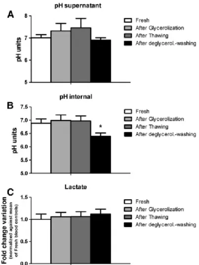
In the present study, we investigated cryostorage of RBCs through monitoring of cell processing steps (from fresh blood, to glycerolization, thawing and deglycerolization/washing) through repeated assays of standard parameters (MCV, RDW-SD) and scanning electron microscopy. Cell processing for cryostorage resulted in increased RBC volumes. Shape alterations caused an increase in osmotic fragility and permeability to ions. A significant pH drop was observed which could not to be attributed to a higher metabolic rate, since the levels of lactate did not show substantial fluctuation during the cell processing steps tested in this study. Membrane anomalies are likely related to the hemolysis observed which preferentially affected the densest and oldest cell sub-populations, as confirmed by means of discontinuous density gradients.
Our results indicate that cryostorage itself in presence of glycerol does not significantly affect RBCs. Most of the alterations observed were related to cell processing and, in particular, to the increase of cytosolic glycerol as a consequence of the glycerolyzation step.
Further studies might profitably investigate replacing glycerol with non-penetrating cryoprotectants.
2.1.1 Materials and methods
Sample collection
Whole blood (450 mL±10%) was collected at the “Celio” Military Hospital in Rome (Italy) from 10 healthy donor volunteers into CPD anticoagulant (63 mL) and leukodepleted. After separation of plasma by centrifugation, RBCs were suspended in 100 mL of Saline, Adenine,
35
Glucose, Mannitol (SAG-M) solution. Ten leukoreduced RBC units were then prepared and cryopreserved, according to the high-glycerol freezing method [Lagerberg et al., 2007].
Briefly, RBC units with a Hct of approximately 60% and fewer than 106 white blood cells were obtained from the blood bank and stored at 2 to 6 °C for 2 h, after which glycerolization and freezing was performed. Glycerolization to a final concentration of 40% glycerol (wt/vol) was accomplished using the Haemonetics ACP 215 device. All glycerolized RBC units were frozen and stored at −80±10 °C in a mechanical freezer for at least 12 months. Frozen RBC units were thawed in a temperature-controlled water bath at 40 °C, until the units reached a temperature between 25 and 30 °C. Thawed RBCs were deglycerolized using the Haemonetics ACP 215 device and resuspended in SAGM.
Thawed deglycerolized RBCs were stored in polypropylene tubes at 2 to 6 °C. The supernatant osmolarity of all the thawed deglycerolized units was below 400 mOsm/kg H2O, indicating an efficient removal of glycerol.
Samples were collected at four sequential stages: (i) fresh blood, within 2 h from collection; (ii) after glycerolization; (iii) after thawing; and, within 2 h after thawing, (iv) after deglycerolization through repeated washing cycles.
RBC analysis
The RBC mean cell volume (MCV), red cell distribution width-standard deviation (RDW-SD), the mean cell hemoglobin concentration (MCHC), and the hematocrit (Hct) were determined with a hematology analyzer (CA 530-Oden, Medonic, Stockholm, Sweden).
Determination of intracellular pH, lactate and glycerol
Red cell pellets obtained by centrifuging 600 μL of suspension in a nylon tube at 30,000×g for 10 min, were frozen, thawed during 5 min and then refrozen. Triplicate measurements of pH were then made with a Radiometer pH glass capillary electrode maintained at 20 °C and linked to a Radiometer PHM acid–base analyzer. To prevent the acid shift seen when samples are kept unfrozen these pH measurements were made immediately after a second thawing of each lysate.
Lactate and glycerol determinations was performed upon methanol/chloroform/water sample extraction through rapid-resolution, reversed phase, high performance liquid chromatography and mass spectrometry, according to the method of D'Alessandro et al. [D’Alessandro et al., 2011]. Results were plotted as mass spectra counts, which are proportional to the metabolite concentrations within the linearity range of the instrument, or fold-change variations upon
36
normalization to the results obtained through testing of fresh RBCs. Rapid resolution, reversed phase, high performance liquid chromatography (RR-RP-HPLC) was performed to separate low-molecular weight compounds, as previously reported [D’Alessandro et al., 2011]. The RR-RP-HPLC directly eluted into an ion trap Rapid resolution, reversed phase, high performance liquid chromatography (RR-RP-HPLC) was performed to separate low-molecular weight compounds, as previously reported [D’Alessandro et al., 2011]. The RR-RP-HPLC directly eluted into an ion trap mass spectrometer (HCT Bruker, Bruker Daltonics, Bremen, Germany), where the compounds were monitored through Multiple Reaction Monitoring (MRM). Mass to charge ratio for precursor and fragment ions to be selected, monitored and quantified were determined as previously reported [D’Alessandro et al., 2011], in agreement with international online available databases (Metlin, Scripps Center for Biotechnology - available at http://metlin.scripps.edu/metabo_info.php?molid=105 – Last accessed on January 30, 2012).
Hemolysis
Hemolysis was calculated according to Harboe [Harboe, 1959]. Samples were diluted in distilled water and incubated at room temperature for 30 min to lyse RBCs. Samples from lysed RBCs were diluted 1⁄300 while supernatants were diluted 1⁄10 in distilled water. After a 30 min stabilization and vortex mixing (Titramax 100, Heidolph Elektro, Kelheim, Germany), the absorbance of hemoglobin was measured at 380, 415 and 450 nm (PowerWave 200 Spectrophotometer, Bio-Tek Instruments, Winooski, Vermont, USA). The mean blank was subtracted and the corrected OD (OD*) was calculated as follows: 2×OD415−OD380−OD450.
Osmotic fragility
The osmotic fragility of RBCs reflects the ability of the membrane to maintain structural integrity. Osmotic fragility was determined by stepwise dilution through PBS solutions ranging from 0.90% to 0.35%.
RBCs with a Hct of 30% to 40% were diluited 1:100 in each PBS solution, mixed and incubated for 30 min at 4°C, followed by centrifugation for 12 min at 1100×g. The free Hb in the supernatant was measured using a spectrophotometer. The concentration of PBS necessary to induce 50% hemolysis defined the osmotic fragility index of the RBCs [Gyongyossy-Issa et al., 2005]. With this method, a larger osmotic fragility index corresponds to more fragile cells.