Alma Mater Studiorum – Università di
Bologna
DOTTORATO DI RICERCA IN SCIENZE CARDIO NEFRO TORACICHE
CICLO: 32°
Settore Concorsuale: Medicina e Chirurgia
Settore Scientifico disciplinare: 06/E1 CHIRURGIA
CARDIO-TORACO-VASCOLARE
TITOLO DELLA TESI: “LEFT ORTHOTOPIC LUNG TRANSPLANT IN
RATS: THE LEARNING PROCESS”
Presentata da: Dr. Davide Zampieri
COORDINATORE DOTTORATO:
Ch.mo Prof. Gaetano Domenico Gargiulo
SUPERVISORE: Prof. N. Daddi
5
INDEX
INDEX 5
ABSTRACT 6
1) INTRODUCTION 7
2) AIM OF THE RESEARCH 38
3) MATERIALS AND METHODS 39
4) RESULTS 42
5) DISCUSSION 49
6 ABSTRACT
INTRODUCTION: The orthotopic left lung transplantation model in rats has been developed to answer a variety of scientific questions in transplant immunology and in the related fields of respiratory diseases. However, its widespread use has been hampered by the complexity of the procedure.
AIM OF THE RESEARCH: Our purpose is to provide a detailed description of the procedure of this technique, including the complications and difficulties from the very first microsurgical step until the ultimate successful completion of the transplant procedure.
MATERIALS AND METHODS: The transplant procedures were performed by two collaborating transplant surgeons with microsurgical and thoracic surgery skills. A total of 150 left lung transplants in rats were performed. Twenty-seven syngeneic (Lewis to Lewis) and 123 allogeneic (Brown-Norway to Lewis) lung transplants were performed using the cuff technique.
RESULTS: In first 50 transplant procedures, post-transplant survival rate was 74% of which 54% reached the end-point of 3 or 7 days post-transplant; whole complication rate was 66%. In the subsequent 50 transplant surgeries (from 51 to 100) post-transplant survival rate increased to 88% of which 56% reached the end-point; whole complication rate was 32 %. In the final 50 transplants (from 101 to 150) post-transplant survival rate was confirmed to be 88% of which 74% reached the end-point; whole complication rate was again 32 %.
CONCLUSIONS: One hundred-fifty transplants can represent a reasonable number of procedures to obtain a satisfactory surgical outcome. Training period with simpler animal models is mandatory to develop anesthesiological and microsurgical skills required for successfully develop this model. The collaboration between at least two microsurgeons is mandatory to perform all the simultaneous procedures required for completing the transplant surgery.
7
1) INTRODUCTION
1.1 Lung allograft dysfunction: clinical and pathological aspects
Lung transplantation represents the only therapeutic option for many incurable pulmonary diseases, such as cystic fibrosis, idiopathic pulmonary fibrosis, and chronic obstructive pulmonary disease. Remarkable progress has been made in improving outcomes, although the 5-year graft survival is still less than 50% primarily because of the development of chronic allograft dysfunction (CLAD) [1]. CLAD developed as a consequence of different early pathological processes including acute lung allograft dysfunction whose specific treatment is crucial for their impact on CLAD.
1.2 Acute lung allograft dysfunction (primary graft dysfunction, infection, acute rejection) Lung allograft dysfunction may be an acute phenomenon (acute lung allograft dysfunction, ALAD), leading to an acute decline in forced expiratory volume, FEV1 (with or without forced vital capacity decline) and may be due to various conditions that affect the graft, such as primary graft dysfunction (PGD), respiratory infections and acute rejection. In some cases, spirometry will not be available, and ALAD may be diagnosed by other tools such as radiology, oxygenation status, and biopsy specimen.
PGD after lung transplantation represents a multifactorial injury that develops in the first 72 hours after transplantation; it is referred to “ischemia-reperfusion injury”, “early graft dysfunction” and “reimplantation edema”. PGD is characterized by severe hypoxia, lung edema and diffuse pulmonary opacities at radiography without other identifiable causes. The typical pathological pattern of PGD is diffuse alveolar damage (DAD). The incidence of PGD is reported to be in the range of 10 to 25%. Despite significant advantages in organ preservation, surgical technique and post-operative care, PGD still remains an important cause of morbidity and mortality [2,3,4]. Infections are very important and common complications of lung transplantation. Bacterial pneumonias are the major infection complications in the early, intermediate, and late post-operative
8
periods. Most of the infections occur in the first 11 months post-transplantation. The underlying native lung may predispose to infection as it occurs in end-stage suppurative diseases such as cystic fibrosis and bronchiectasis. In the late post-operative period, the major predisposing factor is the presence of CLAD. The diagnostic approach to suspected pneumonia at any time post-transplant includes sputum, blood cultures and often bronchoscopy with bronchoalveolar lavage (BAL), sterile brush and sometimes biopsy. The role of new biomarkers such as procalcitonin for diagnosis or follow-up has not been well established. Viral infection after lung transplantation is common and most frequently caused by cytomegalovirus (CMV) or by other community-acquired respiratory viruses. CMV seronegative organ recipients are more susceptible to the infection. The lowest risk occurs in donor /recipient negative patients [5]. The majority of CMV episodes occurs within the first 3 months following lung transplant, while the majority of the later infections is due to influenza and occurs after 1 year. Mounting evidence suggests a relationship between CMV pneumonitis and chronic rejection in the form of bronchiolitis obliterans syndrome (BOS) and is associated with decreased survival despite treatment [6]. The incidence of CMV infection has been reported to range from 30% to 86%, with a mortality of 2–12% [7]. CMV may coexist with rejection. Both of these individual processes induce a cytokine cascade that promotes the development of the other. Tumour necrosis factor-alpha, a key signal in the reactivation of CMV from latency, is released during allograft rejection, thereby facilitating the onset of viral replication and subsequent infection. Conversely, infection of the vascular endothelium and smooth muscle by CMV leads to an upregulation of adhesion molecules promoting an increase influx of inflammatory cells in the graft and subsequently the development of rejection. Additionally, molecular mimicry and the production of anti-endothelial antibodies during CMV may also play a role in the development of rejection [8]. Recent diagnostic tools have ptomoted a shift in the diagnosis of CMV infection and disease. The previous method of diagnosis, pp65 antigen detection, has been replaced by quantitative nucleic acid-based amplification testing via polymerase chain reaction (PCR) for the recognition of viremia by most centers, with 85% of institutions using this method for monitoring and diagnosis [9]. There
9
are no universally accepted viral load cut-offs for positive and negative results, and the reported values may be dissimilar between different laboratories. Despite this, current guidelines on the management of CMV in solid organ transplant patients do not clearly favor one test over the other and cite both as acceptable options for diagnosis. There are two accepted approaches for the prevention of disease from CMV, the universal prophylaxis and the pre-emptive therapy. The universal prophylaxis involves administration of antivirals to all transplant patients considered to be at high risk by their serostatus. The pre-emptive therapy consists in monitoring viral replication in high-risk patients and administering antivirals at a predetermined level of replication prior to the onset of disease. Although there are no randomized trials comparing one strategy versus the other, most centers favor the prophylaxis or may sometimes employ both [9]. A Cochrane Review comparing prophylaxis with antivirals versus placebo or no treatment in different groups of solid organ transplant patients showed a significant reduction in disease (relative risk 0.42), infection (relative risk 0.61), mortality from CMV disease (relative risk 0.26) and all-cause mortality (relative risk 0.63). Interestingly, the review also found a decrease in the risk of developing herpes-simplex virus, varicella-zoster virus and bacterial infections [10]. Prophylaxis may not only be beneficial in decreasing direct morbidity and mortality from CMV disease but may also have secondary effects by decreasing the morbidity and mortality of both acute and chronic rejection. Although the Cochrane Review failed to show a difference in the incidence of acute rejection episodes, other small studies have shown that prevention of CMV decreases the risk for acute rejection, including lung transplant patients [11-15]. Fungal infections are a common complication after lung transplant with an estimated incidence of 15–35% and an overall mortality of 80% [16]. Complications at the site of the anastomosis (i.e. stenosis or necrosis) create the ideal environment for these infections. Other risk factors include the immunomodulatory effect of coexistent infections (i.e. viral) and neutropenia [17,18]. Pre-transplant fungal colonization is common, especially in patients with cystic fibrosis and chronic obstructive pulmonary disease, and it has been associated with post-transplant fungal infection and BOS although not all colonized patients develop active/invasive
10
infection [19]. The most common fungal pathogens are Candida and Aspergillus species, while Zygomycetes, Scedosporium, Fusarium, Cryptococcus species, histoplasmosis and coccidiomycosis occur less commonly. These infections, more prevalent during the first few months after transplantation, can manifest as an invasive disease with a reported 1-year cumulative incidence of 8.6%. Diagnosis of invasive aspergillosis may require aggressive procedures (i.e. biopsy) to verify tissue involvement; however, this is not always possible, and often, the diagnosis is based on the evaluation of computed tomography chest findings and fungal staining/culture from bronchoscopy (i.e. BAL). There are limited data on the role of minimally invasive tests such galactomannan, PCR and 1,3-β-D-glucan assay for the diagnosis of invasive aspergillosis [20]. Lung allograft rejection can be a hyperacute, acute or chronic process and it occurs through immunologic mechanisms that include the activation of the innate and the adaptive immune systems [21].
When considering pathological aspects, acute rejection is characterized by perivascular mononuclear cell infiltrates, which may be accompanied by sub-endothelial infiltration, so-called endothelialitis or intimitis, and also by lymphocytic bronchitis and bronchiolitis.
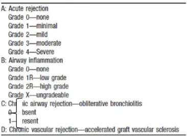
Histological pulmonary allograft rejection is now graded according the revised working formulation for classification and grading of pulmonary allograft rejection as described in the following table [22].
11
The intensity of the perivascular mononuclear cell cuffs and the distribution of the mononuclear cells, including extension beyond the vascular adventitia into adjacent alveolar septa, form the basis of the histological grade. Acute rejection usually affects more than one vessel (particularly in adequate transbronchial biopsy samples) but is occasionally seen as a solitary perivascular infiltrate [22].
Grade A0 (No Acute Rejection): normal pulmonary parenchyma is present without evidence of mononuclear cell infiltration, hemorrhage or necrosis.
Grade A1 (Minimal Acute Rejection): there are scattered, infrequent perivascular mononuclear infiltrates in alveolated lung parenchyma.
Grade A2 (Mild Acute Rejection): more frequent perivascular mononuclear infiltrates are seen surrounding venules and arterioles and are readily recognizable at low magnification.
Grade A3 (Moderate Acute Rejection): easily recognizable cuffing of venules and arterioles by dense perivascular mononuclear cell infiltrates, which are commonly associated with endothelialitis.
Grade A4 (Severe Acute Rejection): diffuse perivascular, interstitial and air space infiltrates of mononuclear cells with prominent alveolar pneumocyte damage and endothelialitis.
12
The revised working formulation allowed airway inflammation to be graded from B0 (no inflammation) to B2R (high-grade small airway inflammation) and main histological features are summarized below [22].
Grade B0 (No Airway Inflammation): no evidence of bronchiolar inflammation.
Grade B1R (Low-grade Small Airway Inflammation): mononuclear cells within the sub-mucosa of the bronchioles, which can be infrequent and scattered or forming a circumferential band.
Grade B2R (High-grade Small Airway Inflammation): the mononuclear cells in the sub-mucosa appear larger and activated, with greater numbers of eosinophils and plasmacytoid cells.
Grade BX (Ungradeable Small Airways Inflammation): the changes are ungradeable due to sampling problems, infection, tangential cutting, artifact, etc.
13
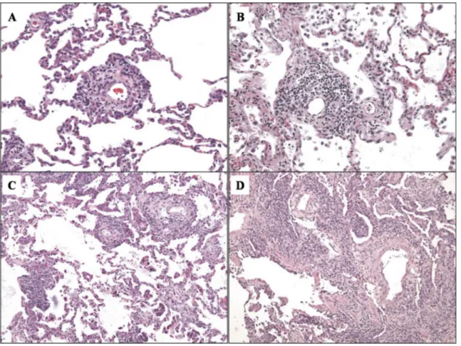
Figure 1.1 Emblematic cases of parenchymal acute rejection graded as A1 (A), A2 (B), A3 (C) and A4 (D). From Stewart S. et al., J Heart Lung Transplant 2007;26:1229–42.
14
Figure 1.2. Emblematic cases of bronchiolar acute rejection graded as B1R (A) and B2R (B). From Stewart S.et al., J Heart Lung Transplant 2007;26:1229–42.
15
1.3 Chronic lung allograft dysfunction – clinical aspects
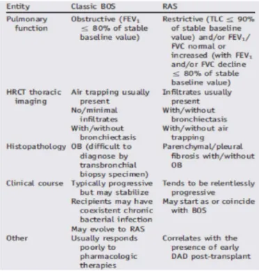
When the post-transplant pulmonary function decline is persistent and not restored to 90% of baseline, chronic lung allograft dysfunction (CLAD) may be suspected [23]. CLAD following lung transplantation is a heterogeneous condition that includes an obstructive form (bronchiolitis obliterans syndrome, BOS) and a restrictive allograft dysfunction (restrictive allograft syndrome, RAS). Although BOS, characterized clinically by irreversible obstructive deficits in pulmonary function tests, remains the major cause of late mortality, RAS accounts for 25–35% of CLAD [24]. The term “chronic” implies a certain duration of time, and in analogy with the BOS definition [23], it has been suggested that a minimum of 3 weeks is a sufficiently prolonged period of time for labelling allograft dysfunction as “chronic”. Diagnostic criteria of the two different types of CLAD are summarized in the following Table.
16
BOS is clinically characterized by progressive (often fatal) airflow obstruction (FEV1 falls below 80% of the best value achieved after transplantation), the absence of parenchymal infiltrates on chest radiographs, a mosaic pattern of perfusion on high-resolution computed tomographic scan, poor responsiveness to therapy, and high mortality rates [25]. Based on these criteria, BOS affects 48% of recipients at 5 years and 76% at 10 years after lung transplantation. Although treatment with azithromycin can sometimes stabilize and even reverse the progressive decline in lung function associated with CLAD, frequently this treatment fails, leaving re-transplantation as the only treatment option.
RAS is defined as CLAD with an irreversible decline in total lung capacity (TLC) to < 90% of baseline as determined using the method explained below. BOS was strictly defined as CLAD without restrictive changes of RAS. Thus, the diagnosis of RAS was not made until FEV1 dropped to meet the criteria of CLAD, even if TLC had already declined to meet the threshold. The diagnosis of BOS was not made until a valid TLC measurement was done to rule out RAS, even if spirometry showed a decline in FEV1 meeting the criteria of CLAD [26].
17
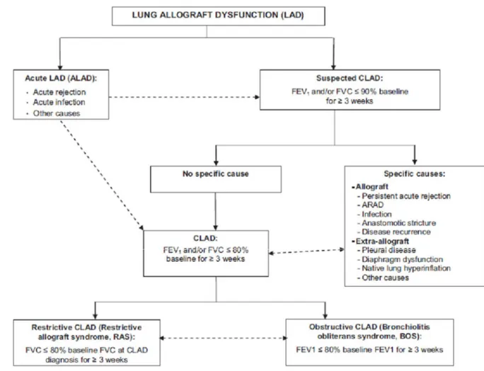
Figure 1.3. This flow chart suggests an approach that can be used to evaluate a lung transplant recipient’s decline in post-bronchodilator forced expiratory volume in 1 second (FEV1) with or without a decline in forced vital capacity (FVC) of Z 10%. This may be acute lung allograft dysfunction (ALAD) and may normalize with treatment. However, when the lung function decline persists for at least 3 weeks without the FEV1 and/or FVC returning to 90% of the post-operative best values, chronic lung allograft dysfunction (CLAD) is suspected. Extended pulmonary function tests (PFT), including spirometry and lung volumes, high-resolution computed tomography (HRCT) of the thorax, and bronchoscopy with bronchoalveolar lavage (BAL) and transbronchial biopsy specimens may identify a cause or causes of suspected CLAD that may still be (completely) reversible upon specific treatment. If the FEV1 and/or FVC declines further to 80% of the post-operative best values despite treatment or without identifying a clear cause, a specific CLAD
18
phenotype should be identified. (Suspected) CLAD could also be a consequence of ALAD if the lung function decline persists. Some patients never develop suspected CLAD but may already have CLAD when they are diagnosed. BOS: bronchiolitis obliterans syndrome; CXR: routine chest X-ray; FEV1: forced expiratory volume in 1 second; SLT: single lung transplant; ARAD: azithromycin-responsive allograft dysfunction; RAS: restrictive allograft syndrome. From Verleden GM, J Heart Lung Transplant 2014; 33:127–133.
1.4 Chronic lung allograft dysfunction – pathological aspects 1.4.1 Obliterative bronchiolitis
The pathological term obliterative bronchiolitis (OB) was introduced in 1984 to describe airway lesions observed in five patients suffering from BOS after lung transplantation [27]. OB is characterized by dense eosinophilic hyaline fibrosis in the sub-mucosa of membranous and respiratory bronchioles, resulting in partial or complete luminal occlusion. This tissue can be concentric or eccentric and may be associated with fragmentation and destruction of the smooth muscle and elastica of the airway wall. It may extend into the peri-bronchiolar interstitium. Mucostasis and/or foamy histiocytes in the distal air spaces are commonly associated with obliterative bronchiolitis and may be observed in transbronchial biopsies in the absence of bronchiolar occlusion or any bronchiolar tissue [22]. The consensus in 2006 was that the distinction between active and inactive obliterative bronchiolitis is no longer useful and the condition should be designated merely as C0, indicating a biopsy with no evidence of obliterative bronchiolitis, and C1, indicating that obliterative bronchiolitis is present in the biopsy. Histological OB is graded as described in the following table [22].
19
Transbronchial biopsy is an insensitive method for detecting OB, its functional grading is the preferred mean for diagnosing and monitoring CLAD [22]. A very recent work by Verleden et al., demonstrated, by using micro-computed tomography, that the constrictive bronchiolitis targets conducting airways while sparing larger airways as well as terminal bronchioles and the alveolar surface [28].
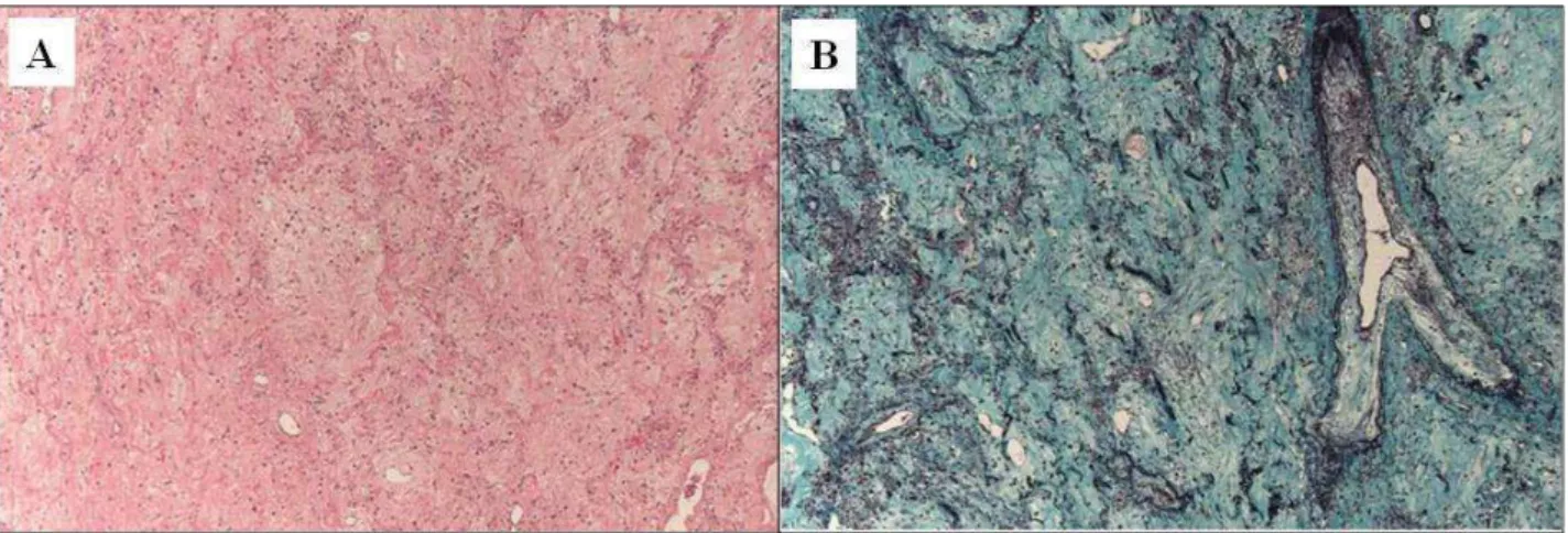
Figure 1.4. A) This small bronchiole shows eccentric scarring of the submucosa of the small airway associated with an inconspicuous peribronchiolar mononuclear infiltrate. The overlying epithelium appears attenuated, while the lumen of the airway is distorted. Such partial occlusion of the small
20
airways may be responsible for significant increases in airflow resistance. H&E. B) The hint to underlying obliterative bronchiolitis in this case is the interrupted cords of smooth muscle forming a tubular structure associated with dense scar tissue in a position adjacent to a pulmonary artery. H&E. From Stewart S.et al., J Heart Lung Transplant 2007;26:1229–42.
1.4.2 RAS
RAS is characterized by restrictive changes in pulmonary function tests that may correlate with inflammatory and fibroproliferative processes in peripheral lung tissue: extensive pulmonary interstitial fibrosis is dominant in the upper lobes of transplanted lungs and was initially reported based on radiographic and histological evidences [26]. A recent study in the largest single series of pleuroparenchymal fibroelastosis cases reported pleuroparenchymal fibroelastosis as a major histopathologic correlate of RAS [24]. RAS is characterized radiologically by features suggestive of a chronic interstitial pneumonia with upper lobe predominance, and histologically by pleural fibrosis and parenchymal fibroelastosis in a predominantly subpleural distribution, with a sharp demarcation between fibroelastotic and unaffected lung parenchyma, and with the presence of fibroblastic foci at this interface. A limited number of cases with similar radiologic and pathologic features have also been reported, including a very recent article by Reddy et al, suggesting a broader spectrum of histopathologic findings [24]. Consistent with the recent finding that onset of RAS is often preceded by the presence of DAD in biopsies, it has been found that pleuroparenchymal fibroelastosis in RAS patients was very often present concurrently with features of DAD. Specimens obtained 1 year after clinical onset of CLAD typically demonstrated features of DAD, whereas those obtained at intervals of a year or more after CLAD onset showed DAD less frequently. These findings, together with the finding in some cases of DAD appearing to merge into areas of pleuroparenchymal fibroelastosis, support a temporal sequence of DAD preceding the development of pleuroparenchymal fibroelastosis in the natural history of RAS [24].
21
Figure 1.5. Pleuroparenchymal fibroelastosis: areas of pleuroparenchymal fibroelastosis characterized by confluent areas of hypocellular collagen deposition with preservation and thickening of the alveolar septal elastic framework. (A) Hematoxylin and eosin stain, original magnification X50; (B) Elastic trichrome stain, original magnification X50. From Ofek E. et al., Modern Pathology (2013) 26, 350–356.
22
1.5 Etiopathogenesis of bronchiolitis obliterans syndrome
The main factors that seem to be etiologically related to BOS are both immunological and non-immunological: prolonged ischemia time, PGD, CMV pneumonitis, aspergillus colonization, respiratory virus infection and gastro-esophageal reflux.
Although the pathogenesis of this progressive airway obstruction is unknown, different immunological mechanisms seem to be involved in the development of BOS. Thus, BOS is thought to represent a final common pathway of a process triggered by both alloantigen dependent and independent mechanisms.
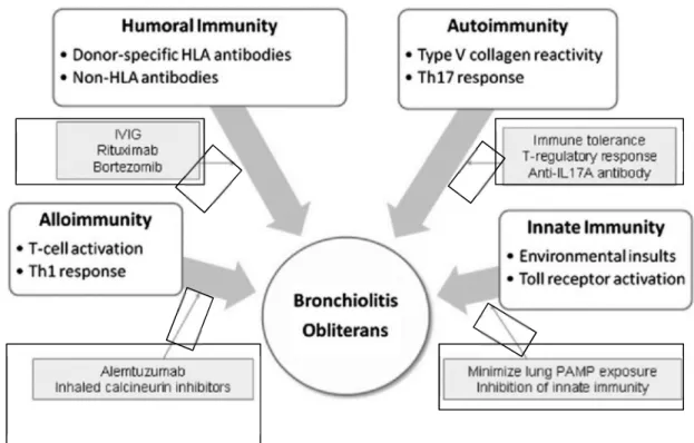
Figure 1.6. Multiple immune mechanisms contribute to the development of OB. Potential therapeutic targets are highlighted. HLA 5 human leukocyte antigen; IVIG 5 IV immunoglobulin; PAMP 5 pathogen-associated molecular pattern; Th 5 T helper. Modified from Todd J et al., Chest 2011; 140(2): 502 – 508.
24
1.5.1 Innate Immunity and Response to Environmental Insults
In recent years, the central importance of innate immunity in host defense has been recognized, particularly with the identification of Toll-like Receptors (TLRs). Innate immunity relies on recognition of highly conserved microbial pathogen-associated molecular patterns (PAMPs) and damage-associated molecular patterns (DAMPs) by innate pattern recognition receptors (PRRs). TLRs, the prototypic family of innate PRRs, are found on pulmonary antigen presenting cells and lung epithelium where they regulate the pulmonary response to inhaled toxins and infections. Both exogenous and endogenous ligands for these receptors have been described, including lipopolysaccharide (LPS), high-mobility group box 1, and hyaluronan fragments. In the context of lung transplantation, genetic studies support the importance of innate immunity and TLRs in the pathobiology of BOS. Taken together, published data suggest a constant interplay among environmental stimuli, the innate immune response, recipient genetic susceptibilities, and adaptive immunity. In fact, many of the previously identified and emerging clinical risk factors for BOS are factors that would likely activate the pulmonary innate immunity [29].
1.5.2 Alloimmune T-Cell Reactivity
The rarity of OB finding in patients without transplantation emphasizes the fundamental role of alloimmune T-cell reactivity in the development of this condition. Acute cellular rejection is the most consistently described risk factor for BOS. Specifically, both acute vascular (A-grade) rejection, especially if histologically severe, and lymphocytic bronchiolitis (B-grade) rejection are associated with a significantly increased risk of BOS. In animal models of tracheal transplantation, the initial alloimmune response is of the T helper (Th) 1 type, with interferon-γ being the predominant cytokine. Interferon-γ upregulates the expression of adhesion and costimulatory molecules by airway epithelial cells, thus further augmenting the alloimmune response by stimulating lymphocyte infiltration and priming T-cell responses. The airway epithelial cell itself, once activated, generates a profibrotic milieu, producing growth factors that ultimately result in
25
tracheal obliteration. The presence of obliterative disease in allogeneic, but not syngeneic, tracheal transplantations supports the importance of alloimmunity in airway fibrosis [29].
Despite clinical and basic evidence supporting a central role for alloimmune reactivity in the development of BOS, the failure of T-cell-based immunosuppressive regimens to prevent the onset of BOS or to stabilize the lung function after its onset supports the importance of other mechanisms of disease pathogenesis. Several additional immune- and nonimmune-related mechanisms likely contribute to the high burden of OB after lung transplantation. Clearly, increased understanding of these factors is critical to the development of improved approaches to prevent and treat BOS [29].
1.5.3 Humoral Immunity
Laboratory advancements in the detection and characterization of anti-human leukocyte antigen (HLA) antibodies by flow cytometry in conjunction with tissue immunostaining for complement fixation have provided clinical evidence that antibody-mediated rejection occurs in lung transplantation. The development of post-transplant anti-HLA antibodies in lung transplant recipients is correlated with an increased risk for BOS and worse overall survival. Recognition of the role of humoral, or antibody-mediated, processes in the pathogenesis of BOS has had a substantive impact on the clinical approach to its prevention and treatment [29]. Indeed, B-cell-modulating therapies are now used to reduce the humoral immune response in lung transplant recipients who develop donor-specific HLA antibodies in an effort to decrease the occurrence or progression of BOS. This treatment can have the benefit of being preemptive when given prior to the onset of acute rejection or BOS in patients with donor-specific antibody (DSA) [29].
1.5.4 Autoimmunity
The discovery of autoimmunity as a mediator of BOS is one of the most exciting novel cellular mechanisms recently described. Sumpter and Wilkes have developed the concept that rejection is biphasic, with the first phase representing tissue injury and the second representing autoimmunity. Tissue injury (from immune or nonimmune insults) exposes normally sequestered self-antigens,
26
and their fragments are released into the lung, acting as triggers for autoreactive T-cell proliferation and autoantibody production [30]. The exposed self-antigens can thus sustain rejection even in the absence of persistent alloimmunity [29].
Type 5 collagen [col(V)], which resides beneath the basement membrane in the perivascular and peribronchiolar tissues of the lung, was the first described potential self-antigen.
Patients with evidence of elevated col(V)-specific cell-mediated immunity have a fivefold to 10-fold increased risk of developing high-grade BOS.
Further investigation suggested that autoreactive Th17 cells, known to be associated with chronic fibrotic autoimmune diseases in humans, partially mediate this response. In support of this concept, a separate study found increased levels of several cytokines crucial for Th17 cell development in BAL fluid from patients with BOS compared with control subjects. Recently, other novel autoimmune targets on the epithelial cell surface, including K-α1 tubulin, have been identified, and binding of autoantibodies to these targets has been shown to promote fibroproliferative events in vitro. The concept of inducible immune tolerance to col(V) or other self-antigens is highly intriguing, and exploitation of this idea may represent a future novel approach to the prevention or treatment of BOS [29].
Th17 cells, a subset of T helper cells distinct from Th1 and Th2 cells, play a key role in the production of several cytokines such as IL-17, IL-21 and IL-22. First described in 1983, IL-17a is the first member of the IL-17 family that is comprised of six isoforms. Produced by T lymphocytes, these cells promote neutrophil growth and activation in the lungs, joint space, central nervous system, and intensities. IL-17a and IL-17f specifically have been shown to play an important role in host defense and autoimmunity [31].
When Vanaudenaerde et al. looked at biopsies and BAL specimens from lung transplant recipients undergoing acute rejection, higher levels of IL-17 were correlated with increased neutrophils and lymphocytes, demonstrating the potential role of IL-17 in acute rejection. Additionally, this group then demonstrated that higher IL-17 mRNA and protein levels in BALs from transplant recipients
27
were associated with the development of BOS [32]. Another mechanism by which IL-17 may contribute to rejection was postulated with IL-17 inducing iBALT, which may contribute to autoimmune reaction in allograft lungs. Finally, data in a murine orthotopic lung transplantation model demonstrated that neutralizing IL-17 prevented OB, down-regulated acute rejection, and up-regulated systemic IL-10. These studies support the multiple roles by which IL-17 may mediate immune responses and rejection [31].
IL-17 has also been implicated in the development of immune responses to self-antigens. Autoantibodies to col(V) from lung transplant recipients were IL-17 dependent and associated with the development of OB after transplantation. Interestingly, the adoptive transfer of lymph node cells reactive against col(V) from immunized donors into isograft recipients induced OB without an alloimmune response. IL17 has been found to contribute to the autoimmune response to K-α1 tubulin, as well. Among mice who were administered antibodies to donor MHC class I antigens, inhibition of IL-17 resulted in decreased levels of autoantibodies to col(V) and K-α1 tubulin. Combined, these results propose a key role for IL-17 in the development of autoimmunity. However, recent work has highlighted that not all Th-17 cells are pathogenic and that other key cytokines such as IL-23 is necessary to induce autoimmune disease. Thus, the connection between IL-17 and autoimmunity in human studies needs to be further and deeply investigated [31].
28
Figure 1.7. Autoimmunity in lung transplantation. After transplantation, exposure of collagen type V [col(V)] and K-α1 tubulin triggers autoimmune responses, both humoral and cell mediated, which contribute to chronic rejection and obliterative bronchiolitis. APC, antigen presenting cell. From Weber DJ and Wilkes DS. Am J Physiol Lung Cell Mol Physiol. 2013;304(5): L307-11.
1.6 Animal models of BOS
To better understand the underlying mechanisms of OB development, a research model that mimicked the phenomenon of pulmonary chronic rejection was introduced in 1993 [33]. In this model, tracheal rings were heterotopically implanted under the skin or into the abdominal cavity of rats or mice, and then developed the typical features of OB histology. This model possesses the advantage of reproducibility, and is easy to perform. However, researchers argued that this model did not sufficiently reflect the complexity of clinical OB. In search of more physiological models, various other techniques were introduced in rodents, including the orthotopic tracheal segment interposition and the implantation of a donor trachea into a recipient lung [34]. With the introduction
29
orthotopic single-lung transplantation model in the mouse, the physiological ventilation and perfusion resembling the human transplantation condition can be achieved . Other preclinical models of OB development have been proposed, such as the miniature swine transplantation into minor histocompatibility complex (MiHC) antigen–mismatched combinations. This model provides several advantages: is closest to human OB, is anatomically similar to the transplant, provides the possibility to monitor the transplant function by repeated biopsies, by bronchoalveolar lavage collection and computed tomography imaging. However, the swine model is limited in its availability because of the need for special breeding facilities, and the high cost. The main characteristics of rat and murine models of OB are summarized in the following table [34].
Table 1.2. From Jungraithmayr W et al., Am J Respir Cell Mol Biol. 2013;48(6):675-84.
1.6.1 Heterotopic tracheal transplantation
The development of heterotopic tracheal transplantation model in 1993 enabled the reproduction of the phenomenon of OB after transplantation for the first time. Tracheal segments were either implanted into a subcutaneous pouch in the neck, or placed intraperitoneally. This approach is technically effortless, reproduces representative results in-vivo, and resembles the identical histopathological changes of human OB. The majority of published studies so far were performed using this model. However, its shortcomings are not negligible. Implanted tracheal segments undergo severe initial ischemia, relying only on diffusion from the surrounding tissue. Furthermore,
30
physiological ventilation as a central functional aspect in lung transplantation does not occur, and large airways instead of bronchioles are investigated, thereby not reflecting the pathological hallmarks of OB. Finally, the observed changes occur within a short span of time, in contrast with the slowly developing OB in human lung-transplant recipients [34].
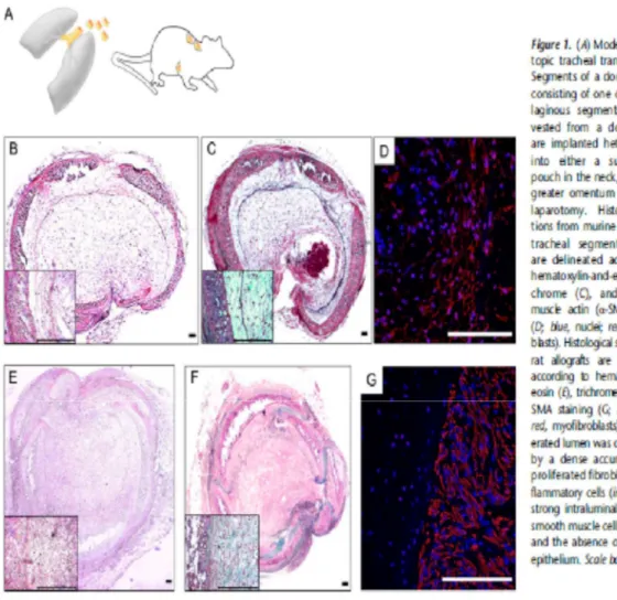
Figure 1.8. (A) Model of heterotopic tracheal transplantation. Segments of a donor trachea, consisting of one or two cartilaginous segments, are harvested from a donor. These are implanted heterotopically either into a subcutaneous pouch in the neck, or into the greater omentum via a small laparotomy. Histologic sections from murine heterotopic tracheal segment allografts are delineated according to hematoxylin-and-eosin (B), trichrome (C), and a–smooth muscle actin (a-SMA) staining (D; blue, nuclei; red, myofibroblasts). Histological sections from rat allografts are
31
also shown according to hematoxylin-andeosin (E), trichrome (F), and a- SMA staining (G; blue, nuclei; red, myofibroblasts). The obliterated lumen was characterized by a dense accumulation of proliferated fibroblasts and inflammatory cells (insets) with a strong intraluminal staining of smooth muscle cells (D and G), and the absence of respiratory epithelium. Scale bars, 100 mm. From Jungraithmayr W et al. Am J Respir Cell Mol Biol. 2013;48(6):675-84.
1.6.2 Orthotopic tracheal transplantation
The disadvantages of the heterotopic tracheal model promoted the search for a more physiological setting. A new model of orthotopic tracheal transplantation was introduced by Ikonen and colleagues in 2000, and was later refined by Schrepfer and colleagues, in which a segment of a donor trachea was interposed into a recipient trachea to provide physiological ventilation [35,36]. These authors described a long-term patency of fully histoincompatible allografts in non immunosuppressed rats, as was also observed in mice. Despite acute alloimmune injury and the induction of myofibroblast proliferation, epithelial regrowth from the host limited the progression of OB, thus emphasizing the role of the epithelium in the control of airway obliteration [34].
32
Figure 1.9. (A) Model of orthotopic tracheal transplantation. The whole trachea is harvested from a donor and orthotopically implanted by suturing the trachea at the cranial and caudal lumen by an end-toend running suture into the recipient. Histologic sections from murine orthotopic tracheal allografts are delineated according to hematoxylin-and-eosin (B), trichrome (C), and a-SMA staining (D; blue, nuclei; red, myofibroblasts). Histological sections from rat allografts are also shown according to hematoxylin-and-eosin (E), trichrome (F), and a-SMA staining (G; blue, nuclei; red, myofibroblasts). On Day 60 after implantation, orthotopic allografts did not obliterate,
33
but show a mild epithelial regrowth (insets in B and C) and a proliferation of myofibroblasts and smooth muscle cells (insets in E and F), with strong staining of smooth muscle cells (G). Scale bars, 100 mm. From Jungraithmayr W et al. Am J Respir Cell Mol Biol. 2013;48(6):675-84.
1.6.3 Orthotopic lung transplantation
The model of orthotopic lung transplantation has the advantage of being not only a transplantation model with physiologic ventilation and perfusion, but also to best reproduce the surgical procedure of human lung transplantation In this model, the recipient’s artery, vein, and main bronchus are cuff-anastomosed, or alternatively sutured to the respective vessels and bronchus of the donor graft, thus mimicking the human transplantation procedure [34].
Orthotopic rat lung transplantation is currently used to investigate ischemia–reperfusion injury and acute rejection. However, due to the strong alloimmune response and the difficulty of control it, this model did not consistently develop OB. Without immunosuppression, major histocompatibility (MHC)-fully mismatched lung allografts (e.g. Brown Norway to Lewis rats) are acutely rejected and become necrotic within several days, while short-term immunosuppression (e.g. cyclosporine for the first few days) enables long-term acceptance of allografts. A commonly used rat lung transplant model of ‘chronic rejection’ is a moderately histoincompatible strain combination, from Fisher 344 (MHC type RT1lvl) rats to Wistar Kyoto (RT1l) rats without immunosuppression [37]. However, many reports have indicated that the chronic lesions in this model are not the typical OB lesions [38].
Two important issues are still largely discussed in rodent orthotopic lung models: a) the reproducibility of surgical procedure; b) the identification of the best genetic strain combination that develops immunological lesions similar to those in humans. Although the orthotopic lung transplantation model is technically demanding, it holds great promise for boosting clinically relevant research. To reach this goal, a wider use of this model must be achieved, because so far only a few centers worldwide can successfully implement this model. Based on this research model,
34
the identification of novel therapeutic targets will be achieved through systematic exploration and meticulous analyses in therapeutic proof-of-concept studies [34].
1.6.3.1 Surgical technique of left orthotopic lung transplant in rat
The orthotopic single lung transplantation model in rat has been developed to answer a variety of scientific questions in transplant immunology and the related fields of respiratory diseases. The first lung transplantation in the rat was achieved by Asimacopoulos et al [41] using sutured anastomoses in 1971. However, the high complication rates and technical difficulties with the sutured anastomosis prevent the widespread adoption of this technique. Subsequent development of a cuffed technique for the anastomoses by Mizuta et al. [42] in 1989 represented a breakthrough that resulted in simplification of the procedure and shorter warm ischemic times. [43]. Since then, a number of further variations on the technique of rat lung transplantation have been described, each of them differing in some anesthesiological or technical details. In spite of this, the procedure remains technically demanding and involves a long learning curve [44].
Although virtually anyone can succeed in this technique, investigators with pre-existing microsurgical, small animal model and thoracic surgery skills should handle the procedure faster and require a shorter training period to master the surgical technique, with learning concepts (i.e., the presence of an instructor, consulting helpful tools, carefully following protocols, and repeatedly watching key video features).
A recent review focuses on technical safeguards to further evolve the surgical technique for cuffed anastomoses in rat lung transplantation [45], but this complex model has other aspect to consider: general anesthesia protocol management, post-operatory cares.
35
Figure 1.10. (A) Orthotopic single lung transplantation. The left lung is removed from a donor animal, the artery (blue), bronchus (yellow), and vein (red) are separated, and each is equipped with a specially designed plastic cuff. The lung is then introduced into the recipient’s respective bronchus and vessels, to obtain a perfused and ventilated transplant. (B) Histologic sections from rat orthotopic lung allografts show typical obliterative bronchiolitis lesions on day 60 after transplant. Only small parts of the respiratory epithelium are intact, and the smooth muscle layer has vanished. Instead, increasing amounts of fibrous tissue have obliterated the bronchial lumen (I, hematoxylin-and-eosin; II, Trichrome). (C) Histological sections from murine orthotopic lung allografts show obliterative bronchiolitis, 70 days after transplantation. Mononuclear cells are prominently present within fibrotic plugs that protrude into the airway lumen (I, hematoxylin-and-eosin stain), with intense staining of collagen within the plugs (II, Sirius red). The polarized Sirius red light clearly indicates the difference in younger, rather reddish collagen and older, whiter collagen (III, Sirius red polarized). From Jungraithmayr W et al. Am J Respir Cell Mol Biol. 2013;48(6):675-84.
36 1.7 From bench to bedside
Each of the proposed lung transplant small animal models provided interesting insights for the interpretation of the pathogenesis of OB. For almost 20 years, the technique of heterotopic tracheal transplantation was the leading experimental model in OB research. Important insights about how and when OB changes occur were achieved soon after the introduction of the heterotopic tracheal transplantation model. Hertz and colleagues demonstrated that, within 21 days after transplantation, murine allografts developed airway fibroproliferation, whereas isografts showed normal respiratory epithelia. Boehler and colleagues then showed that the development of OB was alloantigen-dependent. Only allogeneic grafts showed typical OB lesions, whereas isografts were reconstituted with a normal epithelial lining after recovery from ischemia [46]. Hertz and colleagues later showed that OB lesions progressed if the initial period of alloimmune injury was sufficient, even if the alloimmune stimulus was removed [47]. To address the need for a more physiologic experimental setup, a variety of small animal models have been proposed during the past two decades, such as the orthotopic tracheal transplantation model or the intrapulmonary trachea implantation model. Answers to the remaining questions could be obtained via the newly introduced model of orthotopic murine lung transplantation, which not only reflects the full physiology of a transplanted graft, but also allows for the investigation of the influence from other factors that are relevant in the evolution of OB, such as acid aspiration or other non-immunologic stimuli. Beyond the possibility of genetic modifications in the mouse through which human diseases can be explored, transplant-related complications such as PGD or ischemia–reperfusion injury and their potential therapeutic options could be investigated in this model. Moreover, the investigation of non–heart-beating donor organs as well as the ex vivo reconditioning of potentially transplantable organs, which plays an increasing role in the retrieval of organs, can be performed in this model. Moreover, the establishment of OB in minor mismatched recipients could also provide the last opportunity for testing novel therapeutic interventions such as inhibition of crucial mediators involved in EMT development.
37
Figure 1.11. A cartoon describing the principal evidences in clinical lung transplantation derived from animal studies.
38
2) AIM OF THE RESEARCH
The aim of the project was to set up a model of orthotopic lung transplant in rat. In this thesis is also reported the detailed learning process, including complications and difficulties.
39
3) MATERIALS AND METHODS
3.1 Animals
Male Brown-Norway (BN, RT1n) and Lewis (RT1l) rats weighing 250-350 g (Charles River, Italy) were used as donors of allogeneic and syngeneic lung grafts, respectively; Lewis rats were used as recipients. Rats were housed in a conventional facility with constant temperature and a 12h light-dark cycle with free access to water and food.
All animal experiments were approved by the Institutional Animal Care and Use Committee of the Mario Negri Institute. All procedures were conducted in compliance with national (D.L. n.26, March 4, 2014) and International Laws and Policies (EEC Council Directive 2010/63/EU, 9/22/2010; Guide for the Care and Use of Laboratory Animals, National Academy Press, 1996). A dedicated evaluation system (score sheet system [48]) was adopted to monitor clinical signs of pain, distress and discomfort.
We performed first syngeneic graft transplants (n=27) and then allogeneic graft transplants (n=123). All recipient animals underwent left orthotopic lung transplant procedure performed, using the Reis technique [49].
Transplant recipients that survived after the lung transplant procedure were sacrificed at the end-point of 3-7 days post-transplant.
3.2 Surgical Team
Two expert in the field of surgery/microsurgery combined their skills. The first surgeon has a long-standing experience in small animal models of orthotopic kidney and heart transplant, the second surgeon is a thoracic surgeon with animal microsurgical experience. The staff dealing with monitoring animal welfare post tx has a 25-year expertise in working with laboratory animals and in their pharmacological treatments, and is able to rapidly recognize animal suffering.
40 3.3 Donor left lung retrieval
Rats were anesthetized with 5% Isofluorane, intubated using a 16-gauge intravenous catheter, and mechanically ventilated with inhaling 2% Isoflurane. Laparo-sternotomy was performed and heparin was administered via the inferior vena cava. The thymus was then excised and inferior and superior caval veins are identified and legated. A 16-gauge Teflon PEF intravenous catheter was introduced into the pulmonary trunk via subvalvular myocard, and left atrium was transected. Donor lungs were flushed with 25 ml of cold (4°C) Servator P® through the main pulmonary artery. The left bronchus, artery and vein (including an atrial cuff) are isolated and excised. The left lung was harvested at end-tidal volume by placing a micro-surgical-clamp before bronchus excision. The inflated lung was placed in a Petri dish and kept on ice, the artery, vein and bronchus are everted over the cuff (consisting of Teflon PET intravenous catheter 18G for artery and 16G for vein and bronchus of a 1-mm cylinder and a 1-mm handle) and fixed using a 7-0 silk suture ligature. At the end of this procedure the lung was preserved in cold (4°C) Servator P® in an ice-bath.
3.4 Recipient procedures
Recipient animals were anesthetized, intubated and ventilated like the donors. Recipient animals were placed in the right lateral decubitus position and a skin incision was performed 1 cm below the inferior margin of the scapula. The lateral chest wall was opened at the fourth intercostal space. The left inferior pulmonary ligament was cut, and the left native lung was exposed outside the thoracic cavity and fixed to animal skin with a clamp. The pulmonary artery, bronchus and vein were clamped separately, with particular attention given to placing the vein clamp as close as possible to the left atrium to gain a longer element to perform anastomosis (this is important in cases in which the vein is divided into two branches that are joined very close to the atrium). However, it was not placed too deep in the atrium, in order to avoid the risk of also clamping the left superior vena cava and post-cava pulmonary vein, causing cardiac arrhythmias and arrest. The
41
donor lung was placed into the recipient thoracic cavity and constantly cooled throughout the implant procedure by covering it with cooled wet gauze.
3.4.1 Bronchus, arterial and vein cuff anastomosis
Pre-knotted 7/0 silk suture ligatures were placed around the bronchus, artery and vein. Then a small incision, close to the native lung, was made in the recipient bronchus. The donor bronchial cuff was then inserted into the recipient bronchus and fixed with the suture; the same procedure was repeated for the artery and vein. Since the donor cuffed element tends to slide out of the recipient element, one surgeon grips the cuff and the second surgeon fixes the suture. If the vein divides into two branches very close to the atrium, the smaller one is closed with 7/0 silk thread and the bigger branch is used to insert the cuff.
At the end, bronchial, vein and arterial clips were removed in sequence. After re-ventilation and reperfusion, glucocorticoids (20 mg i.p.) were administered to all rats. The native lung was removed. The thorax, muscles and skin were sutured and a 16-gauge intravenous catheter was placed for thorax drainage and then removed at extubation. The rats were left in a heated box for complete recovery and then given buprenorphine (0.15 mg/kg subcutaneously).
3.5 Assessment of transplant viability
After surgery, the animal welfare was monitored at least three times a day using the dedicated evaluation system (score sheet system [48]) to assess clinical signs of pain, distress and discomfort. Experimental end-points were chosen to verify graft function and early rejection process between 3 and 7 days post-transplant. Animals were sacrificed before reaching the experimental end-point in case of clinical discomfort. At sacrifice, the entire heart-lung block was explanted, gently inflated with a 16-gauge intravenous catheter and carefully examined. The anastomoses were analyzed and their patency evaluated. Each lung graft was photographed, sampled and fixed in formalin.
42
4) RESULTS
4.1 Anesthesiological Setting
Optimal assessment of oro-tracheal intubation using a 16-gauge intravenous catheter, protocol of mechanical ventilation and general anesthesia protocol management required a training period. Problems in oro-tracheal intubation were represented by esophageal placement of intravenous catheter and impossibility to check the correct position of the catheter in the trachea with an ultrasound. During the first 50 transplant, we experimented esophageal placement in 4% of animals, from 51 to 100 and from 101 to 150 tx we did not have problem during intubation. We decided to perform oro-tracheal intubation both in donor and recipient, even if in donor was not necessary to perform the surgical procedure, as training. Incorrect tracheal tube placement did not represent a problem in donor, because animal was placed in supine position and after sternotomy was easily possible to check lung ventilation and eventually perform a rescue tracheostomy. In the recipient incorrect tracheal tube placement represented a more serious problem as animal was in right lateral decubitus with difficult access to airways and impossibility to perform tracheostomy.
4.2 Intraoperative complications of donor lung preparation
During the first 50 transplant, the main cause of failure in donor preparation was injury to the pulmonary hilum structures (8%), especially the wall of the pulmonary vein (PV) (2 of 4), followed by the pulmonary artery (PA) (1 of 4), the pulmonary bronchus (Br) (one of 4), (Table 1). In the following 50 tx we recorded 6% injury to PV vein during dissection and on the latest 50 tx 4% of injury always to PV.
Failure of PV preparation occurred principally during dissection, because the dissection of the space between the PV and the Br requires a similar fine preparative ability in both the donor and the recipient, because the PV and Br are thinner structures than the PA. The distribution of complication types in donor procedure execution is given as proportions in Table 3.
43 4.3 Complications of graft preparation
Complication rate at back-table procedures was 20% in first 50 transplant procedures (PA damage in 2 of 10 and PV damage in 8 of 10); 12% in tx from 51 to 100 (PV damage in all cases); 14% in Tx from 101 to 150 (PV damage in all cases).
Problem in graft preparation at back table were mainly due to the necessity to remove all fat and exceeding connective tissue in order to obtain a thinnest tissue to pull through the cuff the PV. PV wall is very thin and in these procedures is very easy to damage it. The only problem with the PA occurred in finding the lumen while cuffing the structure. When the lumen of the PA cannot be properly identified, a small portion of the end should be cut with straight, sharp scissors so that the lumen becomes clear. From the 51th transplant onward, the incidence of complications decreased to 12-14%. The main problem remained injury to the PV.
All these problems did not prevent the lung transplantation and the graft implant was possible in all cases. Injuries to PV or PA wall were successfully repaired with 10/0 nylon suture. The donor preparation time during the first 50 transplant was 29 ± 16.5 min, from 51 to 100 transplants 20 ± 7.9 min, and from 101 to 150 transplants 16 ± 3.1 min with an improvement of almost 50% in time execution.
The complications encountered during graft preparation execution are shown in Table 3.
4.4 Complications in graft implantation
The complications in recipients included: wrong thoracotomy space execution in 3 cases in first 50 tx, with rib rupture in one case due to excessive depressor traction to gain hilum structures, (2 in fifth intercostal spaces; 1 in third intercostal space); 1 in tx from 51 to 100 (in fifth intercostal space); none in Tx from 101 to 150.
Rupture of a hilum structure during dissection was encountered at the beginning of the implant procedure execution in particular: 3 cases in first 50 tx (one PV damage and 2 Br); one case of PV in tx from 51 to 100 and no cases in the following tx.
44
Rupture of the PV, PA, or Br during anastomosis occurred in 13 cases in first 50 tx (4 PA; 9 PV); 6 cases in tx from 51(all PV) to 100 and 7 cases in tx from 101 to 150 (all PV).
PV structure in very thin and fragile so that only one attempt of cuff recipient vessel introduction is possible;. In very few cases is possible to repair PV injury 10/0 nylon suture. Generally, the shortness of the vein and the irreversible damage prevent the repair., The complication enocunterd in recipient procedure execution is shown in Table 3.
4.5 Transplant outcome
In first 50 transplants, post-transplant survival rate was 74% of which 54% reached the end-point. Whole complication rate was 66%. Tx failure was due to complications during graft implantation in 13 cases, in particular: problems of PA anastomosis execution in three cases and problems of PV anastomosis.Ten transplanted animals did not reached suited end-point after successful surgical procedure: 4 for bronchial secretions and 9 for unexpected death.
From 51 to 100 transplant procedures, post-transplant survival rate was 88% of which 56% reached the end-point. Sixteen animals did not reach the end-point: 5 for bronchial secretions and 11 for unexpected death. Whole complication rate was 32 %; tx failure was determined by complications in 6 cases: 1 PV cuff problem execution and 5 PV anastomosis execution.
From 101 to 150 transplants, post-transplant survival rate was 88% of which 74% reached the end-point. Seven animals did not reach the end-point: 2 for clutter secretions; one case of pneumothorax and 4 for unexpected death. Whole complication rate was 32 %; tx failure was determined by complications in 6 cases: 2 PV cuff problem execution and 4 PV anastomosis execution.
The post-mortem analysis in animals that did not reach the end-point evidenced in all cases patent anastomosis, and diffuse lung edema in rat with bronchial secretions compatible with ischemia/reperfusion injury.
Outcomes are reported in Table 4.
45 TABLES
Table 1. Donor procedure complications
TX 0-50 TX 51-100 TX 101-150
PA damage 1 / /
PV damage 2 3 2
Br damage 1 / /
whole donor complication rate 4 (8%) 3 (6%) 2 (4%)
Table 2. Cuff preparation complications.
TX 0-50 TX 51-100 TX 101-150
PA 2 / /
PV 8 6 7
Br / / /
whole cuff preparation complication rate 10 (20%) 6 (12%) 7 (14%)
Table 3. Recipient procedure complications.
TX 0-50 TX 51-100 TX 101-150 Wrong thoracotomy space execution 3 1 /
Rupture of a PA during dissection / / / Rupture of a PV during dissection 1 1 / Rupture of a Br during dissection 2 / /
Rupture of PA during anastomosis 4 / /
46
Rupture of Br during anastomosis / / /
whole recipient complication rate 19 (38%) 7 (14%) 7 (14%) Table 4. Tx outcomes.
TX 0-50 TX 51-100 TX 101-150 Surgical success 37 (74%) 44 (88%) 44 (88%) Reached suited end-point 27 (54%) 28 (56%) 37 (74%) Tx not executed for donor problems / / /
Tx not executed for graft preparation problems / 1 (2%) 2 (4%) Tx not executed for recipient problems 13 (26%) 5 (10%) 4 (8%) Whole complication rate 33 (66%) 16 (32%) 16 (32%) Complication that determinantes tx failure 13 (26%) 6 (12%) 6 (12%) Table 5. Time spent for procedures execution.
TX 0-50 TX 51-100 TX 101-136 Graft preparation 29 min ± 16.5 min 20 min ± 7.9 min 16 min ± 3.1 min Cold (Whole) ischemia time 66 min± 7.6 min 55 min± 6.5 min 48 min± 2.1 min Warm ischemia time 30 min± 2.6 min 21 min± 3.4 min 17 min± 1.8 min
The warm ischemia time is defined here as the time between taking the graft from +4° C storage solution and putting it on the recipient till its complete reperfusion and reventilation
47
Figure 1. Learning curve for orthotopic single lung rat transplantation. Operative time, time to perform both arterial and vein anastomoses (warm ischemia time).
Figure 2. Learning curve for orthotopic single lung rat transplantation: number of complications divided for single procedures
48
Figure 3. .Learning curve for orthotopic single lung rat transplantation: surgical success rate, animals that reached suited end-point.
49 5) DISCUSSION
Orthotopic lung transplant in rats represents an important model to study patterns of rejection, which negatively influence clinical outcome of patients that underwent lung transplant. Only few centers in the word have developed the necessary expertise to reproduce this model. Its world-wide dissemination would be very important to permit the study of the different mechanisms of lung acute and chronic rejection, ischemia/reperfusion injury, viral and bacterial infections and possibly tolerance. For this reason the availability of a simple and easy model with high reproducibility is of great importance. Although the Mizuta’s cuff technique, and its further subsequent improvement, facilitates anastomosis during the operation, many drawbacks remain.
In the present study, we have described in detail the setting-up of a rat model of orthotopic left lung transplantation. Surgical success of the model was achieved thanks to microsurgical skills previously acquired in simpler rat model and in thoracic surgery by the two surgeons. Lung transplantation in rat represents a challenging model and a large number of animals for the learning
50
curve has been required to obtain a satisfactory survival rate. Previous acquired microsurgical and anesthesiological skills are required to perform lung tx.
A better survival can be achieved by less complication rate during procedures (66% at the beginning vs. 32% at the end) and with improvement of cold and warm ischemia times.
Cold ischemia time is dependent from the time required for cuff preparation, in particular PV cuff. In order to obtain a PV long enough, a part of left atrium has to be removed with PV during donor procedure. Left atrium is thicker than PV and is useful to handle the element to avoid PV wall damages during the insertion of the cuff.
Another crucial aspect to consider is the general anesthesia management: intubation procedure can require some training in naive animals, because while in the donor is possible to perform a rescue tracheostomy, in recipient this is not feasible. Pharmacological management of anesthesia can be challenging, as other authors reported in different protocols (41-45), and it is necessary to modify it during the experiment. We didn’t need a period to develop our anesthesiological protocol because we already optimized it in a simpler model of rat thymectomy through median sternotomy. In the lung transplant model we encountered some problems with incorrect tracheal intubation and cardiac arrest due to anesthesia overdose, so we started adapting anesthesiological protocol during surgery relying on vital parameters (in particular heartbeat, that can be easily monitored during thoracotomy). For this reason, the presence of two operators is mandatory during tx: the first surgeon performs surgical procedures, while the other manages anesthesia and help the fist surgeon in performing anastomosis as reported before.
A recent review underlines that PV anastomosis is the most difficult technique because the recipient vein tears easily and suggests to create the PV anastomosis first [45]. In our experience the PV wall is very thin and inelastic compared to other veins, handling of this structure must be done very carefully, indeed incautious maneuvers can easily provoke dissection. Thus, we performed the PV anastomosis after PA an Br, to avoid excessive tension. Additionally, maintaining the connective tissue around the vein can alleviate tensions during cuff insertion procedures.
51
Moreover, PV anastomosis is challenging due to the presence of a collateral vein in the PV. When collateral vein is close to the atrium its ligation is mandatory. However, this procedure reduces the PV diameter of the vein used to insert donor cuff with increased risk of PV recipient dissection during anastomosis. In addition, performing the ligation increases the warm ischemia time. One report has described the details of the rat lung transplantation model including its complications and mistakes [44]; the authors described the experience in 197 rat lung tx reporting only number of complications and warm ischemia times, without providing other details. In our learning experience we obtained a warm ischemia time comparable to that of Kubisa et al [44].
Furthermore, it is fundamental to perform vein anastomosis at the first attempt (repeated attempts induce vein wall dissection). In this phase, the collaboration by two operators is crucial: the first surgeon keeps the cuff into the recipient vein and the second fixes it, in order to avoid the cuff slip or erroneous apposition of the knot (from our previous experience these procedures can damage vascular wall and favor thrombosis). Vein thrombosis can be avoided manipulating as little as possible the vase and choosing an appropriate cuff size, permitting a good blood flow.
Damages of artery or bronchus are rare, because these structures are plastic and tolerate well the tension necessary to insert the cuff into the recipient element.
Postoperative complications occurred in a high percentage of recipient animals: 20% in first 50 tx, 32% in tx from 51 to 100, 16% in tx from 101 to 150. In some cases, the cause of post-operatory tx failure was ischemia/reperfusion injury. Otherwise, the cause of post-operatory tx failure remained unknown, probably infections, thrombosis or embolism. This cannot be verified in-vivo because we lacked post-tx strumental evaluation instruments.
In conclusion, left orthotopic lung transplant in rat is a complex model that requires a setting up period, even in the hands of surgeons with microsurgical and thoracic surgery skills. One hundred-fifty transplants can represent a reasonable number of procedures to obtain a satisfactory surgical outcome. Previously acquired microsurgery skills with simpler animal models are mandatory to properly perform anesthesiological and microsurgical procedures to succeed in this model.
53
6) REFERENCES
1. Grossman EJ, Shilling RA. Bronchiolitis obliterans in lung transplantation: the good, the bad, and the future. Transl Res. 2009 Apr;153(4):153-65.
2. Christie JD, Bavaria JE, Palevsky HI, Litzky L, Blumenthal NP, Kaiser LR, Kotloff RM. Primary graft failure following lung transplantation. Chest. 1998 Jul;114(1):51-60.
3. Christie JD, Kotloff RM, Pochettino A, Arcasoy SM, Rosengard BR, Landis JR, Kimmel SE. Clinical risk factors for primary graft failure following lung transplantation. Chest. 2003 Oct;124(4):1232-41.
4. King RC, Binns OA, Rodriguez F, Kanithanon RC, Daniel TM, Spotnitz WD, Tribble CG, Kron IL. Reperfusion injury significantly impacts clinical outcome after pulmonary transplantation. Ann Thorac Surg. 2000 Jun;69(6):1681-5.
5. Humar A, Snydman D; AST Infectious Diseases Community of Practice. Cytomegalovirus in solid organ transplant recipients. Am J Transplant. 2009 Dec;9 Suppl 4:S78-86.
6. Snyder LD, Finlen-Copeland CA, Turbyfill WJ, Howell D, Willner DA, Palmer SM. Cytomegalovirus pneumonitis is a risk for bronchiolitis obliterans syndrome in lung transplantation. Am J Respir Crit Care Med. 2010 Jun 15;181(12):1391-6.
7. Kotton CN, Kumar D, Caliendo AM, Asberg A, Chou S, Snydman DR, Allen U, Humar A; Transplantation Society International CMV Consensus Group. International consensus guidelines on the management of cytomegalovirus in solid organ transplantation. Transplantation. 2010 Apr 15;89(7):779-95.
8. Zamora MR. Cytomegalovirus and lung transplantation. Am J Transplant. 2004 Aug;4(8):1219-26.
54
9. Zuk DM, Humar A, Weinkauf JG, Lien DC, Nador RG, Kumar D. An international survey of cytomegalovirus management practices in lung transplantation. Transplantation. 2010 Sep 27;90(6):672-6.
10. Hodson EM, Ladhani M, Webster AC, Strippoli GF, Craig JC. Antiviral medications for preventing cytomegalovirus disease in solid organ transplant recipients. Cochrane Database Syst Rev. 2013 Feb 28;2:CD003774.
11. Jaksch P, Zweytick B, Kerschner H, Hoda AM, Keplinger M, Lang G, Aigner C, Klepetko W. Cytomegalovirus prevention in high-risk lung transplant recipients: comparison of 3- vs 12-month valganciclovir therapy. J Heart Lung Transplant. 2009 Jul;28(7):670-5.
12. Snydman DR, Limaye AP, Potena L, Zamora MR. Update and review: state-of-the-art management of cytomegalovirus infection and disease following thoracic organ transplantation. Transplant Proc. 2011 Apr;43(3 Suppl):S1-S17.
13. ERS 2012 Balestro E, Rossi E, Lunardi F, Damin M, Nannini N, Loy M, Marulli G, Rea F, Calabrese F. Combined CMV prophylaxis reduces short term complications after lung transplantation.
14. NIT 2013 Calabrese F, Lunardi F, Nannini N, Balestro E, Andriolo L, Loy M, Rea F, Calabrese F. Trattamento profilattico combinato anti-CMV: impatto sulle complicanze a breve termine dopo trapianto di polmone.
15. AMIT 2013 Lunardi F, Nannini N, Balestro E, Rossi E, Loy M, Rea F, Calabrese F. Impact of combined CMV prophylaxis on short term complications after lung transplantation.
16. Solé A, Salavert M. Fungal infections after lung transplantation. Curr Opin Pulm Med. 2009 May;15(3):243-53.
17. Danziger-Isakov LA, Worley S, Arrigain S, Aurora P, Ballmann M, Boyer D, Conrad C, Eichler I, Elidemir O, Goldfarb S, Mallory GB Jr, Michaels MG, Michelson P, Mogayzel PJ Jr, Parakininkas D, Solomon M, Visner G, Sweet S, Faro A. Increased mortality after pulmonary

![Figure 1.7. Autoimmunity in lung transplantation. After transplantation, exposure of collagen type V [col(V)] and K-α1 tubulin triggers autoimmune responses, both humoral and cell mediated, which contribute to chronic rejection and obliterat](https://thumb-eu.123doks.com/thumbv2/123dokorg/8082145.124444/28.892.141.811.103.500/autoimmunity-transplantation-transplantation-autoimmune-responses-contribute-rejection-obliterat.webp)