A
A
l
l
m
m
a
a
M
M
a
a
t
t
e
e
r
r
S
S
t
t
u
u
d
d
i
i
o
o
r
r
u
u
m
m
–
–
U
U
n
n
i
i
v
v
e
e
r
r
s
s
i
i
t
t
à
à
d
d
i
i
B
B
o
o
l
l
o
o
g
g
n
n
a
a
DOTTORATO DI RICERCA IN
SCIENZE MEDICHE VETERINARIE
Ciclo XXX
Settore Concorsuale: Area 07 - Scienze agrarie e veterinarie > 07/H - Medicina veterinaria > 07/H2 Patologia veterinaria e ispezione degli alimenti di origine animale Settore Scientifico Disciplinare: VET/03 Patologia generale e anatomia patologica veterinaria
Expression of P-glycoprotein and Breast Cancer Resistance Protein in Canine Mammary Tumors and in a Chemoresistant Mast Cell Tumor
Presentata da: LEVI MICHELA
Coordinatore Dottorato Supervisore Chiar.mo Prof GENTILE ARCANGELO Chiar.ma Prof.ssa BENAZZI CINZIA Co-supervisore Chiar.mo Prof SARLI GIUSEPPE
2
ABSTRACT
Multidrug resistance (MDR) consists in the ability of cancer cells to become resistant towards different drugs and is frequently mediated by ABC-transporters efflux pumps, such as P-glycoprotein (P-gp) and Breast Cancer Resistance Protein (BCRP), which are also infamous for conferring cancer cell stemness and aggressiveness, thereby imparting a poor prognosis. MDR has been extensively studied in human oncology, but less is known in veterinary medicine. The aims of the past three years of investigation on canine mammary tumors have been to determine the distribution of P-gp and BCRP in the different cellular components of hyperplasia and neoplasia, to compare P-gp and BCRP expression in the histological stages and grades of canine mammary carcinomas (CMSs), to describe P-gp and BCRP expression in the stroma associated with neoplasia, and to examine P-gp and BCRP expression in two aggressive types of CMSs, namely canine inflammatory mammary cancer and histological grade 3 non-inflammatory carcinomas.
P-gp and BCRP immunohistochemical expression was significantly higher in malignant vs benign epithelial cells and hyperplastic epithelium of the mammary gland, in aggressive histotypes (simple vs complex carcinomas; inflammatory carcinoma vs non-inflammatory carcinoma, only for P-gp), and in histological grade 2 and 3 carcinomas vs grade 1. Neoplasia-associated fibroblasts showed an increased expression in stage II and grade 2 and 3 carcinomas compared with stage I and grade 1.
An increased expression of P-gp and BCRP was found in a canine relapsing and chemoresistant cutaneous mast cell tumor after chemotherapy with Vinblastine e Prednisolone. Chemoresistance in this case could be related to an increased efflux of the drugs mediated by these transmembrane pumps.
Evaluation of P-gp and BCRP could help in the identification of aggressive, invasive and chemoresistant canine tumors, and the dog could provide a useful spontaneous model for chemoresistant human tumors.
3
RIASSUNTO
La resistenza multifarmaco (MDR) conferisce alle cellule neoplastiche resistenza verso diversi composti chemioterapici ed è frequentemente dovuta all’azione di pompe di efflusso transmembrana (ABC-transporters), tra le quali la glicoproteina-P (P-gp) e la Breast Cancer Resistance Protein (BCRP), conosciute inoltre, per conferire caratteristiche di malignità e “staminalità” associate ad una prognosi infausta. La MDR è oggetto di molteplici studi in oncologia umana, mentre poco è noto in veterinaria. Gli obbiettivi di questi tre anni di ricerca sui tumori mammari della cagna sono stati: determinare l’espressione di P-gp e BCRP nelle componenti cellulari della mammella iperplastica e neoplastica, confrontarne l’espressione tra i diversi gradi e stadi istologici dei carcinomi, descriverne l’espressione nello stroma associato alla neoplasia, ed esaminarne e confrontarne l’espressione in due gruppi di neoplasie mammarie aggressive quali il carcinoma infiammatorio e il carcinoma di grado istologico 3.
Mediante l’immunoistochimica è emerso che l’espressione di P-gp e BCRP era significativamente più elevata nei tumori mammari maligni (nelle cellule epiteliali maligne rispetto all’epitelio iperplastico), negli istotipi più aggressivi (nei carcinomi semplici rispetto ai complessi e nei carcinomi infiammatori rispetto ai carcinomi non-infiammatori, per P-gp), e nei carcinomi di grado istologico 2 e 3 rispetto al grado 1. I fibroblasti esprimevano maggiormente P-gp e BCRP nello stroma associato ai carcinomi di stadio II e di grado 2 e 3, rispetto a quelli di stadio I e grado 1.
Un aumento dell’espressione di P-gp e BCRP è stato riscontrato in un cane con mastocitoma cutaneo recidivante dopo chemioterapia con Vinblastina e Prednisolone. La chemioresistenza sviluppata potrebbe essere dovuta all’aumento dell’efflusso dei farmaci dal comparto intracellulare mediato da P-gp e BCRP. Determinare l’espressione di P-P-gp e BCRP potrebbe essere utile ad identificazione le neoplasie aggressive e chemioresistenti, ed il cane potrebbe fornire un valido modello spontaneo per lo studio della chemioresistenza nei tumori dell’uomo.
4
INDEX
ABSTRACT ... 2 RIASSUNTO ... 3 Ringraziamenti ... 6 ABBREVIATIONS ... 8MULTIDRUG RESISTANCE IN HUMAN NEOPLASIA ... 9
Pharmacological and Cellular Mechanisms of Drug Resistance .... 10
Multidrug Resistance Mediated by ATP-binding Cassette Transporters Efflux Pumps ... 13
Induction and Regulation of ABC-Transporters Expression ... 16
P-Glycoprotein in Human ... 17
Breast Cancer Resistance Protein in Humans ... 19
VETERINARY ONCOLOGY – Canine Mammary Tumors ... 21
Incidence ... 21 Risk Factors ... 21 Cytologic Examination ... 22 Histologic Classification ... 23 Histological Grading ... 29 Histological Staging ... 32
Phenotypic Classification of Canine Mammary carcinomas ... 36
Prognostic Markers ... 41
VETERINARY ONCOLOGY – Chemotherapy in Dogs ... 43
Chemotherapy in Canine Mammary Tumors ... 47
P-glycoprotein in the Dog ... 51
Breast Cancer Resistance Protein in the Dog ... 54
Assessing ABC-Transporters Expression and Function ... 55
BIBLIOGRAPHY - General Section ... 57
Experiment 1. IMMUNOHISTOCHEMICAL EXPRESSION OF P-GLYCOPROTEIN AND BREAST CANCER RESISTANCE PROTEIN IN CANINE MAMMARY HYPERPLASIA, NEOPLASIA AND SUPPORTING STROMA ... 71
Introduction ... 71
Aims ... 72
Material and Methods ... 73
5
Discussion ... 78
Conclusions ... 81
Figures and Graphs ... 82
Experiment 2. CHEMORESISTANCE MARKERS P-GLYCOPROTEIN AND BREAST CANCER RESISTANCE PROTEIN IN CANINE INFLAMMATORY AND GRADE 3 MAMMARY CARCINOMA ... 86
Introduction ... 86
Aims ... 88
Material and Methods ... 88
Results ... 90
Discussion ... 92
Conclusions ... 95
Figures and Graphs ... 97
Experiment 3. ORIGINAL PURPOSE AND TECHNICAL PITFALLS ... 102
IHC technical pitfalls with Estrogen Receptor ... 102
IHC technical pitfalls with Human Epidermal Growth Factor Receptor 2... 103
Experiment 3. IMMUNOHISTOCHEMICAL EXPRESSION OF P-GLYCOPROTEIN AND BREAST CANCER RESISTANCE PROTEIN IN CANINE MAMMARY CARCINOMAS ... 104
Introduction ... 104
Aims ... 105
Material and Methods ... 105
Results ... 107
Discussion ... 109
Conclusions ... 112
Figures and Graphs ... 113
Case Report. INCREASED EXPRESSION OF THE CHEMORESISTANCE MARKERS P-GLYCOPROTEIN AND BREAST CANCER RESISTANCE PROTEIN IN A CANINE CUTANEOUS MAST CELL TUMOR TREATED WITH CHEMOTHERAPY AND TYROSINE KINASE INHIBITOR ... 117
Introduction ... 117
Description of the Case ... 117
Results ... 118
Discussion and Conclusions ... 118
Figures... 121
6
Quando la vita scorreva lentamente come un pigro fiume, la complessità esisteva ma non veniva percepita. Oggi tutti se la sentono addosso come un torrente vorticoso. De Toni, Comello
Prede o ragni. Uomini e organizzazioni nella ragnatela della complessità.
Ringraziamenti
Ringrazio la Prof.ssa Cinzia Benazzi e il Prof Giuseppe Sarli per l’opportunità e la fiducia datami nell’intraprendere questo percorso, per la grande esperienza e professionalità. La Prof.ssa Laura Peña per la calorosa accoglienza ricevuta e la proficua collaborazione intrapresa.
Barbara Brunetti e Giancarlo Avallone per essere i miei maestri in questo mestiere, per la loro competenza e comprensione. Siete, per me, infallibili.
Luisa Vera Muscatello che rappresenta la ricercatrice ideale: incorrotta, abile e generosa. Valeria Pellegrino per portarmi sorrisi, calore, consigli e per la solare operosità.
I miei genitori per sostenermi stabilmente e assolutamente da 30 anni ormai.
Tutte le persone grazie alle quali in questi anni ho appreso ad esprimermi in un nuovo linguaggio, sia esso una descrizione ed una diagnosi istologica, un articolo scientifico o una lingua straniera.
Chi ha sopportato il mio spirito critico. Le persone che amo e che ho amato.
Dedico questa tesi, con la quale termino il mio percorso da studentessa, al mio gruppo di amiche veterinarie, compagne negli anni universitari ed oggi professioniste disperse per il mondo, che trovo sempre vicine.
8
ABBREVIATIONS
ABC, ATP-binding Cassette Transporters ATP, Adenosine Triphosphate
BBB, Blood Brain Barrier
BCRP, Breast Cancer Resistance Protein IC, Canine Inflammatory Mammary Cancer CK, Cytokeratin
CMC, Canine Mammary Carcinoma CMT, Canine Mammary Tumor Cox-2, Cyclo-oxygenase-2 CSC, Cancer stem cells
EGFR, Epidermal Growth Factor Receptor ER, Estrogen Receptor
FF PE, Formalin Fixed and Paraffin Embedded GH, Growth Hormone
HE, Hematoxilin and Eosin HPF, High Power Field
IC-50, 50% Inhibitory Concentration IGF-I, Insulin-Like Growth Factor-1 IHC, Immunohistochemistry
MDR, Multidrug Resistance
MRP1, Multidrug Resistance-associated Protein 1 MTD, Maximum Tolerated Dose
P-gp, P-glycoprotein
PXR, Pregnane X Receptor qRT, quantitative real-time
9
MULTIDRUG RESISTANCE IN HUMAN NEOPLASIA
“When will there be a cure for cancer?”
It is so difficult to find an answer to this question because cancer is not one disease but many disorders with widely different natural histories and responses to treatments (Kumar et al., 2014; Nunney et al., 2015). Cancer cells fail to contribute to the tissue function in a multicellular being and behave selfishly with uncontrolled reproduction of themselves, until they overwhelm the complexity of the organism, often leading to the death of the individual (Nunney et al., 2015).
Paul Ehrlich was a pioneer in the field of chemotherapy introducing about 110 years ago this cure as a “magic bullet” against infectious diseases using trypan red as the drug to target African trypanosomes (Kaufmann, 2008). Soon from that moment, sadly, drug-resistant bacterial strains and neoplastic cells promptly arose as major therapeutic obstacles (Lage, 2008).
Patients with cancer must be treated with a chemotherapeutic drug maximizing its efficacy and minimizing the adverse effects of the treatment (Holohan et al., 2013; Lage, 2008).
At first patients use to respond positively to chemotherapy but, still, there is a subgroup that from the beginning does not show a remission of cancer (intrinsic chemoresistant) and another that after an initial remission no longer responds
to further treatments (acquired chemoresistant) (Bonavida, 2013).
Conventional chemotherapy also presents the side effect of killing rapidly dividing cells, mainly of the bone marrow and gastrointestinal tract thus a break between treatments is necessary to prevent serious toxicity. Without these pauses severe drug toxicities occur but this also permits the development of neoplastic cell clones characterized by acquired drug-resistance capacities often followed by tumor recurrence and metastases (Biller, 2014; Holohan et al., 2013).
Moreover, neoplastic cells often gain cross resistance to several agents by developing the so called multidrug-resistance (MDR) phenotype (Chen et al., 2016; Ferreira et al., 2015; Lage, 2008). MDR is thus defined as the resistance of cancer cells to structurally and mechanistically unrelated classes of anticancer drugs (Gottesman et al., 2002).
The complex phenomenon of MDR is extensively studied and some of the major mechanisms concerning in vitro MDR have been characterized, nevertheless put this knowledge into the clinic practice still represents a major challenge (Gillet and Gottesman, 2010).
10
Delineating the biochemical, molecular and genetic mechanisms that regulate chemotherapy resistance of neoplastic will be at the basis of the development of strategies in order to answer the immediate need for an effective cure against cancer (Bonavida, 2013).
The mechanisms of drug resistance can be classified into (1) pharmacological mechanisms of drug resistance, strictly related to pharmacological aspects, and
(2) cellular mechanisms of drug resistancewhich involve the cellular capacity
to interact with the drug molecule (Colmegna et al., 2017; Ferreira et al., 2015; Lage, 2008).
In this introductive part we will briefly list the former, and then we will focus on the cellular mechanisms of drug resistance, specifically on ATP-binding cassette (ABC) transporters with a special emphasis on P-glycoprotein (P-gp) and Breast Cancer Resistance Protein (BCRP) efflux transporters, which are the objects of this study.
Pharmacological and Cellular Mechanisms of Drug Resistance
Many tumors show resistance to drugs because of pharmacokinetic reasons, as inefficient and heterogeneous tumor drug distribution, related to a deficient vascularization and high interstitial pressure resulting in an inadequate exposure to anticancer drugs (Colmegna et al., 2017).
In order to eliminate the highest number of neoplastic cells traditional chemotherapeutic protocols assume that the anti-proliferative drug must be administered at the maximum tolerated dose (MTD). The tumor is considered clinically resistant to the MTD if the drug concentration during the time of drug exposure at MDT, is not sufficient to achieve a clinically ascertained complete or partial response (Lage, 2008).
The pharmacological mechanisms of drug resistance include:
1. The application of the drugs, e.g.inadequate infusion or, in the case of orally
administered drugs, the bioavailability can be insufficient because of inconstant intestinal absorption, or because of the “first pass effect” in the liver or in the gut;
2. low metabolic activation/drug biotransformation when prodrugs are administered, e.g. cyclophosphamide must be converted into the active metabolite 4-hydroxycyclophosphamide by cytochrome P450 oxidases;
11
3. the pharmacokinetics of the drug molecule (e.g. molecular weight, lipid solubility, total net charge) and the pharmacokinetics in the plasma, i.e. metabolisms and excretion of the drugs, and binding to plasma proteins; 4. the tumor microenvironment, e.g. tumor fibrovascular stroma characterized by
structural and functional anomalies, intermittent hypoxia, compression of intratumoral microvessels by growing neoplastic mass, disorganized lymphatic network, high tumor interstitial fluid pressure, diffusion across the so called “desmoplastic fibroinflammatory stroma” that can hinder the penetration of drugs through the tumor bulk. The fibrovascular stroma can be subjected to the action of the drug molecule as well as the tumor (Fuso Nerini et al., 2014; Minchinton and Tannock, 2006; Padera et al., 2004; Tlsty and Coussens, 2006). The acidification of the extracellular compartment also has important effects on the success of chemotherapy (Gillet and Gottesman, 2010; Trédan et al., 2007);
5. the availability in a specific tissue district, i.e. the peculiar structure of barriers such as the blood brain barrier (BBB) (Chen et al., 2016; Colmegna et al., 2017; Fuso Nerini et al., 2014; Gillet and Gottesman, 2010; Lage, 2008).
Cellular drug resistance is based on various mechanisms that can be classified as:
• drug activation and inactivation • expression of drug efflux pumps • deregulation of apoptosis
• alteration of drug target.
Cellular mechanisms of drug resistance are considered a pleiotropic phenomenon involving countless mechanisms that take place directly into neoplastic cells and are closely interconnected to the cell response to drug molecules (Gillet and Gottesman, 2010; Lage, 2008).
A first type of resistance directly connected to the neoplastic cell is the so called kinetic resistance. It occurs when in a tumor just a small growth fraction of neoplastic cell is present and can be targeted by the anti-proliferative drug. Often in large primary solid tumors a large proportion of cells are in the G0
phase/quiescent state which is especially a barrier for the action of cell cycle
12
1. Drug entry. The plasma membrane is the first structure that a drug must cross to reach the intracellular compartment. Therapeutic species can enter by passive diffusion, endocytosis, or facilitated transport (uptake transporters); the uptake of drug molecules can be reduced by various mechanisms involving alteration in lipid metabolism (e.g. the ceramide pathway) and by the overexpression of ATP-dependent drug efflux pumps (Gillet and Gottesman, 2010; Lage, 2008).
2. Drug metabolism. Inside the cell three phases of enzymatic reaction can alter drug molecules: Phase I enzyme or oxidative metabolism that is mainly mediated by cytochrome P450 enzymes (CYPs) and epoxide hydrolases and consist of conversion of drug species into highly mutagenic aromatic metabolites (epoxide). Phase II enzymes including glutathione-S-transferases and UDP-glucuronosyltransferases, conjugate with epoxides. In the third phase ABC-transporters efflux pumps eject these conjugated metabolites (Deeley et al., 2006).
3. Drug sequestration. Cytoplasmic organelles as endosomes, lysosomes, golgi, and secretory compartments can entrap drugs molecules thanks to the influx mediated by ABC-transporters (Zapf et al., 2008). Moreover metallothioneins that are cysteine-rich molecules and have high affinity for metal ions and reactive oxygen species, can lead to resistance to metal-based therapy and radiation (Theocharis et al., 2004). Drug sequestration is an important phenomenon but does not seems to confer the same extent of resistance to drugs as mechanisms mediated by efflux transporters do (Gillet and Gottesman, 2010).
4. Even after nuclear entry many mechanisms can be activated in order to evade drug effects. Efflux from the nucleus can be achieved through the action of vault ribonucleoprotein particles, which have shown an important role in non-P-glycoprotein multidrug-resistant cells (Kickhoefer et al., 1998). When, within the nucleus, drugs molecules form damaging adducts with DNA, a complex network of interacting pathways is initiated. The efficacy of therapeutic agents can be significantly reduced by DNA repair. The intricate network of repair systems includes: the direct reversal pathway (MGMT, ABH2, ABH3), the mismatch repair (MMR) pathway, the nucleotide excision repair (NER) pathway, the base excision repair (BER) pathway, the homologous recombination (HR) pathway, and the nonhomologous end joining (NHEJ) pathway. The upregulation of these pathways can repair DNA damage induced by some chemotherapeutic agents preventing cancer cell death (Hakem, 2008).
13
5. Evasion of drug-induced apoptosis. Disruption of apoptotic pathways is one of the hallmark of cancer and is a major obstacle to the success of chemotherapy. If the damage of DNA caused by the drug is extensive, rather than repair itself the cell will enter one of these states: senescence, apoptosis, or necrosis (Kumar et al., 2014). Moreover, other nonapoptotic mechanisms, including autophagy, mitotic catastrophe, necrosis, and senescence can lead to the cell death and can be altered in a drug resistant cell (Okada and Mak, 2004).
6. Altered signal transduction pathways, governed via integrin receptors, growth factor receptors, frizzled receptors, and smoothened-patched receptors as well as chromosomal abnormalities can lead to the blockage of apoptosis and expression of MDR-linked genes (Gillet and Gottesman, 2010).
7. Cancer stem cells (CSCs) also known as cancer-initiating cells have the capacity to initiate and sustain the growth of a heterogeneous cancer through self-renewal and differentiation (Alkatout et al., 2008; Gillet and Gottesman, 2010). An important feature of stem cells is that they have most of the MDR mechanisms previously discussed. ABCB1 and ABCG2 are well-characterized ABC-transporters expressed in both cancer and normal stem cells and are considered markers associated with immature cell types (Bunting, 2002; Hirschmann-Jax et al., 2004; Holohan et al., 2013; Moitra, 2015; Zhou et al., 2001).
8. Hypoxia induces in mammalian cells the expression of multidrug transporters, upregulating the expression of numerous MDR-linked genes such as ABC-transporters, Bcl2 family genes, glutathione etc, mainly through the activation of the transcription factor Hypoxia Inducible Factor-1(Colmegna et al., 2017).
Multidrug Resistance Mediated by ATP-binding Cassette Transporters Efflux Pumps
One of the most important mechanisms underlying MDR is the overexpression of adenosine triphosphate (ATP)-binding cassette (ABC) transporters. This super-family of protein complexes, at present, consists of 48 members and is classified into 7 subfamilies from ABC-A through to ABC-G based on their sequence similarities (Borst and Elferink, 2002; Chen et al., 2016). ABC-transporters are transmembrane pumps which efflux both cytotoxic agents and targeted anticancer drugs pumping them out of the cell using ATP driven energy (Chen et al., 2016; Fletcher et al., 2010; Gillet and Gottesman, 2010; Kathawala et al., 2015). Their function in drug resistance is to lower intracellular drug concentrations and compromise the success of
14
chemotherapeutic regimen (Ferreira et al., 2015).They are one of the largest and oldest families of membrane proteins, their abundance varies between species, but they are highly conserved in sequence and often demonstrate similar structure and functions across prokaryotic and eukaryotic organisms. At the base of their function there is a drug-binding site that can shift from a high-affinity state upon drug binding into a low-high-affinity state that, with the conformational changes driven by ATP hydrolysis, releases the substrate into the extracellular medium (Ferreira et al., 2015; Glavinas et al., 2004; Kathawala et al., 2015).
The nucleotide-binding domains (NBDs) define the membership to the ABC-protein superfamily, while substrate recognition is a function of the trans-membrane domains (TMDs), and sequence and protein homologies in this region define which subfamily the ABC-proteins belong to (Leitner et al., 2007). Their structure consists of the nucleotide-binding domains (NBDs) and transmembrane binding domains (TMDs). TMDs have the function of substrate recognition. On the NBDs, also known as ABC-domains, is based the topological classification of these pumps (Borst and Elferink, 2002; Kathawala et al., 2015; Leitner et al., 2007).
ABC-pumps transport important substrates across extracellular and intracellular membranes, such as amino acids, cholesterol and its derivatives, sugars, vitamins, peptides, lipids, some important proteins, hydrophobic drugs and antibiotics (Dean and Annilo, 2005; Gottesman and Ambudkar, 2001; Ifergan et al., 2004). Secretory epithelial cells use ABC-transporters to excrete many substances, sometimes against a steep concentration gradient. Several human diseases involving errors in liver metabolism are related to mutations in one of the genes encoding these pumps (Borst and Elferink, 2002).
Fifteen members of the ABC-transporter family have been named “infamous” (Kumar et al., 2014), because they have shown the capacity of efflux cancer chemotherapeutic molecules and render cancer cells resistant to treatment (Fletcher et al., 2010).
MDR development in neoplastic cells is explained by various mechanisms. Firstly, cells natively expressing drug-efflux proteins retain their phenotype throughout the process of malignant transformation (Chen et al., 2016). Secondly, chemotherapeutics have been shown to induce genetic PGP expression in non-expressing cells (Ambudkar et al., 2005).
Levchenko and colleagues (2005) have been the first to show that, under the selective pressure of colchicine, the intercellular transfer of functional P-gp
15
could occur between different tumor cell types, namely co-cultures of sensitive and resistant human neuroblastoma and adenocarcinoma cells. The result was an increase in drug resistance both in vitro and in vivo. The resistance that cells acquired permitted them to survive toxic drug concentrations long enough to develop stable genetic P-gp-mediated resistance (Levchenko et al., 2005). Cell-to-cell transfer of functional P-gp has been then confirmed by other studies (Pasquier et al., 2011; Zhou et al., 2013). Some authors suggested that the intercellular transfer of P-gp during the course of treatment could be mediated by membrane microparticles (Bebawy et al., 2009; Zhou et al., 2013). In another study trogocytosis was firstly reported to occur between a cancer cell (epithelial ovarian cancer) and an original type of stromal cell ("Hospicells"). This interaction induced autonomous acquisition of chemoresistance to platin and taxanes mediated by multi-drug resistance proteins (Rafii et al., 2008). These findings introduce a new modality of chemoresistance onset and possible spread in a neoplastic population that could involve the fibrovascular stroma and may have important implications in the diagnostic value of PGP expression.
Figure 1. Function of ABC-transporters. ABC-transporters are energy-dependent transporters; they exhibit a conformational change upon substrate binding and ATP hydrolysis which drives the transport process of the substrate. Modified from (Chen et al., 2016).
16
Induction and Regulation of ABC-Transporters Expression
In human oncology the induction of ABC-transporters has been extensively investigated. Exposure to xenobiotics (as carcinogens and cytotoxic drugs), hypoxia, heat shock, irradiation and inflammation has been correlated to the upregulation of ABC-transporter in many cells types, normal and neoplastic (Scotto, 2003).
Two members of the nuclear receptor superfamily, the Pregnane X Receptor (PXR) and Constitutive Androstane Receptor (CAR), have been recognized to up-regulate multiple target genes including Phase I (CYP3A4, CYP2B6) and Phase II enzymes (UDP-glucuronosyltransferases, sulfotransferases), as well as various ABC-transporters including P-gp (Urquhart et al., 2007).
The function of PXR and CAR is to sense xenobiotics and to regulate their degradation and clearance. The activation of PXR is induced by many compounds including a wide variety of drugs (i.e. dexamethasone, rifampicin, spironolactone tamoxifen, vinca alkaloids, taxanes and alkylating agents) (Wang et al., 2012).
ABC-transporter expression has also been linked to well-known oncogenes and tumor suppressor (Zandvliet and Teske, 2015).
• P-gp expression id modulated by p53, both mutant and wild type (Chin et al., 1992), by the Ras/Raf signaling pathway (Miltenberger et al., 1995) and the APC (adenomatous polyposis coli) gene (Yamada et al., 2000).
• A decreased BCRP expression is correlated to the activation of the MAPK/ERK and JNK pathway (Tomiyasu et al., 2014).
Nevertheless, epigenetic regulation and microRNA appear to be of importance in the expression of ABC-transporters (Scotto, 2003).
Three main human ABC-transporters primarily associated with the MDR phenomenon are P-gp (ABCB1 encoded), multidrug resistance-associated protein 1 (MRP1, ABCC1 encoded), and the most recently described BCRP (ABCG2 encoded) (Robey et al., 2007).
In this thesis we will focus on P-glycoprotein and Breast Cancer Resistance Protein and we will briefly summarize their significance in humans in the following pages.
17
P-Glycoprotein in Human
With the discovery of Permeability-glycoprotein or P-glycoprotein (P-gp), also known as ABCB1 and MDR1, in 1976 by Juliano and Ling a clear relation between efflux pumps and MDR was effectively established (Juliano and Ling, 1976; Ueda et al., 1986; Zandvliet and Teske, 2015). This transporter was expressed in Chinese hamster ovary cells selected for colchicine resistance and it was proposed that such protein would modulate some properties of hydrophobic membrane regions (Gillet and Gottesman, 2010; Juliano and Ling, 1976). Later studies demonstrated that P-gp was involved not only in MDR but also in compromising drug access to sensitive compartments such as brain, testes and ovaries, protected by additional barriers with high P-gp expression (Ferreira et al., 2015). ABCB1 is encoded by ABCB1 gene localized to chromosome 7p21, and has a molecular weight of 170-kDa.
P-gp is located in the kidney, placenta, liver, adrenal glands, intestine (where it limits the uptake of compounds from the gastrointestinal tract), and stimulates excretion of compounds in the liver, kidney, and intestine. (Schinkel, 1997). Important endogenous compounds such as nucleotides, folate, steroids and eicosanoids rely on ABC-transporters to be secreted, and at the BBB and other blood-tissue barriers P-gp protects sensitive organs from exposure to toxic compounds present in the blood compartment (Gottesman et al., 2002; Ifergan et al., 2004; Kathawala et al., 2015; Leitner et al., 2007; Schinkel, 1997).
Many neutral and cationic hydrophobic chemotherapeutic molecules are substrates of P-gp, therefore the overexpression of P-gp confers resistance to taxanes (e.g. paclitaxel, docetaxel), epipodophyllotoxins (e.g. etoposide and teniposide), vinca alkaloids (e.g. vinblastine and vincristine), anthracyclines (e.g. doxorubicin and daunorubicin) antibiotics (e.g. actinomycin D), breakpoint cluster region-abelson (BCR-ABL) tyrosine kinase inhibitors (TKIs) (e.g. imatinib), and epidermal growth factor receptor (EGFR) TKIs (e.g. erlotinib) (Gottesman et al., 2002; Kathawala et al., 2015; Marchetti et al., 2008; Peng et al., 2012).
High expression of P-gp has been observed in many tumors including hematopoietic malignancies and numerous carcinomas including renal, colon, hepatocellular, adrenal, mammary and ovarian carcinoma, frequently bearing intrinsic chemoresistance (Cordon-Cardo et al., 1990).
18
PGP expression in breast cancer, especially when associated with PGP expression in stromal fibroblasts, is suggestive of a particularly malignant phenotype. P-gp expression in tumor cells, and especially when accompanied by P-gp expression in fibroblasts in desmoplastic stroma, has a prognostic value in primary breast cancer patients and is likely to be a marker of a high malignant phenotype. P-gp expression of tumor cells might play a role in tamoxifen resistance. These findings may have important implications for the treatment of breast cancer patients, and warrant further prospective investigation (Linn et al., 1995).
Stem cells, both in normal tissue and in tumors are characterized by a high level of ABC-transporter expression, including P-gp (Fletcher et al., 2010). P-gp has been found to be expressed in practically all hematopoietic progenitor cells, and the highest levels of P-gp are associated with cells displaying characteristics of pluripotent stem cells (Chaudhary and Roninson, 1991). Normal stem cells have multiple mechanisms to protect them from cytotoxic insults, that include highly active drug-efflux pumps (Klopfleisch et al., 2016; Moitra, 2015).
19
Breast Cancer Resistance Protein in Humans
Doyle and colleagues discovered BCRP in 1998 examining a MCF7 breast cancer cell line (MCF-7/AdrVp) that showed resistance to doxorubicin. The responsible for chemoresistance in this case was therefore called breast cancer resistance protein (Doyle et al., 1998; Miyake et al., 1999; Staud and Pavek, 2005).
BCRP is encoded by the ABCG2 gene which is located on chromosome 4q22(Mao and Unadkat, 2015, 2005). It is also known as ABCG2, mitoxantrone resistance protein (MXR) because it has been shown to cause mitoxantrone resistance (Rocchi et al., 2000), or placenta ABC-protein (ABC-P) because of its presence at the blood tissue barriers of the placenta (Litman et al., 2000). BCRP is a 72-kDa plasma membrane glycoprotein and is activated upon homodimerization or oligomerization with itself or other transporters. Differently from P-glycoprotein and MRP1, which are structured in 2 repeated halves, BCRP has just one TMD and one NBD to mediate MDR, and six putative transmembrane domains, suggesting that BCRP is therefore a half-transporter, which may function as a homodimer or heterodimer (Litman et al., 2000; Maliepaard et al., 2001; Mao and Unadkat, 2005; Rocchi et al., 2000).
BCRP has a wide distribution and is expressed mainly at the level of the plasma membrane, in placental syncytio-trophoblasts, the apical surface of small intestines, colon epithelium, liver canalicular membrane, luminal surfaces of microvessel endothelium of human brain and in the veins and capillaries of blood vessels limiting cellular accumulation of various noxious compounds (Diestra et al., 2002; Doyle et al., 1998; Kathawala et al., 2015; Maliepaard et al., 2001). BCRP in lactating mammary glands mediates the transfer of its substrates into milk thereby increasing the exposure to potential noxes of a breastfed newborn (Meyer zu Schwabedissen and Kroemer, 2011).
Substrates of BCRP include topotecan and mitoxantrone, organic anion conjugates, nucleoside analogs, organic dyes, TKIs, anthracyclines, camptothecin-derived topoiso-merase I inhibitors, methotrexate topotecan and flavopiridols (Burger et al., 2004; Kathawala et al., 2015; Mao and Unadkat, 2005; Rocchi et al., 2000).
The majority of the work regarding BCRP significance in human cancer has been done with leukemia, particularly acute myeloid leukemia. Several studies have shown a positive correlation between high levels of BCRP expression and poor clinical outcomes, even if there are some discrepancies among studies that
20
are still awaiting to be clarified (Mao and Unadkat, 2015). Solid tumors of the gastrointestinal tract, endometrium, lung and melanoma have shown to express this glycoprotein (Diestra et al., 2002). T-cell lymphomas seem to very commonly express BCRP (Saglam et al., 2008).
Importantly, BCRP has been recognized to be upregulated in a subset of stem cells, termed the "side population", characterized by high efflux capability for antimitotic drugs. However the role of ABCG2 in stem cell biology remains to be elucidated (Fletcher et al., 2010; Hirschmann-Jax et al., 2004; Kathawala et al., 2015)
21
VETERINARY ONCOLOGY – Canine Mammary Tumors
The aim of the following introduction regarding canine mammary tumors is to give the reader a general frame of this wide topic, including the latest updates regarding this important disease in veterinary medicine from the pathologist’s point of view. Part of this chapter and the images are taken from a review that my colleagues and I wrote during my first year of PhD research activities and that has been published in the journal Veterinaria (Levi et al., 2016).
Incidence
Mammary neoplasia is among the first three most common tumors in the female dog (Sorenmo, 2003) and for this reason canine mammary tumors (CMTs) have been the subject of intense research by histopathologists and oncologists during the last few decades (Goldschmidt et al., 2017).
Two Italian studies have accounted for a prevalence of 56% in female dogs in the provinces of Venice and Vicenza, and 70% of tumors in the registries of the city of Genoa (Merlo et al., 2008; Vascellari et al., 2009).
In the United Kingdom the prevalence has been reported to be 205/100.000 cases considering both male and female dogs every year respectively (Dobson et al., 2002), while in the United States the prevalence was 198/100000 cases accounting for 41.7% of all neoplasms in intact bitches (Dorn et al., 1968). The geographical incidence of CMT varies depending on the degree of use of early sterilization (Carolyn, 2014). In fact, the role of ovarian hormones in promoting neoplasms, at least initially, has been well established (Goldschmidt et al., 2017; Millanta et al., 2005; Sorenmo et al., 2011) and for this reason early ovariectomy, commonly performed in the USA, greatly reduces the risk of mammary tumors developing in female dogs and cats.
Risk Factors
The incidence of mammary tumors increases with age, which is considered the most important risk factor, and the mean age of development has been reported to be 8.5 years for benign tumors, and 9.5 years for malignant tumors (Chang et al., 2005; Goldschmidt et al., 2017; Sorenmo et al., 2009).
Breed is another risk factor.Large breeds develop mammary neoplasms at a
younger age than small breeds (Goldschmidt et al., 2017), but small breeds bear a higher incidence of CMT, namely poodles, English Springer Spaniel, Brittany
22
Spaniel, Cocker Spaniel, Maltese, Yorkshire Terrier and dachshund are the most represented. An exception is the German Shepherd that is highly represented too (Sleeckx et al., 2011; Sorenmo et al., 2011).
Sexual hormones are of great importance in the development of CMT, especially estrogen and progesterone (Goldschmidt et al., 2017). In fact in young bitches ovariectomy performed before their first heat cycle, the risk of developing a mammary neoplasm is 0.05% compared to the intact ones. The risk is decreased by 92% and 74% if ovariectomy is performed prior to the second and third heat cycles; no statistically significant benefits are reported if ovariectomy occurs after the third cycle (Goldschmidt et al., 2017; Sleeckx et al., 2011; Sorenmo et al., 2011, 2000). However, in a systematic review on this topic, Beauvais and colleagues questioned that early neutering has such a powerful protecting effect on the risk of developing CMT because of limited available evidence and risk of bias in previous studies (Beauvais et al., 2012). Exogenous hormones, i.e. progestins and estrogens, seem to promote the development of both benign and malignant CMTs (Goldschmidt et al., 2017; Misdorp, 1991).
Obesity at a young age and a diet rich in animal proteins are thought to be correlated with the development of breast cancer (Cleary, 2013), and also CMTs seem to be affected by these factors (Goldschmidt et al., 2017; Pérez Alenza et al., 1998).
In a recent study Lim and colleagues have examined obesity-related molecules, namely aromatase, leptin, and insulin-like growth factor 1 receptor (IGF-1 R) in canine mammary carcinomas on the basis of the body condition score and found a higher expression of aromatase in the overweight or obese group, in correlation with the expression of ER and PR. In addition, they found higher proportion of poorly differentiated tumors in the overweight or obese female dogs (Lim et al., 2015).
Cytologic Examination
Cytological examination of mammary tumors is considered inexpensive and easy to perform in the veterinary practice. It has been recommended that cytological studies are used prior to surgery to distinguish benign from malignant tumors, inflammatory-type mammary carcinoma from mastitis, tumors other than mammary ones (for example, mast cell tumors) and non-neoplastic lesions (Morris, 2013).
23
However, Cassali and colleagues highlighted that 25% of cytological preparations from dogs were inadequate for making a diagnosis, often because of insufficient cells in the smear as well as technically unsatisfactory sampling or the intrinsic characteristics of the tumor, i.e. necrosis, and osteo-cartilaginous matrix present in the samples (Cassali et al., 2007). Furthermore, although evaluation of an adequate sample can enable to distinguish benign from malignant lesions, based on examination of fine needle aspirates of mammary neoplasms, malignant lesions are often underestimated in dogs because, typically, benign lesions coexist next to focal malignant lesions in this species. For such reason, the finding of a benign lesion in a bitch does not exclude the presence of poorly represented, unsampled malignant lesions (Cassali et al., 2007).
Cytology is also used to look for neoplastic cells in fine needle aspirates of regional lymph nodes, nearby reactive tissue or surgical scars to distinguish local tumor recurrence from an inflammatory process. Given the above-mentioned drawbacks of cytology and the fact that it does not enable histological typing or grading of the neoplasm, tissue examination continues to be essential, particularly if the tumor is malignant, because it helps to determine the prognosis and allows the evaluation of surgical margins of the excised tumor (Cassali et al., 2007). If the margins do not appear to be infiltrated by the neoplasm, it is recommended that the smallest distance between the tumor and the margin is reported; if, on the other hand, an excision margin is positive, the histology report should specify whether this is due to the presence of isolated cells of a continuation of the lesion right up to the margin (Cassali et al., 2011). Over the years various authors have encouraged a more pertinent and standardized method of evaluating canine mammary tumors in order to obtain homogeneous data, which can be more easily compared in scientific research and usefully applied in clinical practice (Cassali et al., 2011; Matos et al., 2012; Peña et al., 2014).
Histologic Classification
The treatment of mammary tumors is still predominantly surgical: the tissue excised is fixed immediately in formalin and processed in the laboratory, then paraffin embedded, and sectioned with a microtome. The sections are stained with hematoxylin and eosin (HE) and are then available for evaluation by the histopathologist, who classifies the lesion according to the currently used
24
histological classification and collects any other information that could be useful for estimating the prognosis and, if possible, establishing the treatment (Morris, 2010). In fact, the histologic report can provide the veterinarian not only with a diagnosis of the tissue type of the tumor, but also some important indications on the prognosis (Goldschmidt et al., 2011).
Innovative diagnostic techniques are often used in human oncology, although so far they are employed alongside the histopathological examination, which remains indispensable for the diagnosis of most tumors. The purpose of the histological classification is not only to assign the neoplasm to one of the known types of mammary tumor, but also to provide prognostically useful information; in fact the histological classification attributes specific biological behaviors to different types of tumor (Cassali et al., 2007; Kumar et al., 2014) The alveoli of the normal mammary gland are composed of two layers of cells: secreting or luminal epithelial cells and basal or myoepithelial cells. These components can proliferate alone or together, giving rise to the so-called complex and mixed tumors in the bitch. The elevated frequency of tumors showing myoepithelial and luminal epithelial proliferation is a unique feature of canine mammary tumors (Gama et al., 2003).
Morphologically tumors can form simple (epithelial luminal or myoepithelial cells), complex (epithelial luminal and myoepithelial cells), mixed (epithelial luminal and/or myoepithelial cells, and osseous/cartilaginous metaplastic tissue), and mesenchymal neoplastic proliferations (Peña et al., 2014). Benign mixed tumors are very common and are formed by the proliferation of benign glandular epithelial and myoepithelial cells with mesenchymal metaplastic elements, primarily cartilage and bone (Beha et al., 2012b). The proliferating myoepithelial cells may exhibit a fusiform or stellate appearance, and these cells are often enveloped within an abundant extracellular myxoid matrix (Peña et al., 2014). Mixed tumors are one of the most common tumor types in the female canine mammary glands (Cassali et al., 2012). The neoplasm is characterized by the presence of three or more cell populations supported by a fibrovascular stroma and consisting of the epithelial component and benign mesenchymal component (cartilage and/or bone and/or adipose tissue) (Goldschmidt et al., 2011). The cartilage contains low or moderate numbers of chondrocytes and chondroblasts rarely exhibiting cellular morphological alterations. When bone tissue is present, it comprises osteoid matrix-forming osteoblasts and mineralized bone. Certain cases also exhibit bone marrow (Cassali et al., 2012).
25
Mixed tumors are characterized by the presence of benign epithelial elements (ductal and/or acinar and myoepithelial cells) and mesenchymal cells with cartilage and/or bone formation eventually combined with myxoid fibrous tissue (Goldschmidt et al., 2011).
In the dog slightly more than 50% of the tumors are malignant (Goldschmidt et al., 2011). Canine neoplasms are characterized by pronounced morphological heterogeneity and the most commonly found malignant tumor is complex carcinoma; in fact, in this species there is frequently proliferation of both the myoepithelial component and luminal epithelial cells (Carolyn, 2014; Hellmén, 2005; Sorenmo et al., 2011).
It has been hypothesized that the myoepithelial proliferation influences the biological behavior of the tumor, inhibiting the replication, invasion and angiogenesis of the neoplastic luminal epithelial cells (Hellmén, 2005). These neoplasms are biologically less aggressive than the other, so-called simple tumors, in which there is no proliferation of the myoepithelial component (Beha et al., 2012a; Hellmén, 2005; Sorenmo et al., 2011).
One of the first classifications of feline and canine mammary tumors was the “International Histological Classification of Tumours of Domestic Animals” by Hampe and Misdorp, published in 1974. This classification was modified in 1999 by Misdorp et al., approved by the World Health Organization and published by the Armed Forces Institute of Pathology (Misdorp et al., 1999). To date, the most recent classification of canine mammary tumors is that published by Goldschmidt et al. in 2011 (Table 1). The innovative concepts of this classification can be summarized as follows:
1) Emphasis is given to separating complex adenomatous neoplasms with carcinomatous microfoci (called carcinoma arising in a complex adenoma or benign mixed tumor) from those in which the luminal epithelial component is wholly carcinomatous (called complex carcinomas), with the former being a precursor of the latter (Goldschmidt et al., 2011).
2) New histological entities are introduced and the marked malignant potential of some, such as micropapillary carcinoma, is highlighted (Goldschmidt et al., 2011).
3) It is pointed out that neoplastic myoepithelium can be malignant and, consequently, malignant myoepithelioma and carcinoma associated with malignant myoepithelioma are recognised as new histological entities (Goldschmidt et al., 2011).
4) For the first time in a classification system for mammary tumors to be used in veterinary medicine, it is suggested the use of IHC markers to identify the
26
luminal epithelial and myoepithelial components, since there are cases in which it is not easy to differentiate the two components through staining with HE (Goldschmidt et al., 2011).
Luminal epithelial proliferation is identified by the expression of the following IHC markers: cytokeratin (CK)8, CK18, CK19 and CK7; markers of myoepithelial cells are CK5/6, CK14, CK17, smooth muscle -actin, calponin, vimentin, and p63 (Beha et al., 2012b; Peña et al., 2014). Thus, in order to obtain a definitive diagnosis of the histological type of tumor according to this classification system, the use of immunohistochemical markers could be necessary, in addition to the routine histological examination with HE staining, to optimize the identification of the two components (Peña et al., 2014).
Interestingly Rasotto and colleagues have recently investigated the prognostic significance of canine mammary tumor histologic subtypes in a 2-year prospective study (Rasotto et al., 2017). The results pointed out that the currently applied classification of Goldschmidt and colleagues (2011) is an independent prognostic indicator identifying subtype-specific median survival times (MST) and local recurrence/distant metastasis rates. An excellent prognosis was associated with a histologic diagnosis of benign tumors and carcinoma arising in benign mixed tumors, and also complex carcinoma and simple tubular carcinoma were considered to have a good prognosis. The risk of tumor-related death was ten times increased for dogs bearing simple tubulopapillary carcinoma, intraductal papillary carcinoma, and carcinoma and malignant myoepithelioma, and an even more unfavorable prognosis was attributed to adenosquamous carcinoma, comedocarcinoma and solid carcinoma associated with a median survival time of 18, 14 and 8 months respectively. Adenosquamous carcinoma as well had the highest local recurrence rate. The worst outcome was that of anaplastic carcinoma and carcinosarcoma, both having a median survival time of only 3 months. The anaplastic and carcinosarcoma groups also had the highest metastatic rates (89% and 100%, respectively).
This study is the first to highlight that canine intraductal papillary carcinoma had a more favorable prognosis than that of other subtypes of carcinomas
namely anaplastic carcinoma, carcinosarcoma, solid carcinoma,
comedocarcinoma and adenosquamous carcinoma. Another interesting finding of this study is that tumor diameter (< 1 cm, between 1 and 2 cm, between 2 and 5 cm, and > 5 cm) can be a strong predictor of local recurrence/distant metastasis and an independent prognosticator of survival according to
27
recurrence, but lymphatic invasion and histologic grade were predictive of local recurrence/distant metastasis and survival in univariate analysis (Rasotto et al., 2017).
Human inflammatory breast cancer is the most aggressive mammary cancer and its canine counterpart is represented by canine inflammatory carcinoma (IC), which has similar epidemiologic, histopathological and clinical features (Camacho et al., 2014; de Andrés et al., 2013; Marconato et al., 2009; Peña et al., 2003; Pérez Alenza et al., 2001; van Uden et al., 2015). CIC is a distinct form of mammary neoplasia and the diagnosis is achieved considering both the clinical presentation, characterized by signs of severe dermal erythema and edema, histologically associated with massive embolization of superficial dermal lymphatic vessels by neoplastic cells. It is the most aggressive and lethal type of mammary cancer in women and dogs with a fulminant clinical course. Some authors have hypothesised that this form of mammary neoplasia has a unique pathogenesis compared to other forms of canine mammary neoplasia: a distinct metastatic pattern (namely to the urinary bladder and reproductive tract) compared to mammary non-inflammatory carcinomas was pointed out (Clemente et al., 2010) and a high expression of vascular factors was seen (Camacho et al., 2014, 2013). CIC has been proposed as a valid spontaneous model to study human inflammatory breast cancer with a higher prevalence, necropsy availability and larger samples, the majority of which can be acquired prior to chemotherapy (Peña et al., 2003).
28
Table 1. Proposed Histologic Classification: 2010, according to Goldschmidt et al 2011.
Malignant Epithelial Neoplasms— Carcinomas
Carcinoma non-invasive (in situ) Carcinoma–simple a. Tubular b. Tubulopapillary c. Cystic-papillary d. Cribriform Carcinoma–micropapillary invasive Carcinoma–solid Comedocarcinoma Carcinoma–anaplastic
Carcinoma arising in a complex adenoma/mixed tumor
Carcinoma–complex type
Carcinoma and malignant myoepithelioma Carcinoma–mixed type
Ductal carcinoma
Intraductal papillary carcinoma Malignant Epithelial
Neoplasms—Special Types
Squamous cell carcinoma Adenosquamous carcinoma Mucinous carcinoma
Lipid-rich (secretory) carcinoma Spindle cell carcinomas
a. Malignant myoepithelioma b. Squamous cell carcinoma–spindle cell variant c. Carcinoma–spindle cell variant Inflammatory carcinoma Malignant Mesenchymal Neoplasms— Sarcomas Osteosarcoma Chondrosarcoma Fibrosarcoma Hemangiosarcoma Other sarcomas Carcinosarcoma— Malignant Mixed Mammary Tumor
Benign Neoplasms Adenoma–simple
Intraductal papillary adenoma (ex duct papilloma) Ductal adenoma (ex basaloid adenoma)
• With squamous differentiation
(keratohyaline granules) Fibroadenoma
Myoepithelioma
Complex adenoma (adenomyoepithelioma) Benign mixed tumor
Hyperplasia/Dysplasia Duct ectasia
Lobular hyperplasia (adenosis) a. Regular
b. With secretory activity (lactational)
c. With fibrosis–interlobular fibrous connective tissue d. With atypia Epitheliosis Papillomatosis Fibroadenomatous change Gynecomastia Neoplasms of the Nipple Adenoma Carcinoma
Carcinoma with epidermal infiltration (Paget-like disease)
Hyperplasia/Dysplasia of the Nipple
29
Histological Grading
The most important information that can be gained from the histopathological examination of surgically excised tissue is relative to the prognosis (Matos et al., 2012; Morris, 2010). Histological grade is the most popular system for quantifying the histological malignancy of mammary carcinomas, since it is significantly associated with disease-free interval and survival and, at least for mammary cancer, is now considered essential information to be included in the histology report (Peña et al., 2013). It enhances the information already provided by the morphological-histological classification, complementing it (Karayannopoulou et al., 2005; Peña et al., 2013). Most grading systems of mammary tumors proposed for domestic animals are drawn from human medicine and are based on the application of the method by Elston and Ellis (EE) (Elston and Ellis, 2002; Goldschmidt et al., 2011; Karayannopoulou et al., 2005; Peña et al., 2013).When adapting a human histological grading system to pets, it is important to give relevance to the mentioned inter-specific differences among the histological types of mammary tumors: in bitches there is a clear prevalence of proliferation of both luminal epithelial cells and myoepithelial cells (complex tumors), while in the woman simple tumors prevail, characterized by luminal epithelial proliferation alone (Peña et al., 2013).
The most recent grading system for canine mammary tumors is that proposed in the study of Goldschmidt and colleagues (2011) and which has shown the
best predictive value in the study byPeña and colleagues (2013) (Table 2).
With this system the following three parameters of the neoplasia are examined: ▪ glandular tubule formation;
▪ nuclear pleomorphism;
▪ mitotic count, evaluated in ten high-power fields (400x).
This last parameter is determined in the most mitotically active areas of the neoplastic tissue sample. A score is given to each parameter and the sum of the scores gives a total for the neoplasm, defining its grade of increasing malignancy: from well differentiated or grade 1, moderately differentiated or grade 2, to poorly differentiated or grade 3 (Peña et al., 2013). In the study by Karayannopoulou and colleagues, which assessed the application of the EE grading system, it was found that the 2-year post-operative survival of dogs with grade 3 carcinoma was clearly inferior to that of dogs with grade 2 or 1 tumors (Karayannopoulou et al., 2005). The system proposed by Peña and
30
colleagues takes into account the heterogeneity of canine mammary carcinomas, updating some aspects of the previous system, as discussed below. As far as complex and mixed neoplasms are concerned, “tubule formation” is evaluated only in epithelial areas and, in the case of tumors with a heterogeneous appearance, in the most malignant areas. The myoepithelium must not be evaluated because the neoplastic myoepithelial component, when present, is unlikely to display tubule formation, a feature which, if considered in the final score would erroneously indicate a poorly differentiated neoplasm (Peña et al., 2013). By convention, malignant myoepitheliomas are assigned an intermediate score of 2. The nuclear pleomorphism of complex and mixed neoplasms is assessed in all the malignant components. The importance of identifying the myoepithelial component is reiterated by this indication necessary to establish the histological grade of the carcinoma (Peña et al., 2014, 2013). Instead the mitotic count must be conducted at the periphery of the nodule or in the most mitotically active parts of the tumor and not only in luminal epithelial cells (Peña et al., 2013). Recently the great utility of this histological grading system was confirmed by Nguyen and colleagues who included grade 3 carcinomas as significant negative prognostic factor for overall survival (Nguyen et al., 2017).
31
Table 2. Criteria Histological Grading System for Canine Mammary Cancer. According to Peña et al. 2013.
TUBULES FORMATION NUCLEAR PLEOMORFISM MITOSES PER 10 HPF (diameter of a field= 0.55 mm) >75% Uniform or regular small
nucleus and occasional nucleoli
< 9 mitoses SCORE→ 1
10%–75%, moderate formation of tubular arrangements admixed with
areas of solid growth
Moderate degree of variation in nuclear size and shape, hyperchromatic nucleus, presence of nucleoli (some of
which can be prominent)
10 - 19 mitoses i SCORE →2
<10%, minimal or no tubule formation
Marked variation in nuclear size, hyperchromatic nucleus,
often with more than 1 prominent nucleolus
> 20 mitoses SCORE → 3
TOTAL SCORE GRADE OF MALIGNANCY
Death related to mammary cancer during a follow-up
period of 28 months 3-5 I (low, well differentiated) 0% 6-7 II (intermediate, moderately
differentiated)
15.8 %
32
Histological Staging
The histological stage, a term introduced by Gilbertson and colleagues in 1983 (Gilbertson et al., 1983), is the parameter through which the invasiveness of a tumor is evaluated. Histological stage 0 indicates a malignant, non-infiltrating neoplasm, i.e a tumor that does not breach the basement membrane on which the epithelium of the gland lies; at this stage the tumor is also called carcinoma in situ. Histological stages I, II and III represent malignant, infiltrating neoplasms that invade, respectively, local stroma (I) (Figure 2), regional lymph nodes (Figure 3) with or without the presence of neoplastic emboli (II) (Figure 4), or systemic metastases (III) (Gilbertson et al., 1983). The histological assessment, which is usually based on samples of the primary tumor and regional lymph nodes, cannot go beyond the classification of histological stage II. It is then delegated to the clinician to determine whether systemic metastases are present using further diagnostic investigations to classify the patient more correctly as having stage II (no systemic metastases) or stage III (presence of systemic metastases) (Gilbertson et al., 1983). Furthermore, it should be stressed that in order to appropriately classify a tumor as histological stage II, a careful cytological examination of the draining lymph nodes should be performed, prior to surgery, and/or a histological examination after surgical excision of the tumor. This information is essential for determining the spread of the neoplastic disease (Cassali et al., 2011). Although standard staining of the lymph node with HE enables the detection of most micrometastases, Matos and colleagues reported that staining lymph nodes with an anti-pancytokeratin antibody, which specifically identifies epithelial cells, revealed isolated malignant cells or microfoci of metastatic cells in almost 10% of the lymph nodes examined, which had been considered to be free of metastases (Matos et al., 2012). Faced with a diagnosis of a malignant, infiltrating epithelial tumor, it is therefore desirable to perform immunohistochemical studies on the lymph nodes to detect metastatic epithelial cells in samples considered negative according to the first screening with HE (Figure 5) (Carolyn, 2014; Cassali et al., 2011; Matos et al., 2012). It should be mentioned that although some studies (de Araújo et al., 2015; Szczubiał and Łopuszynski, 2011) have revealed that the presence of macrometastases (>2 mm) in regional lymph nodes is a negative prognostic factor, there are conflicting data concerning the presence of micrometastases (metastatic foci with a diameter between 0.2 and 2 mm) and isolated tumor cells (groups of cells with a diameter <0.2 mm). As for the macrometastases, the already mentioned study by Szczubiał and Łopuszynski
33
highlighted that the post-operative survival of bitches differed only between the group of animals without metastases and the group in which the animals had evident lymph node macrometastases (Szczubiał and Łopuszynski, 2011). On the other hand Araújo and colleagues confirmed the prognostic importance of finding macrometastases, particularly if their diameter exceeded 7 mm, and more than one lymph node was involved. However, these authors noted that the
presence of isolated tumor cells, identified unequivocally by
immunohistochemical studies, was associated with a shorter survival time when compared to that of animals without metastases, and probably these neoplasms had a more aggressive histological subtype. Thus, the importance that should be given to lymph node micrometastases is presently uncertain (de Araújo et al., 2015; Szczubiał and Łopuszynski, 2011).
34
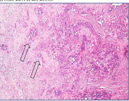
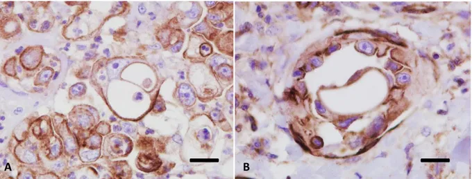
Figure 2 - Bitch, simple tubulo-papillary mammary carcinoma. Histological stage I indicates tumors that are locally invasive; in this field it is clear that extensions of neoplastic cells (indicated by the arrows) are invading the surrounding stroma. (Haematoxylin-eosin, 40X). (From: Levi et al., 2016).
Figure 3 - Bitch, simple tubulo-papillary mammary carcinoma. Histological stage II indicates those tumors with neoplastic emboli and/or metastases in regional lymph nodes. Carcinomas metastasise predominantly via the lymphatic system: panel (a) a large embolusinside a vessel; panel (b) numerous emboli within the lymphatic system (arrows), but no embolization in blood vessels (empty arrows). (Haematoxylin-eosin, 100X). (From: Levi et al., 2016)
35
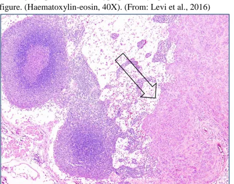
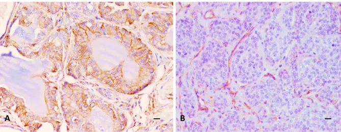
Figure 4 - Bitch, inguinal lymph node. Histological stage II refers to those tumors with neoplastic emboli and/or metastases in regional lymph nodes. In the right of the figure there is an evident metastasis (indicated by the arrow) of a mammary carcinoma expanding the draining lymph node. Two hyperplastic lymphoid follicles can be seen in the left of the figure. (Haematoxylin-eosin, 40X). (From: Levi et al., 2016)
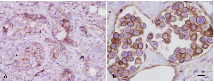
Figure 5 - Bitch, inguinal lymph node excised during mastectomy. IHC staining performed with an antibody against pancytokeratin shows a microfocus of metastatic neoplastic epithelial cells at low (a) and high magnification (b). (DAB stain, counterstaining with Papanicolaou haematoxylin; a:100X, b:400X). (From: Levi et al., 2016).
36
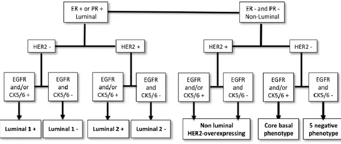
Phenotypic Classification of Canine Mammary carcinomas
Similarly to breast cancer in women, mammary carcinomas in bitches and queens constitute a heterogeneous group of tumors with regards to both morphology and biological behavior, independently of histological type (Abadie et al., 2017; Brunetti et al., 2013; Gama et al., 2008; Peña et al., 2013). Studies of the gene expression profile of breast cancer have identified molecular subtypes with different pathobiology and prognosis (Kumar et al., 2014; Perou et al., 2000; Sørlie et al., 2001a; Yang et al., 2007). Gene expression profiling is considered the gold standard for identifying subtypes of breast cancer, but it is impractical for routine clinical use and cannot be applied to tissue samples embedded in paraffin. For this reason it has been suggested to use a panel of immunohistochemical markers to divide the various carcinomas into subgroups mirroring those determined by gene expression studies. The suggested antibody panel contains antibodies targeting estrogen receptor (ER), progesterone receptor (PR), epidermal growth factor receptor 2 (HER2), CK5/6 and CK14, enabling tumor subtypes to be defined as luminal-like if they express receptors for either estrogen or progesterone, and non-luminal-like if they do not express hormone receptors (Kumar et al., 2014). This latter category is further divided into HER2-overexpressing tumors, which overexpress the HER2 receptor (an important receptor for the corresponding growth factor, often amplified in malignant neoplasms), and basal-like tumors which have a phenotype similar to basal cells; finally, the normal-like subtype comprises the neoplasms that are negative for all three markers (Perou et al., 2000; Sørlie et al., 2001). This classification has been consistently used in human medicine: each subtype has a different prognosis, requires different treatment and has a different metastatic pattern. The non-luminal-like subtypes are those with the more aggressive clinical behavior (Kumar et al., 2014). Being able to apply this new molecular-based system of classification to mammary tumors in veterinary medicine would be profitable: the possible role that each subtype would have as a prognostic indicator independently of the histological subtype, and more reliably, given the already described limitations of histological typing for prognostic purposes, it would open the way to a targeted treatment (Abadie et al., 2017; Peña et al., 2014; Sassi et al., 2010). In fact, in human medicine the cornerstone of treatment of breast cancers with a luminal phenotype (expressing hormone receptors) is inhibition of the ER through targeted therapy (estrogen modulators, aromatase inhibitors) (den Hollander et al., 2013). In contrast, for those tumors that show amplification of