UNIVERSITA’ DELLA CALABRIA
Dipartimento di Farmacia e Scienze della Salute e della Nutrizione
Dottorato di Ricerca in
MEDICINA TRASLAZIONALE (XXX
CICLO
)
Targeting systems vulnerabilities in uveal melanoma by
CRISPR Cas/9 focal adhesion kinase (FAK) genome
editing and therapeutic inhibition
Settore Scientifico Disciplinare
MED/04
Coordinatore del Dottorato:
Ch.mo Prof. Sebastiano Ando’
Tutor:
Ch.mo Prof. Marcello Maggiolini
Dottorando:
Dott. Damiano Cosimo Rigiracciolo
Index
Italian Abstract………...………... ……. ……..1
English Abstract………...………..2
Chapter 1
Introduction
1.1
Uveal Melanoma:clinical burden……….3
1.2
Epidemiology………...3
1.3
Etiology and risk factors………..4
1.4
Clinical presentation and diagnosis……….4
1.5
Primary treatment of Uveal Melanoma………...5
1.6
Metastatic Uveal Melanoma treatments………..7
1.7
Systemic therapies in Uveal Melanoma………..9
1.8
Genetic mutations in Uveal Melanoma……….10
1.9
Clinical implications………..13
1.10 Focal adhesion kinase (FAK)………....14
1.11 FAK structure organization………...15
1.12 Regulation of FAK expression………..16
1.13 Regulation of FAK activity………...17
1.14 Role of FAK in cancer cells………...18
1.15 Novel strategies targeting FAK with pharmacological inhibitors……….20
Chapter 2
Materials and methods
2.1 Cell lines, culture procedures, and chemicals………24
2.2 Mutational analysis………24
2.3 Immunoblot analysis………..24
2.4 Immunoprecipitation assay……….25
2.5 siRNA transfection……….25
2.6 Cell prolifeartion analysis………..25
2.7 Focal adhesion assay………..25
2.8 Methylcellulose growth assay………25
2.9 Genetic editing with CRISPR/Cas9………...26
2.10 In vivo mice experiments and VS4718 treatment……….26
2.11 Data and statistical analysis………..26
Chapter 3
Results
3.1 PTK2 gene is overexpressed in Uveal Melanoma………..27
3.2 GNAQ activates FAK in UM cancer cells ……….29
3.3 FAK mediates the proliferation of uveal melanoma cells through the
PI3K/AKT/mTOR signaling pathway ………..32
Chapter 4
Discussion
……….36
References
………....39
Publications
……….54
Abstract
Il Melanoma Uveale rappresenta la neoplasia intraoculare più frequente nell’età adulta. Colpisce circa 2,500 individui ogni anno negli USA ed il 50% dei pazienti affetti da tale neoplasia sviluppa metastasi entro 5 anni dalla diagnosi. Non essendo state ancora identificate terapie efficaci, la sopravvivenza in presenza di metastasi è di circa 6 mesi. Il Melanoma Uveale è geneticamente caratterizzato dalla presenza di mutazioni somatiche attivanti a carico degli oncogeni GNAQ e GNA11, che codificano per due diverse subunità α delle proteine G. Tali mutazioni sono state identificate rispettivamente in circa il 94% dei casi di Melanoma Cutaneo ed il 4% dei casi di Melanoma Uveale.
Sulla base di tali osservazioni, nel presente lavoro di tesi è stato valutato il ruolo esercitato da una proteina citoplasmatica ad attività tirosin-chinasica associata ai recettori per le integrine denominata FAK (focal adhesion kinase), nella progressione del Melanoma Uveale, sia in vitro che in vivo. In particolare, mediante analisi bioinformatica (www.cbioportal.com) delle alterazioni genomiche di campioni estratti da pazienti affetti da melanoma uveale (n=80), è stato inizialmente determinato che il gene codificante per FAK (PTK2) risulta over-espresso nel 56% dei casi. Inoltre, il presente studio condotto in cellule di Melanoma Uveale OMM1.3 (GNAQ/11 mutate) e in cellule ingegnerizzate per l’espressione di un recettore di membrana accoppiato a proteine-G (Gαq) attivato esclusivamente da ligandi sintetici denominate HEK293 DREADD/Gq, ha dimostrato il coinvolgimento di segnali mediati da GNAQ nell’attivazione di FAK attraverso il reclutamento del fattore coinvolto nello scambio di nucleotidi guaninici denominato TRIO e la proteina appartenente alla super-famiglia di Ras denominata Rho-A. A riprova, saggi biologici hanno dimostrato l’efficacia di specifici inibitori di FAK nei processi di proliferazione cellulare sia in cellule di Melanoma Uveale derivanti da lesioni primarie che da metastasi epatiche. Attraverso l’innovativo approccio genetico denominato CRISPR/Cas 9 genome editing (Clustered Regularly Interspaced Short Palindromic Repeats), il silenziamento dell’espressione di FAK ha ridotto significativamente la crescita del melanoma uveale in modelli sperimentali utilizzati in vivo. Collettivamente, i risultati ottenuti indicano che FAK può essere considerato un potenziale target terapeutico per il trattamento del Melanoma Uveale e di altre neoplasie caratterizzate da mutazioni oncogeniche a carico delle subunità αq/α11 dei recettori di membrana accoppiati a proteine G.
Abstract
Uveal melanoma (UM) is the most common primary cancer of the eye in adults. It is diagnosed in about 2,500 adults in USA every year and approximately 50% of UM patients develop liver metastasis mostly within 5 years after diagnosis, independently of the successful treatment of the primary lesions. The survival of metastatic UM patients is often only few (2-6) months. To date, there are not effective options to treat or prevent UM metastasis. UM is genetically characterized by mutually exclusive activating mutations in the
GNAQ and GNA11 oncogenes, which encode heterotrimeric Gαq family members. These mutations have
been identified in about 92% and 4% of uveal and skin melanomas, respectively. Here, we focused on a cytoplasmic protein tyrosine kinase associated with integrins namely focal adhesion kinase (FAK), which modulates important cell processes such as growth, survival, migration and angiogenesis. By the analysis of UM genomic alterations (TCGA), we found that the gene encoding FAK (PTK2) is amplified or overexpressed in >56% of all UM lesions. UM represents the human cancer harboring the highest levels of FAK, which we confirmed by immune histochemical analysis of a large collection of UM lesions. We found that Gq-GPCRs triggers a rapid phosphorylation of FAK at position Y397, which reflects its activation, through a guanine nucleotide exchange factor (TRIO)/Rho-A signaling circuitry. This promotes the assembly of focal adhesions, independently of the PLCβ/Ca2+ second messenger system and the canonical PKC pathway. Next, we have assessed whether Gαq promotes the FAK-dependent proliferation of uveal melanoma cells. Both, CRISPR/Cas9 genome editing of FAK as well as the use of FAK inhibitors under clinical evaluation for other diseases resulted in reduction of UM tumor growth in vivo. The impact of these FAK inhibiting strategies in UM metastasis is under current evaluation. Collectively, our findings support the potential clinical benefit of targeting FAK as a precision therapy approach for UM and other GNAQ-driven malignancies and pathological conditions.
Chapter 1
Introduction
1.1 Uveal Melanoma: clinical burden
Uveal melanoma (UM) is the most common primary intraocular malignancy in adults displaying a high propensity for metastasis to the liver within 5-10 years after diagnosis, independently of the successful treatment of the primary lesions [1]. UM is a rare subtype of melanoma, representing ~5% of all melanoma tumors. Approximately 90% of all uveal melanomas involve the choroid, with the remainder arising in the ciliary body or the iris of the eye (Fig. 1.1) [1]. The terms choroidal melanoma and ocular melanoma are alternative terms for this cancer, because most of the uveal tract is choroidal. However, the term ocular melanoma should be avoided, because it implies the inclusion of conjunctival and adnexal melanomas, which behave like cutaneous rather than uveal primaries. UM can also rarely arise in melanocytes in the conjunctiva–melanoma of the conjunctiva accounts for ~2% – 3% of all eye neoplasms.
Figure 1.1: schematic representation of uveal melanoma origin sites
1.2 Epidemiology
The incidence rate of UM ranges from 0.2 to 0.3 per million individuals in African/Asian populations to up to 6 per milion individuals in white population [2]. The average age of diagnosis is ~60 years and it affects both sexes equally or slightly more frequently males as per some reports [2] (Fig. 1.2). More recently, an increase in the mean age at diagnosis for the interval between 1973 and 2009 was described based on the analysis of 7043 UM patients from the Surveillance, Epidemiology, and End Results Program (SEER) database [3]. Furthermore, there is a strong difference in the incidence of UM for different ethnic groups: the annual age-adjusted incidence is 0.31 for Afro-Americans, 0.38 for Asians, 1.67 for Hispanics, and 6.02 for non-Hispanic whites [4], yet prognosis does not differ for ethnic groups [4]. The European Cancer Registry-based study on survival and care of cancer patients (EUROCARE) for the years 1983–1994 reported similar incidence rates with a characteristic increase from south to north Europe, from <2 per million in Spain and Southern Italy to >8 per million in Norway and Denmark [5].
Figure 1.2: epidemiology of uveal melanoma in both sexes
1.3 Etiology and risk factors
The etiology of UM is still unclear. A variety of risk factors have been identified, incuding the presence of light eyes, fair skin color, red/blond hair, and BAP1 mutations [6]. In contrast to Cutaneous Melanoma, the impact of ultraviolet (UV) light exposure is less clear for UM: although there are effective therapeutic strategies to eradicate primary UM. Cornea, lens, and vitreous body absorb almost all wavelengths below 300 nm and much of the spectrum between 300 and 400 nm. However, age-dependent alterations of the vitreous body [might alter the absorptive capacity of the latter. The associations between UM risk and blue iris or a generally weakly pigmented phenotype and sun exposure suggest a role for UV radiation in the etiology of UM [6]. If there is a role for UV light in UM etiology, it is certainly by far weaker than that for CM. The etiologic effect of UV radiation for UM is likely too weak to overcome confounding factors such as co-distribution of weakly pigmented skin and iris and latitude, co-occurrence of UV radiation with light of longer wavelengths, and protective, vitamin D-mediated effects of sun exposure. Interestingly, a recent study has demonstrated that posterior choroidal melanomas occurring in illuminated areas were associated with frequent adenine-to-cytosine mutations, whereas ciliochoroidal melanoma arising from unilluminated areas are associated with frequent adenine-to-thymine mutations and light eye color, suggesting that both light eye color and sunlight may be indipendent risk factors associated with different anatomic and mutation profiles [7].
1.4 Clinical presentation and diagnosis
The most common presenting symptom in patients with primary UM is blurred vision (37.8%); howewer, many patients are asymptomatic at the time of diagnosis (30.2%). Other common symptoms at presentation include: painless loss or distortion of vision (metamorphopsia) (2.2%), a serous (fluid) retinal detachment, (photopsia) (8.6%), floaters (7%), visual filed loss (6.1%), visible tumor (3.1%) and pain (2.4%). Other diagnoses to be considered when assessing lesions concerning for UM are dependent upon location based on
the evaluation of iris or posterior lesions of the eye (Fig.1.4). For example, when UM affects the anterior segment of the eye, patients may notice discoloration of the iris or persistent injection of the episclera; also, chronic conjunctivitis may represent a referring diagnosis. Rarely, a blind eye or an eye with a dense cataract may harbor an occult melanoma. Patients with suspicious pigmented lesions should be assessed by an ophthalmologist who has clinical expertise in ocular tumors. The presence of subretinal fluid and orange pigment and the documented growth on fundus photography are findings that support the diagnosis of UM. Drusen and pigment epithelial changes are more suggestive of a benign lesion. Fluorescein angiography can demonstrate an intrinsic secondary vasculature of the choroid; however, ocular echography is the single most effective diagnostic tool available to the clinician. Melanomas tend to exhibit low internal reflectivity as well as an intrinsic acoustic quiet zone on ultrasound. Most are dome-shaped, but a collarstud or mushroom configuration is highly suggestive of melanoma. The shape occurs after a break in Bruch’s membrane, a structure of the retina. Some reports suggest a correlation between increased tumor thickness and the risk of distant metastasis. Most experts agree that a lesion measuring >3 mm in apical height is likely a melanoma. Rarely is a clinical biopsy necessary to confirm the diagnosis of UM. The Collaborative Ocular Melanoma Study reported >99% diagnostic accuracy for eyes with typical features that were enucleated [8]. In some instances, a diagnostic biopsy may be indicated, particularly when the lesion is amelanotic or difficult to assess because of vitreous hemorrhage or debris. Fine-needle aspiration can be performed but requires the assistance of a skilled cytologist who is familiar with ocular pathology.
Figure 1.4: different diagnosis of uveal melanoma by location
1.5 Primary treatment of Uveal Melanoma
Before ocular therapy, a systemic workup should be performed to demonstrate lack of distant metastasis; once it is confirmed that disease is limited to the eye, local ophthalmic therapy can be focused on the primary neoplasm. Distant metastasis is rare at the time of initial UM presentation, occurring in <5% of patients. If distant disease is present, a local therapy for the eye may be deferred in favor of systemic treatment, although this depends on the symptomatology of the patient with regard to the eye. It cannot be emphasized enough that the management of UM is highly individualized; what follows are general guidelines and principles used by leading ocular oncologists in North America and Western Europe.
Close serial observation: in most instances, it is best to consider this approach for patients who have ocular lesions with indeterminate findings that are not typical of melanoma. The ophthalmologist will 5
often monitor patients for definitive features, such as rapid growth or the development of subretinal fluid. In very rare instances, observation may be the preferred approach if the patient is too frail for surgical intervention to either enucleate or place a radionuclide plaque.
Laser therapy: diode laser therapy, also konwn as transpupillary thermo therapy (TTT) and photodinamyc laser photocoagulation are modalities that direct focused energy to destroy tumor vascular supplies and reduce local recurrences by injecting and activating light-sensitive compounds and free readicalsis well tolerated but has limited value, because local relapse rates are as high as 20%. TTT has been effective as primary therapy in up to 80% of cases of small or indeterminate lesions with few risk factors. Howewer, the rate of tumor control with laser therapy varies inversely with tumor size. Therefore, it is best considered for small tumors (<3mm thickness) arising at a distance from the macula and the optic nerve. More commonly, this modality is used in an adjuvant setting after radiation.
Radiation therapy: focal radiation therapy is the most common globe-salvaging approach used by ocular oncologists. The Collaborative Ocular Melanoma Study medium-size trial randomized patients who had tumors that measured from 2.5 to 10.0 mm in apical height to primary brachytherapy (with iodine-125 plaques) or enucleation. In that trial, no statistically significant difference was observed in melanoma-related mortality between the 2 cohorts [9]. Since then, ocular brachytherapy has emerged as the most common globe-sparing modality for tumors within these parameters. Patients who have melanomas with an apical height >10.0 mm can be treated using this approach but are more likely to experience severe radiation retinopathy and visual loss. In USA, iodine-125 is the most commonly used isotope. Other radioisotopes are also used; ruthenium-106 is the preferred isotope in many European centers. If available, charged-particle and proton-beam radiation are alternative to brachytherapy. Most clinicians agree that both modalities have high rates of local tumor control, as high as 98% in some series.
Surgery: enucleation (removal of the globe) is the most common surgery performed for UM, and is appropriate for patients with vision loss, extensive extraocular growth, circumferential tumor invasion and large tumor diameter. Alternative surgical modalities include transretinal endoresection and trans-scleral resection: both procedures are site-and surgeon-dependent, with the majority of data coming from single-institution case series. Uvectomy (selective excision of the tumor with retention of the globe) is best limited to small anterior tumors of the eye, such as iris melanomas. Patients who undergo selective choroidal resection of their melanomas may benefit from adjuvant plaque radiotherapy to reduce the risk of ocular recurrence. Surgery affords the most detailed histopathologic assessment and can confirm microscopic extraocular extension. Epithelioid cell type and the presence of microvascular loops are associated with a worse prognosis. Exenteration (surgical removal of the globe and adjacent orbital contents) is rarely indicated and is limited to extreme cases of massive orbital involvement and patients who are receiving palliative care.
In addition, in patients with UM, it’s believed that survival and the risk of distant relapse are independent of the method selected for primary tumor management. Some authorities have suggested that micrometastatic disease precedes local therapy. However, because metastatic risk correlates with the size of the primary tumor, some ocular oncologists now take a more aggressive local approach to the management of smaller primary and indeterminate tumors. A few adjuvant studies have been conducted in UM in an attempt to prevent metastatic disease. However, none of these studies demonstrated meaningful metastasis free survival benefit or overall survival (OS) benefit, including a small phase 3 using methanolextracted residue of bacillus Calmette-Guerin [10] and 2 single-arm studies of interferon-α using matched historic controls. Adjuvant intra-arterial hepatic infusion of the alkylating agent, fotemustine, was studied in an effort to reduce the occurrence of liver metastasis, because the liver is the most common site of metastasis and cause of death from UM [11]. In that study, 22 UM patients with choroidal involvement, and longest basal dimension >20 mm, extrascleral extension or apical height >15 mm were treated with fotemustine for 6 months. In this contest, adjuvant intra-arterial hepatic fotemustine was not shown to improve survival compared with matched historical controls. There are several ongoing adjuvant clinical trials in the USA such as ipilimumab, sunitinib, valproic acid, and crizotinib for high-risk patients. These agents or classes of agents have been chosen for study based on the molecular characteristics of UM cells or expected immunomodulatory or microenvironment effects.
1.6 Metastatic Uveal Melanoma treatments
Since the liver represents the first and only site of metastasis in most patients with UM, and the prognosis of patients with metastatic UM is highly dependent on the presence of liver metastasis and disease progression in the liver [12], liver-directed local treatments such as surgical resection, hepatic artery embolization, hepatic arterial infusion of chemotherapy, and radiofrequency ablation have been used in patients with metastatic uveal melanoma. A survival benefit has been demonstrated in patients who undergo surgical resection of liver metastasis compared with non-surgical control groups in multiple retrospective studies, but this benefit is limited to those who have minimal tumor volume that is limited to the liver and who also are fit enough for surgery (in a population with an average age at diagnosis of 70 years); and, according to these criteria, <10% of patients who have metastatic UM are candidates for liver resection [13].
Direct targeting of the hepatic arterial circulation is an enticing anatomic option for patients with liver-predominant disease, because the normal liver will receive blood supply from the portal system, whereas metastatic tumors generally are supplied predominantly by the hepatic artery. Hepatic arterial infusion of fotemustine (Fig. 1.6.1) has been studied in patients with UM metastatic to the liver in a randomized phase 3 study that assigned 171 patients to receive fotemustine either intravenously or by hepatic arterial infusion [14]. Although hepatic arterial infusion of fotemustine significantly improbe progression-free survival (PFS) compared with intravenous administration (median PFS, 4.5 vs 3.5 months; hazard ratio [HR], 0.62; 95% 7
confidence interval [CI], 0.45-0.84; P 5 .002), there was no improvement in OS (median OS, 14.6 vs 13.8 months; P5.59).
Figure 1.6.1: chemical structure of fotemustine
Isolated hepatic perfusion (IHP) is another option. In contrast to hepatic arterial infusion, IHP is a closed-circuit perfusion that delivers high doses of chemotherapy to the liver with minimized systemic drug exposure (Figure 1.6.2). IHP is an invasive and precise operative procedure that requires great skill and a period of extracorporeal circulation. A simple derivative percutaneous procedure known as percutaneous hepatic perfusion (PHP) has been developed. In a phase 3 trial, 93 patients were randomized to either PHP with melphalan or best supportive care, and crossover to PHP was allowed for those in the best supportive care arm after hepatic progression [15]. In that trial, there was a significant improvement in the median hepatic PFS (245 vs 49 days; P < .001) and the overall response rate (34.1% vs 2%; P < .001) compared with best supportive care.
Figure 1.6.2: isolated hepatic perfusion circuit
Chemoembolization which combines hepatic artery embolization with infusion of concentrated doses of chemotherapeutic agents like cisplatin and 1,3-bis (2-cholorethyl)-1-nitrosourea, is another commonly used liver-directed therapy for metastatic UM. Gelatin sponge and non spherical polyvinyl alcohol particles have been used as embolic agents for chemoembolization. Because these embolic agents have an unpredictable level of arterial occlusion on account of their irregular shape and heterogeneous calibration, spherical embolic agents and drug-eluting microspheres have been developed and increasingly are being used. In a retrospective study of 201 patients with UM involving the liver, systemic chemotherapy, intra-arterial hepatic chemotherapy, and chemoembolization were compared [12]. In that study, although the rate of objective response to systemic treatment was <1%, chemoembolization induced a 36% objective response rate with a median response duration of 6 months.
Also, another important liver-directed approach is the radioembolization by using yttrium-90 (90Y) radiospheres. 90Y is small enough to pass deep into tumor vessels (2-4 mm tissue penetration of radiation) but not through the capillary, thus sparing normal liver surrounding the tumor (Figure 1.6.3). Two retrospective studies of 90Y produced high response rates of up to 62% (8 of 13 patients) with a median OS of 7 to 10 months [16-17]. Currently, a prospective phase 2 study of 90Y plus ipilimumab is ongoing to evaluate the clinical activity in patients with UM metastatis to the liver.
Figure 1.6.3: Yttrium-90 PET following radioembolization
1.7 Systemic therapies in uveal melanoma
Chemotherapy: UM is highly resistant to systemic cytotoxic chemotherapy. Several clinical trials of single-agent or combination chemotherapy have produced disappointing results, with objective response rates of <1%. [18-20]. On the basis of promising activity in patients with advanced cutaneous melanoma, biochemotherapy with either bleomycin, vincristine, lomustine, dacarbazine, and interferon-a or with fotemustine, interferon-a, and interleukin-2 has been studied in 4 phase 2 trials for patients with metastatic UM [21-22]. Objective response rates in those studies were <20%, and the median OS was only 12 months with significant pulmonary toxicities, neurotoxicities, and myelosuppression. To date, no clear role has been established for chemotherapy or biochemotherapy in patients with metastatic UM.
Immunotherapy: in March 2011, ipilimumab, a human antibody that blocks the immune checkpoint interaction between cytotoxic T-lymphocyte–associated protein 4 (CTLA4) and B7 protein 1 (B7.1) on antigen-presenting cells and/or target tumor cells, was approved for the treatment of advanced melanoma based on improved OS in a large, randomized, phase 3 study [23]. Although ipilimumab has been extensively studied in cutaneous melanoma, only limited data for ipilimumab are available in UM. For example, in a retrospective review of 39 patients with metastatic UM who received ipilimumab at doses of either 3 mg/kg (N 5 34 patients) or 10 mg/kg (N 5 5 patients) [24], the objective response and disease control rates (with the addition of patients who had stable disease at 12 weeks to objective responders) were 2.6% and 46%, respectively, at week 12 and 2.6% and 28.2%, respectively at week 23. The median OS was only 9.6 months. The efficacy of ipilimumab in UM also was investigated in other studies.
The programmed death 1 protein (PD-1) is another important immune checkpoint receptor expressed on the surface of T cells. PD-1 has 2 known ligands, PD-L1 (B7-H1) and PD-L2 (B7-DC). The ligation of PD-1 with PDL1 inhibits T-cell proliferation and activation and induces apoptosis of antigen-specific T cells to suppress antitumor immunity. Recently, pembrolizumab (humanized) and nivolumab (human) immunoglobulin G4 (IgG4) anti-PD1 monoclonal antibodies were approved by the Food and Drug Administration for the treatment of metastatic melanoma after ipilimumab (and v-Raf murine sarcoma viral oncogene homolog [BRAF] inhibitor therapy for patients who have melanoma with BRAF mutations). The efficacy of PD-1 blockade has not yet been reported in metastatic UM. In a single-center EAP, 7 patients with metastatic UM received 2 mg/kg of pembrolizumab every 3 weeks [25]. Two patients had objective responses (1 complete response and 1 partial response), and the median PFS was 12.2 weeks in that report. Currently, several clinical trials of immunotherapy including adoptive cell therapy and combined ipilimumab and nivolumab, are under way to identify effective immunotherapeutic approaches in metastatic UM.
Targeted therapy: 80% of UMs have oncogenic mutations in GNAQ or GNA11, and these mutations are potential drivers of mitogen-activated protein kinase (MAPK) activation, similar to oncogenic BRAF mutations in cutaneous melanoma [26]. Therefore, inhibition of the MAPK pathway has been studied in metastatic UM. A randomized phase 2 trial of selumetinib, a selective MAPK kinase (MEK) inhibitor, produced promising preliminary outcomes for UM [27]. In that study, 101 patients with metastatic UM were randomized to receive either selumetinib or temozolomide (or dacarbazine). The median PFS in the selumetinib group was 15.9 weeks with a median OS of 11.8 months, whereas the chemotherapy group had a median PFS of 7 weeks and a median OS of 9.1 months (P 5 .09 for OS). This was the first randomized study to demonstrate improved PFS in patients with metastatic UM, although an OS benefit was not observed. Currently, several MEK-inhibitors based clinical trials are underway or have been completed, including a randomized, double-blind, phase 3 study called SUMIT comparing selumetinib plus dacarbazine versus placebo plus dacarbazine (completed; no difference in PFS) and studies of MEK inhibitors with protein kinase C or AKT inhibitors. These studies will provide insight into the role of MEK inhibitors, their molecular targets, and other interacting pathways in the treatment of metastatic UM.
1.8 Genetic mutations in uveal melanoma
Oncogene and tumor-suppressor mutations that are common in other cancers are mostly absent in UM, a disease that is characterized by a low mutation burden. UM also differs in its genetic mutation profile from conventional cutaneous melanoma, in which BRAF and NRAS mutations dominate. These driver mutations, which control the biology of up to 70% of cutaneous melanomas, are absent/rare in UM [28]. UM has, in many cases, a poor prognosis since about half of all patients develop metastatic disease, predominantly to the liver. By using gene expression profile (GEP) classification, UM can be stratified into two distinct molecular classes with a significant difference in prognosis [29]. Class 1 tumors can be further divided into two
subgroups (class 1A and 1B) and has in general a good prognosis and low metastatic risk, whereas class 2 tumors have high metastatic risk and thereby a worse prognosis. The risk of metastasis has been determined to be 2% for class 1A tumors, 21% for 1B tumors, and finally 72% for class 2. The different molecular classes are also associated with mutations in different UM driver genes [30]. The most frequently mutated genes that are considered to be drivers in UM development and progression are: BAP1, EIF1AX, GNA11, GNAQ, and SF3B1 [26, 30-32]. Also, activating mutations occur in PLCβ4 (phospholipase C β4), the downstream effector of Gaq signaling [26, 31] and in cysteinyl leukotriene receptor 2 (CYSLTR2), a seven-transmembrane G-protein-coupled receptor (GPCR), which functions in leukotriene-mediated signaling [33]. Microarrays and gene copy numbers from single nucleotide polymorphism arrays revealed that 42.2% of uveal melanoma samples harbored mutated GNAQ, 32.6% harbored mutated GNA11, 31.5% harbored mutated BAP1, 9.7% harbored mutated SF3B1, 18.9% harbored mutated EIF1AX and 1% harbored mutated TERT [34]. Of these, GNAQ and GNA11 mutations are usually mutually exclusive, but both can coexist with BAP1 or SF3B1 mutations [52]. Likewise, BAP1 and SF3B1 mutations are mutually exclusive, as are EIF1AX and SF3B1 mutations, whereas TERT mutations appear to coexist specifically with GNA11 or EIF1AX mutations [32, 34].
• BRCA1-associated protein 1 (BAP1): BAP1 encodes a deubiquitinating hydrolase with multiple cellular functions, such as regulation of chromatin dynamics, DNA damage response, cell cycle regulation, and cell growth. It is involved in the polycomb multiprotein repressor complex that is critical for transcriptional silencing of target genes by removing ubiquitin molecules from histone H2A. As a consequence of this functional loss, an accumulation of monoubiquitinated histone H2A has been revealed, which in turn was found to cause a more de-differentiated phenotype [35]. Furthermore it has been shown that BAP1 may interact with promoters regulated by the E2F transcription factor 1 gene (E2F1) and, thus, may affect the cell cycle progression genes controlled by E2F [36]. BAP1 is able to display tumor suppressor capacity by binding to the BRCA1 protein and thereby enhancing BRCA1-mediated tumor suppression [37]. BAP1 mutations strongly correlate with metastatic disease in UM: over 80% of metastasizing UM has been found to carry a mutation in this gene [30]. Most of the BAP1 mutations are truncating variants or missense variants affecting the ubiquitin carboxyl-terminal hydrolase domain. In some cases, BAP1 is not altered by a sequence mutation but by hemizygous deletion of one or more exons. Such alterations may be missed by traditional Sanger sequencing because of the presence of normal DNA in the sample. Also, BAP1 is frequently mutated in other tumor types, including cholangiocarcinoma, renal cell carcinoma, mesothelioma, and bladder cancer (www.cbioportal.org). Several of these cancer types are part of the hereditary cancer syndrome known as tumor predisposition syndrome that is characterized by germline mutations of BAP1 in patients belonging to cancer-prone families. Currently, there is no consensus among genetic counseling groups regarding who should be screened and surveillance strategies for patients who have germline BAP1 aberrancy.
• Splicing factor 3b subunit 1 (SF3B1): SF3B1 located at chromosome 2 is another driver gene identified by whole-exome sequencing of UM tumors. SF3B1 is essential in pre-mRNA splicing by encoding the unit of the splicing factor 3b protein complex that is a critical part of both major (U2-like) and minor (U12-like) spliceosomes. SF3B1 has recently also been designated as a factor involved in DNA damage repair [38]. Missense mutations in specific regions of the SF3B1 gene have been found to alter the splicing of many target genes [39]. Uveal melanoma is among a small group of cancers associated with SF3B1 mutations: these mutations define a genetic subset of uveal melanoma that have been reported to be associated with favorable prognostic features and to be nearly always mutually exclusive of BAP1 mutations [30]. SF3B1 mutations are observed in codon 65 and mostly in tumors without loss of all or part of chromosome 3. Furney et al. have reported an association of these mutations with alternative splicing of transcripts [40].
• Eucaryotic Translation Initiation Factor 1A X-Linked (EIF1AX): EIF1AX, located on the Xp22 chromosome, encodes the eukaryotic translation initiation factor 1A (eIF1A). This factor is essential in the initiation phase of translation of eukaryotic cells by the transfer of methionyl initiator tRNA to the small (40S) ribosomal unit and stabilizes the formation of the ribosome around the AUG start codon, which enables translation. EIF1AX was identified as a UM driver gene by whole-exome sequencing [32]. Approximately 14%–20% of all UM carries a mutation in this gene, with most mutations found in exons 1 and 2 [32, 34]. EIF1AX mutations usually occur in non metastatic cases, are associated with class 1 GEP tumors and good prognosis, and are inversely associated with metastasis [41]. EIF1AX mutations are usually mutually exclusive with BAP1 mutations and to a large extent also to SF3B1 mutations. As expected most EIF1AX mutations are identified in tumors with disomy 3 (48%) and rarely occur in monosomy 3 tumors (3%) [32]. In contrast to, for example, BAP1 mutations, which mainly are truncating and loss-of-function variants, the majority of the EIF1AX mutations are heterozygous non synonymous variants, or in some cases splicing variants, leading to deletions of one or two amino acids. Thus, in most cases, the core protein remains unchanged.
• GNAQ and GNA11: GNAQ encodes the alpha subunit (Gaq) and GNA11 the alpha subunit 11 (Ga11), both being guanine nucleotide–binding proteins belonging to the heterotrimeric protein family, which are of importance in transmembrane signaling systems. The alpha subunits serve as a switch between the G-proteins active state – when bound to guanosine triphosphate (GTP) – and the inactive state – when GTP is hydrolyzed to guanosine diphosphate. Activating mutations in GNAQ/GNA11 were the first described driver mutations in UMs. GNAQ and GNA11 mutations occur in a mutual exclusive pattern and are exclusively found in codon 209 and in some cases in codon 183 [26, 31]). Mutations at these positions lead to a constitutive activation of the Gaq and Ga11 subunits by abolishing their intrinsic GTPase activity, thereby preventing the return to an inactive state. In total, ~85% of all UMs carry a mutation in either of these genes. Both GNAQ and GNA11 have been found to upregulate the MAP kinase pathway when constitutively activated in a similar fashion as BRAF and NRAS mutations (Figure 1.8.1).
The activation of the MAPK pathway in the absence of BRAF/NRAS mutations in UMs was at first unforeseen until the identification of GNAQ and later GNA11 mutations that had the same effect as the
V600EBRAF mutation. Cell lines with a GNAQ Q209L mutation have also been found to be highly
sensitive to mitogen-activated protein kinase (MEK) inhibition [31]. Mutations in these genes have not been associated with the two different molecular classes of UM tumors. In addition, GNAQ/GNA11 mutations have not been reported to be of prognostic value and they occur at similar frequencies in metastatic and non-metastatic lesions. Furthermore, they have not been linked to patient outcomes. Taken all this data into consideration is supportive of GNAQ/GNA11 being early events. The hotspot mutations in GNAQ or GNA11 are also commonly found in benign nevi such as blue nevi [26, 42]. Actually, GNAQ Q209 was most frequently found in blue nevi, observed in 55% of the lesions, whereas 45% of the primary UMs and 22% of the metastatic UM, respectively, carry this mutation [26]. Inverse relationship was seen for the GNA11 Q209 mutation where metastatic lesions showed the highest number (56%) followed by primary UM tumors (32%) and lastly blue nevi (6%) [26]. Mutations affecting codon R183 are less frequent, present in 2% of the blue nevi and 5% of primary UM tumors, GNAQ and GNA11 mutations combined [26].
Figure 1.8.1: signaling pathways in uveal melanoma
1.9 Clinical implications
The identification of driver genes has led to the identification of novel treatment targets and several clinical trials are ongoing investigating these targets in UM therapy. Targeting mutated GNQ/GNA11 directly is difficult because of the molecular nature of the mutations causing an inactivation of intrinsic GTPase within the cell. However, for several downstream molecules of GNAQ/GNA11, targeted therapies have become 13
available. These include mitogenic activated protein kinase/extracellular signal-regulated kinase (MEK) that is shown to be upregulated in GNAQ/GNA11 mutated tumors [43]. Inhibition of MEK has actually been found to decrease the proliferation of UM tumors both in vivo and in vitro [44]. Furthermore, a clinical Phase II trial has shown a prolonged progression-free survival of nearly 9 weeks when treating patients with the MEK inhibitor selumetinib compared to chemotherapy (temozolamide) [45]. However, in another Phase II trial, there was no significant effect on overall survival when treating with semurafenib compared to chemotherapy, although there was a modest increase in response rate and progression-free survival [27]. Other putative downstream targets of GNAQ/GNA11 mutated tumors are protein kinase C and molecules of the protein kinase B (AKT)/mammalian target of rapamycin pathway [46-47]. Also BAP1 mutations are difficult to target directly because of their recessive nature. However, the effects of the mutations are possible to target by the use of histone deacetylase (HDAC) inhibitors in tumors with loss of BAP1 function. The absence of functional BAP1 protein leads to hyperubiquitination of H2A in the cells [35]. The use of HDAC inhibitors can reverse this phenotype, thereby causing a shift from aggressive, de-differentiated class-2 UM cells to more differentiated and less aggressive cells [35]. HDAC inhibitors have also been suggested as adjuvant treatment in high-risk patients [48].
In familial cancer combining clinical and genetic information can be used to improve prognostic estimates and to improve strategies for early diagnosis. By genetic testing of cancer-prone families, the clinical outcome can in many situations be improved, by detecting precursor lesions and tumors at an early stage in members of mutation-positive families. Often, however, this is not straight forward, as in familial melanoma, where a low frequency of mutations in high penetrance genes is seen and risk estimates for mutation carriers have not been well established at this point. Genetic testing is often only recommended when the result is of importance in the management of the patient and where there is a possibility of improving the clinical outcome. However, in families exhibiting the phenotype specific for BAP1 tumor predisposition syndrome, genetic testing should be offered. Additional research will be of importance to elucidate the penetrance and risk of developing different types of cancer in mutation carriers. Identifying the susceptibility factor in cancer-prone families will be of importance for choice of surveillance programs and follow-up of the patient and their relatives. For families with a high cancer burden but without mutation in any known high predisposing gene, next-generation sequencing will be the natural choice to search for novel susceptibility genes. This will subsequently increase the knowledge about genetic susceptibility and may in the future be the basis for improved early detection and prevention of UM as well as lead to the development of new targeted treatments.
1.10 Focal adhesion kinase (FAK)
Focal Adhesion Kinase (FAK) is a 125 kDa non-receptor tyrosine kinase also known as PTK2, that resides at the sites of integrin clustering, known as focal adhesions. FAK plays a significant role in adhesion, survival, 14
motility, metastasis, angiogenesis, lymphangiogenesis, cancer stem cell functions [49-50], tumor microenvironment and epithelial to mesenchymal transition (EMT) [51]. FAK was first discovered in 1992 by several different groups [52], the next year human FAK was isolated [53] and later it was found in Drosophila and other species [54]. Once FAK was isolated in humans it was directly linked to cancer due to its high expression in primary tumors and overexpression in nearly all metastatic tumors, with no detectable FAK mRNA or protein in normal tissues [53].
1.11 FAK structure organization
The human gene encoding FAK namely PTK2 is localized on chromosome 8q24, a region characterized by frequent aberrations in human cancers [55]. Structurally, FAK consists of an amino-terminal regulatory FERM domain, a central catalytic kinase domain, two proline-rich motifs, and a carboxy-terminal focal adhesion targeting domain FAT (a four helix bundle) (Fig. 1.11.1).
Figure 1.11.1: FAK domain structure model
The N-terminal FERM domain of FAK contains one nuclear export sequence (NES) and one nuclear localization sequence (NLS), whereas the FAK kinase domain contains another NES sequence close to the major FAK phosphorylation sites Y576/Y577 suggesting the regulation of FAK through nucleo-cytoplasmic shuttling and supporting nuclear functions of FAK (binding with p53 [56], and Mdm-2 [57] and other partners) [58].
The C-terminal domain of FAK contains two proline-rich regions that function as binding sites for SRC-homology (SH)3-domain-containing proteins. SH3-domain-mediated binding of the adaptor protein p130Cas to FAK is important in promoting cell migration through the coordinated activation of Rac at membrane extensions [59]. The SH3-mediated binding of other proteins, such as GRAF (GTPase regulator associated with FAK) and ASAP1 (Arf GTPase-activating protein (GAP) containing SH3, ankyrin repeat and pleckstrin homology (PH) domains-1), connects FAK to the regulation of cytoskeletal dynamics and focal contact assembly. However, the downstream connections of GRAF and ASAP1 remain undefined. Furthermore, the C-terminal domain encompasses the FAT region which promotes the co-localization of FAK with integrins at focal contacts. The FAK FAT domain also binds directly to an activator of Rho-family GTPases that is known as p190 RhoGEF, and FAK-mediated tyrosine phosphorylation of p190 RhoGEF might be a direct 15
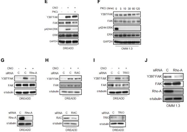
link to RhoA activation [60]. Interestingly, FAK is phosphorylated (P) on several tyrosine residues, including Tyr397, 407, 576, 577, 861 and 925. Tyrosine phosphorylation of FAK on its major site Tyr397 creates a Src-homology-2 (SH2) binding site for Src [61-62]. The interaction between Y397-activated FAK and Src leads to a cascade of tyrosine phosphorylation of multiple sites in FAK, as well as other signaling molecules such as p130CAS and paxillin, resulting in cytoskeletal changes and activation of other downstream signaling pathways [59]. Also, FAK phosphorylation leads to activation of phospholipase Cγ (PLCγ), suppressor of cytokine signalling (SOCS), growth-factor-receptor bound protein 7 (GRB7), the Shc adaptor protein, p120 RasGAP and the p85 subunit of phosphatidylinositol 3-kinase (PI3K) [55]. Phosphorylation of Tyr576 and Tyr577 within the kinase domain is required for maximal FAK catalytic activity, whereas the binding of FAK-family interacting protein of 200 kDa (FIP200) to the kinase region inhibits FAK catalytic activity [63]. Also, FAK phosphorylation at Tyr925 creates a binding site for GRB2 [63]. These structural studies provided a basis for developing of small molecule inhibitors targeting FAK.
1.12 Regulation of FAK expression
Nuclear factor κB (NFκB) and p53 are well-characterized transcription factors which activate and repress the PTK2 promoter, respectively [64-65]. Also, other transcription factors like Nanog [66], Argonaute2 (Ago2) [67], and PEA3 [68] increase PTK2 promoter activity. Nanog promotes FAK expression in colon carcinoma cells and as part of a signaling loop, Nanog activity is increased by FAK phosphorylation [66]. Ago2, a part of the cellular RNA interference machinery, is amplified in hepatocellular carcinoma and induces FAK transcription [67]. Ago2-silencing reduces FAK levels and concomitantly blocks tumorigenesis and metastasis in mice. Elevated PEA3 and FAK levels correlate with metastatic stages in human oral squamous cell carcinoma [68]. PEA3 induces FAK expression in vitro and silencing of either PEA3 or FAK reduces metastasis of human melanoma xenografts. Given the complexity and size of the PTK2 promoter region, it is likely that transcription factor combinatorial effects regulate PTK2 transcription.
FAK is also subject to alternative splicing as PTK2 with deletion of exon 33 (FAK amino acids 956–982), identified in a subset of breast and thyroid patient samples, results in enhanced cell motility and invasion [69]. However, this deletion disrupts FAK linkage to integrins and it is unclear how truncated FAK may function. At the protein levels, FAK is subject to proteasomal or calpain-mediated degradation [70]. Poly-ubiquitination by the E3 ligase mitsugumin 53 (also known as TRIM72) promotes FAK proteasomal degradation during myogenesis, but this has not been tested in tumor cells [71]. However, in general, FAK protein levels are elevated in advanced stage of solid tumors. Together, these results support the notion that elevated FAK expression is connected to several tumor-associated phenotypes.
1.13 Regulation of FAK activity
Several types of signaling events initiate FAK activation. The best-characterized mechanism promoting FAK activation involves engagement of integrins with the ECM and the subsequent co-clustering of proteins like talin and paxillin with the cytoplasmic tail of integrin [72]. This leads to the recruitment of FAK to sites of integrin clustering via interactions with integrin-associated proteins, leading to FAK activation. Other examples of signaling stimuli promoting FAK activation include stimulation by specific growth factors like epithelial growth factor (EGF), hepatocyte growth factor (HGF), vascular endothelial growth factor (VEGF) and platelet derived growth factor (PDGF), activation of particular G-protein-coupled receptors, and the binding of interacting partners of the FAK FERM domain such as ezrin in an integrin-independent manner [63, 70, 72].
The first step in FAK activation involves displacement of the FERM domain from the kinase domain, presumably reflecting binding of a phospholipid or peptide ligand to the FERM domain, allowing rapid autophosphorylation of Y397 [72]. This then creates a high affinity binding site for the SH2 domain of Src, or other SFKs, leads to exposure of the activation loop, and prevents further interactions between the FERM and kinase domains. Src then transphosphorylates additional sites on FAK (Y576 and Y577) on the kinase domain activation loop, leading to full activation [72]. The phosphorylated activation loop also precludes the inhibitory docking of the FERM domain.
Recent studies provide additional mechanistic insights into FAK activation, one of which establishes a phospholipid, phosphatidylinositol-4,5-bisphosphate (PI(4,5)P2), as an important signaling messenger linking integrin signaling to FAK regulation [74]. Integrin-mediated local production of PI(4,5)P2 promotes binding of PI(4,5)P2 to a basic region (K216AKTLRK222) of the regulatory FAK FERM domain, inducing FAK clustering in focal adhesions [74]. FAK subsequently transits into a partially open conformation where the autophosphorylation site Y397 is exposed without re-leasing the autoinhibitory interaction of FERM-kinase domains, but this is sufficient to facilitate autophosphorylation and subsequent Src recruitment to Y397 [74]. Src-phosphorylation of the activation loop then releases the FERM/kinase domain leading to a fully active conformation. Using a variety of complementary approaches including structural and biophysical analyses, Brami-Cherrier et al, identified that autophosphorylation of Y397 requires FAK dimerization, mediated via FERM:FERM and FERM:FAT interactions, and occurs in trans FERM:FAT interaction involves binding of FAT to a basic patch on the FERM domain. Interestingly, paxillin contributes to positive regulation of FAK activity by clustering FAK at focal adhesions and reinforcing FERM:FAT association [75]. In addition, different types of cellular stimuli impact upon FAK activation. For example, elevated intracellular pH positively regulates FAK, in which deprotonation of the His58 FAK residue at high intracellular pH initiates conformational changes that may enhance Y397 autophosphorylation [76].
1.14 Role of FAK in cancer cells
FAK is at the intersection of various signaling pathways promoting cancer growth and metastasis (Fig. 1.14.1). These include kinase-dependent control of cell motility, invasion, cell survival, and transcriptional events promoting epithelial-mesenchymal transition (EMT) [70, 72]. Additionally, kinase-independent scaffolding functions of FAK can influence cell survival or cancer stem cell proliferation
[59, 70,
72]. Understanding the balance between kinase-dependent and -independent functions is the key to the interpretation of FAK-related phenotypes.Figure 1.14.1: Kinase dependent and independent-FAK signalling pathways
Conditional tissue-specific FAK floxed mouse models and chemical FAK inhibitors have allowed the delineation of several FAK-associated pathways. For example, several groups have used polyoma virus middle T antigen (PyMT)-driven breast tumor models combined with tissue-specific FAK knockout through the mouse mammary tumor virus (MMTV) promoter (MMTV-PyMT model) [49, 77] to assess FAK function in tumor progression.
Survival and apoptosis: the first demonstration of a survival function for FAK was performed by Frisch et al, where expression of active FAK was shown to suppress anoikis in Madin Darbin canine kidney (MDCK) and immortalized keratinocyte HaCat epithelial cells [78]. Then, FAK was linked to apoptosis in cancer cells, in which the inhibition of FAK with anti-sense oligonucleotides or with dominant negative FAK (FAK-CD (FAK C-terminal domain)) caused loss of adhesion and apoptosis in tumor cells [79]. Also, down-regulation of FAK with FAK siRNA decreased MCF-7 breast cancer viability and inhibited tumor growth [80]. In addition, FAK was shown to interact with p53 and inhibit its apoptotic activity [56]. Furthermore, nuclear FAK regulated survival through its direct binding to Mdm-2, which promoted p53 ubiquitination and degradation [57]. The anti-apoptotic function of FAK was demonstrated in HL-60 leukemia cells, where
FAK activated the PI-3-Kinase/AKT pathway and induced functions of NF-kappa B and inhibitor of apoptosis proteins (IAPs) [81].
Motility: FAK was shown to play a pivotal role for motility in cancer cells [82]. FAK-null embryos exhibited decreased cell motility [83], whereas overexpression of FAK induced cell motility [84]. The Src and PI-3 Kinase downstream signaling pathways have been shown to be an important role for FAK-mediated cell motility [70].
Invasion and metastasis: tumor cell invasion into the surrounding microenvironment is an important step in cancer progression, allowing cancer cells to form metastasis at secondary locations. This requires transition to a motile phenotype through changes in focal adhesons (FAs) and cytoskeletal dynamics, and alterations in matrix metalloproteinase (MMP) expression or activation to facilitate ECM invasion. EMT, which is driven by a transcriptional program, supports the progression to these invasive properties. FAK was shown to mediate cell invasion and metastasis through promotion of EMT (Fig. 1.14.2). For example, FAK promotes cellular membrane expression of MT1-MMP, a matrix metalloproteinase, which serves to degrade the ECM [85]. Also, FAK activity increases MMP-9 expression and spontaneous breast carcinoma metastasis in a syngeneic and orthotopic mouse model [86]. Other studies show that MMPs regulation and surface presentation in cancer cells involves multiple downstream pathways such as p130Cas58 and the PI3K/Akt/mTOR cascade [87]. Thus, FAK plays an important role in EMT, invasion and metastasis and the details of the down-stream molecular mechanisms of FAK-regulated EMT and E-cadherin mediated cell-cell adhesions or integrin-ECM mediated adhesions and their cross-talk and role in metastasis remain to be discovered.
Figure 1.14.2: FAK promotes survival and invasion in cancer
Angiogenesis: angiogenesis is critical to malignant progression and involves the local formation of nascent blood vessels from pre-existing vasculature through stimulation of ECs and subsequent mobilization, proliferation and sprout formation [63]. FAK integrates angiogenic signals from vascular endothelial growth factor receptors (VEGFRs) and integrin receptors, and directs the migration and growth of endothelial cells 19
to promote angiogenesis [88]. The requirement of FAK in angiogenesis was initially suggested by early observations of restricted patterns of enriched FAK expression in the embryonic vasculature and the embryonic lethality conferred upon FAK gene ablation in mice, which is due to cardiovasculature defects [89]. VEGF-induced FAK activation promotes rapid localization of FAK to endothelial adherens junctions and binding of FAK to vascular endothelial (VE)-cadherin via its FERM domain and FAK-mediated phosphorylation of β-catenin. This subsequently induces β-catenin/VE-cadherin dissociation and increased junctional breakdown. In the context of cancer, the angiogenic function of FAK was demonstrated using EC-specific FAK-null melanoma- or lung-carcinoma bearing mice, which exhibit suppressed VEGF-mediated tumor angiogenesis and growth [90]. Additionally, elevated FAK expression was observed in the vascular and tumor cell compartments of invasive breast cancer specimens [91]. A key mechanism underpinning the pro-angiogenic role of tumoral FAK is induction of VEGF expression. Inhibition of FAK catalytic activity in breast carcinoma cells by stable expression of FRNK reduces FAK Y925 phosphorylation, the ability of the Grb2 adaptor protein to bind to FAK, as well as Erk2 activation [92]. The concomitant impairment of FAK– Grb2–Erk2 signaling results in decreased VEGF expression in vitro and in vivo together with small avascular tumors in mice without affecting cell survival or proliferation in vitro [92]. Reconstitution experiments with a FAK Y925F or impaired kinase activity mutants in Src-transformed FAK-null fibroblasts confirmed the role of this FAK phosphorylation site and catalytic activity in regulating VEGF-associated angiogenesis. Suppression of FAK expression in neuroblastoma, breast and prostate carcinoma cells also results in reduced VEGF expression [72]. Overall, these findings indicate that FAK can play contrasting roles within cancer cells and the surrounding tumor microenvironment, and highlight novel rationales for therapeutic targeting of FAK.
1.15 Novel strategies
targeting FAK with pharmacological inhibitors
FAK has long been considered as a potential target for cancer therapeutics, reflecting its pivotal role in governing malignant characteristics and the evidence of its high expression and activity in both preclinical tumor models and human cancers. A number of inhibitory approaches were initially employed to functionally interrogate the oncogenic role of FAK. These include antisense oligonucleotide, siRNA- and shRNA-based abrogation of FAK expression), and overexpression of FRNK [93-95]. Attenuation of FAK signaling through these approaches led to decreased cell viability by induction of apoptosis, as well as impaired migratory and angiogenic capacity of cancer cells in vitro and in vivo, and provided proof-of-principle for the development of more clinically relevant pharmacologic approaches such as small molecule inhibitors. Over the past decade, a number of preclinical and clinical studies have employed a variety of pharmacologic agents with different mechanisms for the blockade of FAK signaling in cancer (Fig. 1.15.1). Of these, several orally bioavailable ATP-competitive FAK inhibitors have entered early clinical testing. FAK small molecule inhibitors can be divided into two main groups: the first group includes inhibitors 20
targeting enzymatic or kinase-dependent functions of FAK, such as inhibitors targeting the ATP-binding site domain and allosteric inhibitors that target other sites of FAK yet still block kinase activity, and the second group includes inhibitors that target the scaffolding function of FAK [96].
Figure 1.15.1: Anti-cancer compound targeting FAK
• Small molecule ATP-competitive kinase inhibitors: small molecules inhibitors that bind within the active site of kinases compete with relatively high levels of ATP present in cells. These inhibitors are designed to make binding interactions with residues surrounding the ATP binding pocket of kinases and the advantage of this approach is that it blocks the FAK enzymatic activity with high efficiency. The best characterized cellular-active and selective nanomolar affinity FAK inhibitors are comprised of pyrimidine (TAE-226, PF
573,228, PF 562,271) or pyridine (VS-4718) ATP site hinge binders [97-99].
• TAE-226 effectively inhibited in vitro kinase activity of recombinant FAK with an IC50 of 5.5 nM, caused apoptosis and decreased tumor growth in glioma and ovarian cancer xenograft models in vivo [100]. TAE226 not only caused tumor regression, but also affected tumor microenvironment, blocked production of VEGF and reduced micro vessel density [100]. TAE-226 also was shown to inhibit IGFR-1 at 120 nM [101]. Since FAK and IGFR-1 were shown to interact and increase cancer cell survival [102], their dual targeting with TAE-226 inhibitor can be very effective. In fact, TAE226 inhibited pancreatic cell growth and decreased phosphorylation of ERK and AKT as well [102];
• PF-573,228 is the mother compound for the derivative FAK-directed drugs (VS-6062 and VS6063) that are currently being evaluated by Verastem. PF-573,228 exhibits an IC50 value of 4 nM, and inhibits cell migration by blocking focal adhesion turnover, but has no effect on cell growth or survival in fibroblast or prostate cancer cell lines [103]. Despite its potent efficacy in FAK inhibition, PF-573,228 showed limited anticancer effects possibly due to the compensatory role of the FAK homologue namely Pyk2 [104]. There is no report that further evaluates this compound in the pre-clinical or clinical settings. • PF-562,271 effectively inhibited in vitro FAK activity with an IC50 of 1.5 nM, and also inhibited Pyk2
kinase activity (IC50=13 nM) [98]. Since Pyk2 was shown to compensate for FAK functions in angiogenesis, metastasis and tumorigenicity in FAK-deficient cells [105], dual-targeting of FAK and Pyk-2 is beneficial for effective therapy. PF-56Pyk-2,Pyk-271 effectively decreased tumor growth in many xenograft
models [98], and also it inhibited pancreatic tumor growth, invasion and metastasis in an orthotopic murine model [106]. This inhibitor blocked not only tumor growth and proliferation, and also inhibited pancreatic tumor microenvironment components such as tumor associated fibroblasts and macrophages [106].
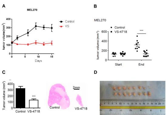
• VS-4718 is the newest FAK inhibitor acquired by Verastem. VS-4718 is a potent reversible inhibitor of FAK, exhibiting an IC50 of 1.5 nM, and is abel to induce a robust FAK inhibition in cultured breast carcinoma cells at a concentration of 0.1 μM [99]. An initial preclinical study indicated that VS-4718 showed limited effects on cell proliferation in adherent cancer cells, whereas it induced marked inhibition of FAK and p130Cas phosphorylation in cells grown in suspension or as spheroids, resulting in caspase-3 activation and apoptosis [99]. Additionally, VS-4718 exerted anti-tumor and anti-metastatic effects in orthotopic breast and ovarian carcinoma mouse tumor models (4T1 and MDA-MB-231) without conferring animal morbidity, death or weight loss [99]. The efficacy of VS-4718 was demonstrated by a marked reduction in both sub-cutaneous tumor growth of breast carcinoma cells and their metastasis to lungs that was accompanied by inhibition of FAK Y397 and p130Cas phosphorylation and elevated caspase-mediated apoptosis [99]. VS-4718 inhibitor is currently in clinical trials in subjects with metastatic non-hematologic malignancies (phase I clinical trial # NCT01849744, http://www.clinicaltrials.gov).
Another important approach is to target the FAK autophosphorylation site, which was reported recently with allosteric FAK inhibitor 14 or compound Y15 to block tumor growth [107-108]. The inhibitor effectively inhibited FAK autophosphorylation activity at 25 nM-1 uM, did not inhibit Pyk-2, EGFR, Src, IGFR-1 and other enzymes in vitro and decreased tumor growth in vivo at 30 mg/kg by intraperitoneal delivery or at 120 mg/kg by oral delivery using breast, pancreatic, neuroblastoma, glioblastoma and colon cancer xenograft mice models. The inhibitor caused apoptosis and decreased proliferation in xenograft tumors in vivo. The advantage of this approach is the high specificity of targeting Y397, the main autophosphorylation site of FAK. This inhibitor also decreased Y418-phosphorylation of down-stream Src in colon cancer cells [108], which can be also benefitial for development of future therapies. FAK has additional functions independent of its autophosphorylation activity or kinase activity [109], which is evident from the different phenotypes of FAK−/−cells and cells with deleted Y397 site [110], or with knock-in point mutation (lysine 454 to arginine) [109], which is important for future therapeutic design of the enzymatic inhibitors.Takeda also developed non-ATP-competitive FAK allosteric inhibitors that efficiently decreased FAK functions [111]. The Takeda identified tricyclic sulfonamides (compund 1 and compund 2) that targeted a novel allosteric site in the C-lobe of the FAK-kinase domain, caused conformational changes of the kinase domain and induced disruption of ATP pocket formation and inhibition of FAK kinase activity with an IC50 of 4.2 and 8.7 μM, respectively [111]. The compounds required pre-incubation of kinase with inhibitor to cause allosteric ATP-non competitive inhibition. Both allosteric inhibitors were highly selective among 288 kinases, with only 10 kinases other than FAK inhibited by >50% and Pyk-2 inhibited by 25%. The compound 2 was more selective
than compound 1, as it did not inhibit 10 kinases >50% and did not inhibit Pyk-2 at 10 μM. This approach reveals novel allosteric and selective way of FAK inhibition. The in vivo studies in xenograft mice models need to demonstrate efficacy on these allosteric inhibitors.
• Inhibitors targeting FAK scaffold: FAK also signals via non-kinase scaffold functions that cannot be affected using conventional small molecule FAK inhibitors. Since FAK has so many binding partners such as Src, EGFR, Her-2, c-Met, PI-3K, disruption of these complexes is an additional therapeutic approach to disrupt signals that FAK integrates and effectively block FAK regulated functions. A small number of compounds developed by CureFAKtor Pharmaceutical are currently undergoing pre-clinical testing. In particular, a compound known as C4 disrupts FAK and VEGFR4 interactions [112], whereas the M13 compound blocks FAK and Mdm-2 interaction [113]. Other inhibitors of FAK-scaffold functions include INT2-31 that blocks FAK and c-Met/IGFR1 interactions [114] and R2 which targets the FAK–p53 interaction [115]. All of these inhibitors effectively reduce cell viability and tumor growth through inhibiting angiogenesis and Akt signaling, or by activating p53 signaling with a resulting enhanced expression of downstream targets of p53 such as p21 and Bax. Also, the inhibitors targeting FAK scaffolding function displayed efficacy at submicromolar and low micromolar doses in vitro and at 15–60 mg/kg in vivo in mice xenograft models. Since there are other many important scaffolding partners of FAK, disrupting FAK-protein interactions based on structural studies is a very perspective approach and will show the efficacy of this approach in future.
Aim of the study
On the basis of the aforementioned findings highlighting the potential involvement of FAK in the development of certain tumors, the present study aimed to address the role of FAK toward the progression of UM in a GNAQ-dependent manner. In particular, our investigation focused on the mechanisms by which
Gaq may activate FAK and the identification of the effectors determining the biological function of the
Gaq-FAK transduction signaling. Next, the benefit of Gaq-FAK inhibition in UM was ascertained both in vitro and in
vivo using CRISPR/Cas9 genome editing as well as specific inhibitors that are currently under advanced
clinical evaluation.
Chapter 2
Materials and methods
2.1 Cell lines, culture procedures, and chemicals: HEK293 DREADD/Gq cells were cultured in DMEM (Invitrogen, CA) containing 10% FBS (Sigma-Aldrich Inc., MO) and 1× antibiotic/antimycotic solution (Sigma-Aldrich Inc., MO). OMM1.3 and Mel270 Uveal Melanoma cells have been described elsewhere [116]. All cell lines were grown in a 37°C incubator with 5% CO2 and underwent to mycoplasma
contamination by using Mycoplasma Detection Kit (Roche) prior to the described experiments. Y-27632 (Tocris Cookson Inc., MO) (10μM), PKC inhibitor (Tocris Cookson Inc., MO) (10μM), Blebbistatin (Tocris Cookson Inc., MO) (100nM), VS-4718/PND-1186 (Selleckchem, CA) (1μM) and PF-562271 (Selleckchem, CA) (1μM) were dissolved in DMSO and were used to treat HEK293 DREADD/Gq cells and uveal melanoma cells for different time points before to perform the experiments.
2.2 Mutational analysis:bioinformatic whole-genome sequencing of UM samples (n=80) derived from The Cancer Genome Atlas (TCGA) (www.cbioportal.org). mRNA expression z-Scores (RNA seq V2 RSEM) selected was ± 1.5
.
2.3 Immunoblot analysis: cells were lysed in RIPA buffer containing Halt protease and phosphatase inhibitor cocktail (Thermo Scientific, Rockford, IL). Protein concentration was measured by Bio-Rad Protein Assay (Bio-Rad, Hercules, CA). Equal amounts of total proteins were subjected to SDS-polyacrylamide gel electrophoresis and transferred to PVDF membranes. Membranes were blocked with 5% non-fat dry milk in T-TBS buffer (50 Mm Tris/HCL, pH 7.5, 0.15 M NaCl, 0.1% [v/v] Tween-20) for 1 hour at room temperature, and then incubated with primary antibodies in blocking buffer at 4°C overnight. Primary antibodies were diluted 1:1000 unless otherwise stated. (Gαq (E-17; Santa Cruz Biotech., CA), anti-RhoA (C-18; Santa Cruz Biotech., CA), anti-Rac1 (BD Biosciences, CA), anti-Trio (H120; Santa Cruz Biotech., CA), anti-GAPDH (14C10; Cell Signaling Technology, MA), anti-FAK (Cell Signaling Technology, MA), anti-pFAK (Y397) (Cell Signaling Technology, MA), anti-pY (Santa Cruz Biotech., CA), AKT (Cell Signaling Technology, MA), pAKT (S473) (Cell Signaling Technology, MA), anti-ERK1/2 (Cell Signaling Technology, MA), anti-pERK42/44 (Cell Signaling Technology, MA), anti-S6 (Cell Signaling Technology, MA), anti-pS6 (Cell Signaling Technology, MA), anti-α-Tubulin (Cell Signaling Technology, MA). Detection was conducted by incubating the membranes with horseradish peroxidase-conjugated goat anti-rabbit and anti-mouse IgG secondary antibodies (Southern Biotech, Birmingham, AL) that were used at a dilution of 1:50,000 in 5% milk-T-TBS buffer at room temperature for 1 hour, and visualized with Immobilon Western Chemiluminescent HRP Substrate (Millipore, Billerica, MA).
2.4 Immunoprecipitation assay: cells were lysed with lysis buffer [10mM Tris-Cl (pH 8.0), 150mM NaCl, 1mM EDTA, 0.3% CHAPS, 50mM NaF, 1.5mM Na3VO4, protease inhibitor (Thermo Scientific, CO), 1mM DTT and 1mM PMSF], and centrifuged at 16,000 rpm for 10 min at 4°C. Supernatants were incubated with primary antibody for 2h at 4°C, and protein G or protein A conjugated resin for another 1 hour. Resins were then washed 3 times with lysis buffer and boiled in SDS-loading buffer.
2.5 siRNA transfection: all human siRNA sequences and providers are described in the following table. All the cells were transfected when 40% confluent using Lipofectamine RNAiMAX Reagent (Invitrogen, CA) according to manufacturer’s instructions, using 50nM of each siRNA, and experiments were performed, when the cells were confluent. Detail of RNAi and oligonucleotide sequences used in this study:
siRNA Source Homology Details siRNA Control Thermo Scientific - SIC001
Hs_GNAQ Thermo Scientific (Smart Pool) Homo Sapiens Human (GNAQ 2776) Hs_Trio Thermo Scientific (Smart Pool) Homo Sapiens Human (TRIO 7204) Hs_RhoA Thermo Scientific (Smart Pool) Homo Sapiens Human (RhoA 387) Hs_Rac1 Thermo Scientific (Smart Pool) Homo Sapiens Human (RAC1 5879) Hs_PTK2 Thermo Scientific (Smart Pool) Homo Sapiens Human (PTK2 5747)
2.6 Cell proliferation analysis: Alamar Blue Cell Viability Reagent was purchased from Life Technologies (Grand Island, NY). In brief, OMM1.3 and MEL270 UM cell lines were cultured in 96-well plates and then treated with VS4718 and PF-562271 for 72 hours. The manufacturer's instructions were followed to complete the assay.
2.7 Focal adhesion assay: cells cultured on fibronectin-coated coverslips were washed three times with PBS, were fixed with 3.7% formaldehyde in phosphate-buffered saline (PBS) for 30 minutes, and permeabilized using 0.05% Triton X-100 for 10 minutes. Cells were blocked with 3% FBS-containing PBS for 30 min, and incubated with anti-pFAK (Y397) (Cell Signaling Technology, MA) and anti-paxillin (Cell Signaling Technology, MA) (in 3% FBS-PBS otherwise stated) for 1 hour at room temperature. The reaction was visualized with Alexa-labeled secondary antibodies (Invitrogen, CA) and using also Alexa Fluor 488 Phalloidin (Invitrogen, CA). Cells were incubated in PBS buffer containing Hoechst 33342 (Molecular Probes, OR) for nuclear staining. Images were acquired with an Axio Imager Z1 microscope equipped with ApoTome system controlled by ZEN 2012 software (Carl Zeiss, NY).
2.8 Methylcellulose growth assay: 5×10³ uveal melanoma cells were embedded in 1% methylcellulose (R&D HSC001) diluted in growth media and plated onto 6-well-poly-hyodroxyethyl methacrylic acid (poly-HEMA) – coated plates (Costar). At the indicated time, colonies were imaged in phase contrast, and



![Figure 2: Representative saturation curve and Scatchard plot of [ 3 H]17β-estradiol (E2) binding](https://thumb-eu.123doks.com/thumbv2/123dokorg/2868163.9175/125.918.239.671.69.866/figure-representative-saturation-curve-scatchard-plot-estradiol-binding.webp)