Dipartimento di Farmacia e Scienze della Nutrizione e della Salute
DOTTORATO DI RICERCA IN BIOCHIMICA CELLULARE E ATTIVITA’ DEI FARMACI IN ONCOLOGIA
(XXVIII CICLO)
Settore Scientifico Disciplinare MED/04
Novel molecular mechanisms involved in the
stimulatory action of zinc in breast cancer
Coordinatore del dottorato: Chiar.mo Prof. Diego Sisci
Docente Tutor: Chiar.mo Prof. Marcello Maggiolini
Dottoranda: Dr.ssa Maria Grazia Perri
A.A. 2015/2016
Index Pag Abstract 1 Chapter 1. Introduction 1.1 Introduction 3 1.2 Breast cancer 4 1.2.1 Tumor Microenvironment 7
1.2.2 Cancer-associated Fibroblasts (CAFs) 10
1.3 The G protein-coupled estrogen receptor (GPR30/GPER) 12
1.4 EGFR/IGF-IR cross-talk 15
1.5 Action of zinc in breast cancer 18
1.6 Aim of the study 19
Chapter 2. Materials and Methods 2.1 Reagents 20
2.2 Cell Cultures 20
2.3 Isolation, cultivation, and characterization of CAFs 21
2.4 Plasmids and luciferase assays 23
2.5 Gene silencing experiments 24
2.6 Gene expression studies 24
2.7 Western blot analysis 26
2.9 ROS production 27
2.10 Cell cycle analysis 27
2.11 Proliferation assay 27
2.12 Migration assays 27
2.13 Statistical analysis 28
Chapter 3. Results 3.1 GPER is involved in the activation of EGFR and IGF-IR by Zn in breast cancer cells 29
3.2 GPER contributes to gene expression changes and growth responses induced by Zn in breast cancer cells 32
3.3 GPER contributes to Zn action in CAFs 36
Chapter 4. Discussion 39
References 42
1
Abstract
Lo Zinco (Zn), minerale essenziale che regola diverse funzioni biologiche, è coinvolto nella progressione del tumore mammario. In particolare, è stato dimostrato che in seguito all’attivazione indotta dallo Zn di recettori ad attività tirosin-chinasica, come IGF-IR, EGFR e IR, vengono innescate vie di trasduzione del segnale che sono alla base della progressione tumorale, quali la via delle MAP-chinasi (MAPK) e la via del fosfatidilinositolo3-chinasi (PI3-K)/AKT. Numerosi studi hanno inoltre dimostrato il coinvolgimento di recettori accoppiati a proteina G (GPCRs) nello sviluppo dei tumori e la loro azione sinergica con vari recettori di membrana . Alla luce di tali osservazioni, una migliore comprensione dei processi attraverso cui i recettori per i fattori di crescita cooperano con segnali mediati da GPCRs potrebbe contribuire allo sviluppo di nuove strategie terapeutiche volte a prevenire e/o ritardare la crescita tumorale. Nel presente studio, è stato dimostrato che lo Zn è coinvolto nel cross-talk funzionale tra IGF-IR, EGFR e GPER in cellule di tumore mammario e fibroblasti tumore-associati (CAFs). In particolare, è stato dimostrato che GPER, IGF-IR e EGFR contribuiscono agli effetti stimolatori indotti da ZnCl2 nella progressione del ciclo cellulare, nella proliferazione e nella migrazione di cellule di carcinoma mammario e dei CAFs. I nostri risultatievidenziano nuovi meccanismi molecolari attraverso i quali lo Zn può indurre effetti stimolatori in cellule di tumore mammario e nei CAF, suggerendo pertanto nuovi potenziali approcci farmacologici nel trattamento del carcinoma mammario.
2
Zinc (Zn) contributes to the regulation of several cellular functions, however it may be also implicated in the progression of breast cancer through different mechanisms. For instance, Zn may activate tyrosine kinase receptors, as insulin-like growth factor receptor I (IGF-IR), epidermal growth factor receptor (EGFR) and the insulin receptor (IR), which then trigger the mitogen-activated protein kinase (MAPK) and phosphatidylinositol 3-kinase (PI3-K)/AKT transduction signaling. These pathways have been largely implicated in cancer growth and invasion together with other important signal molecules like the G-protein coupled receptors (GPCRs). In this regard, the cross-talk between GPCRs and growth factor receptors has been shown to contribute to cancer growth, angiogenesis and metastasis. Recently, both EGF and IGF-I mediated pathways were demonstrated to interact with the G protein estrogen receptor (GPER, also known as GPR30), which has been involved in the proliferation and migration of several types of cancer and stromal cells. Overall, these findings indicate that a better understanding of the molecular mechanisms by which growth factor receptors cooperate with GPCR signals would be highly relevant toward new therapeutic strategies aimed to prevent or delay the progression of several tumors. In the present study, we ascertained that zinc chloride (ZnCl2) triggers a functional crosstalk of GPER with IGF-IR and EGFR in breast cancer cells and in main components of the tumor microenvironment like cancer-associated fibroblasts (CAFs) derived from breast cancer patients. Further corroborating these data, we assessed that GPER along with IGF-IR and EGFR contribute to the stimulatory effects induced by ZnCl2 on cell-cycle progression, proliferation, and migration of breast cancer cells and CAFs. Our results provide novel mechanistic insights through which Zn may induce stimulatory effects in breast tumor cells and CAFs, suggesting further molecular targets in the treatment of breast malignancy.
3
Chapter 1
Introduction1.1 Introduction
Zinc (Zn) is the second most abundant heavy metal in human tissues and contributes to the regulation of crucial cellular functions [1]. As an essential mineral, Zn is required for protein, nucleic acid, carbohydrate and lipid metabolism and is involved in gene transcription, growth, development and differentiation [1]. Zn is normally found in air, water and soil, however, Zn concentrations may be boosted by several industrial activities including mining, coal and waste combustion and steel processing [2]. For instance, soils located in areas where Zn is mined, refined or used as fertilizer, are heavily contaminated with the metal [2]. The Recommended Daily Allowance of Zn in adults is 8–11mg/day, with a tolerable upper intake level of 40mg/day [3-5]. The adverse effects associated with a high Zn intake include acute gastrointestinal effects and headache, impaired immune function, changes in lipoprotein and cholesterol levels, reduced copper levels and zinc-iron interactions as well as various other disorders [6-8]. In addition, Zn has been involved in the development of several types of tumors including breast cancer [9-10]. In this regard, previous studies have reported an association between dysregulated Zn homeostasis and breast cancer progression together with higher Zn levels in breast tumor specimens as compared to normal mammary tissues [11-12]. Compelling evidence has also linked an aberrant expression of Zn transporter proteins with the proliferation and migration of breast cancer cells [13-15]. A recent study has also suggested that specific dysregulations of Zn transporters may characterize grade, invasiveness, metastatic potential and response to therapy in breast cancer [16]. Of note, zinc
4 regulated transporters (ZIP) that control Zn influx into the cytosol, were found to be up-regulated by estrogens [17], and increased ZIP levels in breast tumors resulted to be associated with a poor prognosis [15]. Noteworthy, Zn may activate tyrosine kinase receptors as EGFR, IGF-IR and the insulin receptor, which then trigger the mitogen-activated protein kinase (MAPK) and phosphatidylinositol 3-kinase (PI3-K)/AKT signalling [18-20]. These transduction pathways have been largely implicated in cancer growth and invasion together with other important signal molecules like the G protein-coupled receptors (GPCRs) [21]. Notably, both EGF and IGF-I mediated signalling were shown to functionally interact with the G protein estrogen receptor (GPER, previously known as GPR30) transduction pathway in breast cancer cells [22-23]. In this regard, it has been reported that GPER activation induces important responses like proliferation and migration in several types of cancer cells and stromal cells that contribute to the malignant progression like cancer-associated fibroblasts (CAFs) [24].
1.2 Breast cancer
Breast cancer is the most common malignancy and the leading cause of cancer-related death in women worldwide. Whereas localized disease is largely curable, metastatic or recurrent disease carries an unfavorable prognosis [25].As a greater percentage of breast cancers are being diagnosed at an earlier stage, the medical community has been challenged to develop diagnostic and treatment modalities that maximize benefit reducing the morbidity associated with therapy [26]. The management of breast cancer has changed considerably in the last two decades with improvements in systemic therapy and advances in surgical techniques [27].
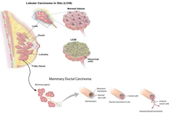
There are two main types of breast cancer:
Ductal carcinoma, it starts in the ducts that move milk from the breast to the nipple. Most breast cancers are of this type.
5 Lobular carcinoma, it starts in the parts of the breast, called lobules that produce milk. In rare
cases, breast cancer can start in other areas of the breast. Breast cancer may be invasive or non-invasive. Non-invasive breast cancer is also called "in situ."
Ductal carcinoma in situ (DCIS) or intraductal carcinoma, is breast cancer in the lining of the milk ducts that has not yet invaded nearby tissues. It may progress to invasive cancer if untreated.
Lobular carcinoma in situ (LCIS) is a marker for an increased risk of invasive cancer in the same or both breasts (Fig. 1.1).
Figure 1.1 | Representation of the anatomy of the Lobular Carcinoma and Mammary Ductal Carcinoma.
There are many risk factors involved in the development of breast tumor:
Age and gender. The risk of developing breast cancer increases with age. Most advanced breast cancer cases are found in women over age 50 [28]. Women are 100 times more likely to get breast cancer than men.
6 Family history of breast cancer. You may also have a higher risk for breast cancer if you have a close relative who has had breast, uterine, ovarian, or colon cancer. About 20-30% of women with breast cancer have a family history of the disease.
Genes. The most common gene defects are found in the BRCA1 and BRCA2 genes. These genes normally produce proteins that protect you from cancer. If a parent passes you a defective gene, you have an increased risk for breast cancer. Women with one of these defects have up to an 80% chance of getting breast cancer sometime during their life [29].
Menstrual cycle. Women who got their periods early (before age 12) or went through menopause late (after age 55) have an increased risk for breast cancer [30].
Other risk factors include:
Alcohol use. Drinking more than 1-2 glasses of alcohol a day may increase your risk for breast cancer [31].
Childbirth. Women who have never had children or who had them only after age 30 have an increased risk for breast cancer. Being pregnant more than once or becoming pregnant at an early age reduces your risk of breast cancer [32].
Hormone replacement therapy (HRT). You have a higher risk for breast cancer if you have received hormone replacement therapy with estrogen for several years or more [33].
Obesity has been linked to breast cancer, although this link is controversial. The theory is that obese women produce more estrogen, which can fuel the development of breast cancer [34].
Radiation. The radiation therapy to treat cancer of the chest area, increase higher risk to develop breast cancer [34].Treatment is based on many factors, including: type and stage of the cancer, whether them cancer is sensitive to certain hormones, whether the cancer over-expresses a gene called HER2/neu.
7 In general, cancer treatments may include: surgery to remove cancerous tissue, lumpectomy removes the breast lump; mastectomy removes all or part of the breast; chemotherapy medicines to kill cancer cells, radiation therapy to destroy cancerous tissue, hormonal therapy. Most women receive a combination of treatments. For women with stage I, II, or III breast cancer, the main aim is to treat the cancer and prevent it from returning. For women with stage IV cancer, the objective is to improve symptoms and help them live longer. In most cases, stage IV breast cancer cannot be cured.
Stage 0 and DCIS Lumpectomy plus radiation or mastectomy is the standard treatment. There is some controversy on how best to treat DCIS.
Stage I and II Lumpectomy plus radiation or mastectomy with some sort of lymph node removal is the standard treatment. Hormone therapy, chemotherapy, and biologic therapy may also be recommended following surgery.
Stage III Treatment involves surgery, possibly followed by chemotherapy, hormone therapy, and biologic therapy.
Stage IV Treatment may involve surgery, radiation, chemotherapy, hormonal therapy or a combination of these treatments. After treatment, some women will continue to take medications such as tamoxifen for a period of time. All women will continue to have blood tests, mammograms, and other tests after treatment. Women who have had a mastectomy may have reconstructive breast surgery, either at the same time as the mastectomy or later [35].
1.2.1 Tumor Microenvironment
The breast cancer microenvironment is a complex combination of several different cell types and molecules and it is a key contributor to malignant progression [36]. The role of tumor microenvironment is becoming more and more important in breast cancer. Several stromal cell
8 types are implicated in promoting ‘hallmarks’ of cancer cells [37]. The microenvironment includes fibroblasts, macrophages, immune cells, adipocytes, endothelial cells, and antigenic vascular cells. Stromal cells surround and interact with tumor cells. Over the last years, a robust body of evidence has highlighted the importance of the crosstalk between tumor and stoma. Tumor microenvironment has been shown to play a crucial role in tumorigenesis, from initiation to progression. Stromal cells promote cancer growth and invasion through the chemokine–chemokine receptor axis [38, 39]. Infiltrating immune cells energize the immune effectors and vascular cells permit nutrients and oxygen uptake by tumors. In a normal mammary duct, there are luminal epithelial cells internally and myoepithelial cells externally delimited by a basement membrane, which maintains the luminal cell polarity [40]. The extracellular matrix (ECM) allows communication with the surrounding stroma. Genetic and epigenetic alterations lead to luminal cell proliferation, loss of epithelial polarity and decrease of myoepithelial cells, and changes in the ECM/basal membrane, finally resulting in mammary tumor development [41]. As opposed to normal fibroblasts, cancer-associated fibroblasts (CAFs) [42]. improve tumor growth and metastasis by producing growth factors and ECM proteins, as well as by modulating immune polarization [43]. Also, the number of CAFs is increased during tumor progression [44]. Accordingly, growth factors, cytokines, chemokines, and matrix metalloproteinases secreted by stromal cells lead to the recruitment of macrophages, endothelial precursor cells, and regulatory lymphocytes, which sustain tumor progression [45]. It is worth noting that stroma has been correlated with clinical outcomes and response to therapy in breast cancer [46]. The expression of ECM genes, uniformly expressed in both neoplastic and adjacent stromal cells, may divide breast cancers into different subgroups with different clinical outcomes [47, 48]. A study performing hierarchical clustering of the gene-expression profile of ECM-related genes classified breast cancer samples into four groups associated with different clinical outcomes [49]. Stromal signatures are highly informative for patients with breast cancer. A
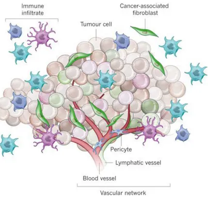
9 serum activated gene-expression signature from activated fibroblasts was identified as a negative prognostic factor in patients with breast cancer [50]. Also, a 26-gene signature called the stroma-derived prognostic predictor was generated by tumor-associated stroma and matched normal stroma from breast cancer samples [51]. This signature was found to be an independent prognostic factor [50]. So tumor microenvironment influences patient outcomes and stromal gene expression signatures represent a strong prognostic value recapitulating the immune, angiogenic, and hypoxic responses [50]. The stromal cells can be divided into three general classes (Fig. 1.2):
Infiltrating immune cells Angiogenic vascular cells
Cancer-associated fibroblastic cells.
Figure 1.2 | Tumor formation involves the co-evolution of neoplastic cells together with extracellular matrix and vascular endothelial, stromal and immune cells. The tumor niche is a dynamic physical topography in which structural support, access to growth factors, vascular supply and immune cell interactions can vary drastically even within the same lesion. The immune infiltrate can include multiple cell types, these cell populations can have both pro- and anti-tumor functions and can vary in their activation status and their localization within the tumor. The vascular network can differ in regard to the vessel's tissue of origin, maturity (extent of pericyte coverage), interstitial pressure and functionality. Cancer-associated fibroblasts can have significant plasticity and diverge with regard to activation status, localization within the tissue, stress response and origin.
10 1.2.2 Cancer-associated fibroblasts (CAFs)
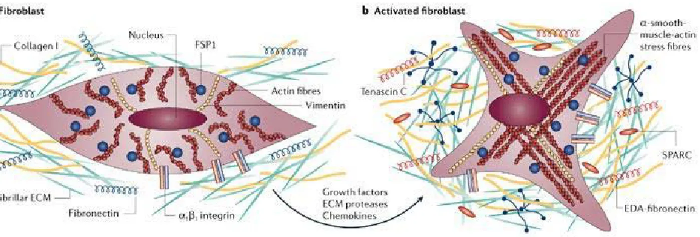
During tumorigenesis, the normal microenvironment ‘niche’ changes to an altered (ie, reactive or desmoplastic) stroma which is composed of non-malignant supporting cells (ie, blood vessels, infiltrating inflammatory cells and blast-like cells) [51, 52]. This altered microenvironment functions by influencing homeostasis of cancer cells via paracrine regulators (eg, growth factors, cytokines and chemokines) and exosomes containing nucleic acids [51, 53-54]. Cancer associated fibroblasts (CAFs), prominent stromal elements in most types of human carcinomas, are α-smooth muscle actin positive, spindle-shaped, blast-like cells. Differentiation of CAFs from other cell types, such as local fibroblasts, hepatic stellate cells, mesenchymal stem cells, endothelial and epithelial cells, is mainly mediated by transforming growth factor-β1 (TGF-β1), but other factors, such as growth hormones (ie, epidermal growth factor (EGF), fibroblast growth factor (FGF) and platelet-derived growth factor (PDGF), chemokines, epigenetic regulators and oxidative stress also may play a role in CAF differentiation. [54, 56, 57] (Fig. 1.3). CAFs, phenotypically, closely resemble normal myofibroblasts, but they express specific markers (ie, fibroblast activation protein (FAP), fibroblast-specific protein 1, neuronglial antigen-2, vimentin, Thy-1, tenascin (TN)-C, periostin (POSTN), palladin or podoplanin (PDPN) and display an increased proliferation and migratory behaviour in vitro [58,59]. CAFs produce and secrete various extracellular matrix (ECM) proteins (ie, collagens I, III, IV), proteoglycans (ie, fibronectin, laminin, TN), chemokines (eg, CXCL and CCL), cytokines (eg, interleukin (IL)-6 and IL-8) and other tumor-promoting factors which affect vascularization (ie, PDGF, vascular endothelial growth factor (VEGF), stromal-derived factor-1 (SDF-1), matrix metalloproteinase (MMPs)), proliferation capacity, tumor cell invasiveness and survival (ie, TGF-β, EGF, hepatocyte growth factor (HGF) or FGF) [51, 60-63]. Regarding anticancer therapy, the frequency of genetic mutations in CAFs is one of the most important issues.
11 Cells with genetic stability may be less prone to escape or resistance to chemotherapy than those with genomic instability [64]. Several studies demonstrated that high percentage of CAFs undergo genetic alterations, such as loss of heterozygosis or mutation of tumor suppressor genes (ie, phosphatase and tensin homolog and P53) [65-68]. The theory of genetic coevolution of CAF and the neighbouring cells (ie, random mutation of CAF generated independently from neoplastic epithelial cells that may support tumor progression) is under debate, due to the potential artefacts caused by the analytical methods used for the identification of these genetic alterations [68]. Interestingly, CAF derived proteins which [51] may have an important role in the development of environment-mediated drug resistance, [52] may act as powerful prognostic markers and [53] may be promising targets of anticancer therapy [68].
Figure 1.3 | A) Normal fibroblasts are embedded within the fibrillar extracellular matrix (ECM) of connective tissue, which consists largely of type I collagen and fibronectin. Fibroblasts interact with their surrounding microenvironment through integrins such as the α1 and β1 integrin. Typically, fibroblasts appear as fusiform cells with a prominent actin cytoskeleton and vimentin intermediate filaments. B) fibroblasts can acquire an activated phenotype, which is associated with an increased proliferative activity and enhanced secretion of ECM proteins such as type I collagen and tenascin C, and also fibronectin that contains the extra domain a (EDA-fibronectin) and SPARC (secreted protein acidic and rich in cysteine). Phenotypically, activated fibroblasts are often characterized as expressing α-smooth-muscle actin. Numerous growth factors such as TGFβ, chemokines such as MCP1, and ECM-degrading proteases have been shown to mediate the activation of fibroblasts
12 1.3 The G protein-coupled estrogen receptor (GPR30/GPER)
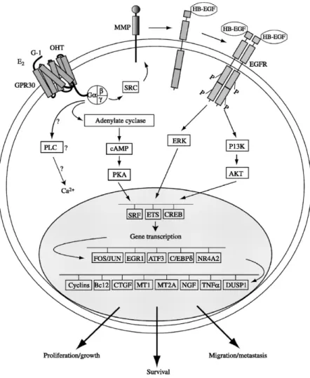
Among the GPCR family members, GPR30/GPER, was recently shown to mediate the multifaceted actions of estrogens in different tissues including cancer cells [69]. GPER was first identified as an orphan member of the 7-transmembrane receptor family by multiple groups in the late 1990s [70-72]. It belongs to the rhodopsin-like receptor superfamily and its gene is mapped to chromosome 7p22.3 [70]. There are four alternate transcriptional splicing variants encoding the same protein which is comprised of 375 amino acids and contains seven transmembrane spanning segments [72]. Although GPER is a seven-transmembrane receptor, its subcellular localization remains to be fully elucidated. Indeed, several studies have reported the presence of GPER at the plasma membrane, in the endoplasmic reticulum, in the Golgi apparatum as well as in the nucleus of CAFs extracted from mammary biopsies [73-75]. Several studies demonstrated that the ligand-dependent activation of GPER trigger the activation of the heterotrimeric G proteins and subsequently Src and adenylyl cyclase (AC), resulting in intracellular cAMP production. Src is involved in matrix metalloproteinases (MMP) activation, which cleave pro-heparan-bound epidermal growth factor (pro-HB-EGF) releasing free HB-EGF. The latter activates EGF receptor (EGFR), leading to multiple downstream events, for example the activation of phospholipase C (PLC), PI3K and MAPK [70]. Activated PLC produces inositol triphosphate (IP3), which further binds to IP3 receptor and leads to intracellular calcium mobilization [76]. The downstream signal of PI3K is AKT pathway, closely related to cancer cell growth as involved in cell survival and proliferation [77]. The activation of MAPK and PI3K results in the activation of numerous cytosolic pathways and nuclear proteins, which in turn regulate transcription factors such as SRF, CREB and members of the E26 transformation specific (ETS) family by direct phosphorylation [78]. These promotes the expression of a second wave of transcription factors such as FOS, JUN, EGR1, ATF3, C/EBPδ, and NR4A2. Cells are literally reprogrammed under the effect of this
13 network of transcription factors and a series of GPER target genes such as CTGF are up-regulated [79] (Fig. 1.4).
Figure 1.4 | Schematic representation of the GPER signaling network
In addition, there may be a variety of signaling crosstalk pathways and both negative and positive feedback loops. For example, it has been demonstrated that EGF up-regulates GPER expression through the EGFR/MAPK pathway in ER-negative breast cancer cells, most likely by promoting the recruitment of the c-FOS-containing transcription factor AP-1 to the GPER promoter [80]. Considering that GPER signaling uses the EGFR/MAPK pathway, a positive feedback loop is conceivable. This mechanism is also operational for EGF and the related growth factor TGFα in ERα-positive breast cancer cells [81]. GPER gene expression has been detected in at least four
14 kinds of human tumor specimens or cell lines, including breast cancer [71, 81-83], endometrial cancer [82-84], ovarian cancer [85-87], thyroid cancer [88], and a rat pheochromocytoma cell line PC-12 [89]. In addition, there is a growing body of evidence supporting that GPER is strongly associated with cancer proliferation [90], migration [91], invasion [92], metastasis [93,94], differentiation [95], and drug resistance [96, 97]. Indeed, as estrogens stimulate the progression of breast cancer in approximately two-thirds of patients who express ER [98, 99], some selective estrogen receptor modulators (SERMs), such as tamoxifen, have been clinically used to antagonize the binding of estrogen to its classic ERs, which is an effective therapeutic strategy in attenuating the growth of ER-positive breast cancers. However, there are around 25% of ER-positive breast cancer patients who do not respond to anti-estrogen therapy (Early Breast Cancer Trialists’ Collaborative Group 2005). It implies that the blockade of classic ERs alone may be not enough to completely abolish estrogen-induced breast cancer cell growth, since estrogen may promote it through other receptors besides classic ERs. Such hypothesis is further supported by the discovery of GPER as the third specific ER with different structure and function respect to ERα and ERβ. GPER has a high binding affinity to not only for estrogen, but also for some ER antagonists, such as tamoxifen and ICI 182,780. Notably, estrogen and the aforementioned anti-estrogens stimulate GPER signaling [100]. These important findings provide a further possible mechanism for the progression of estrogen-related cancers, and raise a novel potential target for anti-estrogen therapy. As it concerns clinical findings, GPER overexpression, was associated with lower survival rates in endometrial and ovarian cancer patients [101, 102] as well as with a higher risk of developing metastatic disease in breast cancer patients [103]. Moreover, in a previous extensive survey, GPER was found to be highly expressed and significantly associated with tumor size (>2 cm), with the presence of distant metastases and increased human EGFR-2 (HER-2)/neu expression [104]. Likewise, a recent study demonstrated that the majority of the aggressive inflammatory breast
15 tumors examined resulted GPER positive [105], suggesting that the expression of the receptor may be considered a predictor of an aggressive disease. In addition to the aforementioned studies on the potential functions of GPER in cancer and possibly other pathological conditions, this receptors was implicated in a broad range of physiological functions regarding reproduction, metabolism, bone, cardiovascular, nervous and immune systems [102]. Estrogen binds to GPER with a high affinity of a reported Kd 2.7 nM (84) or 6 nM [103]. Moreover, in the last few years a great attention was focused on the identification of synthetic ligands of GPER acting as agonists or antagonists. In particular different compounds named G-1 [104] G-15 [105], GPER-L1 and GPER-L2 [106] and MIBE [110], were identified using virtual and bio-molecular screening and are used to evaluate the GPER-mediated signaling and functions. In addition, different studies shows that ICI 182,780 [109] and 4-hydroxytamoxifen (OHT) [109-110] are also able to bind GPER and mimic estrogen effects. It has been reported that a variety of xenoestrogens, including bisphenol A, can bind and activate GPER leading to important biological responses [111].
1.4 EGFR/IGF-IR cross-talk
RTKs are transmembrane proteins involved in the control of multiple physiological processes [112]. These proteins consist of an extracellular ligand binding domain, a single transmembrane domain and a highly conserved cytosolic tyrosine kinase domain [112]. The RTK family includes the majority of receptors for growth factors, like the EGFR, commonly known as ErbBs/HER and the IGF-IR, which belongs to the IGF system. In particular, ErbB family comprises four members: EGFR (ErbB1, also known as HER1), ErbB2 (also known as Neu or HER2), ErbB3 (HER3), and ErbB4 (HER4). ErbBs exist as monomers and upon ligand activation or when overexpressed, form homo- and heterodimers [113,114]. The four ErbB receptors are specifically activated by soluble small peptides: EGF, transforming growth factor-α (TGF-α), heparin-binding EGF-like growth
16 factor (HB-EGF), amphiregulin (AR), betacellulin (BTC), epiregulin (EPR), epigen (EPG) and the neuregulins (NRGs 1–6) often referred as heregulins [115]. The ligand binding and the subsequent formation of an ErbB dimer promote the cross-phosphorylation of the dimer partner, generating a network of intracellular signals that control numerous biological processes [116, 117]. In particular, the ErbB family dimers activate various transduction pathways including the MAPK cascade, which leads to gene transcription, cell proliferation, migration, differentiation, angiogenesis [118, 119], and the phosphatidylinositol 3-kinase (PI3-K)/Akt signaling, which mainly promotes cell survival [120]. Likewise, the IGF system plays a key role in cell growth, differentiation, survival as well as in cell transformation and metastasis [121, 122]. It comprises ligands (IGF-I, IGF-II and insulin), cell surface receptors as the IGF-I receptor (IGF-IR), the IGF-II receptor (IGF-IIR), the insulin receptor (IR) and six soluble IGF-binding proteins (IGF-BPs) [122, 123]. Because of the high sequence similarity between the IR and the IGF-IR, IGFs and insulin are able to cross-bind to each other’s receptor, even though with much weaker binding affinity [124]. The ligand binding induces the receptor phosphorylation and the recruitment of numerous docking proteins, including IRS family members (IRS-1, IRS-2) and adaptors molecules as Shc and Grb2 [125]. These substrates are then involved in the activation of transduction pathways like MAPK, PI3-K and the Janus kinase/signal transduce rand activator of transcription pathway (JAK/STAT), which mediate important biological responses as glucose metabolism, cell growth, survival and inhibition of apoptosis [126] It is generally accepted that the growth-promoting activities of IGF-I, IGF-II and insulin are mainly mediated by IGF-IR, whereas IGF-IIR serves as a clearance receptor as it removes IGF-II from the cell surface [127]. Accordingly, elevated IGF-IR expression has been associated with multiple biological features of cancer progression including the development of metastatic processes and the resistance to chemotherapeutics [128]. The IGF system also acts influencing the signaling and the biological responses mediated by other cell membrane proteins
17 such as EGFR [129]. For instance, in vitro and in vivo studies have indicated that the cross-talk between IGF-IR and EGFR can lead to the resistance against EGFR-targeted drugs with relevant implications for cancer therapy [130, 131]. Moreover, EGFR and IGF-IR induce the proliferation of cancer cells through different mechanisms which include the interaction with GPCRs as well as with downstream transduction molecules [129]. Several studies have suggested that the transduction signaling generated by EGFR and the IGF system interacts with different mediators of the estrogen action toward the resistance to endocrine therapy and cancer progression [132]. For instance, the overexpression of EGFR and IGF-IR, the lack of hormonal targeted cancer therapy, the low survival rate and the poor patient prognosis are hallmark features of the estrogen receptor alpha-negative (ERα-) breast cancer subtype [133-137]. Recent studies have shown that ligand-activated EGFR and IGF-IR mediate the regulation and function of GPER in cancer cells, adding a novel mechanism by which these growth factor receptors may contribute to the estrogen stimulation of malignant cells. The aforementioned findings have also extended our knowledge on the functional cross-talk between EGFR and IGF-IR with a member of the GPCR family, supporting the hypothesis that interfering with such interactions may be an effective pharmacological approach in cancer patients. These data suggest the significance of IGF-IR/EGFR activation via the cross-talk with estrogen receptor to regulate cancer cell phenotype and properties (Fig. 1.5). A better understanding of the molecular mechanisms involved in the cross-talk among these receptors may provide new therapeutical approaches that could guarantee major benefits in patients with hormone-sensitive tumors [138].
18 Figure 1.5 | Schematic representation of the cross-talk between GPER and both ErbBreceptors and IGF-IR. Ligand-induced activation of growth factor receptors mediatesthe up-regulation of GPER through diverse kinase transduction pathways. Agonistactivated GPER triggers stimulatory effects like gene expression changes, cell prolif-eration and migration. Note that GPER is placed somewhat arbitrarily at the plasmamembrane, but signaling would presumably work the same way with GPER localizedwithin cells.
1.5 Action of Zinc in breast cancer
Zinc (Zn) essential for many cellular processes, has also been reported to play a potential role as second messenger in signal pathway linked with various physiological actions [139, 140]. The catalytic activity of hundreds of enzymes of every type is dependent on the participation of Zn [1]. Recently, a regulatory role of Zn in cell signaling has also been recognized [19]. Hershfinkel and colleagues [141] have reported the existence of a Zn-sensing receptor that triggers calcium dependent signaling. Evidence that Zn acts extracellularly to induce this effect, combined with the ability of Zn to organize protein domains in a multidentate manner [142], raises mechanistic questions concerning the mode of interaction of the Zn ligand with its putative receptor. The adverse effects associated with a high Zn intake include acute gastrointestinal effects and headache, impaired immune function, changes in lipoprotein and cholesterol levels, reduced copper levels and zinc-iron interactions as well as various other disorders [143, 144]. Furthermore, the imbalances in
19 Zn homeostasis cause disease states including Alzheimer’s disease [145], diabetes [146, 147, 148], cancer [149] and others [150]. In breast cancer patients, the level of Zn has been found to be lower in serum than healthy subjects and elevated in malignant tissues [151-154], whereas in liver, prostate and gallbladder cancers, the level of Zn in the malignant tissues has been found to be decreased [151, 152, 155-157]. These recent reports suggest that Zn is implicated in breast cancer development. The intracellular Zn level is tightly controlled by several protein molecules called zinc-binding protein and zinc transporters [158]. These behaviors of Zn in and out of cell across membranes are maintained through two families including the ZRT IRT-like protein (ZIP) family that facilitates Zn influx into the cytosol, and zinc transporter (ZnT) family that facilitates Zn efflux from the cytosol. Some ZIP family members are reported to involve in aggressive cancer progression [146, 151, 159-166]. ZIP6 is associated with histological grade of estrogen-receptor-positive breast cancer, and is estrogen-receptor-positively related with the lymph node metastasis [151, 167-169]. ZIP6 also has been reported to regulate epithelial-mesenchymal transition (EMT) [151, 156, 159]. Moreover, ZIP10 has been reported to mediate the migration and invasive behaviors of breast cancer cells [170]. Moreover, the degree of Zn accumulation is associated with cancer progression [139] and malignancy [140], but the mechanisms responsible for Zn accumulation, and the relationship between Zn accumulation and breast cancer subtype are not well understood. Of note, Zn regulated transporters (ZIP) that control its influx into the cytosol, were found to be up-regulated by estrogens [152], and increased ZIP levels in breast tumors resulted to be associated with a poor prognosis [150].
1.6 Aim of the study
In the present study, we have evaluated whether Zn might trigger the transduction signaling mediated by GPER through a crosstalk with IGF-IR and EGFR in breast cancer cells and in main
20 components of the tumor microenvironment like CAFs. In particular, we have determined the molecular mechanisms through which zinc chloride (ZnCl2) involves GPER in the activation of IGF-IR/EGFR-mediated signalling, which in turn triggers downstream pathways like ERK and AKT in breast cancer cells and in CAFs. Further corroborating these findings, we have evidenced that ZnCl2 stimulates a functional crosstalk of GPER with IGF-IR and EGFR toward the transcription of diverse GPER target genes. In addition, we have shown that GPER contributes to the stimulatory effects induced by ZnCl2 on cell-cycle progression, proliferation and migration of
21
Chapter 2
Matherials and Methods2.1 Reagents
We purchased zinc chloride (ZnCl2), zinc sulfate (ZnSO4), wortmannin (WM), N,N,N',N'-tetrakis(2-pyridylmethyl)ethane-1,2-diamine (TPEN), N-acetyl-L-cysteine (NAC) and 2′,7′-dichlorofluorescin diacetate (DCFDA) from Sigma-Aldrich (Milan, Italy); tyrphostin AG1478 from Biomol Research Laboratories (Milan, Italy); PD98059 (PD), and 3-bromo-5-t-butyl-4-hydroxybenzylidenemalonitrile (AG1024) from Calbiochem (Milan, Italy); (3aS,4R,9bR)-4-(6-Bromo-1,3-benzodioxol-5-yl)-3a,4,5,9b-3H-cyclopenta[c]quinolone (G15) from Tocris Bioscience (Bristol, UK); human Connective Tissue Growth Factor (CTGF) Recombinant Protein from MBL International (Eppendorf, Milan, Italy). All compounds were solubilized in DMSO except ZnCl2, ZnSO4, NAC and human CTGF recombinant protein, which were dissolved in water. Treatments with the inhibitors AG1478, AG1024, G15, NAC, PD, TPEN and WM were performed concomitantly with ZnCl2 exposure, as indicated.
2.2 Cell Cultures
SkBr3 breast cancer cells were obtained by ATCC, used less than 6 months after resuscitation and maintained in RPMI 1640 without phenol red supplemented with 10% FBS and 100 mg/mL penicillin/streptomycin (Life Technologies, Milan, Italy). CAFs were extracted from breast cancer primary tumors of patients who had undergone surgery. The specimens were collected and signed informed consent was obtained from all the patients and from the institutional review board(s) of
22 the Regional Hospital of Cosenza. Tissues from tumors were placed in digestion solution (400 IU collagenase, 100 IU hyaluronidase and 10% serum, containing antibiotics and antimycotic) and incubated overnight at 37°C. Cells were separated by differential centrifugation at 90×g for 2min. Supernatant containing fibroblasts was centrifuged at 485×g for 8min, pellet obtained was suspended in fibroblasts growth medium (Medium 199 and Ham’s F12 mixed 1:1, supplemented with 10% FBS and antibiotics) and cultured at 37°C in 5% CO2. All cell lines were switched to medium without serum the day before immunoblots and reverse transcription-PCR experiments.
2.3 Isolation, cultivation, and characterization of CAFs
CAFs were obtained from surgical specimens of breast cancer tissues of 47 patients who underwent mastectomy at the Regional Hospital in Cosenza (Italy). Samples were immediately incised in 5 ml of medium and incubated over-night in digestion solution (400 IU collagenase, 100 IU hyaluronidase and 10% FBS, containing antibiotic and antimycotic solutions). Cells were then separated by differential centrifugation at 90×g for 2 min. The supernatant containing fibroblasts were centrifuged at 485×g for 8 min, the pellet obtained was suspended in fibroblasts growth medium (Medium 199 and Ham’s F12 mixed 1:1 and supplemented with 10% FBS and 1% penicillin) and cultured at 37°C, 5% CO2. At 80% of confluence fibroblasts were stored at -80°C for the next isolation of RNA. Primary cell cultures of breast fibroblasts were characterized by immunofluorescence. Briefly cells were incubated with human vimentin (V9) and human anti-cytokeratin 14 (LL001) (Santa Cruz Biotechnology, DBA, Milan, Italy). In order to assess fibroblasts activation, anti-fibroblast activated protein α (FAPα) antibody (H-56, Santa Cruz Biotechnology, DBA, Milan, Italy) was used (Figure 2.1). All experiments were performed in a mixed population of CAFs obtained from 5 patients with low serum insulin levels. Signed informed consent from all the patients was obtained and all samples were collected, identified and used in accordance with approval by the Institutional Ethical Committee Board (Regional Hospital of Cosenza, Italy).
23 Figure 2.1 | CAFs were immunostained by anti-cytokeratin 14 (A), anti-vimentin (B) and anti FAPα (C)antibody.
2.4 Plasmids and luciferase assays
The luciferase reporter plasmid for c-fos encoding a 2.2-kb 5´ upstream fragment of human c-fos was a gift
from Dr. K. Nose (Hatanodai, Shinagawa-ku, Tokyo). EGR1-luc plasmid, containing the -600 to +12
5’-flanking sequence from the human EGR1 gene, was kindly provided by Dr. Safe (Texas A&M University). The Renilla luciferase expression vector pRL-TK (Promega, Milan, Italy) was used as internal transfection control. Cells (1x10⁵) were plated into 24-well dishes with 500 μl/well culture medium containing 10% FBS. Transfection were performed using X-treme GENE 9 DNA transfection reagent as recommended by the manufacturer (Roche Diagnostics, Milan, Italy), with a mixture containing 0.5 μg of reporter plasmid and 10 ng of pRL-TK. After 24 h, treatments were added and cells were incubated for 18 h. Luciferase activity was measured using the Dual Luciferase Kit (Promega, Milan, Italy) according to the manufacturer’s recommendations. Firefly luciferase activity was normalized to the internal transfection control provided by the Renilla
24 luciferase activity. Normalized relative light unit values obtained from cells treated with vehicle were set as 1-fold induction upon which the activity induced by treatments was calculated.
2.5 Gene silencing experiments
SkBr3 cells and CAFs were plated in 10-cm dishes and transiently transfected by X-treme GENE 9 DNA Transfection Reagent for 24 h before treatments with a control vector, a specific shRNA sequence for each target gene. The short hairpin (sh)RNA constructs to knock down the expression of GPER and CTGF and the unrelated shRNA control constructs have been described previously [27/122]. Short hairpin (sh)RNA constructs against human GPER were bought from Open Biosystems (www.Biocat.de) with catalog no. RHS4533-M001505. The targeting strands generated from the shRNA vectors sh1, sh2, sh3, sh4, and unrelated control are complementary to the
following sequences, respectively: CGAGTTAAAGAGGAGAAGGAA, CTCCCTCATTGAGGTGTTCAA, CGCTCCCTGCAAGCAGTCTTT,
GCAGTACGTGATCGGCCTGTT, and CGACATGAAACCGTCCATGTT. Considering that sh3 showed the highest efficacy, after the first use it has been referred to as shGPER. The shRNA construct for CTGF was obtained from the same supplier (Open Biosystems; www.Biocat.de). It has clone ID TRCN0000061950 and is based on the same lentiviral expression vector pLKO.1 as the other shRNA constructs. The targeting strand generated from the CTGF shRNA construct is TAGTACAGCGATTCAAAGATG.
2.6 Gene expression studies
Total RNA was extracted and cDNA was synthesized by reverse transcription as previously described [28]. The expression of selected genes was quantified by real-time PCR using Step One sequence detection system (Applied Biosystems, Milan, Italy). Gene-specific primers were designed using Primer Express version 2.0 software (Applied Biosystems Inc. Milan, Italy) and are
25
as follows: GPER Fwd ACACACCTGGGTGGACACAA-3′ and Rev
5′-GGAGCCAGAAGCCACATCTG-3’; HES-1 Fwd 5′-TCAACACGACACCGGATAAA-3′ and Rev 5′-CCGCGAGCTATCTTTCTTCA-3′; NOTCH 1 Fwd 5′-AATGGCGGGAAGTGTGAAGC-3′
and Rev 5′-GCATAGTCTGCCACGCCTCT-3′; MTN-2 Fwd
5′-CTCCGAGTGGGCCAGTAAAG-3′ and Rev 5′- CTGGCTCAGATTCTGTTGGCT-3′; FBN-1 Fwd 5′-GCCGCATATCTCCTGACCTC-3′ and Rev 5′-GTCGATACACGCGGAGATGT-3′; 18S
Fwd 5′- GGCGTCCCCCAACTTCTTA-3′ and Rev 5′-GGGCATCACAGACCTGTTATT-3′.
Assays were performed in triplicate and the results were normalized for 18S expression and then calculated as fold induction of RNA expression. For c-fos, CTGF, Cyr61, EGR1, MT1X, MT2A, cyclin D1, cyclin A, GPER and the ribosomal protein 18S, which was used as a control gene to obtain normalized values, the primers were: 5'-CGAGCCCTTTGATGACTTCCT-3' (c-fos forward), 5'-GGAGCGGGCTGTCTCAGA-3' (c-fos reverse); 5'-ACCTGTGGGATGGGCATCT-3'
(CTGF forward), 5'-CAGGCGGCTCTGCTTCTCTA-3' (CTGF reverse);
5′-GAGTGGGTCTGTGACGAGGAT-3′ (Cyr61 forward) and
5′-GGTTGTATAGGATGCGAGGCT-3′ (Cyr61 reverse); GCCTGCGACATCTGTGGAA-3’ (EGR1 forward), 5'-CGCAAGTGGATCTTGGTATGC-3’ (EGR1 reverse); 5'-TGTCCCGCTGCGTGTTT-3' (MT1X forward) and TTCGAGGCAAGGAGAAGCA-3' (MT1X reverse); CCCGCTCCCAGATGTAAAGA-3' (MT2A forward) and 5'-GGTCACGGTCAGGGTTGTACATA-3' (MT2A reverse); 5'-GTCTGTGCATTTCTGGTTGCA-3' (cyclin D1 forward) and 5'-GCTGGAAACATGCCGGTTA-5'-GTCTGTGCATTTCTGGTTGCA-3' (cyclin D1 reverse); 5'-
GCATGTCACCGTTCCTCCTTG -3' (cyclin A forward) and 5'-
GGGCATCTTCACGCTCTATTTT -3' (cyclin A reverse);
CCTGGACGAGCAGTATTACGATATC-3' (GPER forward) and 5'-TGCTGTACATGTTGATCTG-3' (GPER reverse) and 5’- GGCGTCCCCCAACTTCTTA -3’ (18S forward) and 5’- GGGCATCACAGACCTGTTATT -3’ (18S reverse), respectively. Assays were
26 performed in triplicate and the results were normalized for 18S expression and then calculated as fold induction of RNA expression.
2.7 Western blot analysis
Cells were processed according to a previously described protocol [29] to obtain protein lysate that was electrophoresed through a reducing SDS/10% (w/v) polyacrylamide gel, electroblotted onto a nitrocellulose membrane and probed with primary antibodies against antiphosphotyrosine antibody (4G10) (Merck Millipore, Milan, Italy), pEGFR Tyr 1173 (sc-12351), EGFR (1005), phosphorylated ERK1/2 (E-4), ERK2 (C-14), phosphorylated p-AKT1/2/3 (Ser 473)-R, AKT/1/2/3 (H-136), IGF-IR (7G11), GPER (N-15), c-fos (H-125), EGR1 (C-19), CTGF (L-20), cyclin D1 (M-20), cyclin A (H-432) and β-actin (C2) (Santa Cruz Biotechnology, DBA, Milan, Italy). Proteins were detected by horseradish peroxidase-linked secondary antibodies (DBA, Milan, Italy) and revealed using the ECL System (GE Healthcare, Milan, Italy).
2.8 Immunoprecipitation Assay
Cells were lysed using 200 µl RIPA buffer with a mixture of protease inhibitors containing 1.7mg/ml aprotinin, 1mg/ml leupeptin, 200mmol/L phenylmethylsulfonyl fluoride, 200mmol/L sodium orthovanadate, and 100mmol/L sodium fluoride. A total of 100 μg proteins were incubated for 2 h with 2 μg of the appropriate antibody (GPER, N-15; IGF-1R, 7G11) and 20 μl of protein A/G agarose immunopreciptation reagent (Santa Cruz Biotechnology). Samples were centrifuged at 13,000 rpm for 5 min at 4°C to pellet beads. After four washes in PBS, samples were resuspended in RIPA buffer with protease inhibitors and SDS sample buffer. Western Blot analysis was performed as described above.
27 2.9 ROS production
The non-fluorescent 2′,7′-dichlorofluorescin diacetate (DCFDA) probe, which becomes highly fluorescent on reaction with ROS, was used to evaluate intracellular ROS production. Briefly, cells (2×10⁵) were incubated with 10 μM DCFDA (Sigma Aldrich, Milan, Italy) at 37 °C for 30 min, washed with PBS and then exposed to treatments, as indicated. Cells were washed with PBS and the fluorescent intensity of DCF was measured (excitation at 485 nm and emission at 530 nm).
2.10 Cell cycle analysis
Cells synchronized for 24 h in serum-free medium were transfected, treated and subjected to
fluorescence-activated cell sorting (FACS) analysis. Adherent and floating cells were centrifuged and resuspended in PBS containing 20 μg/mL propidium iodide plus 40 μg/mL ribonuclease (Sigma-Aldrich) for 1 h. Cells were then subjected to FACS analysis (FACS Jazz, BD, Milan, Italy) and results were expressed in terms of percentage.
2.11 Proliferation assay
Cells were seeded in 24-well plates in regular growth medium. After cells attached, they were incubated in medium containing 2.5% charcoal-stripped FBS, transfected for 24 h, and treated as indicated, with transfection and treatments renewed every 2 days. Cells were counted using an automated cell counter (Life Technologies) following the manufacturer’s recommendations.
2.12 Migration assays
Migration assays were performed using Boyden chambers (Costar Transwell, 8 mm polycarbonate
membrane, Sigma Aldrich, Milan, Italy). Cells were transfected in in regular growth medium. After 24 h, cells were trypsinized and seeded in the upper chambers. Treatments were added to the medium without serum in the bottom wells where applicable, cells on the bottom side of the membrane were fixed and counted 6 hours after seeding. Wound-healing assays were also
28 performed in order to further assess cell migration. Cells were seeded into 12-well plates in regular growth medium. When at 70% to 80% confluence, cells were transfected in medium without serum. After 24 h, medium was replaced with 2.5% charcoal-stripped FBS and cells were treated. We then used a p200 pipette tip to scratch the cell monolayer. Cells were allowed to migrate for 24 h, the gap area was then photographed and migration distances were measured.
2.13 Statistical analysis
Statistical analysis was performed using ANOVA followed by Newman-Keuls’ testing to determine differences in means. P < 0.05 was considered as statistically significant.
29
Chapter 3
Results
3.1 GPER is involved in the activation of EGFR and IGF-IR by Zn in breast cancer cells
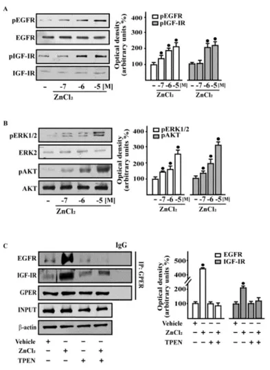
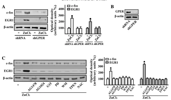
As a dysregulated Zn homeostasis may contribute to breast carcinogenesis through different mechanisms [148], including the activation of growth factors transduction pathways [154-156], we began our study by ascertaining that Zn chloride (ZnCl2) triggers the rapid phosphorylation of EGFR and IGF-IR (Fig. 3.1A) as well as the activation of downstream kinases such as ERK and AKT (Fig. 3.1B) in a dose-dependent manner. Similar results were obtained using Zn sulfate (ZnSO4) (data not shown). On the basis of these findings and considering that Zn serum concentration is approximately 15 μM [170], in subsequent assays 10 µM ZnCl2 were used. As our previous studies have shown that, in cancer cells, both EGFR and IGF-IR transduction signalling are involved in GPER regulation [110, 81-82], we evaluated whether the activation of EGFR and IGF-IR by ZnCl2 may involve GPER. By co-immunoprecipitation studies performed in SkBr3 cells, we ascertained that ZnCl2 increases a direct interaction of GPER with EGFR and IGF-IR, while the Zn chelator TPEN prevented this response (Fig. 3.1C).
Next, we determined that the ZnCl2-dependent activation of EGFR and IGF-IR as well as ERK and AKT requires GPER, as observed silencing its expression (Fig. 3.2A-D). Using the EGFR inhibitor AG1478, the IGF-IR inhibitor AG1024 and the GPER antagonist G15, we also established that EGFR, IGF-IR and GPER are involved in ERK and AKT activation by ZnCl2 (Fig. 3.2E). Likewise, in the presence of the Zn chelator TPEN and the scavenger of reactive oxygen species (ROS) NAC, the phosphorylation of ERK and AKT by ZnCl2 was no longer evident (Fig. 2E), suggesting that the production of ROS may be involved in these effects [155-156]. Therefore, we assessed that the
30 generation of ROS triggered by ZnCl2 in SkBr3 cells is no longer evident in the presence of TPEN or NAC (Fig. 3.2F), nicely confirming the aforementioned findings. Collectively, these observations indicate that ZnCl2 activates a complex transduction signalling that may involve GPER together with EGFR and IGF-IR and downstream effectors like ERK and AKT, hence leading to important biological outcomes.
Figure 3.1 | ZnCl2 triggers rapid responses and stimulates the co-immunoprecipitation of EGFR and IGF-IR with GPER in
breast cancer cells. (B) in SkBr3 cells treated for 15 min with vehicle (-) and increasing concentrations of ZnCl2, as indicated. Side panels show densitometric analysis of the blots normalized to EGFR, IGFIR, ERK2, and AKT that served as loading controls, respectively for pEGFR, pIGF-IR, pERK1/2, and pAKT. (C) Co-immunoprecipitation assays performed in SkBr3 cells treated with 10mM ZnCl2 for 15 min using the antibody against GPER followed by immunoblotting for EGFR or IGF-IR, as indicated. In control samples, nonspecific IgG was used instead of the primary antibody. IP, Immunoprecipitation. Input represents the blots probed with the antibody against GPER. Side panels show densitometric analysis of the blots normalized to b-actin. Data shown are the mean ±SD of three independent experiments. (*) indicates P<0.05 for cells treated with vehicle (-) versus treatments
31 Figure 3.2 | GPER is involved in the rapid action of ZnCl2 in breast cancer cells. (A–D) Phosphorylation of EGFR (A), IGF-IR
(A), ERK1/2 (C) and AKT (C) in SkBr3 cells after silencing GPER expression. Cells were transfected with control shRNA or shGPER, and treated for 15 min with vehicle (-) and 10mM ZnCl2. (B and D) Efficacy of GPER silencing. (E) ERK1/2 and AKT activation in SkBr3 cells treated for 15 min with vehicle (-) or 10mMZnCl2 alone or in combination with 10mMEGFR inhibitor AG1478, 10 mM IGF-IR inhibitor tyrphostin AG1024, 100nM GPER antagonist G15, 20mM zinc chelator TPEN, and 300mM free radical scavenger NAC. Side panels show densitometric analysis of the blots normalized to EGFR, IGFIR, ERK2, and AKT that served as loading controls, respectively for pEGFR, pIGF-IR, pERK1/2, and pAKT. (F) ROS production determined as DCF fluorescence in SkBr3 cells treated for 1 h with vehicle (-) or 10mM ZnCl2 alone and in combination with 20 mM zinc chelator TPEN, and 300 mM free radical scavenger NAC. DCF fluorescence obtained in cells treated with vehicle (-) was set as onefold induction upon which ROS levels induced by treatments was calculated. Data shown are the mean ±SD of three independent experiments. (*) indicates P<0.05 for cells treated with vehicle (-) versus treatments.
32 3.2 GPER contributes to gene expression changes and growth responses induced by Zn in
breast cancer cells
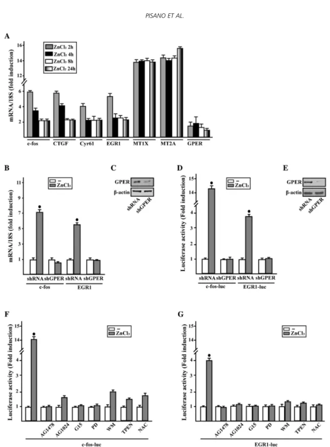
Considering that GPER triggers a specific gene signature [171], we then assessed that in SkBr3 cells ZnCl2 regulates the mRNA expression of certain GPER target genes like c-fos, CTGF, Cyr61, EGR1, MT1X and MT2A, as evaluated by real-time PCR (Fig. 3.3A). Next, we ascertained that the treatment with ZnCl2 does not alter the mRNA expression of GPER (data not shown), however its silencing prevented the mRNA induction of c-fos and EGR1 (Fig.3.3B-C), two main GPER target genes [171]. In accordance with these findings, the transactivation of c-fos and EGR1 promoter constructs upon ZnCl2 exposure was no longer evident knocking down GPER expression (Fig. 3.3D-E). Moreover, the EGFR inhibitor AG1478, the IGF-IR inhibitor AG1024, the GPER antagonist
G15, the MEK inhibitor PD, the PI3K inhibitor WM, the zinc chelator TPEN and the ROS scavenger NAC abolished the luciferase activity of c-fos and EGR1 reporter plasmids induced by ZnCl2 (Fig. 3.3F-G).
In accordance with these findings, c-fos and EGR-1 protein expression triggered by ZnCl2 was prevented by GPER silencing (Fig. 3.4A-B) and in the presence of each of the aforementioned inhibitors (Fig. 3.4C).
33 Figure 3.3 | ZnCl2 regulates the expression of GPER target genes in breast cancer cells. ZnCl2 regulates the expression of GPER
target genes in breast cancer cells. (A) The mRNA expression of c-fos, CTGF, Cyr61, EGR1, MT1X, MT2A and GPER was evaluated by real-time PCR in SkBr3 cells treated with vehicle (-) and 10mM ZnCl2, as indicated. (B) Evaluation of c-fos andEGR1mRNAexpression in SkBr3 cells transfected with shRNA or shGPER, and treated for 2 h with vehicle (-) and 10mM ZnCl2. (C) Efficacy of GPER silencing. Results obtained from experiments performed in triplicate were normalized for 18 S expression and shown as fold change of RNA expression compared to cells treated with vehicle. (D) Evaluation of c-fos and EGR1 luciferase reporter genes in SkBr3 cells transfected with shRNA or shGPER, and treated for 18 hwith vehicle (-) and 10mM ZnCl2. (E) Efficacy of GPER silencing. (F and G) Evaluation of c-fos and EGR1 luciferase reporter genes in SkBr3cells treated for 18 h with vehicle (-) or 10mM ZnCl2 alone or in combination with 10mM EGFR inhibitor AG1478, 10mM IGF-IR inhibitor tyrphostin AG1024, 100nM GPER antagonist G15, 10mM MEK inhibitor PD98089 (PD), 1mM PI3K inhibitor wortmannin (WM), 20mM Zn chelator TPEN, and 300mM free radical scavenger NAC. Luciferase activity was normalized to the internal transfection control; values are presented as fold change (mean ±SD) of vehicle control and represent three independent experiments, each performed in triplicate. (*) indicates P<0.05 for cells receiving vehicle (-) versus treatments.
34 Figure3.4 | GPER is involved in c-fos and EGR1 protein increase induced by ZnCl2 in breast cancer cells. in breast cancer cells.
(A and B) Protein levels of c-fos and EGR1 in SkBr3 cells transfected with shRNA or shGPER, and treated with vehicle (-) or 10mM ZnCl2 for 4 h. (B) Efficacy of GPER silencing. (C) Immunoblots showing c-fos and EGR1 protein expression in SkBr3 cells treated for 4 h with vehicle (-), and 10mM ZnCl2 alone or in combination with 10mM EGFR inhibitor AG1478, 10mM IGF-IR inhibitor tyrphostin AG1024, 100nM GPER antagonist G15, 10mM MEK inhibitor PD98089 (PD), 1 mM PI3K inhibitor wortmannin (WM), 20 mMZn chelator TPEN, and 300mMfree radical scavenger NAC. Side panels show densitometric analysis of the blots normalized to b-actin. Values represent the mean ±SD of three independent experiments. (*) indicates P<0.05 for cells treated with vehicle (-) versus treatments.
As cyclin D1 and cyclin A have been implicated in the development of several tumors including breast cancer [172], we also evaluated the potential of ZnCl2 to induce these cell cycle regulators. We found that ZnCl2 stimulates the expression of both cyclins (Fig. 3.5A-B), however this response was abrogated silencing GPER (Fig. 3.5B-C) as well as in the presence of AG1478, AG1024, G15, PD, WM, TPEN (Fig. 3.5D). As it concerns NAC, its inhibitory action was mainly exerted on cyclin D1 protein increase by ZnCl2 whereas the up-regulation of cyclin A upon NAC treatment was blunted but still evident (Fig. 3.5D). Indeed, although the chelator TPEN does not act in a selective manner, its ability to prevent the aforementioned responses to Zn may further confirm our findings on the biological properties of this metal. On the basis of the results obtained, it could be therefore argued that GPER is involved in Zn-dependent gene expression that occurs through the EGFR and IGF-IR transduction pathways.
35 Figure 3.5 | GPER is involved in the up-regulation of cyclins by ZnCl2 in breast cancer cells. (A) The mRNA expression of cyclin D1 and cyclin A was evaluated by real-time PCR in SkBr3 cells treated with vehicle (-) or 10 mM ZnCl2, as indicated. Results obtained from experiments performed in triplicate were normalized for 18 S expression and shown as fold change of RNA expression compared to cells treated with vehicle. (B) Cyclin D1 and cyclin A protein levels in SkBr3 cells transfected with shRNA or shGPER. and treated with vehicle (-) and 10 mM ZnCl2 for 12 h. (C) Efficacy of GPER silencing. (D) Cyclin D1 and cyclin A immunoblots in SkBr3 cells treated for 12 h with vehicle (-), and 10 mM ZnCl2 alone or in combination with 10mM EGFR inhibitor AG1478, 10mM IGF-IR inhibitor tyrphostin AG1024, 100nM GPER antagonist G15, 10mM MEK inhibitor PD98089 (PD), 1 mM PI3K inhibitor wortmannin (WM), 20mM zinc chelator TPEN, and 300 mM free radical scavenger NAC. Side panels show densitometric analysis of the blots normalized to b-actin. Values represent the mean ±SD of three independent experiments. (*) indicates P<0.05 for cells treated with vehicle (-) versus treatments.
We also found that ZnCl2 triggers SkBr3 cell cycle progression and proliferation through the involvement of GPER (Fig. 3.6A-E), thus paralleling the activation of both cyclin D1 and cyclin A. Likewise, we determined that SkBr3 cell migration induced by ZnCl2 is no longer evident silencing GPER as determined by Boyden chamber assay (Fig. 3.6F-G). Taken together, these data extend the current knowledge on the molecular mechanisms through which Zn may induce stimulatory effects in breast cancer cells.
36 Figure 3.6 | GPER contributes to ZnCl2 induced cell-cycle progression and proliferation of breast cancer cells. (A) Cell-cycle analysis performed in SkBr3 cells transfected with shRNA or shGPER, and treated with vehicle (-) and 10mM ZnCl2 for 18 h. (B) The histograms show the percentages of cells in G1/G0, S, and G2/M phases of the cell cycle, as determined by flow cytometry analysis. (D) The proliferation of SkBr3 cells upon treatment with 10 mM ZnCl2 is prevented knocking down GPER expression. Cells were transfected with shRNA or shGPER and treated every 2 d with vehicle (-) or ZnCl2 as indicated, and then counted on day 6. Proliferation of cells treated with vehicle was set as 100% upon which cell growth induced by treatments was calculated. (F) The migration of SkBr3 cells upon 6 h treatment with 10 mM ZnCl2 is abrogated knocking down GPER expression, as evaluated by Boyden Chamber assay. (C, E, and G) Efficacy of GPER silencing. Each data point is the mean ±SD of three independent experiments performed in triplicate. (*) indicates P<0.05 for cells treated with vehicle (-) versus treatments.
3.3 GPER contributes to Zn action in CAFs.
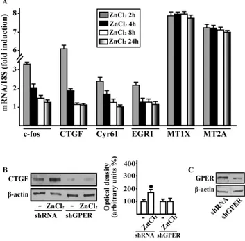
In order to further ascertain whether GPER may contribute to the action of Zn, we used CAFs that play an active role toward the growth, expansion and dissemination of breast cancer cells [173-174]. Remarkably, ZnCl2 increased the mRNA levels of diverse GPER target genes like c-fos, CTGF, Cyr61, EGR1, MT1X and MT2A in CAFs obtained from breast cancer specimens (Fig.7A). Gene expression profile displayed responses to ZnCl2 similar to those observed in SkBr3 cells (Fig. 3A), as the induction of c-fos, CTGF, Cyr61 and EGR1 was rapid (2-4 h) but declined thereafter,
37 whereas the expression of MT1X and MT2A was still evident up to 24 h. Then, we observed that the up-regulation of CTGF protein levels upon ZnCl2 treatment is prevented knocking down GPER expression in CAFs (Fig. 3.7B-C).
Figure 3.7 | GPER is involved in gene expression changes induced by ZnCl2 in CAFs. (A) The mRNA expression of c-fos, CTGF, Cyr61, EGR1, MT1X, and MT2A was evaluated by real-time PCR in CAFs treated with vehicle (-) and 10 mM ZnCl2, as indicated. Results obtained from experiments performed in triplicate were normalized for 18 S expression and shown as fold change of RNAexpression compared to cells treated with vehicle. (B) Immunoblots showing CTGF protein expression in CAFs transfected with shRNA or shGPER, and treated for 4 h with vehicle (-) and 10 mM ZnCl2. Side panel shows densitometric analysis of the blot normalized to b-actin. (C) Efficacy of GPER silencing. Values represent the mean ±SD of three independent experiments. (*) indicates P <0.05 for cells treated with vehicle (-) versus treatments.
As CTGF exerts an acknowledged role in migratory properties of different cell types [171, 175], we evaluated whether GPER signalling through CTGF may trigger the migration of CAFs. Scratch experiments and Boyden chamber assays revealed that ZnCl2-stimulated migration of CAFs is abolished silencing GPER or CTGF expression, whereas adding CTGF the migratory response was rescued (Fig. 3.8). Collectively, the aforementioned results indicate that Zn-activated GPER signaling mediates a similar gene expression profile as well as important biological responses in both breast cancer cells and CAFs. On the basis of these findings, it could be argued that Zn may trigger through GPER a functional interplay between cancer cells and CAFs toward breast tumor progression.
38 Figure 3.8 | GPER and its target gene CTGF contribute to the migration of CAFs induced by ZnCl2. (A–C) The migration of CAFs upon treatment with 10 mM ZnCl2 for 24 h is prevented knocking down GPER and CTGF expression, as assessed by wound-healing assay. Cell migration is rescued in CAFs transfected with shGPER (B) or shCTGF (C) exposed to 10 mM ZnCl2 for 24 h and treated with 100 ng/ml CTGF. Images shown are representative of three independent experiments. (F) The migration of CAFs induced by a 6 h treatment with 10 mM ZnCl2 is prevented knocking down GPER and CTGF expression, as evaluated by Boyden Chamber assay. Cell migration is rescued in CAFs transfected with shGPER and shCTGF, exposed to 10 mM ZnCl2 for 6 h and treated with 100 ng/ml CTGF. Efficacy of GPER (D and G) and CTGF (E and H) silencing. Values represent the mean ±SD of three independent experiments. (*) indicates P<0.05 for cells treated with vehicle(-) versus treatments.
39
Chapter 4
Discussion
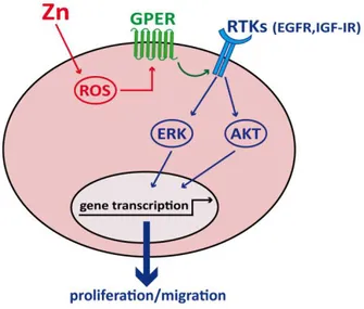
Several human activities as well as natural events can lead to heavy metals pollution and, therefore, increased incidence of various tumors [164-166]. In the present study, we have demonstrated that one important pollutant like Zn may trigger a functional interplay of GPER with EGFR and IGF-IR, which leads to the activation of main transduction pathways, gene expression changes, and important biological responses like proliferation and migration in breast cancer cells and CAFs (Figure 3.9).
Figure 3.9 | Schematic representation of the functional cooperation of GPER with IGF-IR and EGFR upon zinc exposure.
Breast cancers have been reported to show an increased Zn uptake and tissue concentration as compared to the normal breast tissue [10, 167], while patients with advanced breast tumors show decreased serum Zn levels. Thus, the determination of serum Zn levels has been proposed as a prognostic marker in breast cancer patients [145, 168-169]. Of note, tamoxifen-resistant breast
40 cancer cells show increased levels of Zn and its transporter ZIP7 that are involved in the activation of EGFR and IGF-IR transduction signaling toward cell proliferation and invasion [15]. In accordance with these findings, growth factors-mediated effects of Zn promoted the activation of kinases, gene expression changes and growth responses [19-20].
Numerous studies have shown that GPER contributes to the progression of certain tumors including breast cancer [69, 177-180]. In addition, clinical studies have indicated that GPER may be a predictor of aggressive cancer behavior as its expression has been associated with negative clinical outcomes in several cancer histotypes [91, 95, 104, 107]. The activation of GPER has been shown to trigger EGFR transactivation, subsequent transduction events such as the activation of MAPK and PI3K cascades, gene expression changes, and relevant biological responses such as proliferation, migration and angiogenesis in diverse cancer cell types and CAFs [181-182]. In this context, it should be mentioned that the metal cadmium may induce cAMP increase, ERK1/2 activation and proliferation of breast cancer cells in a GPER-dependent manner [183]. Recently, we also demonstrated that copper activates the HIF-1α/GPER/VEGF signalling in cancer cells leading to angiogenesis and tumor progression [182]. Further extending these findings, in the present study we have demonstrated that in breast cancer cells exposed to Zn the activation of GPER leads to rapid signalling events such as the phosphorylation of EGFR and IGF-IR and their downstream effectors ERK and AKT, the up-regulation of c-fos and EGR1, two main GPER target genes largely involved in growth responses. It is worth noting that Zn induced also GPER targets namely metallothioneins MT1X and MT2A, whose overexpression correlates with chemoresistance and poor prognosis in breast tumors [184-185]. Moreover, in line with the known capability of GPER to trigger the transcription of genes associated with cell growth [171], we assessed the potential of Zn to regulate the expression of two members of the cyclin family as cyclin D1 and A. According to their regulatory role of cell-cycle progression, proliferation and notably migration [186], we assessed also that Zn through GPER significantly increases the percentage of SkBr3 cells in the S phase of the cell cycle as well as stimulates cell proliferation and migration.
41 Several studies have suggested the active role exerted by the cancer microenvironment on the growth and spread of neoplastic cells [187]. For instance, CAFs contribute to breast cancer aggressiveness through the production of secreted factors that promote migration, invasion and angiogenesis [187]. Further extending these findings, we have ascertained that Zn promotes the migration of CAFs through GPER and the induction of its target gene CTGF, which has been widely involved in cancer cells dissemination and metastasis [171, 175]. Moreover, we have assessed that Zn may influence analogous transcriptional and functional responses in both breast cancer cells and main components of the reactive stroma like CAFs toward more aggressive tumor features.
Altogether, the present data provide novel insights into the molecular mechanisms through which Zn may elicit stimulatory effects in breast cancer cells and tumor microenvironment components like CAFs. In particular, our findings indicate that GPER may be included together with EGFR and IGF-IR among the transduction mediators of relevant biological responses to Zn in breast cancer cells and the surrounding stroma.