Dipartimento di Inge,gneria per Il'Ambiente e il Territorio e Iingegneria
Chimica
Scienze ed Ingegneria dell'Ambiente, dellJe Costruzioni e dell' Energia (SIACE)
CHIM/07 fONDAMENTI CHIMICI DELLE TECNOLOGIE Ci:clo XXIX
PhD Thesis
PERFORMANCE OF H0LL0W FIBER MEMI8RAN,E
BIOREACTOR AS A BIOARTIFICIAL LIVER
PhO Student
Haysam Mohamed Magdy Ahmed
il
~ ~.,I)
suP~rviso,rsD .sa.
Loredan~artolo
pr~
Coordinator. Prot. Pietro Pantano
Host Institute: Istituto per la Tecnologia delle Membrane Consiglio Nazionale delle Ricerche (ITM-CNR)
February 22,,2017
2
Current research was funded by:
Marie Skłodowska-Curie Actions
Initial Training Network
Project BIOART
3
Contents
Acknowledgement ... 6
Abstract (Italian) ... 7
Abstract ... 10
Preface and Scope of Work ... 12
CHAPTER 1 ... 14
State of the art and research scope ... 14
1.1 Liver anatomy and physiology ... 14
1.2 Cellular components of the human liver ... 17
1.2.1 Hepatocytes ... 18
1.2.2 Sinusoidal endothelial cells ... 18
1.2.3 Stellate cells ... 19
1.2.4 Kupffer cells ... 19
1.2.5 Cholangiocytes ... 20
1.3 Liver support systems ... 20
1.3.1 Artificial liver support systems ... 20
1.3.2 Bioartificial liver support systems ... 22
1.3.3 Hepatocyte transplantation ... 27
1.3.4 Repopulation of decellularized liver ... 28
1.3.5 Organ printing ... 28
1.3.6 Induced organogenesis ... 29
1.4 Cell source as critical issue for BAL devices ... 30
1.4.1 Primary hepatocytes ... 30
1.4.2 Cell lines ... 31
1.4.3 Stem cells ... 31
1.5 Oxygen supply ... 33
1.7 Bioreactor configurations in bioartificial liver designs ... 35
1.5.1 Bioartificial liver systems in clinical trial ... 35
1.5.2 Bioreactor designs for in vitro hepatic culture studies ... 38
CHAPTER 2 ... 41
Long-term Maintenance of Human Hepatocyte Microtissue Spheroids in a crossed hollow fiber membrane bioreactor ... 41
2.1 Introduction ... 41
4
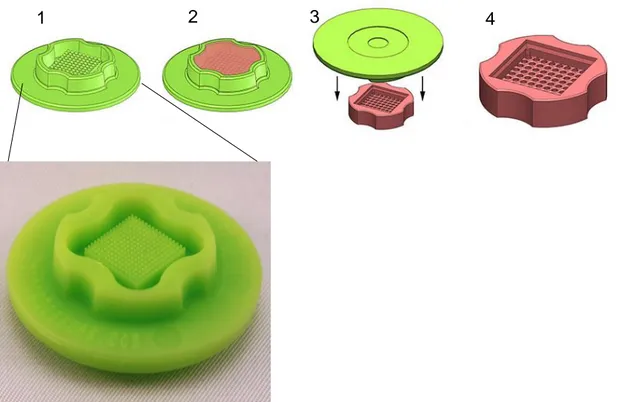
2.2.1 Mold fabrication ... 44
2.2.2 Bioreactor ... 44
2.2.3 Cell Culture ... 46
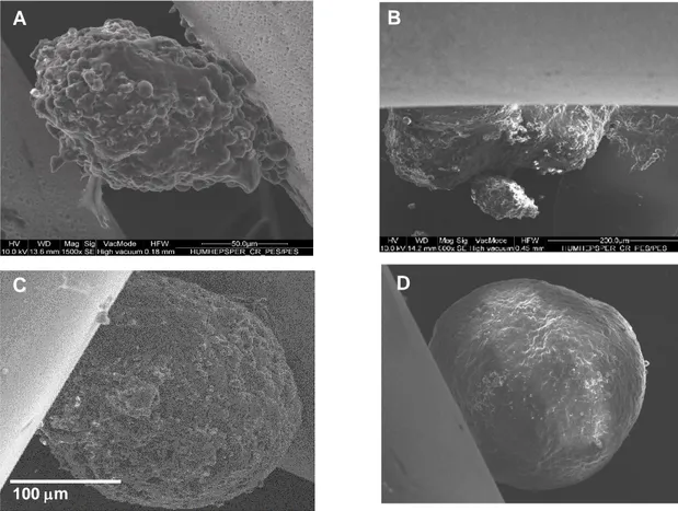
2.2.4 Cell Morphology ... 47
2.2.5 Assay of metabolic functions ... 48
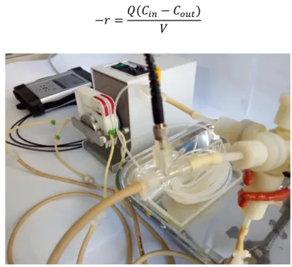
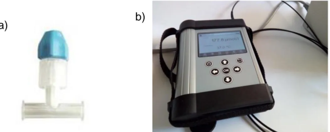
2.2.6 Dissolved oxygen concentration measurement ... 49
2.3 Results ... 50 2.3.1 Morphological evaluation ... 50 2.3.2 Hepatocyte-specific functions ... 56 2.3.3 Oxygen uptake ... 59 2.4 Discussion ... 60 2.5 Conclusion ... 62 CHAPTER 3 ... 63
3D organotypic liver organoid formation in hollow fiber membrane systems . 63 3.1 Introduction ... 63
3.2. Materials and Methods ... 65
3.2.1. Membrane systems ... 65
3.2.2. Cell Culture ... 66
3.2.3 Cell Morphology ... 68
3.2.4 Assay of metabolic functions ... 69
3.2.5. Statistical analysis ... 70
3.3. Results ... 70
3.3.1 Morphological evaluation of organotypic membrane system ... 70
3.3.2 Hepatocyte-specific functions ... 71
3.3.3 Oxygen uptake rate ... 80
3.4. Discussion ... 82
3.5. Conclusions ... 85
CHAPTER 4 ... 86
Expansion and differentiation of stem cells in the hollow fiber membrane bioreactor ... 86
4.1 Introduction ... 86
4.2 Materials and Methods ... 88
4.2.1 Hollow-fiber membrane bioreactor ... 88
4.2.2 Cell Culture ... 88
5
4.2.4 Cell Morphology ... 89
4.2.5 Assay of metabolic functions ... 90
4.2.6 Periodic Acid-Schiff (PAS) Stain for Glycogen ... 90
4.2.7 Total RNA Isolation and Reverse-Transcription Polymerase Chain Reaction ... 90
4.2.8 Statistical analysis ... 91
4.3 Results ... 92
4.3.1 Morphological evaluation of differentiating cells on various coatings ... 92
4.3.2 Gene expression profile of treated cells ... 95
4.3.3 Hepatocyte specific functions ... 96
4.4 Discussion ... 98
4.5 Conclusions ... 99
General Conclusion and Future Perspectives ... 101
6
Acknowledgement
Firstly, I would like to express my sincere gratitude to my advisor Dr. Loredana De Bartolo for the continuous support throughout my PhD study, for her patience, motivation, and immense knowledge. Her guidance helped me in all the time of research and writing of this thesis. I could not have imagined having a better advisor and mentor for my PhD study.
Besides my advisor, I would like to thank Dr Efrem Curcio and Dr. Lidietta Giorno for their insightful comments and encouragement.
My sincere thanks also go to Dr. Simona Salerno, Dr. Antonella Piscioneri, and Dr. Sabrina Morelli, who provided me an opportunity to join their team, and who gave access to the laboratory and research facilities. Without they precious support it would not be possible to conduct this research.
I thank my fellow labmates in for the stimulating discussions, for the sleepless nights we were working together before deadlines, and for all the fun we have had in the last three years.
Last but not least, I would like to thank my family: My wife, parents and brother for supporting me spiritually throughout writing this thesis and my life in general.
7
Abstract (Italian)
C'è una crescente necessità di sviluppare un dispositivo bioartificiale di tipo epatico da utilizzare sia in applicazioni in vitro, per la sperimentazione della tossicità di molecole da parte delle aziende farmaceutiche, e sia in applicazioni cliniche per supportare pazienti con insufficienza epatica in attesa di trapianto di organo. A tale scopo è stato realizzato un bioreattore a membrana a fibre cave incrociate adoperante cellule epatiche umane in grado di favorire il mantenimento a lungo termine di epatociti. Il bioreattore è costituito da due fasci di membrane a fibre cave (HFM), uno deputato all’alimentazione e l’altro alla rimozione di cataboliti e prodotti specifici cellulari. I due fasci di fibre sono assemblati in una configurazione incrociata ed alternata in modo da stabilire una distanza l’una dall’altra di 250 μm. Questa configurazione del bioreattore delinea tre compartimenti separati: due compartimenti all’interno del lumen delle fibre cave dove il mezzo di coltura fluisce e un compartimento extraluminale dove le cellule sono coltivate. I compartimenti intraluminali ed extraluminale comunicano tra di loro attraverso i pori della parete di membrana. Il mezzo che fluisce nel lumen delle fibre di alimentazione permea nel compartimento cellulare, dove i cataboliti ed i metaboliti prodotti dalle cellule vengono rimossi dalle fibre cave deputate all’allontanamento dei molecole di sintesi e di scarto cellulari. In questo dispositivo le membrane a fibre cave consentono la compartimentalizzazione delle cellule in un microambiente controllato a livello molecolare ed il trasporto selettivo di molecole verso e dal compartimento cellulare proteggendo le cellule da eventuali sforzi di taglio. Inoltre, le membrane, grazie alla loro geometria intrinseca, offrono un'ampia superficie per l'adesione e la crescita delle cellule in un volume ridotto.
Epatociti umani rappresentano una fonte cellulare ottimale da utilizzare nelle terapie che sono basate sull’uso di cellule, in quanto riflettono più da vicino le condizioni in vivo. In vivo gli epatociti sono altamente proliferativi all'interno del loro microambiente. Tuttavia, quando sono isolati dal loro microambiente e coltivati in vitro, perdono rapidamente le loro funzioni specifiche. Pertanto, è di importanza fondamentale la realizzazione di modelli in vitro in grado di mantenere gli epatociti vitali e funzionali per lungo tempo. Un aspetto critico è la la scarsa disponibilità di epatociti umani per cui occorre prendere in considerazione fonti cellulari alternative. Gli studi effettuati in questi ultimi anni indicano come una delle migliori fonti cellulari alternativa agli epatociti le cellule staminali, poiché queste cellule sono ampiamente disponibili possiedono in vitro
8 un’elevata capacità proliferativa e possono essere differenziate in epatociti. A differenza delle cellule provenienti da animali e delle linee cellulari, le cellule staminali non costituiscono un rischio di trasmissione virale zoonotica o tumorigenicità.
In questo lavoro, il bioreattore a membrana è stato ottimizzato al fine di creare condizioni di coltura per aggregati cellulari come sferoidi e per sistemi organotipici tridimensionali (co-coltura di epatociti e cellule non parenchimali) che garantiscano il mantenimento a lungo termine della funzionalità dei costrutti epatici umani. A tal proposito, le funzioni specifiche epatiche come l'urea, la sintesi dell'albumina e la biotrasformazione di farmaci sono state valutate nel bioreattore. I cambiamenti morfologici cellulari sono stati analizzati utilizzando il microscopio elettronico a scansione ed il microscopio confocale a scansione laser. Inoltre, il consumo di ossigeno delle cellule poste in coltura nel bioreattore è stato continuamente monitorato nel tempo al fine di assicurare un adeguato approvvigionamento di ossigeno.
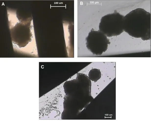
Gli sferoidi epatici umani, posti in coltura nello spazio extracapillare del bioreattore sono andati incontro ad un processo di fusione che ha portato alla formazione di strutture di maggiore dimensione simili a microtessuti. La fusione degli sferoidi è stata osservata sia tra le fibre che intorno alle fibre simulando il processo che avviene in vivo. Questo modello di coltura, grazie alle sue caratteristiche tridimensionali e all'aumentata interazione cellulare, così come avviene in vivo, ha favorito il mantenimento a lungo termine della vitalità e delle diverse funzioni specifiche epatiche come la sintesi di albumina ed urea ed il metabolismo xenobiotico. Allo stesso modo, nel sistema organotipico, le cellule si riorganizzano formando strutture tissutali simili a quelle del tessuto epatico in vivo. Questo è stato reso possibile grazie al piastramento sequenziale sulle membrane di cellule non parenchimali e parenchimali che hanno formato strutture stratificate tridimensionali simili a quelli in vivo. Il bioreattore che è stato ottimizzato in questo lavoro di tesi fornisce un microambiente di coltura ben controllato da un punto di vista molecolare attraverso l'alimentazione continua di sostanze nutritive, di cui una delle più importanti è l'ossigeno, e la rimozione di cataboliti. Ciò è stato confermato dai risultati relativi alla misura della concentrazione di ossigeno nel mezzo di coltura sia nella corrente in ingresso che in uscita dal bioreattore. In entrambi i modelli di coltura, l'approvvigionamento di ossigeno nel bioreattore è risultato essere sufficiente e significativamente maggiore a quello osservato in condizioni di coltura statica. Inoltre, una nuova fonte di cellule staminali, ovvero le cellule staminali mesenchimali derivate dal fegato, è stata utilizzata: le cellule sono state differenziate con successo in epatociti dopo 24 giorni di coltura, sia nel sistema
9 statico che nel bioreattore. Tuttavia, il bioreattore ha mostrato una migliore capacità di mantenere la vitalità delle cellule e di differenziare le cellule staminali mesenchimalinel fenotipo epatico, come dimostrato dall'aumento dell'espressione genica di marcatori epatici specifici (ad es. albumina ed il fattore nucleare epatico alfa-4) e dalle velocità di sintesi di urea e albumina.
Il prototipo di bioreattore realizzato su scala di laboratorio ha mantenuto con successo e funzionalmente attivi gli epatociti umani coltivati come sferoidi e in co-coltura con cellule non parenchimali per quasi un mese. Un aspetto importante è stato il differenziamento epatico delle cellule staminali mesenchimali, che rappresentano una potenziale fonte di cellule alternativa agli epatociti umani primari. Tutti questi risultati sono stati ottenuti utilizzando solo cellule umane, che convalidano le prestazioni del dispositivo che è stato sviluppato come sistema epatico bioartificiale da utilizzare in vitro. Questo bioreattore su scala di laboratorio ha un elevato potenziale applicativo cha va dagli studi in vitro delle malattie epatiche agli studi di tossicità a lungo termine. Inoltre, può essere realizzato su scala clinica ed applicato come fegato biartificiale per sostituire le funzioni epatiche di pazienti affetti da insufficienza epatica in attesa di trapianto.
10
Abstract
There is a growing need for a bioartificial liver device for applications ranging from lab-scale platform for drug toxicity testing by pharmaceutical companies to a clinical-scale temporary support device for liver failure patients pending transplantation.
To this end, a crossed hollow-fiber membrane bioreactor was used in conjugation with various human cells to establish long-term cultures. The bioreactor consists of two bundles of hollow fiber membranes (HFM) that are cross-assembled in alternating manner at distance each other of 250 µm. The bioreactor is comprised of three separate compartments: two intraluminal compartments within the feeding and removing HFM, and an extraluminal compartment or shell outside of the fibers that represents the cell compartment. The intraluminal and extraluminal compartments communicate through the pores of the fiber wall. The medium permeates out of the feeding hollow fiber bundle into the cellular compartment. The catabolites and metabolites produced by cells are removed by the other bundle of hollow fibers. The HFM allow the compartmentalization of cells in a microenvironment controlled at molecular level and the selective mass transfer of molecules to and from the cell compartment without causing shear stress. Owing to its intrinsic geometry they provide a wide surface area for the adhesion and growth of cells in a small volume.
Primary human hepatocytes were used as they are the optimal cell source for cell-based therapies, because they most closely reflect the in vivo situation. Within their in vivo microenvironment, hepatocytes are highly proliferative. However, when they are isolated from their in vivo microenvironment and cultured in vitro they rapidly de-differentiate losing their liver-specific functions. Therefore, there is an urgent need for the development of in vitro models capable of maintaining hepatocytes viable and functional for long periods of time. Moreover, due to the scarcity of human hepatocytes, an alternative cell source needs to be considered. Hepatogenic differentiation of stem cells is considered the best option, since these cells are readily available and can proliferate in vitro. Unlike animal cells and cell lines, stem cells do not pose a risk of zoonotic viral transmission or tumorigenicity.
In this work, culture conditions for 3D microtissue spheroids and organotypic cultures (co-culture of hepatocytes and non-parenchymal cells) were optimized in the bioreactor in order to achieve the long-term maintenance of functional human primary liver constructs. Liver-specific functions such as urea, albumin synthesis and drug metabolism were evaluated. Also, morphological
11 assessment was carried out utilizing scanning electron microscope, confocal laser scanning microscope and light microscope. In addition, oxygen consumption rates were monitored throughout the cultures to ensure an adequate oxygen supply.
The human hepatocyte spheroids cultured in the extracapillary space of the bioreactor fused forming larger microtissue-like structures between and around the fibers that more closely resembles the in vivo situation in comparison to single spheroids. By more closely resembling the in vivo environment, due to its three-dimensionality and cell–cell interaction, this model supported long-term viability and the maintenance of several liver-specific functions including albumin and urea synthesis and xenobiotic metabolism. Similarly, in the organotypic cultures, cells rearranged forming tissue-like structures that better resemble the in vivo liver tissue due to the sequential seeding of cells during the experiment setup resulting in layered structures similar to those seen in vivo. The bioreactor provides a well-controlled microenvironment through continuously feeding nutrients, most important of which is oxygen, and removing catabolites. This was confirmed via dissolved-oxygen monitoring in both inlet and outlet of the bioreactor. In both culture conditions, oxygen in the bioreactor was found to be sufficient and significantly higher than those observed in batch condition.
In addition, a novel stem cell source, namely liver-derived mesenchymal stem cells, was successfully differentiated into hepatocyte-like cells after 24 days in culture both in batch systems and HFMBR. However, the bioreactor showed an improved survival as well as differentiation of the mesenchymal stem cells as demonstrated by the increase of gene expression of specific hepatocyte marker (e.g. Albumin and Hepatocyte Nuclear Factor 4 Alpha), and higher urea and albumin synthesis rates.
The lab-scale prototype hollow-fiber membrane bioreactor (HFMBR) realized in this work successfully maintained viable and functionally active human hepatocytes cultured as spheroids and in co-culture with non -parenchymal cells for almost one month. Moreover, it resulted in enhanced hepatic differentiation of mesenchymal stem cells, providing a potential alternative cell source to the scares primary human hepatocytes. All these results were obtained using human cells only, which validate the performance of the device that was developed as an in vitro bioartificial liver platform. This lab-scale bioreactor has various potential applications ranging from basic studies of liver diseases to long-term toxicity studies. Moreover, it can be scaled-up to a clinical scale to be used as BAL device for liver failure patients.
12
Preface and Scope of Work
Liver disease and the subsequent loss of liver function is an enormous clinical challenge, and is currently the 12th most frequent cause of death in the United
States and the 4th most frequent for middle-aged adults (1). Emergence of new
liver diseases such as steatohepatitis, absence of a hepatitis C vaccine, and increasing number of hepatocellular carcinoma patients, further worsens the situation (2, 3). Liver transplantation is the only established successful treatment for end-stage liver disease, and currently there are over 119,613 people on the waiting list for a donor organ and of those, there are around 14,525 candidates awaiting a liver transplant (based on United Network for Organ Sharing Organ Procurement and Transplantation Network, UNOS OPTN, December 5, 2016). In 2015, there were 5950 liver transplants performed in the United States. The European Liver Transplant Registry (ELTR) reports that in Europe 137,404 liver transplantations have been performed from 1968 to 2015 (European Liver Transplant Registry).
Various surgical options have been pursued, including living-donor partial transplantation and split liver transplants, in order to expand the supply of livers available for transplantation (4). Despite these surgical advances, organ shortage remains a major hurdle, thus it is unlikely that liver transplantation procedures alone will ever meet the increasing demand. Despite the progress made in supportive care, only 20% of liver failure patients survive without liver transplantation, which increases the survival rates to over 80% (5, 6). Moreover, improving the patient’s condition before liver transplantation substantially increases post-transplant survival (1). Lastly, temporary liver support could potentially create time for the diseased liver to regenerate, thereby rendering liver transplantation superfluous. Taken together, there is an urgent need for liver support therapy. For these reasons, many researchers have developed various liver support devices, which can be divided into non-biological (artificial) and biological (bioartificial) systems. Non-biological systems typically use adsorbents and/or dialyzing membranes to detoxify the patients’ plasma. Probably the potential of these systems is limited by their lack of metabolizing and synthesizing capacity, as well as the fact that detoxification in these systems is non-specific, which might lead to the removal of beneficial proteins, such as growth factors and clotting factors.
Generally, a BAL system consists of functional liver cells supported by an artificial cell culture material. In particular, it incorporates hepatocytes into a bioreactor in which the cells are immobilized, cultured, and induced to perform
13 the hepatic functions by processing the blood or plasma of liver-failure patients. BAL devices act as a bridge for the patients until a donor organ is available for transplantation or until liver regeneration (7).
Another important application of the BAL, is the study of drug-induced liver toxicity. Hepatotoxicity is a major cause for drug withdrawals from the market, resulting in huge financial losses for pharmaceutical companies (8, 9). Human-based in vitro models comprising of microsomes, cell lines, primary hepatocytes, and liver slices (10-13) provide additional information to the existing animal models. However, they can be limited by poor stability, and, with the exception of precision cut liver slices, lack the hierarchy and structural components of liver. Monolayer cultures of primary hepatocytes are the most commonly used format for toxicity assessment and provide a suitable model for initial assessment, but are severely hindered by the lack of 3D organization, non-parenchymal cells, and thus cell–cell interactions via contact or paracrine effects.
The aim of this thesis is to refine and further develop more sensitive human in vitro models and methods for longer maintenance of functional hepatocytes by using a crossed hollow-fiber membrane bioreactor that was developed in our laboratory. The work plan constitutes of two different parts:
1) To develop and evaluate new in vitro human liver cell culture models to improve and maintain the hepatic functionality, better extrapolating to human liver biology.
• Development of a 3D liver micro-tissues from hepatocyte spheroids
(CHAPTER 2).
• A liver co-culture model, incorporating hepatocytes and non-parenchymal cells (sinusoidal and stellate cells) (CHAPTER 3).
2) To generate new sources of functional human hepatic cells.
• Establishment of a stepwise, directed, differentiation protocol for hepatic differentiation of human stem cells (Liver Mesenchymal stem cells) (CHAPTER
4).
Evaluation of the beneficial effect of the dynamic crossed hollow-fiber
membrane bioreactor system (developed in our laboratory) on the prolonged maintenance and enhanced functions of the above-mentioned models was carried out in this context.
14
CHAPTER 1
State of the art and research scope
1.1 Liver anatomy and physiology
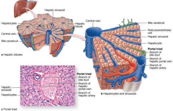
The liver is the largest internal organ in the body and performs several essential functions. These include bile production, plasma protein synthesis, glucose homeostasis and glycogen storage, processing and storage of fats, such as cholesterol, and production of hormones (14). The liver is a highly-specialized tissue that comprises many different cell types, further described below. The liver parenchyma is organized as thousands of small (~0.7 × 2 mm) hepatic lobules, in which hepatocytes form hundreds of irregular plates arranged radially around a small central vein. Peripherally each lobule has three to six portal areas, each of which contains three interlobular structures that comprise the portal triad, namely a branch of the hepatic artery, a branch of the portal vein and a bile ducts (Fig 1.1). The liver is supplied with oxygenated blood from the hepatic artery and venous blood from the portal vein entering the periportal area of the lobule and via branches of small interlobular vessels. Between all of the anastomosing plates of hepatocytes of a hepatic lobule are important vascular sinusoids that emerge from the peripheral branches of the portal vein and hepatic artery and converge on the lobule’s central vein. The mixed blood flows through the sinusoids and leaves the lobule via the hepatic central vein located in the center of the lobule. Bile is secreted by the hepatocytes into bile canaliculi, flows in the opposite direction of the blood and empties into the bile ducts that are lined by epithelial cells called cholangiocytes (15). The bile is ultimately secreted into the duodenum where it facilitates the digestion of lipids (14).
The lobule is divided in zones based on the metabolic functions that liver cells have to perform. The concentration of oxygen, nutrients, insulin and glucagon is highest in the periportal area and decreases towards the central vein. As a result of the concentration gradient, hepatocytes in the different zones have different morphology and functions (16). For example, hepatocytes around the central vein have higher density of endoplasmic reticulum and possess the highest levels of enzymes involved in detoxification and biotransformation (17, 18). Substances from orally consumed food and drugs reach the liver via the venous blood from the intestine, which is filtered through the liver before entering the systemic blood circulation. This makes the liver a central organ in metabolism of both endogenous substances, such as bilirubin and ammonia, as well as exogenous substances, like bacterial toxins and alcohol (14). Most pharmaceutical drugs available on the
15 market today are administered orally which makes the liver a highly-exposed organ for drug toxicity. Due to its central position in the body, the liver also functions as an important immune organ harboring many cells involved in both the innate and the adaptive immune response (19).
Fig. 1.1. The liver, a large organ in the upper right quadrant of the abdomen, immediately below the diaphragm, is composed of thousands of polygonal structures called hepatic lobules, which are the basic functional units of the organ. (a) Diagram showing a small central vein in the center of a hepatic lobule and several sets of blood vessels at its periphery. The peripheral vessels are grouped in connective tissue of the portal tracts and include a branch of the portal vein, a branch of the hepatic artery, and a branch of the bile duct (the portal triad). (b) Both blood vessels in this triad branch as sinusoids, which run between plates of hepatocytes and drain into the central vein. (c) Micrograph of a lobule shows the central vein (C), plates of hepatocytes (H), and in an adjacent portal area a small lymphatic (L) and components of the portal triad: a portal venule (PV), hepatic arteriole (HA), and bile ductule (B). (X220; H&E). Image taken from (15).
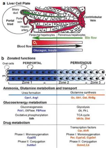
Zonated hepatic functions include glucose metabolism, ammonia detoxification, and the metabolism of drugs and xenobiotics (Fig. 1.2). Glucose metabolism was the first such function to be reported, already in 1977, where gluconeogenesis was found to be mainly periportal; whereas glycolysis was mainly perivenous (20). One of the major roles of the liver is the removal of harmful ammonia arriving from the intestine via the portal vein. Ammonia is first metabolized by periportal hepatocytes, to generate urea. Residual ammonia is then converted by perivenous hepatocytes into glutamine (21). Other zonated processes include: lipid metabolism, with
16 lipogenesis occurring perivenously and fatty-acid degradation periportally (22, 23). Perivenous hepatocytes are predominantly responsible for drug metabolic activities exhibited by the liver through monooxygenation followed by glucuronidation of the drugs converting them into excretable products (22).
Fig. 1.2 Structure and functions of the zonated liver lobule (a) The liver cell plate, with blood circulation indicated in red. Bile is shown in green and circulates in the opposite direction to blood. The concentration of oxygen and hormones decreases along a continuous gradient from the PP area to the PV area. (b) Zonal functions. The proliferation of hepatocytes is achieved mostly by division of the mature hepatocytes themselves (circled arrow), which do not migrate along the portocentral axis, with proliferation from oval cells observed only rarely (dotted circled arrow). The zonated metabolic systems include the ammonia detoxification system, glucose and energy metabolism, and xenobiotic metabolism. The proteins involved in each type of zonal metabolism are indicated.(24).
Not all hepatic processes are zonal. Some functions, such as serum-protein synthesis, appears to occur in all hepatocytes. Albumin is also synthesized in all hepatocytes, with a higher concentration in the periportal area.
Liver disease usually leads to a variety of life-threatening metabolic and physiologic abnormalities. For example, the absence of these functions leads to bleeding
17 abnormalities, accumulation of neurotoxins causing hepatic encephalopathy, accumulation of serum ammonia, and jaundice from elevation of serum bilirubin. It is possible to medically support liver disease patients through therapies targeted at features such as portal hypertension and coagulopathy. However, there are no therapeutic strategies effectively replacing the range of affected functions. Thus, an organ transplant has been the only permanently successful therapy to date. This is different compared to other organs, such as the heart and kidneys, in which patients with failing tissues can be supported by pharmaceuticals and machines, without the need for immediate transplantation.
Consequently, efforts have been focused toward the development of liver support systems that could provide temporary support for patients with liver failure. These measures include extracorporeal support devices analogous to kidney dialysis systems, processing the blood or plasma of liver failure patients (25, 26). They range from non-biological-based systems to cell-based therapies, such as BALs. In vivo, the liver exhibits a unique capacity for regeneration, with the potential for full restoration of liver mass and function even after massive damage (27).
However, a major hurdle to the advancement of cell-based therapeutic strategies is the loss of the proliferative capacity and of the liver-specific functions exhibited by hepatocytes once isolated from the in vivo microenvironment (28). Another obstacle is the limited availability of human hepatocytes. Only a limited supply of primary human hepatocytes is currently available from organs deemed inappropriate for transplantation. Thus, significant research efforts are focused on the potential of alternative cell sources, most notably those based on stem cell differentiation and reprogramming.
1.2 Cellular components of the human liver
The hepatocellular parenchyma accounts for 60% of the total cell population and 80% of the total volume of the organ, with the lobular parenchyma representing approximately 93%, the hepatic veins 4%, and the portal triads 3% of the hepatic parenchyma. Non-parenchymal cells comprise 30–35% of the total number of liver cells, but only 6% of the total liver volume. Almost half (40%) of the non-parenchymal cells are fenestrated endothelial cells. The remainder consists of phagocytic Kupffer cells (33%), extraluminal stellate cells (22%), biliary epithelial cells (4%), natural killer cells (1%) (29).
18
1.2.1 Hepatocytes
The hepatocytes are rich in cellular organelles such as mitochondria, endoplasmic reticulum and Golgi apparatus, a sign of active protein synthesis and secretion from these cells (30). Hepatocytes have a large nucleus and about 25% of the cells are binucleated which often results in polyploidy, suggested to be an important mechanism to restrict liver growth and prolong cell survival (31). The hepatocellular membranes have a complex structure with different membrane sections with different biochemical composition and functional properties: the basolateral section (facing the sinusoids), the lateral (inter-cellular) section, and the apical section facing the bile canaliculi (14). The hepatocytes perform a variety of different functions. They produce bile that is vital for the digestion of lipids. Many serum proteins i.e. albumin and blood clotting factors are synthesized by the hepatocytes and they also regulate the glucose homeostasis in the blood in response to glucagon and insulin signalling. The hepatocytes are also essential for the biotransformation of many endogenous substances, like different serum proteins, lipids and steroids. They also metabolize many exogenous substances, such as alcohol, chemicals and pharmaceuticals. Hepatocytes also play an important role in the hepatic immune response via the production of complement factors and acute phase proteins as a response to cytokine stimuli, like 6 (interleukin-6), IL-1β and TNFα, produced by Kupffer cells and endothelial cells (14). Hepatocytes have also been reported to acquire antigen presenting skills (32) and are generally considered to be both the target and inducer of the innate immune response (33). The liver has a remarkable regenerating capacity both via proliferation of hepatocytes (34) and via activation and differentiation of oval cells (27, 35). Growth factors and cytokines, such as HGF (hepatocyte growth factor), TGF-β, FGF1, IL-6 and TNFα, released by Stellate cells and Kupffer cells, has shown to have hepatoprotective effects and to stimulate liver regeneration (35-37). Additionally, TGFβ signaling, which under normal conditions keeps the hepatocytes in a quiescent state, is suppressed during injury (27).
1.2.2 Sinusoidal endothelial cells
The hepatic sinusoids are lined by fenestrated endothelial cells. The basolateral surface of the hepatocyte is separated from endothelial cells by the space of Disse. The fenestration (0.1-0.3 µm) allows efficient transfer of proteins and other macromolecules between the blood and the hepatocytes. The fenestration also facilitates the communication between cells in the sinusoidal lumen and the
19 hepatocytes as well as other cells in the space of Disse (33). The sinusoidal endothelial cells play an important role in the hepatic immune response as they participate in the clearance of antigens from the circulation by receptor mediated endocytosis, cytokine secretion and by antigen presenting capacities (38). They also collect and present antigens originating from hepatocytes (33). The regulation of endothelial antigen presentation and their role in induction of apoptosis of activated T cells play an important role for the immunologic tolerance in the liver (39).
1.2.3 Stellate cells
The hepatic stellate cells, or fat storing cells, are spindle-shaped cells located in the space of Disse, with extensions into the inter-hepatocellular space. They have an important role in storage and transportation of retinoids (vitamin A compounds) (40) and the have the ability to secret different components of the extracellular matrix (ECM), like collagen, proteoglycans and laminin, all essential for many hepatocellular functions (41). Stellate cells also play a role in hepatic immunoregulation as they are known to express Toll-like receptors for LPS stimuli (42). Activated stellate cells can amplify an inflammatory response in the liver by secretion of cytokines and chemokines (36) as well as by antigen presentation (43, 44). When activated, the stellate cells become depleted of vitamin A and via fibrogenic activities they start synthesizing large amount of ECM components, including collagen and adhesive glycoproteins (36, 45). Chronic liver injury may lead to overproduction of ECM by the stellate cells which ultimately results in liver cirrhosis (27).
1.2.4 Kupffer cells
Kupffer cells together with lymphocytes constitute the major part of the hepatic immune cells. Kupffer cells are resident liver macrophages with migratory, phagocytic, inflammatory and antigen presenting capabilities, believed to be derived from circulating monocytes (33). The major part of the Kupffer cells are found around the periportal veins where the cells are larger and more active in phagocytosis compared to those found around the central veins (46). Kupffer cells reside in the sinusoids where they are in close contact with passing lymphocytes as well as with the hepatocytes via the space of Disse (33). They constitute the first line of defense and their location provides effective clearance of endotoxins like LPS and other infectious agents (47). Thus, Kupffer cells have important regulatory function in the pathophysiological state of the liver.
20
1.2.5 Cholangiocytes
Cholangiocytes, or biliary epithelial cells, line a complex tree-like network of conduits that form the biliary tract. Bile acids and organic solutes secreted by hepatocytes into the canalicular spaces between hepatocytes, form the so-called primary bile. This is then transported via cholangiocyte-lined channels to the gall bladder where it is stored. Cholangiocytes modify pH, fluidity and composition of the bile through various secretory and absorptive processes (48). Moreover, they actively participate in reactive and reparative responses to various pathological stimuli during disease states (49). Many drugs that induce a hepatic toxic response or chronic inflammation, result in dysfunction of the bile formation and bile flow, ultimately leading to cholestasis (50).
1.3 Liver support systems
Over the last three decades, liver support systems have been developed to replace orthotopic liver transplantation, or to complement patient care by promoting liver tissue regeneration, or to provide a bridge to liver transplantation. A successful liver support system should provide sufficient detoxification, synthesis, excretion and biotransformation functionality as performed by the liver. They are commonly divided into two main categories: biological and non-biological systems.
1.3.1 Artificial liver support systems
Accumulation of endogenous hepatotoxic substances is conjectured to induce loss of liver function, which in turn gives rise to accumulation of toxins, production of cytokines, and further damage of the liver (25). Artificial liver (AL) devices are typically designed to emulate detoxification functions of the liver through filtration and adsorption mechanisms only, without employing living components.
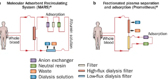
Two of the most prominent and most used AL devices are Molecular Adsorbent Recirculating System (MARS®; Gambro, Stockholm, Sweden) and Prometheus® (Fresenius Medical Care, Bad Homburg, Germany). The MARS® uses a high-flux hollow-fiber dialysis module made up of albumin impregnated polysulfone membranes. The cut-off of the membranes is limited to 50 KDa to prevent the passage of the proteins and albumin in the patient’s blood through the membrane, while membrane-bound albumin act as the acceptor molecule for albumin-bound toxins within the extracorporeal circuit. The toxins are then passed to the 600ml
21 recirculating 20% albumin solution flowing at constant rate in counter-current flow. The dialysate is sequentially cleansed by a haemodialysis/haemofiltration module (removing water soluble substances) and adsorber columns containing activated charcoal and anion exchange resin (removing most of the albumin bound substances). The regenerated dialysate is recirculated to take up more toxins from the blood (51) (Fig. 1.3a).
On the other hand, Prometheus® is based on fractionated plasma separation and adsorption, in which the patient’s plasma is passed through an albumin-permeable polysulfone membrane, which enables the patient’s albumin fraction to pass into a secondary circuit in which the direct removal of albumin-bound toxins by different adsorbers (that is, anion exchanger and neutral resin) takes place (Fig. 1.3b). In addition, conventional, high-flux dialysis is performed within the primary (blood) circuit (52).
Fig. 1.3. Artificial liver support systems. The hypothesis that the hepatocellular dysfunction present in the clinical syndrome of liver failure is primarily caused by the accumulation of toxins not cleared by the failing liver is addressed by filtration and adsorption devices. a) Molecular Adsorbent Recirculating System (MARS®; Gambro, Stockholm, Sweden). b) Fractionated plasma separation and adsorption (Prometheus®, Fresenius Medical Care, Bad Homburg, Germany). Adapted from (52).
ALs are merely based on physico-chemical mechanisms and thus lack synthetic and biochemical functions of the liver. Additionally, hepatic detoxification inside the body is not limited to elimination of albumin-bound toxins. Moreover, Recent clinical
22 trials showed no survival benefit of extracorporeal liver support therapy compared with standard therapy (26).
In search for a device that provides a larger complement of important liver functions, including synthetic and metabolic processes, biohybrid support devices incorporating living hepatic cells have been developed (53).
1.3.2 Bioartificial liver support systems
Cell-based therapies have been extensively studied to overcome the limitations of the ALs and expand the spectrum of hepatic functions available. Among these therapies are isolated cell infusion via the hepatic portal vein for the transplantation of hepatocytes derived from primary or stem cells, extracorporeal bioartificial liver (BAL) devices integrating liver cell culture in a bioreactor, and tissue-engineered graft implantation. It is a significant challenge to sustain and extend hepatocyte function in vitro. Liver functionality is regulated by soluble mediators, interactions between cells and extracellular matrix (ECM), and interactions between cells, which all represent types of microenvironmental signals. Therefore, to gain a comprehensive understanding of these regulating processes, different in vitro hepatic culture models have been created, such as perfused whole-organ and wedge biopsies, precision-cut liver slices, isolated primary hepatocytes, immortalised liver cell lines, and isolated organelles (54).
The BALs have great potential for the creation of a short-term support and framework for drug assessment and in vitro research on hepatic cultures. Various different hepatic functions, including detoxification, metabolism and synthesis, could be undertaken by BALs owing to the fact that they integrate functional hepatocytes.
To preserve the phenotypic functions of cells, it is expected that the in vivo microenvironment will be replicated by in vitro cell cultures. External cues inserted in the microenvironment of isolated hepatocytes with regard to time as well as space determine how viable and functional those hepatocytes are (55). The clinical success of a BAL device depends on its ability to provide suitable mass transport, lengthen cell viability and functionality, and be amenable to scaling-up to a size of clinical relevance.
A number of limitations are presented by the basic static monolayer (2D) cultures based on petri dish: (i) nutrient depletion and aggregation of cellular metabolic products cause ongoing temporal transformations in the cellular microenvironment; (ii) oxygen is poorly soluble and has long diffusion length, which is why it occurs in
23 restricted supply; (iii) unstable interactions between cells and between cells and the ECM; (iv) lack of comparison between the smooth, 2D substrate and the in vivo settings; and (v) the lack of permeability of the substrate causing unidirectional mass exchange.
The various BAL designs that have been proposed to address these limitations can be categorised into four major groups, namely, flat plate systems, hollow fibre membrane bioreactors (HFMBR), perfused packed bed/scaffold systems and suspension/encapsulation-based reactors (Figure 1.4). Furthermore, research has also been conducted recently on microfluidic systems and microfabricated reactor systems for in vitro applications (56).
Fig. 1.4. Schematics of cell-based bioreactor designs. The majority of liver cell-based bioreactor designs fall into these four general categories, each with inherent advantages and disadvantages. Adapted from (57).
1.3.2.1 Flat-Plate System
An additional approach towards overcoming the above-mentioned limitations has taken the form of various strategies explored in relation to bioreactors. Although considerable improvement in mass transfer can be achieved with perfusion, a considerable obstacle is posed by the shear stress imposed on the cells, which has to be kept within a range that is acceptable to the hepatocytes. Membrane bioreactors include flat-plate as well as hollow-fibre systems (Figure 1.5).
Dynamic culture systems gain numerous advantageous features as a result of membrane integration in flat-plate bioreactors. Membranes enable system arrangement into clear-cut sections and mediate the passage of nutrients and oxygen to the cellular part whilst at the same time preventing fluid shear. Furthermore, membranes permit selective mass transfer of molecules to and from
24 cells in every direction by supporting one or both cellular layer surfaces. Moreover, separate application of oxygen gradient from the fluid flow is made possible by membranes with oxygen permeability. Unlike smooth solid plastic, a 3D substrate closely resembling the in vivo one is provided by porous membranes, which is helpful for research on cellular interaction (58).
Among the additional improvements that have been considered are monolayer-based systems with applicability to flat-membrane bioreactors as well, including co-cultures (59) and sandwich co-cultures (60) for enhancing cell-cell and cell-ECM interactions, and substrate micropatterning (61) for cell morphogenesis studies. A radial-flow bioreactor consists of a stacked microfabricated grooved substratum, with hepatocytes being protected against flow shear stress by the grooved glass plate (62). Meanwhile, among the various existing in vitro hepatocyte culture models, sandwich culture is believed to be one of the ideal ones. However, the application of sandwich culture in BAL bioreactor design is hampered due to several technical difficulties, such as cell housing capacity that is not as high as other configurations and arduous bioreactor construction process, that need to be addressed in future bioreactor design.
1.3.2.2 Hollow-Fiber System
The main type of bioreactor configuration is the hollow-fibre system. Tissue engineering applications for cell-based treatments (63) including BAL devices (64) and large scale cell cultures (65) widely employ hollow-fibre membrane bioreactors (HFMBR).
An HFMBR is made up of a cluster of hollow fibres (HF) with partial permeability put in an external housing, like ultrafiltration or dialysis applications. Each configuration can fulfil a distinct function, according to the application and type of cell employed. In general, the cells are bound to the HF external wall, with flow of medium within the lumen of the fibres that exchange nutrients and other elements, comparable to a network of blood capillaries. Conversely, it is possible to seed the cells into the HF lumen with module operation under various patterns of flow (66). In addition, multibore system (67) alginate immobilization (68) and three-compartment multi-coaxial system (69) are among the other configurations of greater complexity that have been proposed.
In terms of the restrictions related to diffusion length, mass transfer rates for nutrient supply as well as for waste elimination can likely be enhanced by HFMBRs.
25 Cell protection against shear and air-liquid interfacial stresses is ensured by compartmentalisation, which keeps the cells apart from the direct fluid flow.
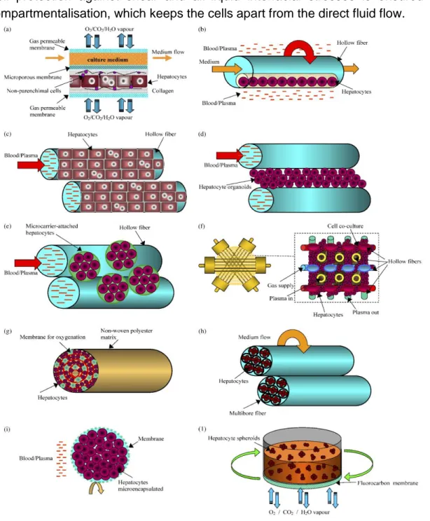
Fig. 1.5 Configuration of membrane bioreactors using hepatocytes cultured (a) between flat-sheet membranes; (b) entrapped in a three-dimensional contracted gel matrix inside of hollow fibre membranes; (c) outside of hollow fibre membranes in monolayer; (d) outside of hollow fibre membranes organised in organoids; (e) outside of hollow fibre membranes attached to microcarriers; (f) in a network formed by four capillary membranes with different functions; (g) in a spirally wound non-woven polyester matrix inside of hollow fibres; (h) in the intraluminal compartment of a multibore fibre bioreactor; (i) in microcapsules and (j) in a rotating-wall gas-permeable membrane system. Image taken from (70).
26 Furthermore, metabolite transport is enhanced and dead volume is reduced by HF designs because, by comparison to flat-plate designs, they offer a greater ratio of surface area to volume. On the downside, the use of closely packed HFs makes it difficult to seed cells into the bioreactor homogeneously (57).
1.3.2.3 Perfused Bed System
Packed bed refers to a hollow vessel that is filled with packing materials. In application of EBAL, this configuration can be used to fill matrix for hepatocyte attachment and perfused with medium or patient’s blood/plasma. Various packing materials for hepatocyte entrapment, such as micro-channeled polyurethane foam (71), polyvinyl resin cubes (72), alginate beads (73), porous hydroxyapatite beads (74), and polyester fabric cell scaffold (75) were explored. Packed bed reactors offer improved mass transfer by allowing direct contact of cells on microcarriers or packing material with the perfusing media (76, 77). A single monolayer culture is easily perfused, but a series of stacked plates may introduce shunting through regions of low resistance. Uniform perfusion of packed bed reactors is a classic engineering problem. Distribution of fluid flow is greatly dependent on the characteristic of the packing material. Larger, rigid particles will yield well distributed flow but a decreased surface area for cells, whereas smaller, porous packing will result in clogging and fluid channeling (78). A packed bed reactor built around a micro-channeled scaffold is an example of one designed explicitly to reduce heterogeneous perfusion and improve the transport characteristics of the devices (79).
1.3.2.4 Encapsulation System
Some designs use encapsulated cells in perfusion systems, which provide immunoisolation, but also increases diffusion resistance (80, 81). Encapsulation-based bioreactors are fairly easy to scale-up and provide a uniform microenvironment for the cells; however, encapsulation presents a diffusion barrier that can lead to cell death in the middle of the beads as well as the degradation of the microparticles over time resulting in exposure of cells to shear stress. Various materials have been used for hepatocyte encapsulation, including hydrogels (80), alginate (81), and copolymer such as hydroxyethyl methacrylate-methyl methacrylate (HEMA-MMA) (82). In some systems, hepatocytes form spheroids before entrapped into a capsule (83, 84), because there are evidences showing that spheroid enhances cell-cell interaction (85), and facilitates the formation of bile-duct structure between cells to improve cell functions.
27 1.3.2.5 Microfluidic Chip Based System
Microfabrication based microfluidic systems for hepatocyte culture has emerged as a promising area for various hepatocyte in vitro applications including BAL. The fine control of hepatocyte microenvironment, which is essential to maintain hepatocyte differentiated functions, is made possible at microscales in microfluidic system. The small fluid volume in microfluidic perfusion allows more efficient mass transfer in terms of delivery and removal of soluble substance (86). Lee et al. (87) created an artificial liver sinusoid with a microfluidic endothelial-like barrier and rat or humanhepatocytes can be cultured within the barrier. Hepatocyte viability and drug metabolism functions can be maintained for 7 days in the system. The first attempt to design a microfluidic system based bioreactor for BAL was by Leclerc et
al. (88). Ten microfabricated polydimethylsiloxane PDMS chips were stacked to
constitute four interconnected cell culture chambers and one oxygen supply compartment. The microfluidic bioreactor can achieve high cell seeding density of 30–40 million cells/cm3 with efficient mass transfer, which demonstrated its
potential of scalability for BAL application. Recently, Pang et al. managed to design a 3D scaffold comprised of 43 culture chambers assembled in a symmetrical pattern on 3 layers. The seeding density was further increased to reach 90 million cells/cm3 (89).
1.3.3 Hepatocyte transplantation
It involves the infusion of donor hepatocytes into the liver of the patient or even into ectopic sites such as the spleen. In principle, the injected cells should be engrafted and proliferate to repopulate the recipient liver restoring its functions (52). This technique was used to temporarily support patients with inborn metabolic liver diseases, such as Crigler–Najjar syndrome type I (90) and urea cycle disorders (91). However, with respect to acute liver failure, currently reported clinical trials have shown inferior results (92). The limited supply of viable hepatocytes, insufficient integration and survival of transplanted hepatocytes, and inadequate immunosuppressive therapy for hepatocyte transplantation patients need to be addressed before establishing this technique as a routine clinical treatment (93).
28
1.3.4 Repopulation of decellularized liver
One approach to engineer a vascularized, functional tissue is the decellularization and recellularization of livers. During decellularization, cells and other immunogenic molecules are removed from tissue or organs to leave the extracellular matrix. Resident cells generate the extracellular matrix which preserves the complex 3D microanatomy of the liver, including its vascular and biliary system frameworks. Furthermore, the extracellular matrix is ideally suited for repopulation with cells, their subsequent engraftment and migration to their specific niches (94). The recellularized grafts are then perfused at 37°C in bioreactors, using either culture media or heparinized blood. In vivo implantations of engineered hepatic tissue have been thus far limited to animal studies, with the first study reported in 2010 (95) in which rat livers were decellularized then recellularized with rat hepatocytes. The grafts were then implanted in rats with arterial blood flow from the renal artery for up to 8 h. Later, segments of decellularized porcine livers were ‘humanized’ via infusions of human fetal hepatocytes with co-cultured human fetal stellate cells followed by perfusion in vitro for up to 13 days (96). Inside the extracellular matrix, the human fetal hepatocytes differentiated into hepatocyte-like cells with some mature hepatocyte properties and bipotential progenitor cells. Although in general implantations of recellularized livers in humans seem possible, various issues need to be addressed. One of the major obstacles is the reconstruction of vascular network via re-epithelialization. This is considered the limiting step since contact between the extracellular matrix proteins and blood components leads to the formation of blood clots. Thus, implantations of recellularized grafts will not be possible until the vasculature is completely re-epithelialized or thrombogenesis within the graft is prevented in a different manner without deleterious systemic effects on the host (52). However, the fact that the vascular extracellular matrix frame is conserved during decellularization is possibly the biggest advantage of this technique in comparison to the other approaches. Preclinical experiments and clinical trials are needed to define relevant quality standards to predict the in vivo behaviour of decellularized tissue of different species in humans.
1.3.5 Organ printing
3D bioprinting is a cutting-edge technique that allows for precise engineering of complex parenchymal organ constructs. 3D organ printing can be defined as layer-by-layer additive biofabrication using liquid bioink (cell suspensions) or
self-29 assembling cellular building blocks (spheroids) (52). 3D spheroid printing represents scaffold-free tissue engineering which enables the precise arrangement of different cell types and other biologic materials (such as extracellular matrix components and growth factors) within organ constructs (97). The spheroid printing technique is based on tissue fusion, a process driven by tension forces between fluids that is observed during embryonic development (98). Tissue fusion means that spheroid blocks of different living cell types that have been placed closely together by a bioprinter melt together to finally represent one entity. Although no reports on printed perfusable hepatic constructs have been published so far, some interesting experimental results demonstrate the potential of 3D printing techniques. The NovoGen MMX Bioprinter™ (Organovo Holdings, Inc., San Diego, CA, USA) was used to print metabolically active 3D hepatic tissue that were stable for over 40 days in vitro (99). Furthermore, compartment-specific organization in a rudimentary microanatomy was shown for hepatocytes, hepatic stellate cells and endothelial cells.
Despite the promising prospect of this technique, these hepatic micro-organs are not designed as organ supportive therapy options. Several hurdles need to be overcome before such a technique can be used to print clinical-scale functional livers. Detailed knowledge of the liver’s micro-anatomy, and software to convert this information into reasonable blueprints are crucial. Furthermore, complex bio-mathematical models to predict the behaviour of biological materials during and after printing are needed. Currently available printing hardware can print a tissue of 1 cm3 in 27 h (100). Thus, enhancement of available printing hardware to quickly
process living cells is a determining step, as the printing process affects the viability of cells. Moreover, maturation processes further limit the scalability of printed tissue, as it can take months until engineered tubules of printed spheroids are functional and perfusable (101). Thus, sustaining the viability of parenchymal cells during maturation and essentially accelerating maturation are fields of particular interest, especially as most experiments so far have been performed with young animal cells—human primary cells are even more complex to handle in vitro (102).
1.3.6 Induced organogenesis
Induced organogenesis is a completely new approach that focuses on the generation of functional, implantable organs using stem cells (alone or with primary cells). These cells are induced in vitro to initiate differentiation towards a certain fate through manipulating extracellular matrix and growth factors, followed by in
30 vivo implantation to allow for maturation of the differentiated cells (103). Recently, human induced pluripotent stem cells were differentiated into endodermal cells and co-cultured with human mesenchymal stem cells and hUVECs in vitro (104). After self-organization of these cells into a kind of vascularized liver bud, they were implanted into immune-deficient mice. Interestingly, the in-vitro generated liver bud was shown to integrate into the vascular system of the recipient only 48 h after implantation. Furthermore, the liver bud matured to functional tissue, resembling adult liver tissue and was able to rescue mice in a drug-induced lethal liver failure model. These encouraging results open up a new field for further research and demonstrate that experimental mimicking of organogenesis might lead to liver support therapies in the future.
1.4
Cell source as critical issue for BAL devices
1.4.1 Primary hepatocytes
Primary human hepatocytes are ultimately the preferred cell type for cell-based therapies, and the development of primary hepatocyte-based approaches is the focus of substantial ongoing research. Within their in vivo microenvironment, hepatocytes have high proliferative capabilities. Following partial hepatectomy of two-thirds of the human liver, the residual mature hepatocytes are able to proliferate and restore the lost liver mass (105). However, primary hepatocytes are phenotypically instable and when they are isolated from their in vivo microenvironment and put in 2D cultures they rapidly de-differentiate into a population of adult liver progenitors (106), losing many of their liver specific functions, in particular CYP enzyme levels (107). In addition, 2D culture limits the survival of the cells to only 1-2 weeks (108). Various attempts have been made to prolong the hepatocyte survival in vitro, for example by culturing primary hepatocytes in a sandwich culture of collagen or matrigel, the hepatocyte life span, morphology and specific hepatic functions can be preserved for longer period of time (108, 109). Unfortunately, the low availability of fresh human liver samples compromise the use of primary hepatocytes in routine testing. Moreover, the resected livers most often originate from medicated patients that may severely affect cell viability and specific functions. Regarding donated livers, the patients have often been subjected to various pharmaceuticals, e.g. for the treatment of brain injury, again potentially affecting the expression of various drug metabolizing
31 enzymes (110). Cryopreserved hepatocytes are often used as they are available and phenotypically characterized which facilitates their use in routine research(111). Moreover, pooled cells from several donors are available which reduce inter-donor variability. However, these cells are expensive and share the same limitations as freshly isolated hepatocytes regarding loss of liver specific functions in culture.
Primary porcine hepatocytes, have been widely used as the cell source for hybrid artificial livers. Porcine hepatocytes exhibit liver-specific functions including biotransformation functions, ammonia detoxification, and synthesis of urea and albumin (112). Although these cells can be readily obtained in large quantities and demonstrate similar functions and therapeutic effects to human hepatocytes, their major drawbacks are the risk of xenogeneic infections such as porcine endogenous retrovirus and the lack of metabolic compatibility (113).
1.4.2 Cell lines
Immortalized hepatocyte cell lines, such as HepG2 (human hepatoblastoma) (114), the HepG2-derived line C3A (115) HepLiu (SV40 immortalized) (116), immortalized fetal human hepatocytes (117) and HepaRG (human hepatoma) (118) have been utilized as readily available alternative to primary hepatocytes. The advantages of using established cell lines include the ability to culture large quantities of cells for an extended period of time and the ability to control the degree of hepatocyte functions that are displayed (119). However, due to lack of the full functional capacity of primary adult hepatocytes, and the risk of transmittance of oncogenic factors to the patient, the use of these cell lines in a clinical setup remains a concern (57). Thus, as a potential precautionary measure, the use of conditionally immortalized lines and the incorporation of inducible suicide genes have been considered (54).
1.4.3 Stem cells
An independent approach to generate hepatocytes for therapeutics is to use stem and/or progenitor cells, which may be sourced from various tissues and have a high proliferative capacity. In principle, such cells could be amplified, differentiated into various cell types, and used in diverse applications (120). Recently, researchers have been attempting to identify small molecules that can potentially induce the maturation of hepatocyte-like cells to enhance the chance for using these cells in a clinically relevant setup (121).
32 1.4.3.1 Embryonic stem cells
The breakthrough for stem cell research came about 30 years ago with the first successful isolation of an embryonic stem cell line from a mouse embryo (122, 123). In 1998, the first generation of an in vitro, multi-passaged, culture of human embryonic stem cells (hESC) was reported (124) and since then many different protocols have been developed for the establishment, propagation and characterization of hESC. These cells are pluripotent and can generate all three germ layers, thus capable of differentiating into any kind of cell in the human body (124, 125). With these unique properties, these cells provide a highly interesting model system for basic research on embryonic and organ development, as well as a hepatic cell source for drug discovery and toxicology studies (126, 127). In the future, these cells might also be used in clinical therapies (128). During the recent years, much effort has been put into the development of effective protocols for hepatic differentiation of hESCs, largely based on what is known about the embryogenesis (129-131). Various directed differentiation strategies have been applied to hESC and iPSC cultures, and have yielded populations that exhibit some functional and phenotypic characteristics of mature hepatocytes, thus termed “hepatocyte-like cells” (130, 132, 133). Yet, hepatocyte-like cells resemble foetal hepatocytes rather than mature adult cells as evident by their characteristics, such as distinctive cytochrome P450 activities as well as expression of foetal genes such as α-fetoprotein (134). Moreover, all differentiation protocols result in highly variable functionality within the cell population and cells begin to lose hepatic characteristics after a few days much like standard culture conditions. Researchers are developing methods to overcome these disadvantages of functional variability, low expression levels, and loss of functionality over time, however embryonic stem cells are a controversial source of cells for scientific research and this is not likely to change in the near future. A different source of stem cells could eliminate this ethical controversy.
1.4.3.2 Hepatic somatic stem cells
Somatic stem cells have a more limited differentiation potential compared to hESCs but have an important role in tissue homeostasis and injury repair in the multicellular organism (135). The presence of hepatic stem cells (oval cells) were first discovered in fetal mice livers (136) and was later also isolated from human adult livers (137). Oval cells are multipotent stem cells that can give rise to both hepatocytes and biliary cells (35). Hepatic oval cell lines have been generated that retain the progenitor cell features expressing markers for both cholangiocytes and
33 hepatic progenitors after long term cultivation and serial passages (138). These cells might thus serve as an expandable hepatic cell source for research and for cell-based therapy (139, 140). Recently, progress has been made in identifying tissue-resident mesenchymal stem cell–like populations that reside in human liver (141, 142), further investigation into the role of these cells in normal liver physiology and repair is needed to determine whether these cell populations represent a clinically relevant source of hepatocytes.
1.4.3.3 Induced pluripotent stem cells
In 2006, a Japanese research group successfully reprogramed adult mouse fibroblasts into induced pluripotent stem cells (iPSC) by introducing four transcription factors: cMyc, Oct3/4, Sox2 and Klf4, by retroviral transduction (143). A similar approach was also successfully performed with human fibroblasts (144) and later using other cell types from both human and mouse (145). The iPSC are stem cell-like regarding morphology and characteristics, such as pluripotency and genetics, expressing a number of stem cell biomarkers (145). Several groups have subsequently been able to successfully generate hepatocyte-like cells from iPSC (133, 146, 147). The iPSC technology is promising with a future potential in patient- and disease-specific therapy (145). However, in order for these cells to be used in a clinical application several important issues have to be addressed, such as somatic origin memory, donor dependent variations, low reprogramming efficiency, risk of potential teratoma formation, safety concerns regarding transduction delivery methods and the presence of transgenes, like oncogenes (148).
In light of this progress, the strategy of using stem cells as a source for generating hepatocytes as a cellular component in BAL devices holds great promise.
1.5 Oxygen supply
Owing to their various metabolic, synthetic and regulatory activities, hepatocytes have an oxygen consumption rate more than ten-fold higher compared to the majority of other cells, triggering an oxygen concentration gradient along the sinusoids (149). It is believed that metabolic function distribution along the sinusoids influences the concentration distribution of oxygen, hormones, nutrients and ECM molecules (54). There are three metabolic areas along lobule hepatic sinusoids that have been distinguished, namely, the periportal zone surrounding the portal vein and hepatic arteriole (zone 1), an intermediate zone 2, and the