Doctorate Course:
Molecular and Experimental Oncology
‘Effects of immunomodulatory
oligonucleotides on pancreatic
cell lines’
Student: Jara Rocchi
Tutor: Dr. Fabrizio Maggi
Supervisor: Prof. Generoso Bevilacqua
i
Background. Pancreatic cancer is a devastating malignancy with a
mortality rate almost identical with its incidence. Thus, the development of new therapeutic strategies for treating this cancer is absolutely required. A potential novel strategy is targeted immunotherapy. The recent discovery of Toll-like receptors (TLRs) provides new targets to specifically activate this immunity. Among TLRs, TLR9 is expressed by various normal and tumor cells and its activation by DNA containing unmethylated CpG motifs leads to a cascade of molecular events that culminate in the induction of several inflammatory mediators, such as cytokines and nitric oxide (NO).
Aim. The study was performed to demonstrate the presence of TLR9
in four human pancreatic carcinoma cell lines (PCCs: PP78, PP109, PP161, and PP117) and to investigate whether its activation by selected oligodeoxynucleotides containing CpG motifs (CpG ODNs) could affect the in vitro characteristics of PCCs.
Methods. TLR9 expression was assessed by using molecular
(RT-PCR and/or real-time (RT-PCR, and DNA sequencing), and immunological assays (immunofluorescence and Western Blot analysis). Cell viability and cell cycle parameters were checked by trypan blue exclusion and FACS analysis, respectively. NO production was measured by the Griess reagent and cytokine production profiles were evaluated using commercial ELISA kits.
Results. TLR9 mRNA and protein were both expressed at basal
levels in all PCCs. CpG ODNs treatment of PP78 and PP109 PCCs increased the TLR9 expression, and it reduced significantly the cell proliferation. CpG ODNs also induced a delay in S-phase followed by
ii
observed in CpG-ODN treated cancer cells.
Conclusions. Our results indicate that TLR9 ligands produce an
evident anti-proliferative effect in PCCs; such an effect might depend on the CpG-DNA induced modulation of endogenous mediators such as IL-8 and NO (Rocchi et al., 2008).
iii
SECTION I: INTRODUCTION
1
1. Pancreatic carcinoma:
therapeutic potential of toll-like
receptor activation
1
2.TLR9 signalling
5
3. Identification of different
classes
of
CpG-ODNs
9
4. Therapeutic application
of CpG-ODNs in cancer therapy
14
4.1 Monotherapy with CpG-ODNs
17
4.2 Use of CpG-ODNs as adjuvant in
cancer vaccines
19
4.3 Combination therapy with TLR9
agonists
21
4.4 Safety of TLR9 ligands
25
5. Direct effects of CpG-ODNs in TLR9
expressing cancer cells
27
6. Experimental model for
investigation of TLR9 in
pancreatic cancer
31
iv
1.
Cell
lines 34
2. TLR9 agonists
35
2.1 Synthetic ODNs
35
2.2 Viral DNA
36
3.
RT-PCR
analysis
37
4. Quantitative RT-PCR for
TLR9
expression
38
4.1 Semiquantitative RT-PCR
38
4.2
Real-time
RT-PCR
38
5.
TLR9
protein
expression 39
5.1 Immunofluorence analysis
39
5.2 Western Blot analysis
40
6.
FACS
analysis 41
7.
Proliferation
assay
41
8. Assay for cytokine production
42
9.
Nitric
oxide
(NO)
assay
42
v
1.2 Expression of TLR9 protein
46
2. Evaluation of TLR9 mRNA
expression in ODN-treated cells
48
2.1
Semiquantitative
PCR
48
2.2
Real
Time
PCR
49
3. Characterisation of the ODN
effects
on
cell
survival
52
3.1 by trypan blue exclusion assay
52
3.2 by FACS analysis
60
4. Characterisation of the viral
DNA-effects on cell survival
62
5. Secretion of cytokines in
response to CpG-ODN stimulation
65
6. NO production in CpG-ODN
treated
PP78
cells
68
SECTION IV: DISCUSSION
70
1
SECTION I
INTRODUCTION
1. Pancreatic carcinoma: therapeutic potential
of toll-like receptor activation
Pancreatic carcinoma is one of the most devastating and lethal diseases, with most patients dying within 6 months after diagnosis. The main reason for the poor outlook of these patients is that only a small number of these tumors are detected in early stages. Because the pancreas is located deeply inside the body, early tumors cannot be seen or felt by health care providers during routine physical exams. Patients usually have no symptoms until the cancer has spread to other organs.
Many efforts have been made to improve the efficacy of treatment but the prognosis of pancreatic carcinoma still remains poor. Surgery is an option in only 10–15% of the patients, but even after resection, recurrence occurs in the majority of the patients, leading to a median survival of about 18 months. Chemotherapy is considered the conventional systemic treatment of advanced pancreatic cancer. The
2
chemotherapy drugs used to treat pancreatic cancer include gemcitabine, which seems to be the best available treatment option improving the quality of life in many patients (El-Rayes et al., 2003). However, the drug is capable to prolong survival of pancreatic cancer patients of few months only (Burris et al., 1997; Goldstein et al., 2004).
Thus, new antitumor therapies for these patients are needed. One potential strategy for pancreatic cancer treatment is targeted immunotherapy. With growing understanding of the regulation of immune responses, multiple new immunotherapeutic targets have evolved, and some have reported in several trials prolonged survival for immune responders. However, reports of spontaneously regressing pancreatic cancer do not exist; this may be because pancreatic cancer, as reported above, is generally diagnosed in a late stage, when it has already overcome the host immune response. The discovery of a series of innate immunospecific receptors activated by pathogen-associated molecular patterns may provide new possibilities for a targeted activation of this immunity. Among the innate immune-specific receptors, the best characterized are the Toll-like receptors (TLRs; Underhill and Ozinsky, 2002).
TLRs are evolutionarily well conserved trans-membrane proteins that comprise a family of approximately 13 members, depending on species; they initiate inflammatory responses to a different array of
3
microbial products; examples include bacterial peptidoglycans, lipopolysaccaride (LPS), and flagellin, which are recognized by TLRs 2, 4, and 5, respectively, and nucleic acid from bacteria and viruses, which are recognized by TLRs 3, 7, 8, and 9 (fig. I.1; Akira and Hemmi, 2003; Heil et al., 2004; Ishii and akira, 2006).
4
The signals induced by TLRs are critical for eliciting immune responses at both the innate and adaptive levels.
Although several TLRs reside on the cell surface, such as TLRs 2, 4, and 5, nucleic acid recognition by TLRs occurs intracellularly (Matsumoto et al., 2003; Leifer et al., 2004; Schmausser et al., 2004).
In particular, TLR9, the focus of the current work, is retained in endoplasmic reticulum and it responds to bacterial and viral DNA as well as synthetic oligodeoxynucleotides (ODNs) that contain unmethylated CpG dinucleotides in specific sequence contexts (Hemmi et al., 2000). Through TLR9 signaling pathway, immunostimulatory CpG sequences activate a complex cascade that leads to stimulation of an immune response and increased production of proinflammatory cytokines and chemokines (Klinman,
et al., 1996; Krieg, 2002).
In humans, TLR9 has been described to be expressed in B-lymphocytes and plasmacitoid dendritic cells, but an increasing evidence suggests that TLR9 expression is not confined to cells of immune system; in fact, some studies, have been detected its expression in normal epithelial and cancer cells, including breast, brain, lung, and gastric cancer cells (Schaefer et al., 2004; Schmausser et al., 2004; Droemann et al., 2005; Schamausser et al., 2005); interestingly, recent studies on type 1 diabetes revealed TLR9
5
transcript expression by pancreatic islet cells (Wen et al., 2004; Giarratana et al., 2004).
Over the past years there has been an enormous increase in the understanding of the molecular and cellular effects of CpG-ODNs, which have been demonstrated to induce a Th-1 response and to activate dendritic cells (Roman et al., 1997; Sparwasser et al., 2000). In different experimental tumor models and in some clinical trials, treatment with synthetic CpG-ODNs alone or in combination with chemotherapy or radiotherapy was shown to exert antitumor activity (Weigel et al., 2003; Krieg, 2004; Milas et al., 2004).
Recently, Tepel and co-workers (2006) demonstrated specific CpG-ODNs to induce significant growth inhibitory effects on orthotopically xenotransplanted pancreatic tumours in highly immunodeficient mice. Similar results were observed by using chemotherapy combined with CpG-ODNs in the orthotopic mouse model of a human pancreatic tumor xenograft (Pratesi et al., 2005): repeated treatments with a specific CpG-ODN at the end of the complete chemotherapy regimen induced an increasing mice survival. The increased survival obtained by the combined use of CpG-ODNs and chemotherapy suggests the potential of this therapeutic regimen in the clinical setting.
Thus, the objective of the present work was to extend and further investigate these effects of CpG-ODNs on pancreatic cancer.
6
2. TLR9 signalling
TLR9 was first cloned and identified as a receptor for unmethylated CpG-DNA as well as for bacterial DNA in 2000 (Hemmi, et al., 2000). After its identification, the mechanisms of various immune responses and the signal transduction pathway mediated by the engagement of CpG DNA with TLR9 were clarified.
TLR consists of leucine-rich repeats (LRRs), a transmembrane domain and a cytoplasmic Toll/interleukin-1 receptor homology (TIR) domain. TLR9 is localised at the intracellular membrane compartment, such as endoplasmic reticulum (ER) and lysosome (Latz et al., 2004).
TLR9 transmits its signals through a specific interaction with an adaptor molecule, MyD88, which contains a cytosolic TIR domain and a death domain (DD).
MyD88 in turn interacts with interleukin-1 receptor-associated kinase-1 (IRAK-kinase-1) and IRAK-4 through its DD. IRAK-4 and its kinase activity are essential for TLR9-mediated cytokine production (Kawagoe et al., 2007).
IRAK-1 is a well known substrate of IRAK-4, and phosphorilated IRAK-1 up-regulates its kinase activity, and subsequently recruits tumor necrosis factor receptor-associated factor 6 (TRAF6).
7
Down-stream of TRAF6 transforming growth factor β-activated kinase 1 (TAK-1) is activated to phosphorylate IκB kinase (IKK) complex, which phosphorylates IκB to induce nuclear translocation of NF-κB (Wang et al., 2001).
Moreover, the activated TAK-1 is prerequisite for the activation of mitogen activated protein (MAP) kinases, including extracellular receptor kinase (ERK), p38, and Jun N-terminal kinase, responsible for the induction of the activator protein-1 (AP-1; fig. I.2). Transcription factors NF-κB and AP-1 induce in immune cells the transcription of different pro-inflammatory cytokine genes, including TNF-α, IL-6 and IL-12.
TLR9, originally localized in the ER, migrates to the endosome when the cells are exposed to CpG-DNA (Latz et al., 2004); after the translocation of the ligands, endosomal acidification is reported to be critical for the signalling of TLR9: chloroquine and other inhibitors of endosomal acidification and/or maturation completely block the immune effects of CpG-ODN, demonstrating an essential role for this compartment in the interaction with TLR9 (Yi et al., 1998; Hacker et
al., 1998).
8
Fig. I.2: TLR9-mediated signaling pathway.
The cytosolic TIR domain of TLR9 recruits the adaptor molecule MyD88 and other signaling molecules such as IRAK-4, and TRAF6 that are required for the signaling complex. The complex in turn activates different signaling cascades that lead to the activation of NF-κB and AP-1.
These activated transcription factors induce diverse immunity-related genes (Kumagai et al., 2008).
9
3. Identification of different classes of
CpG-ODNs
Several syntetic CpG-ODNs agonists for TLR9 are currently in development for the cancer treatment.
The molecular structure of these DNA sequences is an important parameter capable to affect their biological function.
In addition to the requirement of unmethylated CpG dinucleotides, the activity of the ODNs is dependent upon the sequences flanking both the 5’ and 3’ regions of the CpG dinucleotides (and Agrawal and kandimalla, 2001; Yu et al., 2001; Krieg, 2002). The immunostimulatory effects of the ODNs are enhanced if there is a TpC dinucleotide on the 5’ end and if the ODN is pyrimidine rich on the 3’ side (Hartmann and Krieg, 2000; Hartmann et al., 2000).
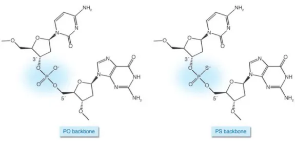
Moreover, the immunostimulatory effects depend on the stability of ODNs. The unmodified phosphodiester (PO) CpG-ODNs are degraded by nucleases within minutes following in vivo administration. Backbone modifications, most notably to phosphorpthioate (PS), decrease the ODN degradation: this nuclease-resistent backbone improves the half-life of ODNs in the body from just a few minutes to about 48 hours (Fig.I.3; Boggs, et al., 1997); thus, for therapeutic application, CpG-ODNs are typically synthesized with at least partially PS backbones.
10
The length of ODN sequences is not directly correlated with their activation; six base-length CpG-ODNs show immunostimulatory activity. Also the immunostimulatory activity of an ODN is determined by the number of CpG motifs it contains (usually two to four are optimal) and may also be affected by spacing of the CpG motifs and by the presence of poly G sequences (Krieg, 1995; Ballas et al., 1996; Hartmann and Krieg, 2000; Vollmer et al., 2004).
Figure I.3: Backbones of native and modified DNA.
PS ODN differ from native PO DNA only in the substitution of a sulfur for one of the nonbridging oxygen atoms (Krieg, 2007).
11
There are at least three classes of CpG-ODNs with distinct structural characteristics. These different classes, through the TLR9-activation, are able to modulate differently the immune system.
CpG ODNs of class-A have poly-G motifs with PS linkages on both ends, with a PO palindromic CpG in the center. CpG-A ODNs induce a strong pDC IFN-α secretion; moreover, they are effective for NK-sensitive tumors, causing NK activation. CpG-ODNs of class-B have the CpG dinucleotide in a fully modified PS backbone. These ODNs are strong B-cell stimulators but weaker inducers of IFN- α secretion, and they are mostly employed into human clinical development in oncology.
A third class of CpG ODNs combines the effects of A-and B-class CpG ODNs. These C-class CpG ODNs are wholly phosphorothioate modified, they have no poly-G motifs, but they are characterised by palindromic sequences combined with stimulatory CpG motifs (table I.1; Boggs et al., 1997; Krieg, 2002; Vollmer et al., 2004).
Recently, a new generation of CpG-ODNs called “immunomers” has been developed: immunomers consist of two CpG molecules linked at their 3’ ends; these have the dual benefits of two 5’ ends, which are necessary for activity, and lack of 3’ ends, which decreases digestion by nucleases (fig. I.4; Yu et al., 2000).
12
Table I.1: Classes of CpG ODN.
Capital letters in ODN sequences indicate 3′ PS internucleotide linkage; lower case letters in ODN sequences indicate 3′ PO internucleotide linkage. (Krieg, 2007).
NNNN X NNNN-O-NNNN X NNNN
X = CpG, YpG, CpR, YpR
O = Linker
Figure I.4: immunomer structure.
ODN class A-class
Example ODN GGgggacgatcgtcgGGGGG (also known as ODN 2216)
Structural features Poly G region at the 3’ and/or 5’
ends; usually with a few PS-modified internucleotide linkages
at the 5’ and 3’ ends for nuclease resistance. Immune effects Induces exceptionally strong pDC IFN-α secretion and moderate expression of costimulatory molecules. Induces very little B
cell activation. B-class TCGTCGTTTTGTCGTTTTGTCGTT
(also known as ODN 2006, CpG 7909 and PF-3512676)
Fully PS-modified backbone, no major
secondary structure; most important CpG motif for
activating human TLR9 is at the 5’ end.
Induces very strong B cell proliferation and differentiation. Induces pDC expression of costimulatory molecules. C-class TCGTCGTTTTCGGCGCGCGCCG
(also known as ODN 2395)
Fully PS-modified backbone, 1 or more
5’ CpG motifs; self-complementary palindrome in middle or 3’ end enables formation of duplex or hairpin secondary structure.
Induces strong B cell proliferation and differentiation Induces pDC IFN- α secretion and expression of costimulatory molecule
5’
3’ 3’
5’
13
ODN internalisation occurs spontaneously in culture without the need for uptake enhancers or transfection. ODN uptake by lymphocytes is energy and temperature dependent and greatly increased by cell activation; it also seems to be receptor mediated, although the specific receptors remain largely unknown (Krieg, 2002). The earliest steps in the CpG-induced signal transduction pathways can be blocked by inhibitors of phosphatidylinositol 3-kinase, which seems to have a role in ODN internalization (Ishii et al., 2002).
14
4. Therapeutic application of CpG-ODNs in
cancer therapy
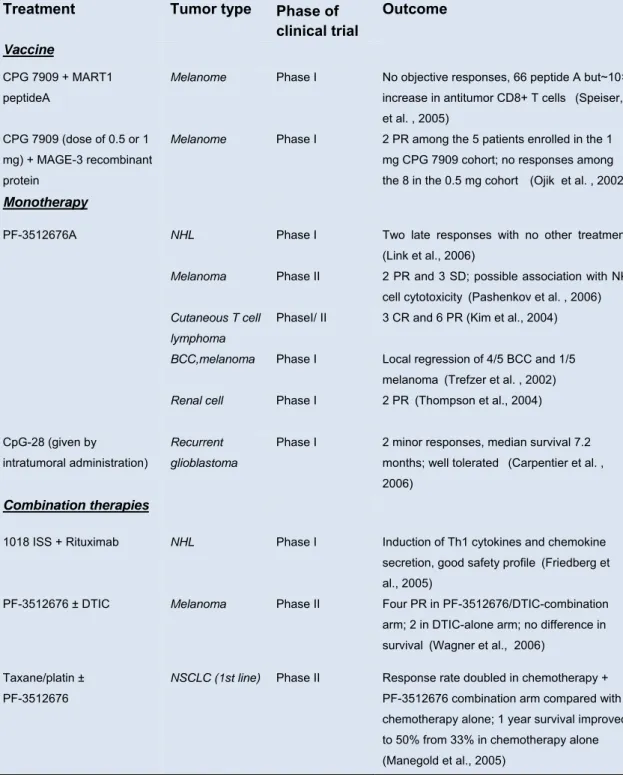
CpG-ODNs may represent a potential novel immunotherapeutic approach to the treatment of cancer. The rationale for investigating TLR9 ligands as possible anticancer agents is based on their ability to strongly activate both the innate and adaptive immunity (fig.I.5); this activation may directly cause antitumor effects and an enhanced tumor antigen presentation in a Th1-like cytokine and chemokine milieu.
The application of TLR9 agonists in human cancer therapy may include different areas; they can be also used alone to activate innate immunity locally and trigger a tumor specific response; they can be also used in combination with tumor antigens, DNA vaccines, monoclonal antibodies, or dendritic cells. In addition, ODNs can be combined with conventional chemotherapy and radiotherapy to increased therapeutic effects, and also to reduce host toxicity and improve patient immunity against secondary infection after chemotherapy and radiotherapy (table I.2).
15
Figure I.5: TLR9 agonists stimulate innate and adaptive antitumor immune
responses.
16
BCC, basal cell carcinoma; CR, complete response; DTIC, dacarbazine; MAGE, melanoma antigen–encoding gene; PR, partial response; SD, stable disease; SLN, sentinel lymph node (Krieg, 2007).
Treatment Tumor type Phase of clinical trial
Outcome
Vaccine
CPG 7909 + MART1 peptideA
Melanome Phase I No objective responses, 66 peptide A but~10×
increase in antitumor CD8+ T cells (Speiser,
et al. , 2005) CPG 7909 (dose of 0.5 or 1
mg) + MAGE-3 recombinant protein
Melanome Phase I 2 PR among the 5 patients enrolled in the 1
mg CPG 7909 cohort; no responses among
the 8 in the 0.5 mg cohort
(Ojik et al. , 2002)
Monotherapy
PF-3512676A NHL
Melanoma
Phase I Phase II
Two late responses with no other treatment (Link et al., 2006)
2 PR and 3 SD; possible association with NK
cell cytotoxicity (Pashenkov et al. , 2006)
Cutaneous T cell lymphoma BCC,melanoma
PhaseI/ II Phase I
3 CR and 6 PR (Kim et al., 2004) Local regression of 4/5 BCC and 1/5
melanoma (Trefzer et al. , 2002)
Renal cell Phase I 2 PR (Thompson et al., 2004)
CpG-28 (given by
intratumoral administration)
Recurrent glioblastoma
Phase I 2 minor responses, median survival 7.2
months; well tolerated (Carpentier et al. ,
2006)
Combination therapies
1018 ISS + Rituximab NHL Phase I Induction of Th1 cytokines and chemokine
secretion, good safety profile (Friedberg et
al., 2005)
PF-3512676 ± DTIC Melanoma Phase II Four PR in PF-3512676/DTIC-combination
arm; 2 in DTIC-alone arm; no difference in
survival(Wagner et al., 2006)
Taxane/platin ± PF-3512676
NSCLC (1st line) Phase II Response rate doubled in chemotherapy +
PF-3512676 combination arm compared with chemotherapy alone; 1 year survival improved to 50% from 33% in chemotherapy alone (Manegold et al., 2005)
17
4.1 Monotherapy with CpG-ODNsThe potential efficacy of CpG-ODN monotherapy for cancer treatment has been demonstrated in a number of animal models. Many of these studies have investigated the ability of TLR9 agonists to control the growth of solid tumors; the results obtained from these experiments suggest that intra-lesional or peritumoral injection of CpG-ODNs is required for an antitumor effect (Heckelsmiller et al., 2002b; Sharma et al., 2003).
For example, in a murine cervical carcinoma model, mice with established subcutaneous tumor treated with CpG-ODNs injected at a distant site showed significant tumor regression with the consequent improvement of survival (Baines and Celis, 2003).
Objective responses have been observed in some patients receiving CpG-ODNs for the treatment of melanoma, renal cells cancer, basal cells carcinoma, or cutaneous T cell lymphoma (Pashenkov et al., 2006). In particular, CpG-ODNs demonstrated significant clinical activity in a phase I trial of intra- and perilesional injections in patients with metastatic melanoma or basal cell carcinoma: treatment was associated with increased levels of serum interleukin (IL)-6 and IL-12p40 in some subjects, and cellular infiltrates of CD8+ were observed in most lesions after treatment (Trefzer et al., 2002).
TLR9 agonists have a variety of effects on B cells that may be relevant in the treatment of hematologic malignancies. Although
18
CpG-ODNs are strong mitogens for normal B cells, they preferentially induce apoptosis in tumor B cells stimulated through TLR9; however, the mechanism responsible of this effect remains unclear.
The activation of TLR9 on primary malignant B cells up-regulates expression of major histocompatibility complex molecules and other surface receptor; this increases their capacity to stimulate T cells which exert an enhanced T-cell-mediated response to tumor expressed on the malignant B cells (Decker et al., 2000; Jahrsdorfer
et al., 2001; Wooldridge and Weiner, 2003).
Several clinical studies of single-agent TLR9 agonists have been completed in patients with hematologic malignancies; monotherapy with TLR9 agonist CpG ODN-2006 (also known as CpG 7909 or PF-3512676) or another B-class CpG-ODN, 1018 ISS, activates NK cells and induces a Th1 cytokine response in patients with B cell lymphomas (Link et al., 2006).
PF-3512676 has also been studied in a phase I trial in patients with refractory non-Hodgkin’s lymphoma; in patients receiving PF-3512676 intravenously up to three times a week, number and activation of NK cells increased in most subjects (Link et al., 2006). Overall, these results derived from clinical trials of CpG-ODNs as a single agent are encouraging for a good safety profile; however, the frequency of objective responses in some tumors has been relatively low, and the aim of ongoing clinical trial consequently has shifted to
19
combination of therapies with the focus to improve the clinical efficacy of administering TLR9 ligands.
4.2 Use of CpG-ODNs as adjuvant in cancer vaccines
The use of antigen pulsed dendritic cells (DC) as tumor vaccines represents a new therapeutic approach for cancer. However, one potential limitation of this approach is that failure to correctly induce the antigen pulsed DC can lead to T cell tolerance or T cell anergy rather than the activation of T cell immunity (Kuwana et al., 2001; Martin et al., 2002).
Different studies have demonstrated the use of CpG-ODNs to efficiently activate antigen pulsed DC resulting in a productive antigen presentation with the consequent induction of strong anti-tumor immune responses (Heckelsmiller et al., 2002a; Wang et al., 2002).
In vivo manipulation of DC using Flt3 ligand (a growth factor for
dendritic cells) and CpG-ODNs has been shown to allow effective presentation of tumor antigen by DC resulting in strong anti-tumor responses capable of rejecting established murine B16 melanoma (Furumoto et al., 2004).
20
Combined DC and TLR9 ligand therapy was also effective to cure large chemotherapy resistant murine renal and colon carcinoma (Heckelsmiller et al., 2002a).
In addition, CpG-ODNs have been used as adjuvant with whole cell vaccines, tumor antigens, antigenic peptides or cell lysates (Krieg, 2004).
The enhancement in antibody responses observed by using TLR9 ligands was similar one producted by complete Freund’s adjuvant (CFA) but without the toxicity associated with CFA (Weiner et al., 1997); CpG-ODNs were able to induce stronger immune responses than those observed by using CFA in combination with the anti-idiotypic antibody 3H1 which functionally mimics carcinoembryonic antigen, a tumor-associated antigen expressed on human colorectal carcinoma and other adenocarcinomas (Baral, et al., 2003).
Moreover, TLR9 agonists have also been shown to increase the effectiveness of cancer vaccines by using autologous tumor cells transduced with genes expressing cytokines including IL-12, granulocyte macrophage colony-stimulating factor (GM-CSF) (Sandler et al., 2003; Switaj et al., 2004), and immunomodulators such as CD154 (Rieger and Kipps, 2003).
More recently, PF-3512676 combined with melanoma antigen A (Melan-A) analog peptide was used to vaccinate melanoma patients (Speiser et al., 2005): all patients that received PF-3512676 in their
21
vaccine showed a rapid and strong antigen-specific T cell response with a frequency of Melan-A specific circulating CD8+T cells approximately ten fold higher than that observed in melanoma patients receiving the same tumor vaccine without PF-3512676. These data demonstrate the capacity to CpG-ODNs to promote efficacy antigen-specific antitumor CD8+ T cell responses in human with advanced cancer.
4.3 Combination therapy with TLR9 agonists
A further method by which CpG-ODN can be used for tumor immunotherapy is its combination with monoclonal antibodies (mAb). Antitumor antibodies are able to bind to the tumor surface and are thought to mediate at least some of their activity through the mechanism of antibody-dependent cellular toxicity (ADCC). The innate activation by CpG-ODN treatment should improve the antitumor effect of antibody immunotherapy by enhancing ADCC (Carpentier et al., 2003; Wooldridge and Weiner, 2003). Moreover, the addition of CpG-ODNs leads to the activation of NK cells and monocytes/macrophages and this capacity plays a substantial role in the improvement of antibody therapy efficacy.
22
The combination of CpG-ODN with antibody immunotherapy improved in a murine lymphoma model the long-term survival from approximately 10% to 80% (Wooldridge et al., 1997).
Recently, a phase I clinical trial has investigated the combination of a CpG-ODN with rituximab, a mAb against CD20, in patients with relapsed/refractory non-Hodgkin’s lymphoma. In this study, 50 patients received PF-3512676 weekly for 4 weeks in combination with rituximab: twelve patients (24%) showed an objective response (Leonard et al., 2007).
Combination therapy with TLR9 agonists is being further evaluated for antitumor activity in a phase I/II trial in combination with radiation therapy and in a phase II trial in combination with rituximab.
Several preclinical models have also suggested that TLR9 ligands can synergize with cytotoxic chemotherapy. CpG-ODNs have been reported to enhance the antitumor effects of a number of different chemotherapeutic agents such as the topoisomerase I inhibitor, topotecan (Balsari et al., 2004), the alkilating agent cyclophosphamide (Weigel et al., 2003) and the anti-metabolite 5-fluorouracil (Wang et al., 2005b).
Chemotherapeutic agents are known to cause tumor cell death. Thus, it’s possible that the cancer bulking, as a result of chemotherapy, can provoke the release of tumor-associated antigen resulting in an in situ vaccine.
23
The employ of chemotherapeutic agents in combination with CpG-ODNs, which are potent adjuvants, could lead to the activation of strong tumor-specific immune responses that would be capable of mediating tumor rejection.
TLR9 ligands were tested in combination with either cyclophosphamide or topotecan in an orthotopic rhabdomyosarcoma model; the results showed that the combination therapy using CpG-ODNs and either of chemotherapy drugs enabled the long-term survival of 15-40% of the mice with a large tumor. This survival benefit required principally the presence of T cells, but not NK cells, suggesting that the CpG-ODNs may have induced the development of anti-tumor T cell response; probably this response may have been sufficient to eliminate the residual tumor after chemotherapy (Weigel
et al., 2003) .
Balsari et al., (2004), showed in a human prostate carcinoma xenograft model therapeutic benefits by using CpG-ODNs in combination with topotecan chemotherapy. All combined treatment resulted in a delay of tumor growth, compared to the effects in topotecan-treated mice.
In some recent studies PF-3512676 in combination with standard chemotherapy was evaluated in a randomized phase II clinical trial in patients with advanced non-small cell lung cancer. Preliminary data demonstrated that patients receiving PF-3512676 plus chemotherapy
24
achieved objective tumor responses more often than subjects given chemotherapy alone. Moreover, PF-3512676 was well tolerated and it did not lead to any clinical significant increase in chemotherapy related-toxicity (Murad et al., 2007; Manegold et al., 2008).
More recently, was also evaluated the efficacy of immunomers in combination with the chemotherapeutic agents docetaxel and doxorubicin in melanoma and breast carcinoma models (Wang et al., 2004): this approach resulted in synergistic antitumor effects in both tumors. This second-generation of immunomer compounds possesses antitumor activity in a broad spectrum of tumor models, predominantly mediated through induction of strong Th1 immune responses.
25
4.4 Safety of TLR9 ligandsIt is generally accepted that TLR9 agonists have a favorable safety profile.
Recently, many progresses have been made in understanding the immunological and pharmacological effects of the first-generation ODN molecules. However the data available on optimized CpG-ODNs in human clinical trials aren’t yet sufficient.
Preliminary results from early-stage clinical trials of CpG-ODNs used as monotherapy or adjuvant in cancer treatment, suggest that this therapeutic approach is generally well tolerated; moreover, the toxicity observed in humans appears limitated.
The most common adverse events observed following administration of TLR9 agonists are local injection-site reactions (for example, erythema, edema, inflammation and pain) or systemic flu-like symptoms (for example, headache, rigors, pyrexia, nausea and vomiting). These symptoms typically develop 24 hours after dosing and are transient, generally lasting for less than 2 days (Krieg, 2006). Because of the strong immune stimulatory effects obtained after CpG-ODN-treatment, one may expect that CpG would cause autoimmune diseases. However, in spite of early suggestions that TLR9 ligands may be a trigger for systemic lupus erythematosus, subsequent studies have revealed that treatment with CpG-ODNs
26
neither causes nor aggravates autoimmune disease, but on the contrary, this therapy reduces the severity of disease (Krieg, 2002).
27
5. Direct effects of CpG-ODNs in TLR9
expressing cancer cells
CpG ODNs may have an additional mechanism of antitumor activity in the treatment of tumors that express TLR9.
In fact, although it is generally assumed that TLR9 is principally expressed in certain immune cells, tumors may also express this receptor.
Cancer cells expressing TLR9 can be stimulated by CpG-ODNs, resulting in similar up-regulation of cytokines and activation of other molecules such as AP1 and NF-kB, as seen on normal antigen-presenting cells (Decker et al., 2000). However, the mechanisms that regulate this activation may be different between cancer cells expressing TLR9 and immune cells.
CpG-ODNs have been reported to up-regulate the expression of MHC class I and II molecules as well as a variety of primary malignant B cells, including various lymphomas and B-cell chronic lymphocytic leukaemia (B-CLL) cells (Decker et al., 2000; Jahrsdorfer et al., 2001).
In a recent study, Jahrsdorfer and co-workers (2005) demonstrated that in contrast to the classic understanding of TLR9 ligands as inhibitors of apoptosis in healthy B cells (Krieg et al., 1995; Bernasconi et al., 2002), immunostimulatory ODNs including
CpG-28
ODNs induce apoptosis in B-CLL cells; this effect was observed not only with CpG-ODNs but also with stimulatory ODNs lacking the classical CpG motif. The authors also explored the potential mechanism responsible for this effect: they found that apoptosis in B-CLL cells was dependent on the activation of caspases and was accompanied by up-regulation of different members of tumor necrosis factor (TNF) receptor family.
A pilot screening study for both protein and mRNA of TLR9 revealed that this receptor is also expressed in lung cancer A549 and in prostate cancer PC3 cells. Wang and co-workers examined whether TLR9 ligands could have direct effects on A549 cancer cells; treatment with TLR9 agonists resulted in increased apoptosis, decreased proliferation, and decreased survival of cancer cells (Wang et al., 2006). Similar results were observed in PC3 cancer cells treated with a second generation immunomer oligonucleotide (IMO): this TLR9 agonist induced apoptosis and decreased proliferation and survival of PC-3 cells (Rayburn et al., 2006).
Rayburn and co-workers demonstrated TLR9 expression also in human colon cancer cells, and that in vitro cell treatment with a TLR9 agonist results in decreased proliferation and increased apoptosis in a dose-dependent manner. The authors also investigated whether the tumor suppressor p53, which is involved in many anti-cancer pathways and mutated in more than 50% of cancers, may be
29
necessary for the antitumor effects observed following treatment with TLR9 agonists: the results revealed that p53 does not play a major role in the apoptosis induced by CpG-ODNs (Rayburn et al., 2007). In summary, in vitro CpG-ODN treatment of cancer cells expressing TLR9 results in antitumor effects. However, CpG-ODN effects may be different in the different types of cancer. With regards to this aspect, recent results from a study on TLR9 agonists and breast cancer cells, revealed that TLR9 ligands induce cellular invasion by increasing matrix metalloproteinase activity and suggested that infections may promote cancer progression through a novel TLR9-mediated mechanism (Merrell et al., 2006).
Very little is currently known about TLR9 expression in pancreatic carcinoma cells; TLR9 is highly expressed and functional in normal pancreatic tissue but its presence and its biological significant in pancreatic carcinoma cells are poorly investigated.
Currently, there is only one study on human pancreatic adenocarcinoma cell line PancTuI treated with TLR9 ligands; the results derived from this study revealed no significant effect of CpG-ODNs on cancer cell viability. The only detectable effect under ODN-treatment was a reduction of thymidine incorporation of about 40%, but this reduction was unaffected by concentration or oligonucleotide characteristics. However, unlike the in vitro results, the authors demonstrated CpG-ODNs to induce significant growth inhibitory
30
effects on orthotopically xenotransplanted pancreatic tumors in highly immunodeficient mice (Tepel et al., 2006).
Taken together, these results indicate that the biological significant of TLR9 expression in malignant cells requires further investigations. Understanding TLR9 function in some tumor cells may provide an addition explanation for the antitumor activity of CpG-ODNs observed in immune deficient mice, and for differences observed in the responses of different tumors to similar CpG-ODN treatments.
31
6. Experimental model for investigation of TLR9
in pancreatic cancer
Because there is little information available on the biological significant of TLR9 in pancreatic cancer, in the present study, we decided to focus on TLR9 expression and function in PCCs; furthermore, pancreatic cancer for its aggressiveness and low survival rate, represents an interesting model to verify if TLR9 ligands can really affect the in vitro characteristics of cancer cells.
For this study we have used 4 human pancreatic carcinoma cell lines (PCCs: PP78, PP109, PP117, and PP161) provided by Departments of Oncology (University of Pisa) and previously established from tumor specimens surgically removed from patients with primary Pancreatic Ductal Adenocarcinoma (PDA).
PCCs reflect primary tumor characteristics at both molecular and phenotipic levels. Thus, they represent a useful research tool to investigate the role of TLR9 in pancreatic tumor.
In particular PP117, PP109 and PP161, are derived from ordinary PDA. In contrast, PP78 cell line has been established from an adenosquamous carcinoma. This tumor is a rare variant of PDA characterized by a worse clinical outcome than classic PDA. The reason for this abnormal aggressiveness is currently under investigation.
32
A very recent work has provided a complete characterization of PCCs with regard to their genetic, cytostructural and functional profiles (Chifenti et al., 2008): some results derived from this study revealed the presence of K-Ras, TP53, and CDKN2A gene alterations in all 4 of them; these findings agree with the high frequency of these mutations in PDA. Moreover, each cell line was characterized by a complex and unique karyotype with numerous structural and numeric chromosomal abnormalities.
Cytokeratin 19 positivity, which represents a specific marker for the pancreatic duct terminal differentiation, was present in all cell lines but undetectable in PP78 cells suggesting a lower level of differentiation in this cell line compared to the remaining cells. Unlike cytokeratins, Vimentin, intermediated filament of mesenchimal cells, generally absent from differentiated epithelial cells was expressed only in PP117 and PP78 cells. These data indicate a different degree of differentiation among PCCs and confirm the pancreatic ductal adenocarcinoma heterogeneity.
33
AIM
Primary AIM:
to investigate the experimental impact of specific
CpG-ODNs or viral DNA on TLR9 transduction
pathway in human PCCs.
Secondary AIM:
to propose synthetic CpG-ODNs as specific drugs for
cancer immunotherapy.
34
SECTION II
MATERIALS AND METHODS
1. Cell lines
Human PCCs (PP78, PP109, PP117, and PP161), previously established from tumor specimens surgically removed from patients with primary PDA (Funel et al., 2007), were cultured as monolayer in 25-cm2 or 75-cm2 flask, routinely passaged by trypsinization and
maintained at 37° C in a CO2 incubator in complete culture medium (RPMI 1640 supplemented with 10% fetal bovine serum, 1% L-Glutamine and 1% ampicillin-streptomycin).
PCCs were provided for this study by Departments of Oncology, Division of Surgical, Molecular and Ultrastructural Pathology
(
University of Pisa).35
2. TLR9 agonists
2.1 Synthetic ODNs
The following synthetic ODNs used for our experiments were purchased from InVivoGen (San Diego, CA): ODNs 2006, AP1, and 1826 containing one or more CpG motifs and ODN 1826-control, without CpG motifs, used as negative control. The ODNs were dissolved in ultrapure pyrogen-free diluent water and stored in aliquots at -20°C until use. All ODNs were synthesized with PS backbone which confers both nuclease resistance and improved cellular uptake to the ODN.
The ODN sequences are indicated in Table II.1.
Table II.1: ODN sequences.
ODNs Sequence
2006 5'-TCGTCGTTTTGTCGTTTTGTCGTT- 3’AP1 5'- GCTTGATGACTCAGCCGGAA - 3’
1826 5'- TCCATGACGTTCCTGACGTT- 3’
1826-control 5'- TCCATGAGCTTCCTGAGCTT - 3’
36
2.2 Viral DNAA plasma sample found to be positive for Torque Teno Virus (TTV) was used to generate the TTV genome to be used in this study. Briefly, an inverse PCR was carried out to obtain a complete TTV genome. PCR product was cloned into pDrive Cloning Vector (Qiagen, Hilden, Germany) and grown in E. coli cells. After recombinant clones extraction, PCR insert was excised by restriction enzyme digestions and purified by electrophoresis on 1.5% agarose gel. Gel-extracted TTV DNA was purified, concentrated in endotoxin-free water, and quantified by spectrophotometer (Maggi et al., manuscript in preparation).
Endotoxin contamination of TTV DNA was excluded by Limulus amebocyte assay (LAL Pyrotell; International PBI, Milan, Italy).
TTV DNA was tested with pre-incubation in serum-free medium with lipofectin (Lipofectin® Transfection Reagent Life Technologies, Carlsband, CA) for 15 minutes at room temperature. The cationic lipid is being used to facilitate uptake of TTV genome into cells.
37
3. RT-PCR analysis
Total mRNA was extracted from PCCs by using the RNeasy Mini kit (Qiagen) and transformed in cDNA by RevertAid First Strand cDNA Synthesis Kit (Fermentas). TLR9 amplification was carried out using both a standard set of primers (Pedersen et al., 2005) and a custom made set designed in our laboratories (Table II.2); PCR product was visualized by electrophoresis on agarose gel and ethidium bromide staining. Glyceraldehyde-3-phosphate dehydrogenase (GAPDH) was co-amplified with TLR9 to verify the quality and the expression level of the mRNA. The specificity of the TLR9 PCR products was confirmed by sequencing.
Target
mRNA Forward primer Reverse primer PCR product size
H-TLR9 TTCCTCTATTCTCTGAGCCG1 GTAGGAAGGCAGGCAAGGTA1
223bp
H-TLR9 TGGGATGTAGGCTGTCTGAG2 TGGGCGGGTGGGCAAAGTC2
547bp
H-GAPDH TGAAGGTCGGAGTCAACGGATTTGGT CATGTGGGCCATGAGGTCCACCAC 960bp
Table II.2: Oligonucleotide primers used for RT-PCR
1 Primers from Pedersen et al., 2005. 2 Primers from our laboratories.
38
4. Quantitative RT-PCR for TLR9 expression
4.1 Semiquantitative RT-PCR
PP109 cells were seeded in 25 cm2 culture flasks (1.5 x 10⁶ cells/flask), cultured for 24 hours and then stimulated with 3µM ODNs for 4 hours. Total RNA was extracted from untreated and ODN-treated cells. Two µg of total RNA was reverse transcribed into cDNA and used as template for PCR. Semiquantitative PCR was then performed by using undiluted or 15-fold diluted cDNA samples to determine the differential level of TLR9 mRNA expression.
4.2 Real-time RT-PCR
PP78 and PP109 cells were seeded in 25 cm2 culture flasks (1.5 x 10⁶ cells/flask), cultured for 24 hours and then stimulated with 3µM ODNs for 4 and 24 and 72 hours. Total mRNA was extracted from untreated and ODN-treated cells, transformed in cDNA and then used as template for TLR9 Real Time PCR.
Real time quantitative PCR analysis was performed on ABI PRISM 7700 by using commercial available kits with primers and probes specific for TLR9 and GAPDH (TaqMan® Gene Expression Assays, Applied). The relative expression ratio of the target TLR9 gene was
39
computed by the Relative Expression Software Tool (REST; Pfaffl et
al., 2002), that standardizes the expression of a target gene by a
non-regulated reference gene.
5. TLR9 protein expression
5.1 Immunofluorence analysis
For immunofluorescence labelling, PCCs were grown at low density on 8-wells chamber-slides for 24 hours. After washing with PBS the cells were fixed with 4% paraformaldehyde for 10 minutes at room temperature and then permeabilized with 0,2%-0.3% of Triton X-100 in PBS. The non-specific binding sites were blocked by incubation with 1% bovine serum albumin (BSA) in PBS for 45 minutes at room temperature. The cells were then incubated over night at 4°C with the primary anti-TLR9 mouse monoclonal antibody (Santa Cruz Biotecnology) in 0,1% BSA in PBS. After incubation slides were washed with PBS and further incubated with the Alexa fluor-488 conjugated goat anti-mouse secondary antibody (Molecular Probes, Invitrogen) for 2 hours at room temperature. After extensive washes the slides were mounted in ProlongGold antifade (Molecular Probes,
40
Invitrogen) and observed using a confocal scanning radiance plus microscope (Bio-Rad).
5.2 Western Blot analysis
PP78 and PP109 cells at confluence were washed twice with PBS and harvested by trypsinization followed by centrifugation. Cells were lysed in extraction buffer consisting of 8 mol/L Urea, 4% (w/v) CHAPS, 65 mmol/L DTT and the protein concentration was determined using a Bradford assay (Bio-Rad). Fifty µg of the total protein extraction of each sample were separated by 10% SDS-PAGE and transferred to nitrocellulose. The transferred membranes were probed with specific primary anti-TLR9 mouse monoclonal antibody and then with the corresponding HRP-conjugated anti-mouse secondary antibody (Amersham). As internal control a polyclonal antibody anti β-actin (Santa Cruz) was used. Protein bands were visualised by incubating the membranes with enhanced chemiluminescence reagent (Bio-Rad) and exposing the membranes to X-ray films.
41
6. FACS analysis
PP78 cells were cultured for 24 hours and then treated with ODN 2006 (5µM). The treated and untreated cells were harvested after 36, 60 and 72 hours of culture. The cells were fixed in 70% ice-cold ethanol and than they were washed twice in PBS and stained with a 50 µg/ml propidium (Invitrogen) and 100 µg/ml RNase solution for 30 minutes at 4°C.
The cells were analyzed using a FACS flow cytometer.
7. Proliferation assay
For proliferation assay, 2x10⁴ cells for PP78 and PP 109 were plated on 8-wells chamberslides and supplemented with 350 µl of complete culture medium. TLR9 ligands and control ODN were added at 3 different concentrations (1µM, 3µM, 5µM) on day 1 of culture. The treated and untreated cells were harvested after 6, 24 hours and 72 hours of culture, and counted into Neubauer chamber under light microscopy. Cell viability was then checked by trypan blue exclusion.
42
8. Assay for cytokine production
PP78 cells were seeded into six-well plates and supplemented with 1.5 ml of complete culture medium. The next day, they were stimulated with TLR9 ligands or control ODN, with or without chloroquine (InvivoGen) pre-treatment; 24 hours post-stimulation, supernatant was collected, filtered through a 0.22-µm membrane filter, and interleukin (IL)-6, IL-8 and IL-10 concentrations were measured by ELISA using reagents from R&D Systems (Abingdon, UK).
9. Nitric oxide (NO) assay
The production of NO from was determined measuring the quantity of nitrite in the supernatant of PP78 cells exposed to ODN 2006 for 48 hours by using the colorimetric Griess Reagent System (Promega). A standard reference curve with nitrite ranging from 1.56 to 100 µM was prepared for each essay for accurate quantification of nitrite levels in experimental samples.
43
SECTION III
RESULTS
1. TLR9 expression in PCCs
1.1 TLR9 mRNA expression
To investigate the TLR9 expression in PCCs, RT-PCR analysis was performed on samples of PP78, PP109, PP117, and PP161 cells lines. All these samples were first run for GAPDH expression to ensure quality of cDNA.
As shown in figure III.1 all PCCs showed TLR9 mRNA expression. The PCR products were also gel purified and sequenced to confirm size and identity, respectively.
In order to obtain highly sensitive quantification of TLR9 expression in PCCs, we also used Real Time RT-PCR; for this analysis a relative quantification was performed using GAPDH as endogenous control gene.
As shown in figure III.2, PP78 and PP109 cells had TLR9 levels comparable to onesobserved in A549 cells, (human lung cancer cell line) used as positive control (Droemann et al., 2005; Wang et al., 2006). In contrast, we found that TLR9 mRNA expression was higher
44
in PP117 and PP161 cells than in control A549 cells (p = 0.009 and p = 0.0002, respectively).
45
960 bp 547 bp pp78pp161 pp11 7 pp109 pp78pp16 1 pp11 7 pp109GAPDH (mRNA) TLR9 (mRNA)
960 bp 547 bp pp78pp161 pp11 7 pp109 pp78pp16 1 pp11 7 pp109
GAPDH (mRNA) TLR9 (mRNA)
Figure III.1: TLR9 mRNA expression in PCCs determined by RT-PCR
Figure III.2: Quantitative comparison of the TLR9 mRNA expression in PCCs by
Real Time RT-PCR. 0,0 0,5 1,0 1,5 2,0 2,5 A549 PP78 PP109 PP117 PP161 mRNA exp ression 1 (N° fold relative to A5 49 cells) p = 0.009* p = 0.0002*
1: normalized to GAPDH mRNA levels
46
1.2 Expression of TLR9 proteinTo confirm the RT-PCR findings, we further examined the expression of TLR9 protein in PCCs by immunofluorescence analysis using a specific anti-TLR9 mouse monoclonal antibody (Santa Cruz Biotecnology) that targets the extramembranous portion of human TLR9 (residues 1-815). All cell lines showed an intense cytoplasmic fluorescence using the aforementioned Ab (fig.III.3/A).
Moreover, PP78 and PP109 cell lines were evaluated for expression of TLR9 protein by western blot analysis. Total proteins from various human cell lysates were normalized using the β-actin monoclonal antibody. As shown In figure III.3/B PP78 cells expressed TLR9 to a level comparable with A549 cells. In contrast, only weak expression of TLR9 protein was detected in PP109 cells.
47
A
B
Figure III.3: TLR9 protein expression in PCCs. A: Immunoflorescence analysis;
B: Western blot detection in PP109 and PP78 PCCs.
pp78 pp10 9 A54 9
β -a c tin
T L R 9
pp78 pp10 9 A54 9β -a c tin
T L R 9
48
2. Evaluation of TLR9 mRNA expression in
ODN-treated cells
2.1 Semiquantitative PCR
In order to determine whether ODNs treatment was able to modulate the TLR9 expression, we first performed semiquantitative RT-PCR on serial diluted cDNA of the TLR9-specific mRNA in PP109 cells stimulated with ODNs for 4 hours.
All diluted and undiluted samples, were first analysed for GAPDH expression to ensure quality and quantity of cDNA (data not shown). As shown In figure III.4, the results revealed that the CpG ODN treatment induced an increase in TLR9 mRNA expression; in particular, the mRNA expression was increased in PP109 cells treated with ODN 2006 for 4 hours: RT-PCR performed in ODN 2006-treated cells was positive in 3 of 4 replicates, versus 2 of 4, and 1 of 4 positive results in ODN AP1-treated and untreated cells, respectively.
These preliminary data suggested that the CpG-ODNs, such as ODN 2006, might act directly on tumor cells by modulating their own receptor transcription.
49
2.2 Real Time PCRTo confirm the above findings, PP109 and PP78 PCCs were treated with ODNs and TLR9 mRNA expression was examined by Real Time PCR at selected times post ODN-treatment.
In this experiment we used GAPDH to normalise gene expression levels. The results were expressed as fold expression compared with untreated cells.
untreated
treated
ODN 2006 ODN AP1 T
im e p o st tr ea tm en t
4 hrs
Positive Positive results results::1/4
1/4
3/4
3/4
2/4
2/4
No diluted Diluted 1:15 untreated treatedODN 2006 ODN AP1 T
im e p o st tr ea tm en t
4 hrs
Positive Positive results results::1/4
1/4
3/4
3/4
2/4
2/4
No diluted Diluted 1:15Figure III.4: Analysis of TLR9 mRNA expression in ODN-treated PP109 cells by
50
As shown in the graphic, TLR9 mRNA levels significantly increased in PP109 cells treated with ODN 2006 (containing 4 CpG motifs) for 4 hours (fig. III.5/A).
24 hours post ODN 2006-treatment, TLR9 levels decreased until return to normal levels 72 hours after stimulation.
In contrast, no significant effect was observed in PP109 cells exposed to ODN AP1(containing one CpG motifs).
ODN 2006-treatment of PP78 cells also increased the receptor expression 4 hours post ODN-treatment, even if such an increase appeared less marked than in PP109 cells (fig. III.5/B).
ODN 1826-control (without CpG motifs) didn’t induce any significant effect on TLR9 mRNA expression.
51
* p < 0.05 (randomisation test, Pfaffl et al., 2002)
1 normalized to GAPDH mRNA levels
B)
Figure III.5: A) Expression of TLR9 mRNA in PP109 cells treated
with 3µM ODNs; B) Expression of TLR9 mRNA in PP78 cells treated with 3µM ODNs. PP78 0 3 6 9 12 15 18
4
24
72
*
mRNA expression
1 (N° fold relative to un treatedTime post treatment (hrs)
ODN 2006 ODN AP1 ODN 2006 ODN AP1
mRNA expression
1 (N° fold relative to un treatedTime post treatment (hrs)
4 24 72
*
0 1 2 3 4 ODN 2006 ODN 1826-C ODN 2006 ODN 1826-C52
3. Characterisation of the ODN effects on cell
survival
3.1: by trypan blue exclusion assay
To investigate whether ODNs could affect the PCCs survival, PP109 and PP78 cells were treated with ODNs for 6, 24 and 72 hours and cell viability was measured by trypan blue method; figure III.6 shows the viable cell numbers of cancer cells treated with different concentrations of ODN 2006, AP1, 1826 and 1826-control.
As shown in Figure III.6, ODNs doses ranging from 1 to 5 µM were not cytotoxic for PP109 and PP78 cells; in fact, the percentage of cell death in ODN-treated cells was comparable to one observed in control cells.
However, a significant reduction of the proliferation rate was demonstrated in PP109 and PP78 PCCs treated for 72 hours with ODN 2006 and ODN 1826, and in PP78 cells treated for the same time with ODN AP1. The results are plotted in figure III.7 (A-C). On the contrary, as expected, ODN 1826-control did not show any significant effect on PCC proliferation (fig.III.7/D).
In summary, the ODNs containing one or more CpG motifs (ODN 1826, ODN AP1, and ODN 2006) induced a significant reduction of proliferation on both cell lines 72 hours post ODN-stimulation; on the other hand, the control ODN showed no or minimal effect on cell
53
growth. Moreover, PP109 cells appeared to be less responsive to CpG ODNs than PP78 cells: only the highest concentration (5µM) of ODN 2006 was able to induce a significant reduction of PP109 cell proliferation; in contrast, PP78 cells showed a significant reduction of proliferation after treatment with ODN 2006 and ODN AP1 at all doses used. In addition, the percentage reduction induced by 5 µM ODNs was higher than the reduction induced by 1µM ODNs (p = 0.010 for ODN 2006 and p = 0.009 for ODN AP1). Unexpectedly, the ODN 1826 was effective on both cell lines when the lowest concentration was used (Table III.1).
54
0 20 40 60 80 100 0 20 40 60 80 1000 1 3
5
% of viable cells 72 hrs post ODN-treatmen
t
Concentration (µM)
PP78
PP109
ODN 2006 ODN AP1 ODN1826 ODN1826-cFigure III. 6: PP78 and PP109 survival in response to ODNs (1-5µM):
55
0 6 24 72 20˙000 60˙000 100˙000 140˙000 180˙000 220˙000 p = 0, 04 1 0 6 24 72 00 66 2424 7272 20˙000 60˙000 100˙000 140˙000 180˙000 220˙000 20˙000 60˙000 100˙000 140˙000 20˙000 60˙000 100˙000 140˙000 180˙000 220˙000 p = 0, 04 1 20˙000 60˙000 100˙000 140˙000 0 6 24 72 p = 0,0 06 p = 0,0 08 p = 0,0 24 20˙000 60˙000 100˙000 140˙000 20˙000 60˙000 100˙000 140˙000 0 6 24 72 0 6 24 72 p = 0,0 06 p = 0,0 08 p = 0,0 24Number of viable cells (mean ± SD)
Time post ODN-treatment (hrs)
Untreated control 1µM 3µM 5µM Untreated control 1µM 3µM 5µM
A)
ODN 2006
PP78 PP10956
0 6
24
72
00 66
24
24
72
72
0
6
24
72
00
66
24
24
72
72
20˙000 60˙000 100˙000 140˙000 180˙000 220˙000 20˙000 60˙000 100˙000 140˙000 20˙000 60˙000 100˙000 140˙000 180˙000 220˙0000
6
24
72
20˙000 60˙000 100˙000 140˙000 p = 0,0 0 5 p = 0,0 1 3 p = 0,0 1 70
6
24
72
00
66
24
24
72
72
20˙000 60˙000 100˙000 140˙000 20˙000 60˙000 100˙000 140˙000 p = 0,0 0 5 p = 0,0 1 3 p = 0,0 1 7Number of viable cells (mean ± SD)
Time post ODN-treatment (hrs)
Untreated control 1µM 3µM 5µM Untreated control 1µM 3µM 5µM
B)
ODN AP1
PP78 PP10957
Number of viable cells (mean ± SD)
Untreated control 1µM 3µM 5µM Untreated control 1µM 3µM 5µM
C)
ODN 1826
PP78 PP109 0 72 0 72 p = 0,0 04 20˙000 80˙000 140˙000 200˙000 260˙000 320˙000 20˙000 60˙000 0 72 0 72 100˙000 140˙000 180˙000 p = 0,0 2458
D)
ODN 1826-control
Figure III. 7: Survival of PP109 and PP78 PCCs incubated for different time in
presence of ODN 2006 (A), ODN AP1 (B), ODN 1826 (C) and ODN 1826- control (D).
Time post ODN-treatment (hrs)
20˙000 80˙000 140˙000 200˙000 260˙000 320˙000 20˙000 80˙000 140˙000 200˙000 260˙000 320˙000 0 72 20˙000 60˙000 100˙000 140˙000 180˙000 220˙000 260˙000 20˙000 60˙000 100˙000 140˙000 180˙000 220˙000 260˙000 0 72
Number of viable cells (mean ± SD)
Untreated control 1µM 3µM 5µM Untreated control 1µM 3µM 5µM PP78 PP109
59
ODNs
Reduction of PCCs proliferation
(mean ± SD%)
PP78 PP109 2006 1µM 33.2 ± 6.2* # 35.0 ± 18.7 3µM 48.5 ± 4.5* 30.4 ± 12.3 5µM 54.1 ± 4.9* 35.7 ± 3.5* AP1 1µM 37.4 ± 5.9* ## 22.4 ± 12.2 3µM 48.7 ± 10.8* 12.3 ± 20.8 5µM 61.0 ± 6.6* 15.1 ± 3.2 1826 1µM 32.5 ± 2.9* 23.3 ± 14.8* 3µM 21.6 ± 6.2 8.3 ± 2.4 5µM 20.3 ± 19.2 21.4 ± 0.5 1826-c 1µM 23.0 ± 6.9 17.9 ± 5.8 3µM 23.8 ± 2.6 8.5 ± 1.9 5µM 18.3 ± 2.3 0.5 ± 4.7 post-treatment with 1-5 µM ODNs.
* : p < 0.05
# : p = 0.010 versus ODN 2006 5µM ## : p = 0.009 versus ODN AP1 5µM
60
3.2: by FACS analysisTo verify if CpG-ODNs may affect cell cycle of PCCs, PP78 cells were treated with ODN 2006 (5 µM), harvested at different times and analysed by FACS analysis. The cell cycle of PP78 cells showed an evident increase of the cell number in S-phase associated with a simultaneous decrease of cells in phase G0/G1 36 hours post ODN-treatment; this delay in S-phase was followed by a G0/G1 arrest observed after 60 hours of treatment. PP78 cells, exposed to ODN 2006 for 72 hours, showed an accumulation in the G0/G1 phase comparable to that observed in control cells at confluence (Fig. III.8). The FACS analysis with propidium iodide did not show any increase of the percentage of apoptotic cells between the ODN-treated and untreated cells.
Our data indicate that PP78 cells are highly responsive to ODN 2006. In fact, this ODN, particularly rich in CpG motifs, induces an evident anti-proliferative effect in cancer cells and significantly modulates cell cycle progression.
61
Figure III.8:
FACS analysis of
62
4. Characterisation of the viral DNA-effects on
cell survival
Our results demonstrated a significant reduction of cancer cell proliferation after stimulation with CpG-ODNs. To further investigate such a finding we carried out an experiment using, as TLR9 ligand, viral DNA obtained from a recently discovered human virus, TTV, that is highly prevalent in the human population and widely distributed in the host tissues (Bendinelli et al.,, 2002). The use of this viral DNA, containing several unmethylated CpG motifs, represents an useful tool to study the combined effect of these motifs on the TLR9. Thus, PP78 cells were treated for 72 hours with TTV DNA mixed with the cationic liposomes (Lipofectin® Reagent), and cell viability was measured by trypan blue method. Negative and positive controls were PP78 cells exposed to diluent or lipofectin reagent only, and ODN 2006, respectively.
In order to investigate if viral DNA might interfere with the activity of synthetic CpG-ODNs, we have also evaluated the TTV DNA effect in combination with ODN 2006.
The results revealed that the TTV-DNA mixed with lipofectin was able to induce, as well as ODN 2006 alone or combined with lipofectin, a significant reduction of the proliferation rate in PP78 cells