Hypophysitis Induced by Monoclonal Antibodies to Cytotoxic T
Lymphocyte Antigen 4: Challenges from a New Cause
of a Rare Disease
F
RANCESCOT
ORINO,
aA
GNESEB
ARNABEI,
cL
IANAD
EV
ECCHIS,
bR
OBERTOS
ALVATORI,
dS
ALVATOREM. C
ORSELLOea
Department of Internal Medicine and
bDepartment of Neuroscience, University of Rome “Tor Vergata,”
Rome, Italy;
cEndocrinology Unit, National Institute of Cancer “Regina Elena,” Rome, Italy;
dPituitary
Center, Division of Endocrinology, Johns Hopkins University School of Medicine, Baltimore, Maryland,
USA;
eEndocrinology Unit, Università Cattolica, Rome, Italy
Key Words. Hypophysitis • Ipilimumab • Tremelimumab • Anti–CTLA-4 Monoclonal Antibodies
Disclosures: The author(s) indicated no financial relationships.
Section Editors: Keith Flaherty: Roche/Genentech, GlaxoSmithKline, Eisai, Metamark, Otsuka (C/A); Walter Urba: Bristol-Myers Squibb (C/A,H).
Reviewer “A”: None
(C/A) Consulting/advisory relationship; (RF) Research funding; (E) Employment; (H) Honoraria received; (OI) Ownership interests; (IP) Intellectual property rights/inventor/patent holder; (SAB) Scientific advisory board
L
EARNINGO
BJECTIVESAfter completing this course, the reader will be able to:
1. Identify symptoms of hypophysitis as an infrequent immune related side effect of ipilimumab and other anti-CTLA-4 monoclonal antibodies.
2. Select the appropriate diagnostic and therapeutic work-up for patients suspected of having anti-CTLA-4 monoclonal-induced hypophysitis.
This article is available for continuing medical education credit at CME.TheOncologist.com.
CME
CME
ABSTRACT
Specific human monoclonal antibodies antagonize cyto-toxic T-lymphocyte antigen 4 (anti–CTLA-4 mAbs), a neg-ative regulator of the immune system, inducing unrestrained T-cell activation. In patients with advanced or metastatic melanoma, one of these agents, ipilimumab, produced considerable disease control rates and, for the first time, a clear improvement in overall survival out-comes. However, accumulating clinical experience with an-ti–CTLA-4 mAbs identified a novel syndrome of autoimmune and autoinflammatory side effects, desig-nated as “immune-related adverse events,” including
mainly rash, colitis, and hepatitis. Autoimmune hypophy-sitis has emerged as a distinctive side effect induced by an-ti–CTLA-4 mAbs. This condition may be life threatening because of adrenal insufficiency if not promptly recog-nized, but it may easily be diagnosed and treated if clini-cally suspected. Hypopituitarism caused by these agents is rarely reversible and prolonged or life-long substitutive hormonal treatment is often required. The precise mecha-nism of injury to the pituitary triggered by anti–CTLA-4 mAbs is yet to be fully elucidated. The Oncologist 2012;17: 525–535
Correspondence: Salvatore Maria Corsello, M.D., Endocrinology Unit, Università Cattolica, Largo A. Gemelli 8 - 00168 Rome, Italy. Telephone: 39-06-3219418; Fax: 39-06-32500063; e-mail: [email protected] Received November 17, 2011; accepted for publi-cation February 28, 2012; first published online in The Oncologist Express on April 3, 2012. ©AlphaMed Press 1083-7159/2012/ $20.00/0 http://dx.doi.org/10.1634/theoncologist.2011-0404
T
he
O
ncologist
®INTRODUCTION
Immunotherapy abrogating immune regulatory molecules represents a new and promising strategy to induce tumor re-gression and to improve survival in cancer patients. Tremeli-mumab and ipiliTremeli-mumab are two fully human monoclonal antibodies (mAbs) selectively blocking cytotoxic T-lympho-cyte antigen 4 (CTLA-4), hereafter, anti–CTLA-4 mAbs, an immune-inhibitory protein expressed on activated T cells.
Tremelimumab (formerly CP-675,206; Pfizer Inc., New York), a fully human IgG2mAb, produced response rates of
7%–15% in initial clinical trials in patients affected by various cancers (including melanoma) [1]. Conversely, in a large phase III study in 665 patients affected by advanced or meta-static melanoma (mM) and randomized to receive tremeli-mumab (15 mg/kg every 12 weeks) or standard chemotherapy (dacarbazine or temozolomide), the overall survival times and response rates were similar in the two arms [2]. Currently, tremelimumab is under study for the treatment of patients with several types of advanced malignancy [1].
Ipilimumab (formerly MDX-010; Bristol-Myers Squibb-Medarex, New York, and Princeton, NJ), a fully human IgG1
mAb, resulted in cancer regression in⬃15% of patients with mM in early clinical trials. In a randomized phase III trial, ip-ilimumab showed the first-ever overall survival benefit for pa-tients with previously treated mM [3], leading to its approval by the U.S. Food and Drug Administration (FDA). Superior overall survival outcomes and response rate were also seen in previously untreated mM patients who received ipilimumab plus dacarbazine, when compared with those receiving dacar-bazine alone [4]. In addition, promising results have been re-ported from phase II studies in patients with advanced or metastatic renal cell carcinoma (mRCC) and prostate cancer (mPC) [5, 6]. Trials evaluating ipilimumab as neoadjuvant or adjuvant therapy in patients who have undergone radical sur-gery for melanoma are ongoing [7]. The most common adverse events (AEs), affecting⬎10% of patients treated with anti– CTLA-4 mAbs, were diarrhea, rash, pruritus, fatigue, nausea, vomiting, and abdominal pain. However, a novel spectrum of autoimmune–inflammatory toxicities, different from those classically encountered with chemotherapy and even other forms of immunotherapy, has emerged following the adminis-tration of these agents. The pathogenic mechanism of these new AEs seem to be sustained by the positive modulation in-duced by anti–CTLA-4 mAbs on the immune system, and they are defined as immune-related AEs (IRAEs) [8 –11]. The gas-trointestinal tract, liver, skin, and anterior pituitary are more frequently involved with these IRAEs (Table 1). Rarer IRAEs include thyroiditis, primary adrenal insufficiency, polyneuri-tis, Guillan-Barré syndrome, optic ischemic or peripheral neu-ropathy, episcleritis or uveitis, polyarthritis or arthralgias, pneumonitis, pancreatitis, aseptic meningitis, nephritis, RBC aplasia, myocarditis, myastenias gravis, sarcoidosis, and my-ositis [12]. The frequency and severity of IRAEs seem to be dose dependent [12, 13].
Most AEs and IRAEs induced by anti–CTLA-4 mAbs are mild to moderate, and patients recover following brief medical treatments of symptoms. More severe toxicities, particularly
grade 3– 4 toxicities, such as diarrhea, colitis, hepatitis, diver-ticulitis, and hypophysitis (Table 2), have been reported [8 – 10, 14, 15].
In a pooled analysis of 325 patients who received ipili-mumab (10 mg/kg), drug-specific AEs were reported in 84.6% of patients [16]. IRAEs occurred in⬃72% of patients. Grade 3– 4 IRAEs were reported in⬃25% of cases, mainly in the gas-trointestinal tract (12%), liver (7%), skin (3%), and endocrine system (E-IRAEs) (3%).
Similar results were observed in an analysis of safety of six clinical trials involving 786 patients treated with 15 mg/kg tremelimumab [14]. Treatment-related AEs (mainly grade 1 or 2) were reported in 79% of patients; in 23% of cases, AEs were gradeⱖ3. The most common AEs of any grade included diar-rhea (40%), rash (23%), fatigue (23%), pruritus (22%), and nausea (21%). Thyroid abnormalities were reported in 2.4% of patients, whereas hypophysitis or adrenal insufficiency oc-curred in⬍1%.
Occasionally (⬃1%), deaths have occurred as a result of colonic perforation both with tremelimumab and ipilimumab [2, 17, 18].
Different IRAEs seem to manifest at distinct times after starting the drug. Skin IRAEs have an earlier onset (3– 4 weeks) than those involving the gastrointestinal tract and liver (6 –7 weeks). E-IRAEs become apparent even later (9 weeks) [16]. The times to recovery or improvement after drug with-drawal also differ: a median of 2 weeks for gastrointestinal symptoms, 4 weeks for liver toxicity, 6 weeks for skin toxicity, and longer (median, 20 weeks) for E-IRAEs [13, 16]. Impor-tantly, the treatment of IRAEs with immunosuppressive agents, such as corticosteroids, does not appear to affect the antitumor response [12].
In several large analyses, the presence of grade 3– 4 IRAEs correlates with higher rates of clinical response, and among clinical responders IRAEs are more frequent [8, 19 –22]. How-ever, high-grade IRAEs are not required for a clinical re-sponse, nor is a clinical response always associated with a high-grade IRAE [23]. In addition, these associations are likely biased by the longer period of exposure in patients ex-periencing a clinical benefit than in patients who worsen or die early [24]. Presently, reliable predictive factors of response and toxicity following treatment with anti–CTLA-4 mAbs are lacking.
HYPOPITUITARISM ANDHYPOPHYSITISNOTRELATED
TOANTI–CTLA-4 mAbs
Hypopituitarism is thought to be an infrequent disease, with an incidence of 4.2 cases per 100,000 per year, and it increases with age [25]. Hypophysitis is among the rarest causes of hy-popituitarism [25].
Different classifications of hypophysitis have been pro-posed based on the anatomical areas damaged in the gland, the etiology, and the histopathological findings (Table 3) [26, 27]. Hypophysitis can be classified as adenohypophysitis, in-fundibuloneurohypophysitis, or panhypophysitis depending on whether it involves the anterior lobe, the posterior lobe and the stalk of the gland, or both [26].
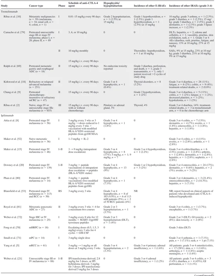
Table 1. Incidence of autoimmune hypophysitis in clinical studies of anti–CTLA-4 monoclonal antibodies
Study Cancer type Phase
Schedule of anti–CTLA-4 agent
Hypophysitis/
hypopituitarism Incidence of other E-IRAEs Incidence of other IRAEs (grade 3–4)
Tremelimumab
Ribas et al. [10] Metastatic malignancies (n⫽ 39) (melanoma,
n⫽ 34; renal cell, n ⫽
4; colon, n⫽ 1)
I 0.01–15 mg/kg every 90 days Grade 2 hypopituitarism,
n⫽ 1 (2.5%) at 15 mg/kg Grade 1 hypothyroidism, n⫽ 1 (2.5%); grade 1 hyperthyroidism, n⫽ 1 (2.5%) at 15 mg/kg 10 mg/kg: grade 3 asthma, n⫽ 1 (2.5%); grade 3 diarrhea, n⫽ 1 (2.5%); 15 mg/ kg: grade 3 diarrhea, n⫽ 2 (5%); grade 3 dermatitis, n⫽ 1 (2.5%); grade 3 lipase increase, n⫽ 1 (2.5%)
Camacho et al. [79] Pretreated unresectable stage III or stage IV melanoma; phase I, n⫽ 28; phase II, n⫽ 89
I 3, 6, or 10 mg/kg 0 0 DLTs: hepatitis, n⫽ 2; edema and cellulitis, n⫽ 1; vasculitis, pruritus, skin exfoliation, rash, n⫽ 1. Grade 3 or 4 AEs (diarrhea, rash, pruritus, fatigue, and nausea), 13% at 10 mg/kg, 27% at 15 mg/kg
II 10 mg/kg monthly 0 Thyroiditis- hypothyroidism,
n⫽ 1 at 10 mg/kg
SAEs, 9% at 15 mg/kg, 23% at 10 mg/ kg; grade 3 diarrhea, 21% at 10 mg/kg, 9% at 15 mg/kg
15 mg/kg i.v. every 90 days Ralph et al. [69] Pretreated metastatic
gastric and esophageal ADC (n⫽ 18)
II 15 mg/kg i.v. every 90 days No endocrine toxicity reported
Grade 3 diarrhea, perforation, and death, n⫽ 1; grade 3 transaminitis, n⫽ 1; only 1 patient received⬎2 cycles of study drug
Kirkwood et al. [18] Refractory or relapsed advanced melanoma (n⫽ 251)
II 15 mg/kg i.v. every 90 days Grade 3 or 4 hypophysitis, n⫽ 1 (0.4%) Thyroid disorders, n⫽ 8 (3.2%) Grade 3 or 4 diarrhea, n⫽ 28 (11%); fatigue, n⫽ 6 (2%); colitis, n ⫽ 9 (4%); treatment-related deaths, n⫽ 2 (0.8%) Chung et al. [9] Pretreated
chemotherapy-insensitive or refractory mCRC (n⫽ 47)
II 15 mg/kg i.v. every 90 days 0 Grade 2 hypothyroidism,
n⫽ 1 (2%)
Grade 3 or 4 diarrhea, n⫽ 5 (11%); colitis, n⫽ 1 (2%); fatigue, n ⫽ 1 (2%); decreased platelets, n⫽ 1 (2%); hypokalemia, n⫽ 1 (2%) Ribas et al. [2] Naïve, stage IV or
unresectable stage IIIc melanoma (n⫽ 655)
III 15 mg/kg i.v. every 90 days with or without chemotherapy
Pituitary or adrenal gland, 3%
Thyroid, 4% Grade 3 or 4 diarrhea, 14%; treatment-related deaths, n⫽ 3 in tremelimumab arm, n⫽ 0 in the chemotherapy arm Ipilimumab
Attia et al. [8] Pretreated stage IV
melanoma (n⫽ 56) I 3 mg/kg every 3 wks or 3mg/kg3 doses reduced to 1 mg/kg every 3 wks⫹ vaccination with modified HLA-A*0201-restricted peptides from gp100 MAA
Grade 3 or 4 hypophysitis, n⫽ 1 (1.8%) 0 Grade 3 or 4 colitis, n⫽ 7 (13%); dermatitis, n⫽ 4 (7%); uveitis, n ⫽ 1 (1.8%); enterocolitis, n⫽ 1 (1.8%); hepatitis, n⫽ 1 (1.8%)
Maker et al. [52] Naïve metastatic melanoma (n⫽ 36)
I 1–3 mg/kg⫹ IL-2 0 0 Grade 3 or 4 colitis, n⫽ 4 (11%); uveitis, n⫽ 1 (2.8%); arthritis, n ⫽ 1 (2.8%)
Maker et al. [15] Pretreated stage IV
melanoma (n⫽ 46) I–II 3dose escalation)3 9 mg/kg (intrapatient
Grade 3 or 4 hypophysitis, n⫽ 8 (17%); 5 mg/kg, n⫽ 1; 9 mg/kg, n⫽ 7
Grade 3 or 4 hypothyroidism,
n⫽ 1 (2.2%) Grade 3 or 4 colitis/diarrhea, n(13%); uveitis, n⫽ 1 (2.8%); arthritis,⫽ 6
n⫽ 1 (2.8%); dermatitis, n ⫽ 1 (2.8%); hepatitis, n⫽ 1 (2.8%); nephritis, n ⫽ 1 (2.8%)
Downey et al. [20] Pretreated stage IV melanoma (n⫽ 139)
I–II 3 mg/kg⫹ peptide vaccinations or intrapatient dose escalation3 peptides (HLA-A*0201 status) Grade 3 or 4 hypophysitis, n⫽ 13 (9%) Grade 1 or 2 hypothyroidism, n⫽ 3 (2%) Grade 3 or 4 enterocolitis, n⫽ 24 (17%); dermatitis, n⫽ 8 (6%); hepatitis, n ⫽ 2 (3%); uveitis, n⫽ 3 (2%)
Phan et al. [80] Pretreated stage IV
melanoma (n⫽ 14) II 3 mg/kgvaccinations (two modified⫹ peptide HLA-A*0201-restricted peptides from gp100) Grade 3 or 4 hypophysitis, n⫽ 1 (7.1%) 0 Grade 3 or 4 dermatitis, n⫽ 3 (21.4%); enterocolitis/colitis, n⫽ 2 (14.3%); hepatitis, n⫽ 1 (7.1%) Blansfield et al. [53] Pretreated stage IV
melanoma (n⫽ 113) and RCC (n⫽ 50) RS 3 mg/kg every 3 wks Grade 3 or 4 hypophysitis, n⫽ 8 of 163 (4.9%), n⫽ 6 of 113 mM patients (5%), n⫽ 2 of 50 RCC patients (4%)
NR NR; report focused on clinical aspects of patients who developed anti–CTLA-4– induced hypophysitis
Royal et al. [81] Metastatic pancreatic
ADC (n⫽ 27) II 3 mg/kg every 3 wksa maximum two courses⫻ 4 for
Grade 2 or 3 hypopituitarism, n⫽ 1 (3.7%)
0 Grade 3 or 4 colitis, n⫽ 1 (3.7%); encephalitis, n⫽ 1 (3.7%) Weber et al. [72] Stage IIIC or IV
melanoma (n⫽ 25) I 3 mg/kg every 8 wks for 12 months⫹ MART-1/gp100/ tyrosinase peptides Grade 2 or 3 hypopituitarism (DLT), n⫽ 1 (4%) 0 Grade 2 or 3 (DLT): GI toxicity, n⫽ 2 (8%); skin toxicity. n⫽ 2 (8%) Fong et al. [70] mHRPC (n⫽ 18) I Escalating doses (0.5, 1.5, 3
mg/kg) every 3 wks for 4 cycles⫹ GM-CSF
0 0 Grade 3 skin (DLT)
Small et al. [71] mPC (n⫽ 14) I 3 mg/kg single dose 0 0 Grade 3 or 4 asthenia, n⫽ 1 (7.1%); pain, n⫽ 1 (7.1%); rash, n ⫽ 1 pt (7.1%) Yang et al. [5] mRCC (n⫽ 61) I 3 mg/kg3 1 mg/kg or all
doses at 3 mg/kg every 3 wks
Grade 3 or 4 hypopituitarism, n⫽ 2 (3.3%)
Grade 3 or 4 primary adrenal
insufficiency, n⫽ 1 (1.6%) All patients: grade 3 or 4 enteritis/colitis,n⫽ 17 (28%); skin, n ⫽ 1 (1.6%); arthralgia, n⫽ 1 (1.6%); aseptic meningitis, n⫽ 1 (1.6%) Weber et al. [21] Unresectable stage III or
IV melanoma (n⫽ 88)
I–II IPI transfectoma-derived, 2.8 mg/kg for 3 doses, or IPI hybridoma-derived, 3 mg/kg for 3 doses; IPI transfectoma-derived 5 mg/kg for 3 doses
0 Grade 3 or 4 adrenal insufficiency, n⫽ 1 (1.2%)
All patients: grade 3 or 4 colitis, n⫽ 3 (3.4%); diarrhea, n⫽ 4 (45%); GI perforation, n⫽ 1 (1.1%)
The etiological classification identifies primary and sec-ondary forms. Primary hypophysitis, the most common form, has an autoimmune pathogenesis with no obvious causative agent [26]. It may occur as an isolated disease or as part of a multiorgan syndrome (i.e., polyglandular autoimmune syn-dromes and IgG-related systemic disease) [27]. Secondary hy-pophysitis includes local and systemic disease, with a clearly identified etiological agent. For local disorders, inflammation of the pituitary appears as a reaction to a sellar disease (i.e., Rathke’s cleft cyst, craniopharyngioma, germinoma, and pitu-itary adenoma). For systemic diseases, hypophysitis stems from the involvement of different organs by infectious or in-flammatory disorders (e.g., Wegener’s granulomatosis, sar-coidosis, tuberculosis, or syphilis).
On pathology, two common forms of hypophysitis (lym-phocytic and granulomatous) and three rarer variants
(xantho-matous, necrotizing, and plasma cell rich) are recognized (Table 3) [27]. Lymphocytic hypophysitis (LYH), often re-ferred to as autoimmune hypophysitis (AH), is the most com-mon. The clinical features of⬃500 patients with primary hypophysitis have been reported so far [28]. The exact inci-dence is unknown and likely underestimated [29]. LYH/AH is mostly seen in striking temporal association with pregnancy or postpartum, but it may also occur in women irrespective of pregnancy, in males, and in children [26, 27].
LYH/AH is characterized by dense diffuse lymphocytic in-filtration of the pituitary that may be organized in lymphoid follicles. Plasma cells are also common, whereas eosinophils, macrophages, and neutrophils are rarer. In a small percentage of patients in whom a biopsy specimen was obtained (4%), mixed lymphocytic and granulomatous lesions were found in the anterior pituitary [26]. These features are thought to repre-Table 1. (Continued)
Study Cancer type Phase
Schedule of anti–CTLA-4 agent
Hypophysitis/
hypopituitarism Incidence of other E-IRAEs Incidence of other IRAEs (grade 3–4)
Gerritsen et al. [73] Chemotherapy-naïve mHRPC patients (n⫽ 28)
I Monthly escalating dose (0.3, 1, 3, or 5 mg/kg)⫹ GVAX immunotherapy Grade 2 or 3 hypophysitis, n⫽ 5 (18%) (5/6 at 3 and 5 mg/kg) Grade 3 adrenal
insufficiency, n⫽ 1 (3.6%) Grade 3 alveolitis, nhepatitis, n⫽ 1 (3.6%)⫽ 1 (3.6%);
Ansell et al. [74] Relapsed or refractory B-cell NHL (n⫽ 18)
I 3 mg/kg3 monthly 1 mg/kg⫻ 3 mos (dose level 1),3 escalation to 3 mg/kg monthly⫻ 4 mos (dose level 2)
Grade 1 or 2 hypophysitis, n⫽ 1 (6%)
0 Grade 3 diarrhea, n⫽ 5 (28%); fatigue,
n⫽ 1 (6%); neutropenia, n ⫽ 1 (6%)
Hodi et al. [3] Pretreated unresectable stage III or IV melanoma (n⫽ 676)
III 3 mg/kg every 3 wks for 4 doses with or without gp100 versus gp100 alone Grade 3 hypophysitis, n⫽ 2 (1.5%) in ipilimumab arm; n⫽ 2 (0.5%) in combination arm; n⫽ 0 in gp100 arm; hypopituitarism, n⫽ 3 (2.3%) in the ipilimumab arm, n⫽ 3 (0.8%) in the combination arm Grade 3 or 4 hypothyroidism, n⫽ 2 (1.5%) in ipilimumab arm, n⫽ 6 (1.6%) in combined arm, n⫽ 2 (1.5%) in gp100 arm; adrenal, n⫽ 2 (1.5%) in ipilimumab arm, n⫽ 3 (0.8%) in combination arm Grade 3 or 4 diarrhea, 30.3%–27.5%; nausea, 39.9%–35.1%; vomiting, 19.7%– 23.7%; abdominal pain, 17.6%–15.3%; colitis, 5.3%–7.6%; constipation, 21.3%– 20.6%; pruritus/rash, 17.8%–24.4%; hepatic, 2.1%–3.8%; fatigue, 36.1%– 42%; pyrexia, 20.5%–12.2%; headache, 17.1%–14.5%.
Hersh et al. [75] Chemotherapy-naïve patients with unresectable stage III or IV melanoma (n⫽ 72)
II 3 mg/kg every 4 wks for 4 doses with or without DTIC (up to 6 cycles; 250 mg/m2
per day⫻ 5 days)
0 Grade 2 adrenal insufficiency, n⫽ 1 (1.4%) in ipilimumab⫹ DTIC arm
Grade 3 or 4 colitis/diarrhea, n⫽ 2 (2.8%); GI hemorrhage, n⫽ 2 (2.8%); vasculitis, n⫽ 2 (2.8%); transaminitis,
n⫽ 2 (2.8%); skin, n ⫽ 2 (2.8%);
multiorgan failure, n⫽ 2 (2.8%) Wolchok et al. [76] Pretreated unresectable
stage III or IV melanoma (n⫽ 217)
II 0.3, 3, or 10 mg/kg every 3 wks for 4 cycles (induction)3 every 3 mos (maintenance).
Endocrine IRAEs are globally reported: grade 3 or 4, 0.3 mg/kg, n⫽ 0; 3 mg/kg, n⫽ 2 (1%); 10 mg/kg, n⫽ 1 (0.5%); in the 3-mg/kg group, hypopituitarism was reported among reasons for stopping treatment.
Grade 3 or 4 GI events: 10 mg/kg, n⫽ 11 (5%); 3 mg/ kg, n⫽ 2 (1%); 0.3 mg/kg, n⫽ 0; liver: 10 mg/kg, n ⫽ 2 (1%); 3 mg/kg, n⫽ 3 (1.4%); 0.3 mg/kg, n⫽ 1 (0.5%); others, n⫽ 2 (1%).
Hodi et al. [77] Unresectable stage III or IV melanoma (may or may not be pretreated,
n⫽ 21)
I 10 mg/kg every 3 wks⫻ 4 3 every 3 mos⫹ bevacizumab 7.5 mg/kg (cohort 1) or 15 mg/kg (cohort 2) every 3 wks
Hypophysitis (grade not specified), n⫽ 3 (14%)
Thyroiditis (grade not specified), n⫽ 4 (19%)
Grade 3 or 4 hepatitis, n⫽ 2 (9.5%); bilateral uveitis, n⫽ 2 (9.5%); giant arteritis, n⫽ 1 (4.8%); grade 2 colitis,
n⫽ 2 (9.5%)
O’Day et al. [78] Pretreated, unresectable stage III or IV melanoma (n⫽ 155)
II 10 mg/kg every 3 wks for 4 cycles (induction)3 every 3 mos (maintenance).
Endocrine IRAEs are globally reported: grade 3, n⫽ 2 (1.3%); grade 4, n⫽ 0 Grade 3 or 4 skin, n⫽ 5 (3.2%); GI, n⫽ 13 (8.4%); liver, n⫽ 11 (7.1%); others, n⫽ 4 (2.6%) Ku et al. [22] Refractory melanoma
(compassionate use, n⫽ 53) II 10 mg/kg every 3 wks for 4 doses3 every 12 wks in case of CB Grade 2 or 3 hypophysitis with adrenal insufficiency, n⫽ 2 (4%) Grade 2 hypothyroidism, n⫽ 1 (2%) Grade 3 or 4 diarrhea, n⫽ 17 (33%); colitis, n⫽ 5 (10%); hepatitis, n ⫽ 4 (8%); pancreatitis, n⫽ 1 (2%) Di Giacomo et al. [55] Pretreated unresectable stage III or IV melanoma (expanded access program, n⫽ 27) II 10 mg/kg every 3 wks for 4 doses3 every 12 wks in cases of CB 0 Grade 1 or 2 hypothyroidism,
n⫽ 2 (7.4%) Grade 3 diarrhea, ntransaminitis, n⫽ 1 (3.7%); grade 4⫽ 2 (7.4%); pancytopenia, n⫽ 1 (3.7%)
Abbreviations: ADC, adenocarcinoma; ANA, antinuclear Ab; anti–CTLA-4, anti– cytotoxic T lymphocyte antigen 4; CB, clinical benefit; DLT, dose-limiting toxicity; DTIC, dacarbazine; GI, gastrointestinal; GVAX, granulocyte-macrophage colony-stimulating factor (GM-CSF) gene-transfected tumor cell vaccine; HLA, human leukocyte antigen; IL-2, interleukin 2; IPI, ipilimumab; IRAE, immune-related adverse event; MAA, melanoma-associated antigen; MART-1, melanoma antigen recognized by T-cells-1; mHRPC, metastatic hormone-refractory prostate cancer; mM, metastatic melanoma; mRCC, metastatic renal cell carcinoma; NHL, non-Hodgkin’s lymphoma; NR, not reported; RS, retrospective study; SAE, serious adverse event.
sent different stages of the same disease, rather than a granu-lomatous form of hypophysitis [26]. In some studies, the predominant lymphocytic subpopulation is represented by cy-totoxic T lymphocyte (CD8⫹) cells, suggesting that T cell– mediated cytotoxicity is critical in the pathogenesis of the disorder [30]. Several other aspects appear to indicate that this condition results from an autoimmune process. Almost 30% of LYH/AH patients have a coexisting autoimmune disease, such as Hashimoto’s thyroiditis, Addison’s disease, type 1 diabetes, or pernicious anemia [26, 31–34]. LYH/AH is considered a component of type 1 polyglandular syndrome [26, 31–33], and the association of LYH/AH with pregnancy has been proposed as further circumstantial evidence supporting an autoimmune pathogenesis [35–37]. In a limited number of animal studies, lymphocytic infiltrates in the pituitary are seen after injections of pituitary extracts [26, 27, 38, 39]. Several candidate pitu-itary autoantigens (growth hormone, ␣-enolase, pituitary gland specific factor 1a and factor 2, secretogranin II) have been proposed, but the pathogenic role of these antigens re-mains to be elucidated [26, 31, 32].
The precise mechanisms by which infiltrates cause loss of function or destruction of the pituitary cells or impairment of vasopressin release have yet to be discovered. It has been sug-gested that LYH/AH may progress through different stages. Initially, the pituitary is inflamed, edematous, enlarged, and in-filtrated by lymphocytes, thus producing mass-effect symp-toms [26, 31–35]. During this phase, endocrine tests may reveal subclinical hypopituitarism. Once the inflammation re-solves, either spontaneously or with the aid of glucocorticoids, and the pituitary parenchyma remains intact, clinical remission may occur. As inflammation progresses, infiltrating cells dis-rupt the normal architecture, eventually leading to destruction of the parenchyma that is replaced by fibrotic tissue and be-comes atrophic. A similar pattern has been reported in a mouse model of LYH [40]. Permanent partial hypopituitarism or pan-hypopituitarism may be the clinical consequence, depending
upon the extent of damage to the different components of the pituitary gland [26].
Usually, LYH/AH is confined to the anterior pituitary, with symptoms such as headache (53%) and impaired vision (43%). Hypopituitarism is present in 44% of patients and, in contrast to other forms of hypopituitarism, is more commonly associ-ated with a deficit of adrenocorticotropic hormone (ACTH) (56%), followed by a deficit of thyroid-stimulating hormone (TSH) (49%), gonadotropins (52%), and growth hormone (GH) (39%) [26]. Hypoprolactinemia (23%) or hyperpro-lactinemia (11%) may be seen, depending on whether the dam-age involves prolactin-producing cells or stalk, respectively [34]. Diabetes insipidus (DI) is less common (1%) and is re-lated to involvement of the posterior pituitary [26, 31–35]. Oc-casionally, hypophysitis may primarily involve the infundibulum and posterior pituitary, causing intracranial mass-effect symptoms, DI, and hyperprolactinemia. In these cases, anterior pituitary function is usually preserved [26, 29, 31–35, 41].
As a result of the enlargement of the gland, headache to-gether with visual field impairment are usually recognized as “sentinel symptoms” at disease onset, followed by hormone function disorders. ACTH deficiency is considered the earliest functional alteration in LYH/AH and is the most frequent “iso-lated” pituitary hormone deficiency [42]. These aspects appear to suggest that antigen(s) targeted by the immune system to trigger autoimmune reactions reside within the corticotroph cells. However, isolated ACTH deficiency may also be ob-served in the absence of LYH/AH, and isolated deficiencies of other anterior pituitary hormones have been described in LYH/AH patients [43– 45]. The greater frequency of ACTH deficiency may simply represent an ascertainment bias, be-cause these patients may come to medical attention more than those with other adenohypophyseal hormone deficiencies be-cause of the more evident symptomatology [26].
Similarly to symptoms, the imaging features of LYH/AH are not specific [34, 46]. Computed tomography and magnetic resonance imaging (MRI) typically reveal a diffuse enlarge-ment of the pituitary gland with loss of normal signal intensity of the posterior pituitary on precontrast images and variable enlargement of the infundibulum. Enhancement is usually uni-form, may also be heterogeneous, and may be delayed or even absent in the posterior pituitary area [46, 47].
Currently, the diagnosis of LYH/AH requires pathological analysis. However, a presumptive clinical diagnosis can be based on the history of gestational or postpartum hypopituitar-ism, a contrast-enhancing sellar mass, a pattern of pituitary hormone deficiency with early loss of ACTH and TSH, rela-tively rapid development of hypopituitarism, and a degree of pituitary failure inconsistent with the size of the mass [26, 48]. Approximately 30% of patients with clinically suspected LYH/AH are diagnosed by combining symptoms and labora-tory and radiological findings [28]. Current immunological tests for LYH/AH, particularly immunofluorescence for anti-pituitary antibodies, offer good sensitivity but lack adequate specificity, and therefore are of limited value in the diagnosis Table 2. Toxicity grading applicable to hypophysitis
according to the Common Terminology Criteria for Adverse Events (CTCAE) of National Institutes of Health, National Cancer Institute
Grade Description
1 Asymptomatic or mild symptoms; clinical or diagnostic observations only; intervention not indicated
2 Moderate; minimal, local, or noninvasive intervention indicated; limiting age-appropriate instrumental ADL 3 Severe or medically significant but not
immediately life-threatening; hospitalization or prolongation of existing hospitalization indicated; disabling; limiting self-care ADL 4 Life-threatening consequences; urgent
intervention indicated
5 Death
and management of LYH/AH patients [28, 31]. However, re-cent advances in this field open promising perspectives [49].
The natural history of LYH/AH is variable [26 –28]. Most patients show improvement in symptoms after mass-reducing treatment (pituitary surgery or high-dose glucocorticoids), but the majority (72%) require some form of long-term hormone replacement. Approximately 4% may improve spontaneously without treatment. When an MRI follow-up was available, re-duction or complete disappearance of the initial pituitary mass was demonstrated in 88% of cases, no significant change was seen in 12% of patients, and 10% of these patients developed an “empty sella.” Unfortunately, it is estimated that 7% of pa-tients affected by LYH/AH die presumably as a result of irre-versible and unrecognized adrenal insufficiency [26 –28].
CLINICALFEATURES OFHYPOPHYSITISINDUCED BY
ANTI–CTLA-4 mAbs
The incidence of hypophysitis induced by anti–CTLA-4 mAbs (hereafter, anti–CTLA-4 –IH) varies considerably (Table 1), reported in 0%–17% of treated melanoma patients [50]. How-ever, accumulating clinical experience demonstrates that this
side effect also occurs in patients with solid tumors of various types, including kidney and prostate cancer [51].
In a trial on 46 patients with mM treated with various doses of ipilimumab, eight patients (17%) experienced hypophysitis, with the majority of cases in patients on the higher drug dose regimen (9 mg/kg) [15]. In a previous study with lower doses of the drug (1–3 mg/kg), a lower incidence of hypophysitis (1.8%) was reported [52].
In a study on 139 subjects with mM receiving ipilimumab at a dose of 1–3 mg/kg every 3 weeks with or without peptide vaccine, screening for E-IRAEs with measurements of ACTH, TSH, cortisol, and free T4 at each cycle of therapy was re-quired [20]. Enterocolitis and hypophysitis were the most com-mon grade 3– 4 IRAEs (17% and 9%, respectively). LYH/AH was clinically diagnosed by Yang et al. [5] in two of 61 patients (3.3%) affected by mRCC who received ipilimumab (1–3 mg/kg every 3 weeks).
In another study on 163 patients with mM or mRCC treated with ipilimumab (3–9 mg/kg every 3 weeks), alone or in com-bination with another type of immunotherapy, hypophysitis was diagnosed in eight patients (4.9%) [53]. Recently, two Table 3. Pathological classification of hypophysitis
Type of hypophysitis Epidemiology and histopathologic essential features
Lymphocytic hypophysitis More common in women (F:M ratio, 3:1); mean age at presentation is 38 (⫾15) yrs. Presents in association with pregnancy and postpartum (⬃40% of women).
Histology: marked infiltration of lymphocytes of the pituitary gland both in a diffuse fashion and occasionally with a focal formation. Lymphocytes are typically accompanied by scattered plasma cells, eosinophils, and fibroblasts, and in later disease stages by fibrosis.
Hypophysitis induced by anti–CTLA-4 mAbs is clinically diagnosed as
autoimmune hypophysitis. It is almost exclusively reported in males (when gender of patients is detailed). Other epidemiologic features are not available (overall, at least 50 cases have been reported). Pathology findings are not available.
Granulomatous hypophysitis More common in women (F:M ratio, 4:1); presents at an older age (44⫾ 16 yrs); not associated with pregnancy.
Histology: multinucleated giant cells forming true granulomas with palisading histiocytes, surrounded by numerous lymphocytes, mainly T cells, and some plasma cells.
Xanthomatous hypophysitis More common in women (F:M ratio, 3:1); mean age at presentation is 37 (⫾ 16) yrs; not associated with pregnancy.
Histology: infiltration with foamy histiocytes and macrophages, accompanied by plasma cells and lymphocytes. In chronic cases, fibrosis and acinar destruction. Necrotizing hypophysitis Involvement of both pituitary lobes and the stalk up to the median eminence of the
hypothalamus.
Histology: extensive necrosis surrounded by lymphoplasmacytic infiltration and fibrosis with scattered areas of glandular tissue.
IgG4-related/ plasma cell rich hypophysitis
Typically part of an IgG4-related systemic disease with multiple organs infiltrated
by polyclonal lymphocytes and IgG4-producing plasma cells, ultimately resulting in
fibrosis and functional impairment.
Histology: abundance of IgG4-producing plasma cells.
Adapted from Caturegli P, Newschaffer C, Olivi A et al. Autoimmune hypophysitis. Endocr Rev 2005;26:599 – 614 and Leporati P, Landek-Salgado MA, Lupi I et al. IgG4-related hypophysitis: A new addition to the hypophysitis spectrum. J Clin Endocrinol Metab 2011;96:1971–1980.
cases of hypopituitarism, presumably resulting from hypophy-sitis, were described in patients submitted to experimental treatment with ipilimumab for mPC [50].
In the large phase III trial that led to the FDA approval of ipilimumab, in which 676 pretreated patients affected by un-resectable stage III or IV melanoma received the drug as a sin-gle agent or in combination with gp100 versus gp100 alone, grade 3 hypophysitis was reported in both groups receiving ip-ilimumab. The incidences of hypophysitis were 1.5% in the combination group (two of 380 patients) and 0.5% in the sin-gle-agent arm (two of 131 patients), with no cases in the gp100 arm [3]. Conversely, no cases of hypophysitis were reported in a phase III trial evaluating dacarbazine with and without ipili-mumab in treatment-naïve patients with mM, in a phase II trial evaluating chemotherapy with and without ipilimumab in pa-tients with non-small cell lung cancer, and in one of two ex-panded access programs to ipilimumab administered at conventional dosages in patients with mM, both in the induc-tion and maintenance phases, with the incidence of this AE be-ing 4% in the other trial (Table 1) [4, 22, 54, 55].
Tremelimumab (15 mg/kg) has been reported to induce hy-pophysitis in 0.4%–2.5% of patients (Table 1) [2, 10, 14, 18]. In contrast to other forms of LYH/AH, patients who expe-rience anti–CTLA-4 –IH are mostly male. These patients usu-ally present with nonspecific symptoms such as headache, visual impairment, fatigue, weakness, confusion, memory loss, erectile dysfunction and loss of libido, anorexia, labile moods, insomnia, temperature intolerance, subjective sensa-tion of fever, and chills [5, 13, 20, 50]. The onset of symptoms usually occurs after 2– 6 months of treatment. Contrast-en-hanced MRI shows marked enlargement of the pituitary gland, often with thickening of the hypophyseal stalk. In some cases, the pituitary gland enhances homogeneously, whereas in other cases there is heterogeneous enhancement. Levels of ACTH, cortisol, TSH and/or free T4, GH, prolactin, insulin-like growth factor I, follicle-stimulating hormone, luteinizing hor-mone, and testosterone are variably altered, indicating differ-ent degrees of hypopituitarism [52]. Very rarely DI has been reported.
Similar to classic primary LYH/AH, the treatment used for anti–CTLA-4 –IH is high-dose corticosteroids, slowly tapered as symptoms and hormone tests improve (Fig. 1). Almost all patients who developed anti–CTLA-4 –IH experience clinical resolution of acute symptoms in a few days following with-drawal of the study drug and starting of corticosteroids [53]. The efficacy of corticosteroids is confirmed by the rapid shrinkage of the pituitary gland on MRI. However, pituitary function may be impaired for a longer period of time. More-over, the duration of replacement therapy with physiological glucocorticoid dosages (mean, 20 weeks) may be considerably longer or even be life long [13, 16, 51]. Hypopituitarism is the only potentially irreversible IRAE induced by anti–CTLA-4 mAbs [56]. In particular, the hypothalamic–pituitary– gonadal and hypothalamic–pituitary–thyroidal axes frequently re-cover, but only a few patients can discontinue glucocorticoid replacement [50, 51]. At the onset of anti–CTLA-4 –IH, it is
impossible to predict which patients will develop persistent hypopituitarism.
The protective role of corticosteroids in reducing the inci-dence and severity of anti–CTLA-4 –IH remains to be ex-plored. In anti–CTLA-4 mAb–induced colitis, preventive administration of budesonide was not found to reduce the in-cidence of this IRAE. Surprisingly, hypopituitarism was ap-parently more frequent in the group receiving budesonide (6.9% versus 3.5%) [57]. High-dose corticosteroid treatment (and replacement therapy) does not appear to decrease the an-titumor effects of CTLA-4 blockade [12]. When indicated, re-treatment with ipilimumab after suspension because of hypophysitis seems to be safe [12].
DISCUSSION
LYH/AH is emerging as a not so uncommon IRAE of anti– CTLA-4 mAbs. Selective deficit of pituitary hormones may be induced by various anticancer treatments [58, 59]. Usually, the clinical onset of these endocrine AEs (E-AEs) is not acute and they progress subclinically [58, 59]. The spectrum of E-AEs experienced by patients treated with anti–CTLA-4 mAbs in-cludes hypopituitarism, primary thyroid disease, and primary adrenal insufficiency. These side effects have been occasion-ally found in the same individual. The pathogenic mechanism of these E-AEs seems to be related to autoimmunity [5, 8, 15, 17, 21]. The prevalence of this autoimmune hypophysitis is variable among different studies (0%–17%) [50]. Autoim-mune hypophysitis has never been reported to be a conse-quence of exposure to other classes of anticancer drug. However, reversible or irreversible hypopituitarism may be a side effect following treatment with other immunomodulatory drugs, such as interferon-␣ [60–62]. A case of granulomatous adenohypophysitis occurring after treatment with interferon-␣2b and ribavirin for hepatitis C was reported [63]. Another patient affected by hepatitis C experienced central hypothy-roidism during treatment with pegylated interferon-␣ and riba-virin and a clinical diagnosis of hypophysitis was made [64].
The clinical presentation of LYH/AH (or other forms of hypophysitis) is similar to that of any expanding sellar mass. In healthy individuals, LYH/AH is suspected if symptoms appear in temporal relationship with pregnancy and postpartum. Sim-ilarly, the diagnosis should be considered when symptoms oc-cur in cancer patients under treatment with anti–CTLA-4 mAbs. Simple clinical guidelines for diagnosis and treatment can be routinely adopted (Fig. 1).
Patients who need to receive anti–CTLA-4 mAbs should be carefully educated on the importance of their vigilance in early detection and prompt reporting of symptoms potentially related to IRAEs, and that these symptoms may occur weeks to months after the start of treatment. In these patients, TSH, free T4, serum electrolytes, serum glucose, and blood cell counts should be assessed before initiating treatment and before each cycle. If the patient develops symptoms such as headache, nau-sea, vomiting, lethargy, or constipation, the drug should be withheld and tests, including morning cortisol, should be re-done. In addition, when anti–CTLA-4 –IH is suspected, refer-ral to an endocrinologist or even admission to a hospital, if
clinically indicated, is advisable. In these cases, a pituitary protocol MRI scan should be performed to evaluate for hy-pophysitis and complete pituitary function should be as-sessed (Fig. 1).
High-dose glucocorticoid therapy is the most widely used treatment for anti–CTLA-4 –IH. If high-dose glucocorticoids are initiated, a suggested regimen is 4 mg dexamethasone ev-ery 6 hours for 7 days, followed by a gradual tapering to 0.5 mg daily and then a change to prednisone or hydrocortisone at re-placement doses under the guidance of an endocrinologist [65]. A brief interruption of anti–CTLA-4 therapy may be war-ranted during the acute stage of hypophysitis. However, once hypophysitis resolves with appropriate treatment and adequate hormone replacement has been tailored, rechallenge with the anticancer treatment should be considered, providing that the anti–CTLA-4 therapy may prolong survival in a patient with an otherwise fatal malignancy. Clearly, this decision should be made on an individual case basis. If the agent is re-started, close monitoring of pituitary function should be done [65].
Several issues concerning anti–CTLA-4 –IH remain to be fully elucidated. The exact incidence of this and other E-IRAEs, the reason for the unusually high prevalence in males,
and the role of CTLA-4 gene polymorphisms, which are known to correlate with the development of autoimmunity, need to be better clarified in larger studies. Also, the lower incidence of anti–CTLA-4 –IH in patients exposed to tremelimumab than in those exposed to ipilimumab remains to be confirmed. In ad-dition, although tumor regression has been frequently associ-ated with IRAEs, correlation between tumor response and the incidence and severity of IRAEs needs to be defined using an appropriate analytical approach [24].
Of major importance, the exact immunologic mechanisms responsible for both anti–CTLA-4 –induced tumor regression and IRAEs have not been clearly explained. It was initially suggested that anti–CTLA-4 mAbs may act by depleting T-regulatory cells (T-regs) [19]. In another study, the antitumor and autoimmune effects were a result of the direct activation of CD4⫹CD8⫹effector cells [15]. Although CD8⫹cytotoxic T lymphocytes are likely to play a major role, the exact tumor and tissues antigen(s) involved in the tumor response and tox-icity are unknown. It is still unclear whether the effects are a result of T cells specifically acting against antigens shared by tumor and normal cells or a result of concomitant activation of multiple populations with separate antihost and antitumor ac-tivities [8, 19, 20, 66]. Melan-A, an antigen shared by mela-Figure 1. Flowchart for the diagnosis and treatment of hypophysitis induced by anti–CTLA-4 mAbs.
*Adrenal insufficiency is considered the hallmark of pituitary damage in anti–CTLA-4 –induced hypophysitis (see text).
Abbreviations: ACTH, adrenocorticotropic hormone; ADH, antidiuretic hormone; anti–CTLA-4 mAb, anti– cytotoxic T lymphocyte antigen 4 monoclonal antibody; DEX, dexamethasone; ECG, electrocardiogram; FSH, follicle-stimulating hormone; fT4, free T4; HD, high-dose; IGF-I, insulin-like growth factor I; LH, luteinizing hormone; MRI, magnetic resonance imaging; TSH, thyroid-stimulating hormone.
noma cells and normal melanocytes, has been associated both with tumor regression and with immune-related skin reactions [66]. In a patient affected by mM and treated with ipilimumab, marked melan-A–specific T-cell reactivity in tumor and skin tissue was found, with CD8⫹T cells localized to nevi and a simultaneous increase in melan-A–specific CD8⫹T cells in the peripheral blood [66].
It has been hypothesized that anti–CTLA-4 –IH may be in-duced by antibodies directed against the pituitary gland [53], but the presence of antipituitary antibodies in patients who re-ceive anti–CTLA-4 mAbs remains to be demonstrated.
To the best of our knowledge, the diagnosis of anti–CTLA-4 –IH has always been made by clinical, laboratory, and radio-logical data. No patient has undergone a pituitary biopsy. Indeed, biopsy of the pituitary gland in cancer patients sus-pected of having developed anti–CTLA-4 –IH raises a series of ethical issues, and it is not necessary either for diagnosis or for treatment. Nonetheless, this remains the only way to obtain es-sential information to improve our knowledge on the patho-physiology of this IRAE. Pituitary autoimmunity is a complex and incompletely defined spectrum of clinical conditions [26], ranging from histologically proven forms of LYH/AH to the presence of pituitary antibodies in apparently healthy individ-uals [28]. Interestingly, Mirocha et al. [67] observed two dis-tinct entities of primary LYH that can be distinguished on the basis of the prevalence of T-regs or T-17 helper lymphocytes (THL-17). One of these entities, in agreement with the classi-cal description of LYH/AIH, demonstrates an autoimmune process with THL-17 dominance and lack of T-regs. The other one appears as a process in which T-regs control the immune response, which may not be self-targeted but foreign targeted (infective agents?). Hypophysitis triggered by an immune ho-meostatic process should not be treated with immunosuppres-sion, whereas autoimmune-sustained hypophysitis may benefit from it [67]. Patients with anti–CTLA-4 –IH usually benefit from corticosteroids and this ex juvantibus criterion, together with other clinical aspects, may indirectly confirm its autoimmune pathogenesis. The potential of the precautionary use of steroids in reducing the long-term sequelae of this E-IRAE, especially in preventing prolonged substitutive treat-ment, still remains to be evaluated.
Because the hurdles in defining the histological character-istics of anti–CTLA-4 –IH persist, anti–CTLA-4 –IH offers a
unique opportunity to assess the fluctuation of the available pi-tuitary antigens and relative antibodies, with the aim to im-prove their reliability as diagnostic and predictive tools. Pituitary antigens and antibodies could be monitored in a ho-mogeneous cohort of patients with a specific disease and known pituitary-damaging agents, such as anti–CTLA-4 mAbs, at baseline, before each cycle of treatment, and during follow-up. Such a study would offer the possibility of defining a series of important clinical, laboratory, and radiological cor-relations, including refinement of the diagnosis and the real in-cidence of anti–CTLA-4 –IH, the potential existence of a subclinical form of anti–CTLA-4 –IH, the impact (if any) of this syndrome on the quality of life of patients, and the possible predisposition of a subgroup of these patients to develop anti– CTLA-4 –IH and other E-AEs. This approach appears even more logical in light of recent data regarding the predictive role of antibodies to thyroglobulin and thyroperoxidase and the TSH receptor in the development of thyroid autoimmune dis-ease [68]. Similarly, in a population of patients with autoim-mune polyendocrine syndrome, measurement of antipituitary antibodies allows the identification of patients at higher risk for developing pituitary autoimmune dysfunction [49].
CONCLUSIONS
Hypophysitis is an infrequent IRAE triggered by anti–CTLA-4 mAbs. Because the clinical suspicion of anti–CTLA-4 –IH can only be based on symptoms, it should be considered when hy-popituitarism or sellar mass-effect symptoms appear in cancer patients under treatment with this class of drugs. This IRAE, if promptly suspected, may be presumptively diagnosed and treated, thus avoiding life-threatening complications, namely, acute adrenal insufficiency. Appropriate correlative studies on anti–CTLA-4 –IH may contribute to improving our knowledge regarding the pathophysiology of pituitary autoimmunity.
AUTHORCONTRIBUTIONS
Conception/Design: Francesco Torino, Agnese Barnabei, Salvatore M. Corsello Collection and/or assembly of data: Francesco Torino, Agnese Barnabei,
Salvatore M. Corsello
Data analysis and interpretation: Salvatore M. Corsello, Francesco Torino, Agnese Barnabei, Roberto Salvatori, Liana De Vecchis
Manuscript writing: Francesco Torino, Agnese Barnabei, Salvatore M. Corsello, Roberto Salvatori, Liana De Vecchis
Final approval of manuscript: Francesco Torino, Agnese Barnabei, Liana De Vecchis, Roberto Salvatori, Salvatore M. Corsello
REFERENCES
1. Ribas A. Clinical development of the anti-CTLA-4 antibody tremelimumab. Semin Oncol 2010;37:450 – 454.
2. Ribas A, Hauschild A, Kefford R et al. Phase III, open-label, randomized, comparative study of tremelimumab (CP-675,206) and chemotherapy (te-mozolomide [TMZ] or dacarbazine [DTIC]) in pa-tients with advanced melanoma. J Clin Oncol 2008; 26(15 suppl):LBA90111.
3. Hodi FS, O’Day SJ, McDermott DF et al. Im-proved survival with ipilimumab in patients with metastatic melanoma. N Engl J Med 2010;363:711– 723.
4. Robert C, Thomas L, Bondarenko I et al. Ipili-mumab plus dacarbazine for previously untreated metastatic melanoma. N Engl J Med 2011;364: 2517–2526.
5. Yang JC, Hughes M, Kammula U et al. Ipili-mumab (anti-CTLA4 antibody) causes regression of metastatic renal cell cancer associated with enteritis and hypophysitis. J Immunother 2007;30:825– 830.
6. Tollefson MK, Karnes RJ, Thompson RH et al. A randomized phase II study of ipilimumab with an-drogen ablation compared with anan-drogen ablation alone in patients with advanced prostate cancer [ab-stract 168]. Presented at the 2010 American Society of Clinical Oncology Genitourinary Cancers Sympo-sium, San Francisco, CA, March 5– 6, 2010.
7. Eggermont AM, Testori A, Maio M et al. Anti-CTLA-4 antibody adjuvant therapy in melanoma. Semin Oncol 2010;37:455– 459.
8. Attia P, Phan GQ, Maker AV et al. Autoimmu-nity correlates with tumor regression in patients with metastatic melanoma treated with anti-cytotoxic T-lymphocyte antigen-4. J Clin Oncol 2005;23:6043– 6053.
9. Chung KY, Gore I, Fong L et al. Phase II study of the anti-cytotoxic T-lymphocyte-associated antigen 4 monoclonal antibody, tremelimumab, in patients with refractory metastatic colorectal cancer. J Clin Oncol 2010;28:3485–3490.
10. Ribas A, Camacho LH, Lopez-Berestein G et al. Antitumor activity in melanoma and anti-self
re-sponses in a phase I trial with the anti-cytotoxic T lymphocyte-associated antigen 4 monoclonal anti-body CP-675,206. J Clin Oncol 2005;23:8968 – 8977.
11. Sanderson K, Scotland R, Lee P et al. Autoim-munity in a phase I trial of a fully human anti-cyto-toxic T-lymphocyte antigen-4 monoclonal antibody with multiple melanoma peptides and Montanide ISA 51 for patients with resected stages III and IV melanoma. J Clin Oncol 2005;23:741–750.
12. Boasberg P, Hamid O, O’Day S. Ipilimumab: Unleashing the power of the immune system through CTLA-4 blockade. Semin Oncol 2010;37:440 – 449. 13. Kaehler KC, Piel S, Livingstone E et al. Update on immunologic therapy with anti-CTLA-4 antibod-ies in melanoma: Identification of clinical and bio-logical response patterns, immune-related adverse events, and their management. Semin Oncol 2010; 37:485– 498.
14. Wallis N, Bulanhagui CA, Dorazio PC et al. Safety of tremelimumab (CP-675,206) in patients (pts) with advanced cancer. J Clin Oncol 2008;26(15 suppl):3040.
15. Maker AV, Yang JC, Sherry RM et al. Intratient dose escalation of anti-CTLA-4 antibody in pa-tients with metastatic melanoma. J Immunother 2006;29:455– 463.
16. Lebbe C, O’Day S, Chiarion-Sileni V et al. Analysis of the onset and resolution of immune-related adverse events during treatment with ipili-mumab in patients with metastatic melanoma [abstract O-015]. Presented at Perspectives in Mela-noma XII, Scheveningen, The Hague, The Nether-lands, October 2– 4, 2008.
17. Wolchok JD, Weber JS, Hamid O et al. Ipili-mumab efficacy and safety in patients with advanced melanoma: A retrospective analysis of HLA subtype from four trials. Cancer Immun 2010;10:9 –14.
18. Kirkwood JM, Lorigan P, Hersey P et al. Phase II trial of tremelimumab (CP-675,206) in patients with advanced refractory or relapsed melanoma. Clin Cancer Res 2010;16:1042–1048.
19. Beck KE, Blansfield JA, Tran KQ et al. Entero-colitis in patients with cancer after antibody block-ade of cytotoxic T-lymphocyte-associated antigen 4. J Clin Oncol 2006;24:2283–2289.
20. Downey SG, Klapper JA, Smith FO et al. Prog-nostic factors related to clinical response in patients with metastatic melanoma treated by CTL-associ-ated antigen-4 blockade. Clin Cancer Res 2007;13: 6681– 6688.
21. Weber JS, O’Day S, Urba W et al. Phase I/II study of ipilimumab for patients with metastatic mel-anoma. J Clin Oncol 2008;26:5950 –5956.
22. Ku GY, Yuan J, Page DB et al. Single-institu-tion experience with ipilimumab in advanced mela-noma patients in the compassionate use setting: Lymphocyte count after 2 doses correlates with sur-vival. Cancer 2010;116:1767–1775.
23. Callahan MK, Wolchok JD, Allison JP. Anti-CTLA-4 antibody therapy: Immune monitoring dur-ing clinical development of a novel immunotherapy. Semin Oncol 2010;37:473– 484.
24. Agarwala SS, Ribas A. Current experience with CTLA4-blocking monoclonal antibodies for the treatment of solid tumors. J Immunother 2010;33: 557–569.
25. Schneider HJ, Aimaretti G,
Kreitschmann-An-dermahr I et al. Hypopituitarism Lancet 2007;369: 1461–1470.
26. Caturegli P, Newschaffer C, Olivi A et al. Au-toimmune hypophysitis. Endocr Rev 2005;26:599 – 614.
27. Leporati P, Landek-Salgado MA, Lupi I et al. IgG4-related hypophysitis: A new addition to the hy-pophysitis spectrum. J Clin Endocrinol Metab 2011; 96:1971–1980.
28. Caturegli P, Lupi I, Landek-Salgado M et al. Pi-tuitary autoimmunity: 30 years later. Autoimm Rev 2008;7:631– 637.
29. Molitch ME, Gillam MP. Lymphocytic hy-pophysitis. Horm Res 2007;68(suppl 5):145–150.
30. Gutenberg A, Buslei R, Fahlbusch R et al. Im-munopathology of primary hypophysitis: Implica-tions for pathogenesis. Am J Surg Pathol 2005;29: 329 –338.
31. Bellastella A, Bizzarro A, Coronella C et al. Lymphocytic hypophysitis: A rare or underestimated disease? Eur J Endocrinol 2003;149:363–376.
32. Landek-Salgado MA, Leporati P, Lupi I et al. Growth hormone and proopiomelanocortin are tar-geted by autoantibodies in a patient with biopsy-proven IgG4-related hypophysitis. Pituitary 2011 Aug 23 [Epub ahead of print]. doi: 10.1007/s11102– 011-0338 – 8.
33. Lury KM. Inflammatory and infectious pro-cesses involving the pituitary gland. Top Magn Reson Imaging 2005;16:301–306.
34. Rivera JA. Lymphocytic hypophysitis: Disease spectrum and approach to diagnosis and therapy. Pi-tuitary 2006;9:35– 45.
35. Thodou E, Asa SL, Kontogeorgos G et al. Clin-ical case seminar: Lymphocytic hypophysitis: Clini-copathological findings. J Clin Endocrinol Metab 1995;80:2302–2311.
36. Bottazzo GF, Pouplard A, Florin-Christensen A et al. Autoantibodies to prolactin-secreting cells of human pituitary. Lancet 1975;2:97–101.
37. Castle D, de Villiers JC, Melvill R. Lympho-cytic adenohypophysitis. Report of a case with dem-onstration of spontaneous tumour regression and a review of the literature. Br J Neurosurg 1988;2:401– 405.
38. Klein I, Kraus KE, Martines AJ et al. Evidence for cellular mediated immunity in an animal model of autoimmune pituitary disease. Endocr Res Com-mun 1982;9:145–153.
39. Landek-Salgado MA, Tzou SC, Kimura H et al. Induction of experimental autoimmune hypophysitis in SJL mice. J Vis Exp 2010;17:2182. doi: 10.3791/ 2182.
40. Lupi I, Zhang J, Gutenberg A et al. From pitu-itary expansion to empty sella: Disease progression in a mouse model of autoimmune hypophysitis. En-docrinology 2011;152:4190 – 4198.
41. Imura H, Nakao K, Shimatsu A et al. Lympho-cytic infundibuloneurohypophysitis as a cause of central diabetes insipidus. N Engl J Med 1993;329: 683– 689.
42. Abe T. Lymphocytic infundibulo-neurohy-pophysitis and infundibulo-panhyinfundibulo-neurohy-pophysitis re-garded as lymphocytic hypophysitis variant. Brain Tumor Pathol 2008;25:59 – 66.
43. Sauter NP, Toni R, McLaughlin CD et al. Iso-lated adrenocorticotropin deficiency associated with
an autoantibody to a corticotroph antigen that is not adrenocorticotropin or other proopiomelanocortin-derived peptides. J Clin Endocrinol Metab 1990;70: 1391–1397.
44. Nagai Y, Ieki Y, Ohsawa K et al. Simultane-ously found transient hypothyroidism due to Hashimoto’s thyroiditis, autoimmune hepatitis and isolated ACTH deficiency after cessation of gluco-corticoid administration. Endocr J 1997;44:453– 458.
45. Barkan AL, Kelch RP, Marshall JC. Isolated gonadotrope failure in the polyglandular autoim-mune syndrome. N Engl J Med 1985;312:1535– 1540.
46. Sato N, Sze G, Endo K. Hypophysitis: Endocri-nologic and dynamic MR findings. AJNR Am J Neu-roradiol 1998;19:439 – 444.
47. Powrie JK, Powell M, Ayers AB et al. Lympho-cytic adenohypophysitis: Magnetic resonance imag-ing features of two new cases and a review of the literature. Clin Endocrinol (Oxf) 1995;42:315–322.
48. Gutenberg A, Larsen J, Lupi I et al. A radio-logic score to distinguish autoimmune hypophysitis from nonsecreting pituitary adenoma preoperatively. AJNR Am J Neuroradiol 2009;30:1766 –1772.
49. Bellastella G, Rotondi M, Pane E et al. Predic-tive role of the immunostaining pattern of immuno-fluorescence and the titers of antipituitary antibodies at presentation for the occurrence of autoimmune hy-popituitarism in patients with autoimmune polyen-docrine syndromes over a five-year follow-up. J Clin Endocrinol Metab 2010;95:3750 –3757.
50. Dillard T, Yedinak CG, Alumkal J et al. Anti-CTLA-4 antibody therapy associated autoimmune hypophysitis: Serious immune related adverse events across a spectrum of cancer subtypes. Pitu-itary 2010;13:29 –38.
51. Di Giacomo AM, Biagioli M, Maio M. The emerging toxicity profiles of anti-CTLA-4 antibod-ies across clinical indications. Semin Oncol 2010;37: 499 –507.
52. Maker AV, Phan GQ, Attia P et al. Tumor re-gression and autoimmunity in patients treated with cytotoxic T lymphocyte–associated antigen 4 block-ade and interleukin 2: A phase I/II study. Ann Surg Oncol 2005;12:1005–1016.
53. Blansfield JA, Beck KE, Tran K et al. Cytotoxic T-lymphocyte-associated antigen-4 blockage can in-duce autoimmune hypophysitis in patients with met-astatic melanoma and renal cancer. J Immunother 2005;28:593–598.
54. Lynch TJ, Bondarenko IN, Luft A et al. Phase II trial of ipilimumab (IPI) and paclitaxel/carboplatin (P/C) in first-line stage IIIb/IV non-small cell lung cancer (NSCLC). J Clin Oncol 2010;28(15 suppl): 7531.
55. Di Giacomo AM, Danielli R, Calabrò L et al. Ipilimumab experience in heavily pretreated patients with melanoma in an expanded access program at the University Hospital of Siena (Italy). Cancer Immu-nol Immunother 2011;60:467– 477.
56. Weber J. Overcoming immunologic tolerance to melanoma: Targeting CTLA-4 with ipilimumab (MDX-010). The Oncologist 2008;13(suppl 4):16 – 25.
57. Weber J, Thompson JA, Hamid O et al. A ran-domized, double-blind, placebo-controlled, phase II study comparing the tolerability and efficacy of
ip-ilimumab administered with or without prophylactic budesonide in patients with unresectable stage III or IV melanoma. Clin Cancer Res 2009;15:5591–5598. 58. Yeung SC, Chiu AC, Vassilopoulou-Sellin R et al. The endocrine effects of nonhormonal antineo-plastic therapy. Endocr Rev 1998;19:144 –172.
59. Darzy KH, Shalet SM. Hypopituitarism follow-ing radiotherapy. Pituitary 2009;12:40 –50.
60. Sakane N, Yoshida T, Yoshioka K et al. Re-versible hypopituitarism after interferon␣ therapy. Lancet 1995;345:1305.
61. Concha LB, Carlson HE, Heimann A et al. In-terferon-induced hypopituitarism. Am J Med 2003; 114:161–163.
62. Chan WB, Cockram CS. Panhypopituitarism in association with interferon-alpha treatment. Singa-pore Med J 2004;45:93–94.
63. Tebben PJ, Atkinson JL, Scheithauer BW et al. Granulomatous adenohypophysitis after interferon and ribavirin therapy. Endocr Pract 2007;13:169 – 175.
64. Ridruejo E, Christensen AF, Mando OG. Cen-tral hypothyroidism and hypophysitis during treat-ment of chronic hepatitis C with pegylated interferon alpha and ribavirin. Eur J Gastroenterol Hepatol 2006;18:693– 694.
65. Hamnvik OP, Larsen PR, Marqusee E. Thyroid dysfunction from antineoplastic agents. J Natl Can-cer Inst 2011;103:1572–1587.
66. Klein O, Ebert LM, Nicholaou T et al. Melan-A-specific cytotoxic T cells are associated with tu-mor regression and autoimmunity following treatment with anti-CTLA-4. Clin Cancer Res 2009; 15:2507–2513.
67. Mirocha S, Elagin RB, Salamat S et al. T regu-latory cells distinguish two types of primary hy-pophysitis. Clin Exp Immunol 2009;155:403– 411.
68. Hutfless S, Matos P, Talor MV et al. Signifi-cance of prediagnostic thyroid antibodies in women with autoimmune thyroid disease. J Clin Endocrinol Metab 2011;96:E1466 –E1471.
69. Ralph C, Elkord E, Burt DJ et al. Modulation of lymphocyte regulation for cancer therapy: A phase II trial of tremelimumab in advanced gastric and esoph-ageal adenocarcinoma. Clin Cancer Res 2010;16: 1662–1672.
70. Fong L, Kavanagh B, Rini BI et al. A phase I trial of combination immunotherapy with CTLA-4 blockade and GM-CSF in hormone-refractory pros-tate cancer. J Clin Oncol 2006;24(18 suppl):2508.
71. Small EJ, Tchekmedyian NS, Rini BI et al. A pilot trial of CTLA-4 blockade with human anti-CTLA-4 in patients with hormone-refractory pros-tate cancer. Clin Cancer Res 2007;13:1810 –1815.
72. Weber JS, Targan S, Scotland R et al. Phase II trial of extended dose anti-CTLA-4 antibody ipili-mumab (formerly MDX-010) with a multi-peptide vaccine for resected stages IIIC and IV melanoma. J Clin Oncol 2006;24(18 suppl):2510.
73. Gerritsen W, van den Eertwegh AJ, de Gruijl T et al. Expanded phase I combination trial of GVAX immunotherapy for prostate cancer and ipilimumab in patients with metastatic hormone-refractory pros-tate cancer (mHPRC) [abstract 5146]. Presented at the 2008 American Society of Clinical Oncology Annual Meeting, Chicago, IL, May 31 to June 3, 2008.
74. Ansell SM, Hurvitz SA, Koenig PA et al. Phase I study of ipilimumab, an anti-CTLA-4 monoclonal
antibody, in patients with relapsed and refractory B-cell non-Hodgkin lymphoma. Clin Cancer Res 2009; 15:6446 – 6453.
75. Hersh EM, O’Day SJ, Powderly J et al. A phase II multicenter study of ipilimumab with or without dacarbazine in chemotherapy-naïve patients with ad-vanced melanoma. Invest New Drugs 2011;29:489 – 498.
76. Wolchok JD, Neyns B, Linette G et al. Ipili-mumab monotherapy in patients with pretreated ad-vanced melanoma: A randomised, double-blind, multicentre, phase 2, dose-ranging study. Lancet On-col 2010;11:155–164.
77. Hodi FS, Friedlander PA, Atkins MB et al. A phase I trial of ipilimumab plus bevacizumab in pa-tients with unresectable stage III or stage IV mela-noma. J Clin Oncol 2011;29(15 suppl):8511.
78. O’Day SJ, Maio M, Chiarion-Sileni V et al. Ef-ficacy and safety of ipilimumab monotherapy in pa-tients with pretreated advanced melanoma: A multicenter single-arm phase II study. Ann Oncol 2010;21:1712–1717.
79. Camacho LH, Antonia S, Sosman J et al. Phase I/II trial of tremelimumab in patients with metastatic melanoma. J Clin Oncol 2009;27:1075–1081.
80. Phan GQ, Yang JC, Sherry RM et al. Cancer regression and autoimmunity induced by cytotoxic T lymphocyte-associated antigen 4 blockade in pa-tients with metastatic melanoma. Proc Natl Acad Sci U S A 2003;100:8372– 8377.
81. Royal RE, Levy C, Turner K et al. Phase 2 trial of single agent ipilimumab (anti-CTLA-4) for lo-cally advanced or metastatic pancreatic adenocarci-noma. J Immunother 2010;33:828 – 833.