Ther Adv Urol 2015, Vol. 7(6) 339 –350 DOI: 10.1177/ 1756287215603274 © The Author(s), 2015. Reprints and permissions: http://www.sagepub.co.uk/ journalsPermissions.nav Therapeutic Advances in Urology
Introduction
Bladder cancer is the seventh most common can-cer in men and in 2015 an estimated 468,351 cases will be diagnosed and 179,753 patients will die of the disease worldwide [Ferlay et al. 2012]. The majority of bladder cancers are nonmuscle invasive (NMIBC), including papillary lesions confined to the urothelium (stage Ta) or invading the lamina propria (stage T1), and carcinoma in
situ (CIS; stage Tis) [Hall et al. 2007]. Although
fatality is unlikely in NMIBC, the high recurrence rate up to 61% within 1 year and 78% within 5 years means significant morbidity for patients concerned, as does possible progression to mus-cle invasive disease in up to 17% at 1 year and up
to 45% at 5 years [Sylvester et al. 2006]. The probability of recurrence and the unpredictability of progression places a substantial burden on patients and health care resources, as patients require long-term surveillance, additional thera-pies in the form of intravesical treatments with various agents, and surgery in the case of recur-rence [Babjuk et al. 2013], making the lifetime costs of bladder cancer the highest of all malig-nant diseases [Svatek et al. 2014].
The high recurrence rate associated with NMIBC has been assigned to poor endoscopic detection and, thus, incomplete resection with standard white-light cystoscopy (WLC), especially in the
Hexaminolevulinate hydrochloride in the
detection of nonmuscle invasive cancer of
the bladder
Savino M. Di Stasi, Francesco De Carlo, Vincenzo Pagliarulo, Francesco Masedu, Cristian Verri, Francesco Celestino and Claus Riedl
Abstract: Clinical trials have shown that hexaminolevulinate (HAL) fluorescence cystoscopy improves the detection of bladder tumors compared with standard white-light cystoscopy, resulting in more efficacious treatment. However, some recent meta-analyses report
controversially on recurrence-free rates with this procedure. A systematic review of literature was performed from December 2014 to January 2015 using the PubMed, Embase and
Cochrane databases for controlled trials on photodynamic diagnosis (PDD) with HAL. A total of 154 publications were found up to January 2015. Three of the authors separately reviewed the records to evaluate eligibility and methodological quality of clinical trials. A total of 16 publications were considered eligible for analysis. HAL–PDD-guided cystoscopy increased overall tumor detection rate (proportion difference 19%, 95% confidence interval [CI] 0.152– 0.236) although the benefit was particularly significant in patients with carcinoma in situ (CIS) lesion (proportion difference 15.7%, 95% CI 0.069–0.245) and was reduced in papillary lesions (Ta proportion difference 5.9%, 95% CI 0.014–0.103 and T1 proportion difference 1.2%, 95% CI 0.033–0.057). Moreover, there were 15% of patients (95% CI 0.098–0.211) with at least one additional tumor seen with PDD. With regard to recurrence rates, the data sample was insufficient for a statistical analysis, although the evaluation of raw data showed a trend in favor of HAL–PDD. This meta-analysis confirms the increased tumor detection rate by HAL– PDD with a most pronounced benefit for CIS lesion.
Keywords: bladder cancer, hexaminolevulinate, meta-analysis, photodynamic diagnosis, tumor detection Correspondence to: Savino M. Di Stasi, MD, PhD Department of Experimental Medicine and Surgery, Tor Vergata University, Via Montpellier 1, 00133 Rome, Italy [email protected] Francesco De Carlo, MD Cristian Verri, MD Department of Experimental, Medicine and Surgery, Tor Vergata University, Rome, Italy
Vincenzo Pagliarulo, MD
Department of Emergency and Organs Transplantation, ‘Aldo Moro’ University, Bari, Italy
Francesco Masedu, PhD
Department of Medicine and Public Health, University of L’Aquila, L’Aquila, Italy
Francesco Celestino, MD
Operative Unit of Urologic Oncology, Policlinico Casilino, Rome, Italy
Claus Riedl, MD
Department of Urology, Landesklinikum Baden-Mödling, Baden, Austria
presence of multifocal disease and/or carcinoma
in situ [Herr et al. 2008]. As a matter of fact,
although cystoscopy remains the gold standard to visualize recurrences, it may miss up to 10–30% of cancer recurrences [Svatek et al. 2005]. Tools to improve visualization of tumors at first trans-urethral resection may result in a more complete resection and thereby reduction of early recur-rence rate.
Photo-dynamic diagnosis (PDD) is a method to detect neoplastic lesions by means of fluorescence [Stokes, 1852] that is caused by the interaction of light with specific molecules (fluorophores), which are naturally present in human tissues (endoge-nous fluorophores), or absorbed by human tissues after external administration (exogenous fluoro-phores) [Wagnieres et al. 1998].
Among clinicians, PDD with 5-aminolevulinic acid (ALA) has raised interest because of the preferential accumulation of protoporphyrin IX (PpIX) in neo/dysplastic tissues. This accumula-tion may be a consequence of dysfuncaccumula-tional heme biosynthesis, precisely a reduction in fer-rochelatase enzyme activity, leading to increased cellular uptake of ALA, increased PPIX synthe-sis and/or reduced PPIX conversion [Peng et al. 1997; Miyake et al. 2009]. However, the low amount of 5-ALA that is internalized in target cells and the low tissue penetration of this drug seems to limit its diagnostic effectiveness and applicability. Since the lipid bilayer of biological membranes is relatively impermeable to charged molecules, the diffusion of intravesical 5-ALA is poor. Thus, more lipophilic 5-ALA derivatives have been explored to enhance bioavailability. Promising results were obtained with hexami-nolevulinate acid (HAL, presently commercial-ized as Hexvix®), an alkyl ester of ALA, which induces a higher PpIX concentration and fluo-rescence twice as high as compared with 5-ALA [Gaullier et al. 1997]. After penetration into the cell, the ester derivative is hydrolyzed into 5-ALA by nonspecific esterases, leading to the forma-tion of PpIX. As compared with 5-ALA, bladder instillation of esterified derivatives of ALA con-fer up to 25 times higher fluorescence [Marti
et al. 2003].
To date, the use of HAL may be regarded the gold standard for bladder fluorescence cystoscopy, for its diagnostic efficiency with clinically managea-ble reduced intravesical exposure times of about 1 h [Klem et al. 2006].
Material and methods Search strategy
A systematic literature search was initially per-formed from December 2014 to January 2015 using the PubMed, Embase and Cochrane data-bases for controlled trials on PDD with HAL. The search included only a ‘free-text’ protocol using the keywords: ‘hexaminolevulinate bladder cancer’ or ‘Hexvix’ or ‘hexaminolevulinate’ or ‘photodynamic diagnosis bladder cancer’ across the ‘Title’ and ‘Abstract’ fields. A language limit was used selecting English as the default.
Three of the authors separately reviewed the records to select the studies comparing WLC with blue-light cystoscopy (BLC) using PDD equip-ment. Studies published only as abstracts and reports from meetings were not included in the review. Discrepancies were resolved by open dis-cussion. Other significant studies cited in the refer-ence lists of the selected papers were evaluated, as were studies published after the systematic search.
Study eligibility
A study was considered eligible to this meta-anal-ysis if it assessed the following: a patient group treated for NMIBC, comparative assessment of transurethral resection of the bladder (TURB) with WLC compared with PDD exclusively with HAL as a photosensitizer, and analysis of tumor detection (evaluated as per lesion or per patient detection) and/or recurrence rates.
Defined end points
The endpoints were: tumor detection rates both at a lesion and patient level, and discrimination between papillary (Ta and T1) and flat lesion (CIS); recurrence rates at 3, 6 and 12 months and false positive detection rates as secondary end point. All data retrieved from the selected studies were recorded in an electronic database.
Statistical analysis
Meta-analysis was carried out by random effects model with proportion differences as outcome variables. Thus, pooled outcome estimates were calculated, accounting for the inter-study vari-ance χ2 [Higgins et al. 2003]. The overall study heterogeneity was assessed using χ2 at p = 5%. Forest plots are provided to show the weight of each study in the overall analysis. The issue of
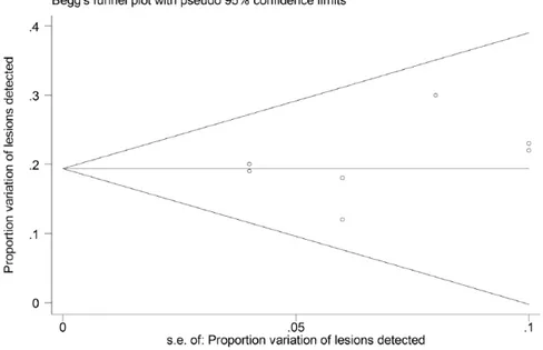
publication bias has been taken into considera-tion with the asymmetry Begg and Mazumdar adjusted rank correlation test [Egger et al. 1997] accompanied by the corresponding funnel plots. The statistical analysis was performed using the statistical software STATA version 13.
Limitations of the study
The lack of standard data collection methods among different papers makes it difficult to obtain undisputable conclusions. Some subgroups were too small for statistical analysis.
Results
The literature searches identified 154 publica-tions up to January 2015; reviewers excluded 130 of these on the basis of title or abstracts; 8 were rejected because they did not conform to inclu-sion criteria. A total of 16 publications were con-sidered eligible for analysis. Among the 16 evaluated papers, seven were unicentric and nine multicentric trials; eight studies were designed as prospective randomized trial and seven as pro-spective within-patient comparison; one more was an observational comparative controlled trial. The meta-analysis provided the funnel plot asso-ciated to the difference of lesions detected using BLC and WLC (Figure 1). The graph does not
involve particular concern about publication bias. The forest plot referred to lesions’ detection dis-plays the observed heterogeneity of the sample of studies which have been selected for the meta-analysis (Figure 2).
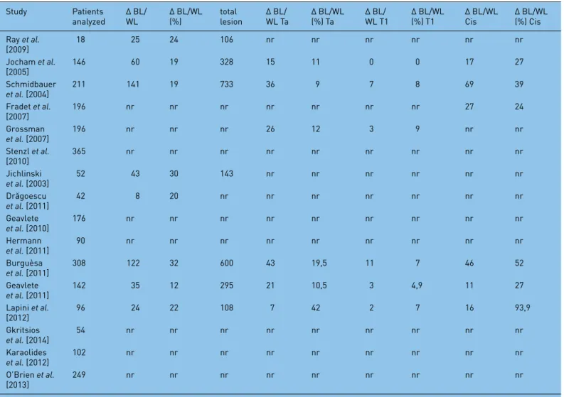
All selected papers and their descriptive baseline characteristics are shown in Table 1, while overall results of meta-analysis are reported in Table 2.
Tumor detection
The evaluation of tumor detection rates between different studies is difficult. This is due mainly to the lack of standard data collection methods: some studies reported only detection at patient level or at lesion level, some other have reported the overall detection or additional detection with BLC only.
We have subdivided results on tumor detection in two main groups: patient detection and lesion detection series. Additional subgroups were defined as: overall additional tumor detection rate with BLC and additional detection rates accord-ing to their histopathological findaccord-ing.
The majority of the evaluated papers have shown the superiority of HAL–PDD-guided cystoscopy over WLC alone in tumor detection. Tables 3 and 4 show comparison of tumor detection rates between BLC and WLC in the selected trials. Figure 1. Begg’s funnel plot for publication bias with the 95% confidence limits. Sample symmetry graphical assessment for the studies comparing the proportion of lesions detected using different lights.
Patient detection rates
Among the 16 evaluated papers, nine reported data on patient tumor detection as shown in Table 3. In 15% (95% confidence interval [CI] 0.098– 0.211) of patients at least one additional tumor was identified only by BLC (overall data on Ta, T1 or CIS). This benefit was observed in patients with Ta tumors (in 4% of patients, 95% CI 0.020– 0.066) and in patients with CIS/flat lesion (5.9%, 95% CI 0.021–0.096). Data on T1 tumor detec-tion rates were insufficient to obtain a statistically significant estimated pool.
The benefit of BLC was particularly significant in patients with Tis lesion and was reduced in papil-lary lesions.
Lesion detection rate
Lesion detection rates of selected studies are reported in Table 4.
A total of 2313 lesions were detected in the patients evaluated (lesion detected by BLC and/ or WLC).
Overall additional lesion detection rate was 19% (95% CI 0.152–0.236, Ta, T1 and Tis lesions). Six studies reported additional Ta tumor detec-tion rates with BLC, ranging from 9% to 42% of the total Ta tumors detected [Jocham et al. 2005; Schmidbauer et al. 2004; Grossman et al. 2007; Burguèsa et al. 2011; Geavlete et al. 2011; Lapini
et al. 2012], with a proportion difference of 5.9%
(95% CI 0.014–0.103) of additional lesion detection rates with BLC. The same studies reported additional T1 tumor detection rates ranging from 4.9% to 9% of the total T1 tumors detected [Schmidbauer et al. 2004; Fradet et al. 2007; Burguèsa et al. 2011; Geavlete et al. 2011; Lapini et al. 2012], only Jocham and colleagues showed no differences in T1 tumors detection rates [Jocham et al. 2005]. Proportion difference for T1 tumors was 1.2% (95% CI −0.033 to −0.057).
The detection of Tis lesions was reported in six studies [Jocham et al. 2005; Schmidbauer et al. 2004; Fradet et al. 2007; Burguèsa et al. 2011; Geavlete et al. 2011; Lapini et al. 2012] showing Figure 2. Forest plot for the graphical examination of the heterogeneity of the studies used to compare lesions’ detection using blue light and white light. The dashed line refers to the difference in the proportion of lesions detected using different lights.
greater additional detection rates with BLC, rang-ing from 24% to 94% of the total Tis lesions detected. Overall additional Tis lesions detection rate was 15.7% (95% CI 0.069–0.245).
Recurrence rates
Only seven papers have reported data on recur-rence rates in patients treated with BLC and WLC. Available recurrence rates are summarized in Table 5.
Recurrence was used to evaluate if an improve-ment in tumor detection could reduce recurrence rates up to 12 months. We have considered the recurrence rate as the number of patients with a recurrence at 3, 6 and 12 months divided by the total number of patients analyzed.
With this definition we have obtained a mean overall recurrence rate of 28.9% and 44.2% for
BLC and WLC, respectively. In addition, we have found a recurrence rate of 10.2% versus 18.1% at 3 months, a recurrence rate of 10.5% versus 22.3% at 6 months and 14.8% versus 32.3% at 12 months for BLC and WLC. Unfortunately, the data sample was insufficient for a statistical analy-sis, however, the evaluation of raw data showed a trend in favor of BLC.
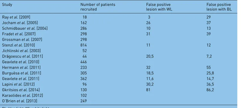
False positive detection rates
The false positive detection rate is the number of suspicious lesions that had negative histology divided by the total number of areas biopsied with each technique.
A T test on the pooled percentage values, weighted on the number of lesions detected in each study of the meta analysis, of the white light false posi-tive and blue light false posiposi-tive was statistically significant (p < 0.05), getting pooled percentage Table 1. Descriptive baseline characteristics of selected paper.
Study Number of patients
recruited Study design Study type Detection/recurrence
Ray et al. [2009] 18 Intra-patient comparison; WL
then BL Prospective D
Jocham et al. [2005] 162 Intra-patient comparison; WL
then BL Prospective multicenter D
Schmidbauer et al. [2004] 286 Intra-patient comparison; WL
then BL Prospective multicenter D
Fradet et al. [2007] 298 Intra-patient comparison; WL
then BL Prospective multicenter D
Grossman et al. [2007] 298 Intra-patient comparison; WL
then BL Prospective multicenter D
Stenzl et al. [2010] 814 Intra-patient comparison; WL
then BL Prospective multicenter D
Jichlinski et al. [2003] 52 Intra-patient comparison; WL
then BL Prospective multicenter D
Drăgoescu et al. [2011] 44 Randomised parallel groups Prospective D
Geavlete et al. [2010] 446 Randomised parallel groups Prospective D/R
Hermann et al. [2011] 233 Randomised parallel groups Prospective
multicenter D/R
Burguèsa et al. [2011] 305 Intra-patient comparison; WL
then BL Prospective multicenter D
Geavlete et al. [2011] 362 Intra-patient comparison; WL
then BL Prospective D/R
Lapini et al. [2012] 96 Intra-patient comparison; WL
then BL Prospective multicenter D
Gkritsios et al. [2014] 130 Randomized parallel groups Prospective D/R
Karaolides et al. [2012] 102 Randomized parallel groups Prospective R
O’Brien et al. [2013] 249 Randomized parallel groups Prospective D/R
Table 2. Meta-analysis summary results.
Outcome Δ Random effects model Model variability Publication
bias
H0: Δ = 0 Pooled Δ Δ CI 95% (χ2, p)(II) τ2(III) Asymmetry
test (z;p)(IV)
Proportion difference of patients with at least one more lesion detected with BL Δ BL/WL 0.000 0.150 [0.098–0.211] 131.96; 0.00 0.0061 0.42; 0.677 Δ BL/WL Ta 0.000 0.040 [0.020–0.066] 12.08; 0.007 0.0041 1.34; 0.180 Δ BL/WL T1 Few data Δ BL/WL Cis 0.002 0.059 [0.021–0.096] 28.5; 0.000 0.0014 1.57; 0.117 Number of lesions detected proportion difference PD lesions detected(I) 0.000 0.190 [0.152–0.236] 3.56; 0.736 0.0022 0.93; 0.351 PD Ta 0.011 0.059 [0.014–0.103] 0.26; 0.992 0.0011 −0.52; 0.602 PD T1 0.590 0.012 [−0.033 to −0.057] 0.09; 0.976 0.0021 0.02; 0.997 PD Cis 0.000 0.157 [0.069–0.245] 13.12; 0.022 0.0073 0.23; 0.822
BL, blue light; WL, white light; Δ, difference; PD, proportion difference.
Table 3. Differential of patient detection rate for BL versus WL cystoscopy (at least one more lesion detected).
Study Patients
analyzed Δ BL/WL Δ BL/WL (%) Δ BL/WL Ta Δ BL/WL (%) Ta Δ BL/WL T1 Δ BL/WL (%) T1 Δ BL/WL Cis Δ BL/WL (%) Cis
Ray et al. [2009] 18 8 44 nr nr nr nr nr nr Jocham et al. [2005] 146 28 19 13 20 1 6 12 41 Schmidbauer et al. [2004] 211 nr nr nr nr nr nr 18 28 Fradet et al. [2007] 196 nr nr nr nr nr nr nr 5 Grossman et al. [2007] 196 31 29 nr nr nr nr nr nr Stenzl et al. [2010] 365 nr nr 41 16 8 13 19 46 Jichlinski et al. [2003] 52 10 23 nr nr nr nr 9 69 Drăgoescu et al. [2011] 42 nr nr nr nr nr nr nr nr Geavlete et al. [2010] 176 18 10,3 12 10,6 1 2,6 5 21,8 Hermann et al. [2011] 90 44 49 nr 45 nr 43 nr nr Burguèsa et al. [2011] 308 72 23,6 nr nr nr nr nr nr Geavlete et al. [2011] 142 14 9,8 7 8 2 5 5 23 Lapini et al. [2012] 96 nr nr nr nr nr nr nr nr Gkritsios et al. [2014] 54 16 30 nr nr nr nr nr nr Karaolides et al. [2012] 102 nr nr nr nr nr nr nr nr O’Brien et al. [2013] 249 nr nr nr nr nr nr nr 12
estimates of 15.65 (SD = 7.06) for the white light and 22.35 (SD = 9.24) for the blue light.
We found a large variation in the false positive detection rate among centers for BLC and WLC (range 7–86% and 3–81%, respectively) as shown in Table 6. This may be explained by a learning curve with the fluorescence technique.
Discussion
Since the first report on PDD by Kriegmair and colleagues in 1994 [Kriegmair et al. 1994], many expectations with regard to improved diagnosis of bladder tumors have been met. Most of the initial reports on the clinical use of PDD in NMIBC showed that the tumor detection rate was increased at about 20% by this new method, and that recurrence, i.e. persister, rates in most cases were reduced on follow-up cystoscopy in the
same amount [Dindyal et al. 2008; Mowatt et al. 2011]. It was also concluded from these findings that a consistent reduction of bladder tumor recurrences would help to significantly reduce the costs for bladder cancer treatment [Dindyal et al. 2008].
After two decades of PDD, reevaluation of its efficacy in the therapy of NMIBC can shed light on the present position of fluorescence endos-copy in global treatment concepts. The results of the present meta-analysis confirm the increased tumor detection rate by BLC in about 20%. The most pronounced benefit was demon-strable for CIS, with a superior detection rate ranging from 24% to 94% compared with WLC. This has been reported by other investigators before [Isfoss, 2011; Kausch et al. 2010]. The benefit for papillary lesions was also demonstra-ble, but minor compared with CIS: it was 9–42% Table 4. Differential of lesion detection rate with BL and WL cystoscopy.
Study Patients
analyzed Δ BL/WL Δ BL/WL (%) total lesion Δ BL/WL Ta Δ BL/WL (%) Ta Δ BL/WL T1 Δ BL/WL (%) T1 Δ BL/WL Cis Δ BL/WL (%) Cis
Ray et al. [2009] 18 25 24 106 nr nr nr nr nr nr Jocham et al. [2005] 146 60 19 328 15 11 0 0 17 27 Schmidbauer et al. [2004] 211 141 19 733 36 9 7 8 69 39 Fradet et al. [2007] 196 nr nr nr nr nr nr nr 27 24 Grossman et al. [2007] 196 nr nr nr 26 12 3 9 nr nr Stenzl et al. [2010] 365 nr nr nr nr nr nr nr nr nr Jichlinski et al. [2003] 52 43 30 143 nr nr nr nr nr nr Drăgoescu et al. [2011] 42 8 20 nr nr nr nr nr nr nr Geavlete et al. [2010] 176 nr nr nr nr nr nr nr nr nr Hermann et al. [2011] 90 nr nr nr nr nr nr nr nr nr Burguèsa et al. [2011] 308 122 32 600 43 19,5 11 7 46 52 Geavlete et al. [2011] 142 35 12 295 21 10,5 3 4,9 11 27 Lapini et al. [2012] 96 24 22 108 7 42 2 7 16 93,9 Gkritsios et al. [2014] 54 nr nr nr nr nr nr nr nr nr Karaolides et al. [2012] 102 nr nr nr nr nr nr nr nr nr O’Brien et al. [2013] 249 nr nr nr nr nr nr nr nr nr
for pTa tumors and 0–9% for pT1 tumors. These findings seem to be in close line with clin-ical experience: while flat lesions are difficult to detect or discriminate from benign morphologic changes of the urothelium, exophytic tumors are
endoscopically more identifiable, except when they are very tiny or multilocular. In these cases, when single small lesions may be missed, PDD is a valuable tool for diagnosis and prevention of tumor persistence.
Table 5. Recurrence rate for BL and WL cystoscopy.
Study Patients
analyzed Overall BL recurrence (%) Overall WL recurrence (%) Δ BL/WL recurrence 3 month (%) Δ BL/WL recurrence 6 month (%) Δ BL/WL recurrence 12 month (%) Ray et al. [2009] 18 nr nr nr nr nr Jocham et al. [2005] 146 nr nr nr nr nr Schmidbauer et al. [2004] 211 nr nr nr nr nr Fradet et al. [2007] 196 nr nr nr nr nr Grossman et al. [2007] 196 nr nr nr nr nr Stenzl et al. [2010] 365 47 56 nr nr nr Jichlinski et al. [2003] 52 nr nr nr nr nr Drăgoescu et al. [2011] 42 nr nr 9,1 13,6 27,3 Geavlete et al. [2010] 176 11,1 31,2 nr nr nr Hermann et al. [2011] 90 30 47 14 nr 31 Burguèsa et al. [2011] 308 nr nr nr nr nr Geavlete et al. [2011] 142 31 45 9 10 10,9 Lapini et al. [2012] 96 nr nr nr nr nr Gkritsios et al. [2014] 54 37,5 46 nr nr 0,8 Karaolides et al. [2012] 102 17 40 10,9 nr 34,7 O’Brien et al. [2013] 249 nr nr −3 nr 6
BL, blue light; WL, white light; Δ, difference; nr, not reported.
Table 6. Comparison of false positive detection rates between WL and BL cystoscopy.
Study Number of patients
recruited False positive lesion with WL False positive lesion with BL
Ray et al. [2009] 18 3 29 Jocham et al. [2005] 162 26 37 Schmidbauer et al. [2004] 286 10 13 Fradet et al. [2007] 298 31 39 Grossman et al. [2007] 298 Stenzl et al. [2010] 814 11 12 Jichlinski et al. [2003] 52 Drăgoescu et al. [2011] 44 20,5 7,2 Geavlete et al. [2010] 446 Hermann et al. [2011] 233 32 55 Burguèsa et al. [2011] 305 18,5 25,8 Geavlete et al. [2011] 362 11,6 14,7 Lapini et al. [2012] 96 30,2 36,5 Gkritsios et al. [2014] 130 81 86,2 Karaolides et al. [2012] 102 O’Brien et al. [2013] 249
Although not statistically significant, the improved tumor detection rate with BLC was well reflected by a decrease of tumor recurrences at 3, 6 and 12 months in the present meta-analysis. The differ-ence was 8%, 12% and 17.5%, respectively. This may be the most relevant finding, since it defi-nitely reduces the number of consecutive opera-tions and the surgery-related morbidity for patients and the costs for the health care system. While expert panels recommend the use of PDD for bladder tumor resection as well in the initial as in the follow-up situation [Malmström et al. 2012; Witjes et al. 2010], this method has not been integrated in the EAU guidelines yet [Babjuk et al. 2013]. The main reason, besides single studies that failed to demonstrate a signifi-cant benefit in recurrence rates with BLC, may be that tumor progression rates have not shown any reduction compared with standard WLC resection. While pTa tumors rarely progress to muscle-invasive disease, and pT1 tumors are also detectable with WLC in most cases, there is a broad consensus that CIS with its potential for stage progression is a domain of BLC at present. Thus, it is surprising, that progression rates have not been markedly improved in the publications on BLC hitherto.
What is the future of PDD? Throughout the last years, novel imaging technologies have been introduced in bladder cancer diagnosis [Lerner
et al. 2015]: narrow-band imaging (NBI), optical
coherence tomography (OCT) and confocal laser endoscopy (CLE) have the potential to detect bladder tumors beyond the borders of macro-scopic visibility. A meta-analysis for NBI showed improved tumor detection rates similar to PDD at about 20% [Li et al. 2013]. Data on improvement and accuracy of tumor detection by OCT and CLE are still lacking [Liu et al. 2012]. These tech-nologies may be used alone or in combination with PDD [Gladkova et al. 2013]. However, they will have to prove equivalence or superiority to PDD, not only in tumor detection rates, but also in recurrence and, possibly, progression rates, as well as cost efficiency, before they may be accepted for clinical routine.
From a patient’s and urologist’s perspective, there is no argument not to use the best available tools to visualize bladder tumors during resection, as no surgeon would perform surgery without his glasses. Even if the benefit of PDD is restricted to only part of the patients, cost efficacy in the course of disease
is a compelling argument for the use of fluores-cence endoscopy for bladder tumor resection. Several new photosensitizing agents and imaging technologies have been developed for improved visibility and detectability of bladder cancer. Hypericin is a photosensitizer with promising applications in photodynamic diagnosis for blad-der cancer, and can be used with the same imag-ing system that is used for the porphyrin-related substrates [Vandepitte et al. 2011].
Technologic improvements may further enhance our ability to detect and stage bladder tumors and distinguish benign from malignant disease [Lerner
et al. 2015; Zlatev et al. 2015]: NBI is a macroscopic
imaging modality that improves WLC by providing increased contrast between normal and abnormal tissue on the basis of neovascularity. NBI cystoscopy has been shown to improve detection rates of blad-der tumors [Cauberg et al. 2010], and is associated with lower recurrence rates and longer recurrence-free survival (RFS) time [Herr et al. 2011] than con-ventional WLC. A single-surgeon randomized study comparing 2-year RFS of patients with NMIBC using NBI or WLC showed that NBI cystoscopy improves completeness of bladder tumor resection, which reduces the frequency of early and late tumor recurrences [Herr, 2015].
OCT, a real-time and high-resolution imaging technology, delineates subsurface microarchitec-ture information from bladder lesions. It has the ability to discriminate between benign or malig-nant lesions [Manyak et al. 2005] and noninvasive and invasive cancers [Goh et al. 2008].
Molecular imaging associates optical imaging technologies with cancer-specific molecular agents to improve the specificity of disease detec-tion [Lerner et al. 2015; Zlatev et al. 2015]. CLE, as OCT, allows in situ tissue characterization with high resolution [Sonn et al. 2009].
The combination of these macroscopic and microscopic visualization techniques has the abil-ity for significant improvement of bladder tumor detection and in situ histological characterization [Lerner et al. 2015].
Additional imaging techniques that currently under development, such as endoluminal high-frequency ultrasound [Yuan et al. 2008], time- and spectral-resolved two-photon imaging [Cicchi
et al. 2010] and coherent anti-Stokes Raman
scat-tering microscopy [Gao et al. 2011], might also become procedures for bladder tumor detection, diagnosis and staging in future.
To improve the results in NMIBC primary objec-tives are to have more accurate diagnostics and more effective therapies. In a more favorable eco-nomic conditions, such as the one before the financial collapse of 2008, it should make availa-ble to every department of urology the best diag-nostic technology (i.e. fluorescence cystoscopy) and the best therapeutic technologies (i.e. intra-vesical electro-osmotic mitomycin [Di Stasi et al. 1999, 2006, 2011]).
Funding
This research received no specific grant from any funding agency in the public, commercial, or not-for-profit sectors.
Conflict of interest statement
The authors declare that there are no conflicts of interest.
References
Babjuk, M., Burger, M., Zigeuner, R., Shariat, S., van Rhijn, B., Compérat, E. et al. (2013) EAU guidelines on non-muscle-invasive urothelial carcinoma of the bladder: update 2013. Eur Urol 64: 639–653.
Burguèsa, J., Condeb, G., Olivac, J., Abascald, I., Iborrae, M., Puertasf, F. et al. (2011) Hexaminolevulinate photodynamic diagnosis in non-muscle invasive bladder cancer: experience of the BLUE group [in Spanish]. Actas Urol Esp 35: 439–445.
Cauberg, E., Kloen, S., Visser, M., de la Rosette, J., Babjuk, M., Soukup, V. et al. (2010) Narrow band imaging cystoscopy improves detection of non-muscle-invasive bladder cancer. Urology 76: 658–663. Cicchi, R., Crisci, A., Cosci, A., Nesi, G.,
Kapsokalyvas, D., Giancane, S. et al. (2010) Time- and Spectral-resolved two-photon imaging of healthy bladder mucosa and carcinoma in situ. Opt Express 18: 3840–3849.
Dindyal, S., Nitkunan, T. and Bunce, C. (2008) The economic benefit of photodynamic diagnosis in non-muscle invasive bladder cancer. Photodiagnosis
Photodyn Ther 5: 153–158.
Di Stasi, S., Giannantoni, A., Massoud, R., Dolci, S., Navarra, P., Vespasiani, G. et al. (1999)
Electromotive versus passive diffusion of mitomycin C into human bladder wall: concentration-depth profiles studies. Cancer Res 59: 4912–4918.
Di Stasi, S., Giannantoni, A., Capelli, G., Giurioli, A., Valenti, M., Zampa, G. et al. (2006) Sequential BCG and electromotive mitomycin C versus BCG alone for high risk superficial bladder cancer: a randomised controlled trial. Lancet Oncol 7: 43–51. Di Stasi, S., Valenti, M., Verri, C., Liberati, E., Giurioli, A., Leprini, G. et al. (2011) Electromotive instillation of mitomycin immediately before transurethral resection for patients with primary urothelial non-muscle invasive bladder cancer: a randomised controlled trial. Lancet
Onocol 12: 871–879.
Drăgoescu, O., Tomescu, P., Pănus, A., Enache, M., Maria, C., Stoica, L. et al. (2011) Photodynamic diagnosis of non-muscle invasive bladder cancer using hexaminolevulinic acid. Rom J Morphol Embryol 52: 123–127.
Egger, M., Davey Smith, G., Schneider, M. and Minder, C. (1997) Bias in meta-analysis detected by a simple, graphical test. BMJ 315: 629–624.
Ferlay, J., Soerjomataram, I., Ervik, M., Dikshit, R., Eser, S., Mathers, C. et al. (2012) GLOBOCAN
2012 v1.0, Cancer Incidence and Mortality Worldwide: IARC CancerBase No. 11. Lyon, France: International
Agency for Research on Cancer.
Fradet, Y., Grossman, H., Gomella, L., Lerner, S., Cookson, M., Albala, D. et al. (2007) A comparison of hexaminolevulinate fluorescence cystoscopy and white light cystoscopy for the detection of carcinoma
in situ in patients with bladder cancer: a phase III,
multicenter study. J Urol 178: 68–73.
Gao, L., Zhou, H., Thrall, M., Li, F., Yang, Y., Wang, Z. et al. (2011) Label-free high-resolution imaging of prostate glands and cavernous nerves using coherent anti-Stokes Raman scattering microscopy.
Biomed Opt Express 2: 915–926.
Gaullier, J., Berg, K., Peng, Q., Anholt, H., Selbo, P., Ma, L. et al. (1997) Use of 5-aminolevulinic acid esters to improve photodynamic therapy on cells in culture. Cancer Res 57: 1481–1486.
Geavlete, B., Jecu, M., Multescu, R., Georgescu, D. and Geavlete, P. (2010) HAL blue-light cystoscopy in high-risk nonmuscle-invasive bladder cancer–re-TURBT recurrence rates in a prospective, randomized study. Urology 76: 664–669.
Geavlete, B., Multescu, R., Georgescu, D., Jecu, M., Stanescu, F. and Geavlete, P. (2011) Treatment changes and long-term recurrence rates after hexaminolevulinate (HAL) fluorescence cystoscopy: does it really make a difference in patients with non-muscle-invasive bladder cancer (NMIBC)? BJU Int 109: 549–556.
Gkritsios, P., Hatzimouratidis, K., Kazantzidis, S., Dimitriadis, G., Ioannidis, E. and Katsikas, V. (2014) Hexaminolevulinate-guided transurethral resection of non-muscle-invasive bladder cancer does not reduce the recurrence rates after a 2-year follow-up: a prospective randomized trial. Int Urol Nephrol 46: 927–933.
Gladkova, N., Kiseleva, E., Streltsova, O., Prodanets, N., Snopova, L., Karabut, M. et al. (2013) Combined use of fluorescence cystoscopy and cross-polarization OCT for diagnosis of bladder cancer and correlation with immunohistochemical markers. J Biophotonics 6: 687–698.
Goh, A., Tresser, N., Shen, S. and Lerner, S. (2008) Optical coherence tomography as an adjunct to white light cystoscopy for intravesical real-time imaging and staging of bladder cancer. Urology 72: 133–137.
Grossman, H., Gomella, L., Fradet, Y., Morales, A., Presti, J., Ritenour, C. et al. (2007) A phase III, multicenter comparison of hexaminolevulinate fluorescence cystoscopy and white light cystoscopy for the detection of superficial papillary lesions in patients with bladder cancer. J Urol 178: 62–67.
Hall, M., Chang, S., Dalbagni, G., Pruthi, R., Seigne, J., Skinner, E. et al. (2007) Guideline for the management of nonmuscle invasive bladder cancer (stages Ta, T1, and Tis): 2007 update. J Urol 178: 2314–2330.
Herr, H. (2015) Randomized trial of narrow-band versus white-light cystoscopy for restaging (second-look) transurethral resection of bladder tumors. Eur
Urol 67: 605–608.
Herr, H. and Donat, S. (2008) Quality control in transurethral resection of bladder tumours. BJU Int 102: 1242–1246.
Herr, H. and Donat, S. (2011) Reduced bladder tumor recurrence rate associated with narrow-band imaging surveillance cystoscopy. BJU Int 107: 396–398.
Hermann, G., Mogensen, K., Carlsson, S., Marcussen, N. and Duun, S. (2011) Fluorescence-guided transurethral resection of bladder tumours reduces bladder tumour recurrence due to less residual tumour tissue in Ta/T1 patients: a randomized two-centre study. BJU Int 108: E297– E303.
Higgins, J., Thompson, S., Deeks, J. and Altman, D. (2003) Measuring inconsistency in meta-analyses.
BMJ 327: 557–560.
Isfoss, B. (2011) The sensitivity of fluorescent-light cystoscopy for the detection of carcinoma in situ (CIS) of the bladder: a meta-analysis with comments on gold standard. BJU Int 108: 1703–1707.
Jichlinski, P., Guillou, L., Karlsen, S., Malmström, P., Jocham, D., Brennhovd, B. et al. (2003) Hexyl aminolevulinate fluorescence cystoscopy: new
diagnostic tool for photodiagnosis of superficial bladder cancer – a multicenter study. J Urol 170: 226–229. Jocham, D., Witjes, F., Wagner, S., Zeylemaker, B., van Moorselaar, J., Grimm, M. et al. (2005) Improved detection and treatment of bladder cancer using hexaminolevulinate imaging: a prospective, phase III multicenter study. J Urol 174: 862–866.
Karaolides, T., Skolarikos, A., Bourdoumis, A., Konandreas, A., Mygdalis, V., Thanos, A. et al. (2012) Hexaminolevulinate-induced fluorescence versus white light during transurethral resection of noninvasive bladder tumor: does it reduce recurrences? Urology 80: 354–359.
Kausch, I., Sommerauer, M., Montorsi, F., Stenzl, A., Jacqmin, D., Jichlinski, P. et al. (2010) Photodynamic diagnosis in non-muscle-invasive bladder cancer: a systematic review and cumulative analysis of prospective studies. Eur Urol 57: 595–606.
Klem, B., Lappin, G., Nicholson, S., van de Wetering, J., de Vries, D., Oosterhuis, B. et al. (2006) Determination of the bioavailability of [14C]-hexaminolevulinate using accelerator mass spectrometry after intravesical administration to human volunteers. J Clin Pharmacol 46: 456–460. Kriegmair, M., Baumgartner, R., Knuechel, R., Steinbach, P., Ehsan, A., Lumper, W. et al. (1994). Fluorescence photodetection of neoplastic urothelial lesions following intravesical instillation of 5-aminolevulinic acid. Urology 44: 836–841.
Lapini, A., Minervini, A., Masala, A., Schips, L., Pycha, A., Cindolo, L. et al. (2012) A comparison of hexaminolevulinate (Hexvix(®)) fluorescence cystoscopy and white-light cystoscopy for detection of bladder cancer: results of the HeRo observational study. Surg Endosc 26: 3634–3641.
Lerner, S. and Goh, A. (2015) Novel endoscopic diagnosis for bladder cancer. Cancer 121: 169–178. Li, K., Lin, T., Fan, X., Duan, Y. and Huang, J. (2013) Diagnosis of narrow-band imaging in non-muscle-invasive bladder cancer: a systematic review and meta-analysis. Int J Urol 20: 602–609.
Liu, J., Droller, M. and Liao, J. (2012) New optical imaging technologies for bladder cancer: considerations and perspectives. J Urol 188: 361–368. Malmström, P., Grabe, M., Haug, E., Hellström, P., Hermann, G., Mogensen, K. et al. (2012) Role of hexaminolevulinate-guided fluorescence cystoscopy in bladder cancer: critical analysis of the latest data and European guidance. Scand J Urol Nephrol 46: 108–116.
Manyak, M., Gladkova, N., Makari, J., Schwartz, A., Zagaynova, E., Zolfaghari, L. et al. (2005) Evaluation of superficial bladder transitional-cell carcinoma by optical coherence tomography. J Endourol 19: 570–574. Marti, A., Jichlinski, P., Lange, N., Ballini, J., Guillou, L., Leisinger, H. et al. (2003) Comparison of aminolevulinic acid and hexylester aminolevulinate induced protoporphyrin IX distribution in human bladder cancer. J Urol 170: 428–432.
Miyake, M., Ishii, M., Kawashima, K., Kodama, T., Sugano, K., Fujimoto, K. et al. (2009) siRNA-mediated knockdown of the heme synthesis and degradation pathways: modulation of treatment effect of 5-aminolevulinic acid-based photodynamic therapy in urothelial cancer cell lines. Photochem Photobiol 85: 1020–1027.
Mowatt, G., N’Dow, J., Vale, L., Nabi, G., Boachie, C., Cook, J. et al. (2011). Photodynamic diagnosis of bladder cancer compared with white light cystoscopy: Systematic review and meta-analysis. Int J Technol
Assess Health Care 27: 3–10.
O’Brien, T., Ray, E., Chatterton, K., Khan, M., Chandra, A. and Thomas, K. (2013) Prospective randomized trial of hexylaminolevulinate photodynamic-assisted transurethral resection of bladder tumour (TURBT) plus single-shot intravesical mitomycin C vs conventional white-light TURBT plus mitomycin C in newly presenting non-muscle-invasive bladder cancer. BJU Int 112: 1096–1104.
Peng, Q., Berg, K., Moan, J., Kongshaug, M. and Nesland, J. (1997) 5-Aminolevulinic acid-based photodynamic therapy: principles and experimental research. Photochem Photobiol 65: 235–251.
Ray, E., Chatterton, K., Thomas, K., Khan, M., Chandra, A. and O’Brien, T. (2009)
Hexylaminolevulinate photodynamic diagnosis for multifocal recurrent nonmuscle invasive bladder cancer. J Endourol 23: 983–988.
Schmidbauer, J., Witjes, F., Schneller, M., Donat, R., Susani, M. and Marberger, M. (2004) Improved detection of urothelial carcinoma in situ with hexaminolevulinate fluorescence cystoscopy. J Urol 171: 135–138.
Sonn, G., Jones, S., Tarin, T., Du, C., Mach, K., Jensen, K. et al. (2009) Optical biopsy of human bladder neoplasia with in vivo confocal laser endomicroscopy. J Urol 182: 1299–1305.
Stenzl, A., Burger, M., Fradet, Y., Mynderse, L., Soloway, M., Witjes, J. et al. (2010) Hexaminolevulinate guided fluorescence cystoscopy reduces recurrence in patients with non-muscle invasive bladder cancer. J Urol 184: 1907–1913.
Stokes, G. (1852) On the change of refrangibility of light. Phil Trans R Soc Lond 142: 463–562.
Svatek, R., Hollenbeck, B., Holmäng, S., Lee, R., Kim, S., Stenzl, A. et al. (2014) The economics of bladder cancer: costs and considerations of caring for this disease. Eur Urol 66: 253–262.
Svatek, R., Lee, D. and Lotan, Y. (2005) Correlation of office-based cystoscopy and cytology with histologic diagnosis: how good is the reference standard? Urology 66: 65–68.
Sylvester, R., van der Meijden, A., Oosterlinck, W., Witjes, J., Bouffioux, C., Denis, L. et al. (2006) Predicting recurrence and progression in individual patients with stage Ta T1 bladder cancer using EORTC risk tables: a combined analysis of 2596 patients from seven EORTC trials. Eur Urol 49: 466–475.
Vandepitte, J., Van Cleynenbreugel, B., Hettinger, K., Van Poppel, H. and de Witte, P. (2011) Biodistribution of PVP-hypericin and hexaminolevulinate-induced PpIX in normal and orthotopic tumorbearing rat urinary bladder. Cancer
Chemother Pharmacol 67: 775–781.
Wagnieres, G., Star, W. and Wilson, B. (1998) ln vivo fluorescence spectroscopy and imaging for oncological applications. Photochem Photobiol 68: 603–632. Witjes, J., Redorta, J., Jacqmin, D., Sofras, F., Malmström, P., Riedl, C. et al. (2010)
Hexaminolevulinate-guided fluorescence cystoscopy in the diagnosis and follow-up of patients with non-muscle-invasive bladder cancer: review of the evidence and recommendations. Eur Urol 57: 607–614.
Yuan, Z., Wang, Z., Pan, R., Liu, J., Cohen, H. and Pan, Y. (2008) High-resolution imaging diagnosis and staging of bladder cancer: comparison between optical coherence tomography and high-frequency ultrasound [serial online]. J Biomed Opt 13: 054007.
Zlatev, D., Altobelli, E. and Liao, J. (2015) Advances in imaging technologies in the evaluation of high-grade bladder cancer. Urol Clin North Am 42: 147–157.
Visit SAGE journals online http://tau.sagepub.com