1 © The Author 2015. Published by Oxford University Press on behalf of the European Orthodontic Society. All rights reserved.
For permissions, please email: [email protected]
Original Article
Bonded versus banded rapid palatal expander
followed by facial mask therapy: analysis on
digital dental casts
Roberta Lione
*
, Luis Tomas Huanca Ghislanzoni
**
,Efisio Defraia
***
,
Lorenzo Franchi
***
and Paola Cozza
*
*Department of Clinical Sciences and Translational Medicine, University of Rome “Tor Vergata”, Italy, **Department of Biomedical Sciences and Health, University of Milan, Italy, ***Department of Surgery and Translational Medicine, University of Florence, Italy
Correspondence to: Lorenzo Franchi, Department of Surgery and Translational Medicine, University of Florence, Via del Ponte di Mezzo, 46-48, 50127 Florence, Italy. E-mail: [email protected]
Summary
Objectives: To compare the dental effects produced by a bonded versus a banded expander
combined with facial mask (FM) in patients with Class III malocclusion by means of digital dental casts.
Materials and methods: Two groups of patients with Class III malocclusion and maxillary transverse
deficiency in the deciduous or early mixed dentition were selected. The first group consisted of 25 subjects (12 females; 13 males) with a mean age of 7.4 years (SD 1.2 years) treated with a bonded expander and FM. The second group consisted of 25 subjects (13 females; 12 males) with a mean age of 8.1 years (SD 1.3 years) treated with a banded expander and FM. For each subject of the two groups, initial (pre-treatment, T1) and final (post-treatment, T2) dental casts were taken and scanned. Maxillary digital models of T1 and T2 were superimposed on the palatal rugae in order to analyse the maxillary anchorage loss. Significant between-group differences were tested with independent sample t-test (P < 0.05).
Results: No statistical differences were found for any of the variables observed.
Conclusion: Orthopaedic treatment of Class III malocclusion with either a bonded or a banded
expander and FM during the deciduous or early mixed dentition induced a significant expansion of the maxillary arch and a slight mesialization of the posterior anchoring teeth with no difference between the two intraoral appliance designs.
Introduction
In recent years, rapid maxillary expansion and facial mask (RME/ FM) therapy has become a common technique to correct Class III malocclusion and the treatment of choice in cases where the etiology of Class III malocclusion is maxillary deficiency (1). Recent data on the long-term effects of RME/FM indicate that the outcome of ortho-paedic treatment of Class III malocclusion is favourable when it is started during the early developmental phases (2). Numerous studies showed that the RME/FM protocol is able to produce significant
craniofacial changes consisting of maxillary advancement combined with a downward and backward movement of the mandible (3–5). In the assessment of overall efficiency for RME/FM treatment, an important variable is the dental component of the correction of Class III malocclusion. To protract the maxilla effectively, the force should be applied to the maxilla as a unit. Since the intraoral appli-ance delivers the force to the maxilla through the elastics attached to the FM, a properly designed appliance is critical for the efficacy of maxillary protraction (6, 7). Several investigation methods such as lateral headfilms, dental casts, and digital dental casts have been
European Journal of Orthodontics, 2015, 1–6
doi:10.1093/ejo/cjv038
by guest on July 18, 2015
used during the last 20 years to examine dental modifications in the maxilla induced by different designs of both RME and FM (6, 8–11). Baik et al. (12) compared the different effects of various intraoral appliances for maxillary protraction, pointing out a more forward movement of the maxillary molars in the RME/FM group than in the group treated with labiolingual arch/FM (12). Tortop et al. (7) showed no significant antero-posterior dental change in prepuberal Class III subjects treated with RME or with lingual arch and maxil-lary protraction. The skeletal contributions to Class III correction were statistically significant, but the dental contribution was similar for both anchorage devices (7).
No previous study evaluated the dental effects of RME/FM pro-tocol comparing the bonded RME with the banded RME by means of digital dental casts. A difference in anchorage loss (AL) between the two devices for RME can be hypothesized on the basis of the dif-ferent number of teeth included in the anchorage units. With respect to plaster dental casts, digital dental models have many advantages, such as storage, transferability, communication, remote diagnosis, and treatment result evaluation. The validity and precision of digi-tal dendigi-tal models have been widely studied, and numerous investi-gations have shown that 3D digital models can be used for model analysis, diagnosis, and treatment planning (13–15). By superimpos-ing pre-treatment and post-treatment models on stable structures as palatal rugae, it is possible to quantify the exact amount of dental movement as results of orthopaedic and orthodontic treatment (16,
17).
The aim of this retrospective study, therefore, was to analyse the dental effects produced by a bonded versus a banded expander com-bined with FM in prepubertal Class III patients by means of digital dental casts.
Subjects and methods
Two groups of patients with Class III malocclusion were selected from the files of the Departments of Orthodontics of the Universities of Rome Tor Vergata and of Florence. The inclusion criteria were the following: European ancestry (white), anterior crossbite or edge-to-edge incisor relationship, Class III molar relationship, Wits appraisal of minus 2.0 mm or more negative, absence of Centric Occlusion-Centric Relation discrepancy (indicating pseudo-Class III maloc-clusion), negative posterior transverse inter-arch discrepancy (18), prepubertal skeletal maturation (CS1–CS2) (19), and deciduous or early mixed dentition. The exclusion criteria were the following: late mixed dentition, cleft lip and/or palate, and other genetic dis-eases. This project was approved by the Ethical Committee at the University of XXXXX (Protocol number: 9314) and informed con-sent was obtained from the patients’ parents.
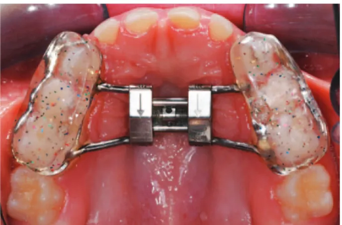
The first group consisted of 25 subjects (12 females; 13 males) with a mean age of 7.4 years (SD 1.2 years) treated consecutively with a bonded expander (Figure 1) and FM (bonded group) at the University of XXXXX (from September 2007 through January 2012). The second group consisted of 25 subjects (13 females; 12 males) with a mean age of 8.1 years (SD 1.3 years) treated con-secutively at the University of XXXXX with a banded expander
(Figure 2), removable posterior bite blocks in the lower arch (20),
and FM (from September 2011 through February 2014). Patients of the bonded RME/FM group received an acrylic splint expander with splints extending from the deciduous canine to the second decidu-ous molar (21). Patients of the banded RME/FM group received a butterfly expander with bands either on second deciduous molars or on the first permanent molars (22). For both appliance designs,
vestibular hooks were present at the level of the deciduous canines. Patients’ parents of both groups were instructed to activate the pala-tal expander 1/4 of a turn per day until overcorrection of the trans-verse width was achieved (palatal cusps of the upper posterior teeth approximating the buccal cusps of the lower posterior teeth). The screw was activated in both groups for an average period of 3 weeks.
At the end of active expansion, patients were given FMs with pads fitted to the chin and forehead for support. Elastics were attached from the soldered hooks on the expander to the support bar of the FM in a downward and forward direction (about 30 degrees to the occlusal plane). Elastics of increasing force were used until a heavy orthopaedic force (400 g per side) was produced. At the time of delivery of the FM, bilateral 3/8 inch 8 ounce elastics typically were used for the first 1–2 weeks of treatment to ease the patient’s adjustment to the appliance. The force generated was then increased with the use of 1/2 inch 14 ounce elastics, and finally 5/16 inch 14 ounce elastics. Patients were asked to wear the FM for 14 hours per day. All patients were treated at least to a positive overjet before discontinuing treatment. Most patients were overcorrected towards a Class II occlusal relationship. The average duration of RME/FM treatment was 1.1 years ± 5 months in both groups.
For each subject of the two groups, dental casts were taken before (pre-treatment, T1) and at the end of RME/FM therapy (post-treatment, T2). In order to analyse the maxillary dentoalveolar struc-tures, the maxillary dental casts were scanned with a tridimensional Figure 1. The bonded expander.
Figure 2. The banded expander.
by guest on July 18, 2015
scanner (D800, 3Shape A/S, Copenhagen K, Denmark, scan time 25 seconds, resolution two cameras 5.0 megapixels, ultra high point accuracy less than 15 μm). Each cast was scanned from 10 or more views that were then combined and rendered into three-dimensions by using a specific software (3Shape-ScanItOrthodontics™ 2010-2p3, 3Shape A/S, Copenhagen K, Denmark). The virtual three-dimensional models were measured with a specific software (VAM, Vectra, Canfield Scientific, Fairfield, New Jersey, USA).
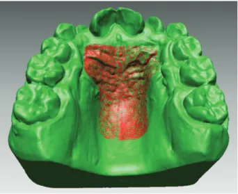
Maxillary digital models taken at T1 and at T2 were superim-posed using the palatal rugae as stable reference structures (16). Superimposition on the palatal rugae area was performed with a double-step procedure. First, 15 points were placed on the T1 and T2 casts on the second and third palatal rugae (three points per rugae per side) and on the palatal raphe between the palatal rugae (Figure 3). A first superimposition through minimization of the distance between homologous points was performed. Then, a T-shape area comprising the palatal rugae and extending posteriorly along the palatal raphe was selected and used to refine the superimposition through a surface-to-surface approach that enhances superimposition precision (Figure 4).
After digital casts were superimposed, a common reference plane (x,
y) parallel to the occlusal plane of the T1 cast was defined (Figure 5) (23). To perform dental arch measurements, homologous points were placed on the centre of the distal marginal crests of right and left sec-ond deciduous molars (ER and EL, respectively). An average point between the two mesial points of the central incisors was then calcu-lated (1-1) and served as anterior reference.
Transversal measurements
1. Intermolar arch width (IAW): linear distance between ER and
EL (Figure 5).
Sagittal measurements
1. Arch depth (AD): linear distance between 1-1 and the line con-necting ER and EL (Figure 5).
2. AL: the mean mesial drift of the right and left second deciduous molar at T2 (calculated on the y-axis).
Statistical analysis
To determine the method error, measurements on the digital models were performed by one trained examiner (LH) and repeated after an interval of approximately 2 weeks. A paired t-test was used to compare the two measurements (systematic error). The magnitude of the random error was calculated by using the method of moments’ estimator (24).
Figure 3. Fifteen points placed on the T1 and T2 digital cast on the second and
third palatal rugae (three points per rugae per side) and on the palatal raphe at the level of the selected palatal rugae.
Figure 4. T-shape area comprising the palatal rugae and extending posteriorly
along the palatal raphe used to refine the superimposition through a surface-to-surface approach.
Figure 5. T1–T2 digital models superimposed. Homologous points placed on
the centre of the distal marginal crests of right and left second deciduous molars at T1 (ER, EL) and T2 (ER′, EL′). The average point between the two mesial points of the central incisors served as anterior reference at T1 (1-1) and T2 (1-1′).
by guest on July 18, 2015
Descriptive statistics were calculated for all the measurements in each group. Exploratory statistics revealed that all variables were normally distributed (Kolmogorov–Smirnov test) with equality of variances (Levene’s test). Differences in gender distribution between the two groups were evaluated with a chi-square test. Significant between-group differences were tested with independent sample
t-tests. The power of the study for the independent sample t-test was
calculated on the basis of the sample size of the two groups and an effect size equal to 0.9 for the variable AD (SD 1.1 mm) (8, 25). The power was 0.88 at an alpha level of 0.05 (26). All statistical compu-tations were performed by using a specific software (SigmaStat 3.5, Systat software, Point Richmond, California, USA).
Results
No systematic error was found between the repeated measure-ments. The P value for the paired t-test ranged from 0.53 (AD) to 0.90 (AL). The random error ranged from 0.22 mm (AD) to 0.29 mm (IAW).
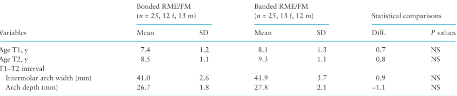
No significant between-group differences were found as to gen-der distribution (chi-square = 0.000; P = 1.000), chronologic age at T1 and T2, and as to duration of T1–T2 interval (Table 1).
The analysis of the baseline characteristics at T1 did not show any significant between-group difference for any of the variables in the bonded group when compared with the banded group.
Descriptive statistics and statistical comparisons on the T2–T1 changes of the variables in the bonded versus banded groups are given
in Table 2. No statistically significant differences were found for any
of the observed variables. The increments in intermolar width were similar in the two groups (IAW: +3.8 mm). AD decreased by 0.7 mm in the bonded RME group and by 0.8 mm in the banded RME group. The mean AL of upper second deciduous molars was 1.5 mm in the bonded group and 0.9 mm in the banded group (Table 2).
Discussion
The aim of this study was to assess the AL of posterior teeth at the end of early treatment with RME/FM by comparing two different expander designs. Maxillary protraction is accomplished by applying elastic forces from a FM to an expander that is attached to the poste-rior teeth. Therefore, the RME/FM protocol typically produces com-bined skeletal and dental effects (3–5, 27). No previous study analysed the dental effects of maxillary protraction by means of superimposi-tion of digital dental casts. This method of investigasuperimposi-tion is particu-larly suitable to evaluate longitudinal dental modifications related to craniofacial growth and/or orthodontic therapy (17, 25). It provides an exact quantification of changes otherwise obscured by the superim-position of bilateral anatomic structures in lateral cephalograms (25,
28). The palatal rugae have been used as stable reference structures to superimpose digital dental casts of both growing and adult patients to test tooth movements in the sagittal plane (16, 29), while their reliabil-ity have been criticized when testing vertical changes as only the third rugae seem to be stable (24). Damstra et al. (30) questioned the use of rugae for patients undergoing an expansion protocol by using a stand-ardized photographic 2D set and considering only the medial part of the rugae. In the literature, the medial part of the second and third rugae and the palatal vault posterior to this zone appear to be the most stable area when orthodontic treatment and growth are involved (16). In this study, the same area described by several authors (16, 25) was used to superimpose the T1 and T2 digital casts and to visualize the transverse and sagittal effects of RME/FM therapy.
When analyzing the post-treatment outcomes of RME/FM, no statistical differences were observed between the two groups with different designs of the expanders. AD decreased by 0.7 mm in the bonded RME group and by 0.8 mm in the banded RME group. These values are very similar to those reported by Ngan et al. (31) that observed a reduction of the maxillary arch length of 0.7 mm during the treatment period with RME/FM in a group of Class III
Table 1. Demographics of the two groups, descriptive statistics, and statistical comparisons at T1 (starting forms). Diff., differences; f,
females; m, males; RME/FM, rapid maxillary expansion and facial mask; SD, standard deviation; y, years.
Variables
Bonded RME/FM (n = 25, 12 f, 13 m)
Banded RME/FM
(n = 25, 13 f, 12 m) Statistical comparisons
Mean SD Mean SD Diff. P values
Age T1, y 7.4 1.2 8.1 1.3 0.7 NS
Age T2, y 8.5 1.1 9.3 1.1 0.8 NS
T1–T2 interval
Intermolar arch width (mm) 41.0 2.6 41.9 3.7 0.9 NS
Arch depth (mm) 26.7 1.8 27.8 2.1 −1.1 NS
NS, not significant.
Table 2. Descriptive statistics and statistical comparisons of the T2–T1 changes. CI, confidence interval; Diff., differences; RME/FM, rapid
maxillary expansion and facial mask; SD, standard deviations.
Measurements
Bonded RME/FM (n = 25)
Banded RME/FM
(n = 25) Statistical comparisons
Mean SD Mean SD Diff. 95% CI P values
Intermolar arch width 3.8 2.2 3.8 1.8 0.0 −1.2 to 1.1 NS
Arch depth −0.7 1.5 −0.8 1.6 0.1 −0.9 to 0.9 NS
Mean anchorage loss of upper ER/EL 1.5 0.8 0.9 1.2 0.6 −0.0 to 1.1 NS
NS, not significant.
by guest on July 18, 2015
subjects with a mean age of 8.4 years (31). Both the bonded and the banded expanders produced a slight mesialization of the poste-rior teeth (1.5 mm in the bonded RME group versus 0.9 mm in the banded RME group). This amount of AL is in agreement with the value reported by other authors by means of cephalometric analysis. Westwood et al. (32) observed a mesialization of anchoring teeth of 1.6 mm at the end of 10 months FM treatment in a group of growing subjects with the same mean age. Vaughn et al. (10) pointed out a difference in AL when the FM is used in combination with RME or not. The maxillary first molar moved forward 1.6 mm in a group of children treated with RME and FM, while the upper molars moved mesially 2.3 mm in a group of subjects with the same mean age treated with the only FM also assisting the Class III correction (10).
The results of this study suggest that in the deciduous or early mixed dentition, either a bonded or a banded expander can be cho-sen as anchorage for maxillary protraction. In the early treatment of Class III malocclusion, no significant antero-posterior dental changes were observed between the two expander designs. RME is frequently needed for the high incidence of maxillary transverse deficiency in the patients with Class III malocclusion (33). The findings of the cur-rent investigation showed that the AL was clinically negligible, thus confirming that the overjet correction has to be ascribed mainly the skeletal changes induced by RME/FM therapy (34).
Conclusion
The orthopaedic treatment of Class III malocclusion with either a bonded or a banded expander combined with FM during the decidu-ous or early mixed dentition produced a slight AL of posterior teeth with no difference between the two intraoral appliance designs.
References
1. Cha, K.S. (2003) Skeletal changes of maxillary protraction in patients exhibiting skeletal Class III malocclusion: a comparison of three skeletal maturation groups. The Angle Orthodontist, 73, 26–35.
2. Baccetti, T., Franchi, L. and McNamara, J.A., Jr. (2004) Cephalometric variables predicting the long-term success or failure of combined rapid maxillary expansion and facial mask therapy. American Journal of Ortho-dontics and Dentofacial Orthopedics, 126, 16–22.
3. Chong, Y.H., Ive, J.C. and Artun, J. (1996) Changes following the use of protraction headgear for early correction of Class III malocclusion. The Angle Orthodontist, 66, 351–362.
4. Ishii, H., Morita, S., Takeuchi, Y. and Nakamura, S. (1987) Treatment effect of combined maxillary protraction and chincap appliance in severe skeletal Class III cases. American Journal of Orthodontics and Dentofacial Orthopedics, 92, 304–312.
5. Mermigos, J., Full, C.A. and Andreasen, G. (1990) Protraction of the max-illofacial complex. American Journal of Orthodontics and Dentofacial Orthopedics, 98, 47–55.
6. Nartallo-Turley, P.E. and Turley, P.K. (1998) Cephalometric effects of com-bined palatal expansion and facemask therapy on Class III malocclusion. The Angle Orthodontist, 68, 217–224.
7. Tortop, T., Keykubat, A. and Yuksel, S. (2007) Facemask therapy with and without expansion. American Journal of Orthodontics and Dentofacial Orthopedics, 132, 467–474.
8. Buongiorno, M., Lione, R., Cozza, P., Franchi, L. (2015) Early treatment of Class III malocclusion with RME and facial mask: evaluation of den-toalveolar effects on digital dental casts. European Journal of Paediatric Dentistry (in press).
9. Cevidanes, L., Baccetti, T., Franchi, L., McNamara, J.A., Jr and De Clerck, H. (2010) Comparison of two protocols for maxillary protraction: bone anchors versus face mask with rapid maxillary expansion. The Angle Orthodontist, 80, 799–806.
10. Vaughn, G.A., Mason, B., Moon, H.B. and Turley, P.K. (2005) The effects of maxillary protraction therapy with or without rapid palatal expansion: a prospective, randomized clinical trial. American Journal of Orthodontics and Dentofacial Orthopedics, 128, 299–309.
11. Williams, M.D., Sarver, D.M., Sadowsky, P.L. and Bradley, E. (1997) Com-bined rapid maxillary expansion and protraction facemask in the treat-ment of Class III malocclusion in growing children: a prospective study. Seminars in Orthodontics, 3, 265–274.
12. Baik, H.S. (1995) Clinical results of the maxillary protraction in Korean children. American Journal of Orthodontics and Dentofacial Orthopedics, 108, 583–592.
13. Fleming, P.S., Marinho, V. and Johal, A. (2011) Orthodontic measure-ments on digital study models compared with plaster models: a systematic review. Orthodontic Craniofacial Research, 14, 1–16.
14. Kusnoto, B. and Evans, C.A. (2002) Reliability of 3D surface laser scan-ner for orthodontic applications. American Journal of Orthodontics and Dentofacial Orthopedics, 122, 342–348.
15. Zilberman, O., Huggare, J.A. and Parikakis, K.A. (2003) Evaluation of the validity of tooth size and arch width measurements using conventional and three-dimensional virtual orthodontic models. The Angle Orthodon-tist, 73, 301–306.
16. Chen, G., Chen, S., Zhang, X.Y., Liu, Y., Shi, F.H. and Xu, T.M. (2011) Stable region for maxillary dental cast superimposition in adults, studied with the aid of stable miniscrews. Orthodontic Craniofacial Research, 14, 70–79.
17. Cho, M.Y., Choi, J.H., Lee, S.P. and Baek, S.H. (2010) Three-dimen-sional analysis of the tooth movement and arch dimension changes in Class I malocclusions treated with first premolar extractions: a guideline for virtual treatment planning. American Journal of Orthodontics and Dentofacial Orthopedics, 138, 747–757.
18. Tollaro, I., Baccetti, T., Franchi, L. and Tanasescu, C.D. (1996) Role of pos-terior transverse interarch discrepancy in Class II, Division 1 malocclusion during the mixed dentition phase. American Journal of Orthodontics and Dentofacial Orthopedics, 110, 417–422.
19. Baccetti, T., Franchi, L. and McNamara, J.A., Jr. (2005) The cervical ver-tebral maturation (CMV) method for the assessment of optimal treatment timing in dentofacial orthopedics. Seminars in Orthodontics, 11, 119–129. 20. Pavoni, C., Mucedero, M., Baccetti, T., Franchi, L., Polimeni, A. and
Cozza, P. (2009) The effects of facial mask/bite block therapy with or without rapid palatal expansion. Progress in Orthodontics, 10, 20–28. 21. Masucci, C., Franchi, L., Giuntini, V. and Defraia E. (2014) Short-term
effects of a modified Alt-RAMEC protocol for early treatment of Class III malocclusion: a controlled study. Orthodontics and Craniofacial Research, 17, 259–269.
22. Cozza, P., Giancotti, A. and Petrosino, A. (1999) “Butterfly expander” in mixed dentition. Journal of Clinical Orthodontics, 33, 583–587. 23. Huanca Ghislanzoni, L., Lineberger, M., Cevidanes, L.H.S., Mapelli, A.,
Sforza, C. and McNamara, J.A., Jr. (2013) Evaluation of tip and torque on virtual study models: a validation study. Progress in Orthodontics, 14, 19–25. 24. Springate, S.D. (2012) The effect of sample size and bias on the reliability
of estimates of error: a comparative study of Dahlberg’s formula. Euro-pean Journal of Orthodontics, 34, 158–163.
25. Christou, P. and Kiliaridis, S. (2008) Vertical growth-related changes in the position of palatal rugae and maxillary incisors. American Journal of Orthodontics and Dentofacial Orthopedics, 133, 81–86.
26. Cohen, J. (1992) A power primer. Psychological Bulletin, 112, 155–159. 27. Saadia, M. and Torres, E. (2000) Sagittal changes after maxillary
protrac-tion with expansion in Class III patients in the primary, mixed, and late mixed dentition: a longitudinal retrospective study. American Journal of Orthodontics and Dentofacial Orthopedics, 117, 669–680.
28. De Luca Canto, G., Pacheco-Pereira, C., Lagrave, M.O., Flores-Mir, C. and Major, P.W. (2015) Intra-arch dimensional measurement validity of laser-scanned digital dental models compared with the original plaster models: a systematic review. Orthodontic Craniofacial Research, 11, 11–18. 29. Almeida, M.A., Phillips, C., Kula, K. and Tulloch, C. (1995) Stability of the
palatal rugae as landmarks for analysis of dental casts. The Angle Ortho-dontist, 65, 43–48.
by guest on July 18, 2015
30. Damstra, J., Mistry, D., Cruz, C., Ren, Y. (2009) Antero-posterior and transverse changes in the positions of palatal rugae after rapid maxillary expansion. European Journal of Orthodontics, 31, 327–332.
31. Ngan, P., Yiu, C., Hu, A., Hagg, U., Wei, S.H.Y. and Gunel, E. (1998) Cephalometric and occlusal changes following maxillary expansion and protraction. European Journal of Orthodontics, 20, 237–254.
32. Westwood, P.V., McNamara, J.A., Jr, Baccetti, T., Franchi, L. and Sarver, D.M. (2003) Long-term effects of Class II treatment with rapid maxillary
expansion and facemask therapy followed by fixed appliances. American Journal of Orthodontics and Dentofacial Orthopedics, 123, 306–320. 33. Franchi, L. and Baccetti, T. (2005) Transverse maxillary deficiency in
Class II and Class III malocclusions: a cephalometric and morphometric study on postero-anterior films. Orthodontic Craniofacial Research, 8, 21–28.
34. Ngan, P. (2005) Early timely treatment of Class III malocclusion. Seminars in Orthodontics, 11, 140–145.
by guest on July 18, 2015