SYSTEMATICS OF LINGULIDE BRACHIOPODS FROM THE END-PERMIAN MASS EXTINCTION INTERVAL
RENATO POSENATO
Dipartimento di Fisica e Scienze della Terra, Università di Ferrara, Via G. Saragat 1, 44121 Ferrara, Italy. E-mail: [email protected] To cite this article: Posenato R. (2016) - Systematics of lingulide brachiopods from the end-Permian mass extinction interval. Riv. It. Paleont. Strat., 122(2): 85-108.
Abstract. The systematics of lingulide brachiopods, from the end-Permian mass extinction interval, is here studied
and discussed. The material has been collected from upper Permian (Changhsingian) beds of Southern Alps and Lower Triassic beds of several Tethyan localities, where the surviving phase following the peak the end-Permian mass extinction is recorded. The study contributes to fill the gap of knowledge regarding the lingulide systematics during a time lapse crucial for the fate of the Mesozoic and Cenozoic marine organisms. The systematics is based both on inner shell morphology and shell microstructure, which are considered to be the most useful taxonomical characters to study the lingulide phylogeny. The specimens have been referred to species of the new genus Trentin-gula, which is characterized by a shell with a secondary layer virgose fabric and a primitive disposition of the ventral
muscle umbonal scar in the Lingulidae phylogeny. Trentingula n. gen. comprises four species: T. lorigae n. gen. n. sp.,
type-species, T. borealis (Bittner), T. mazzinensis n. gen. n. sp., and T. prinothi n. gen. n. sp. The type-species is late
Griesbachian – Dienerian in age and has a wide geographic distribution in the western Tethys (Southern Alps and Hungary). Trentingula prinothi n. gen. n. sp. occurs in the Upper Permian Bellerophon Formation of the Dolomites;
it has a large shell with a short mantle cavity. Trentingula mazzinensis n. gen. n. sp. occurs in the Griesbachian Mazzin
Member of Werfen Formation and is characterized by a small sized shell, about half of the type species, which records the “Lilliput effect” related to the aftermath of the end-Permian mass extinction.
Received:March 11, 2016; accepted: May 20, 2016
Key words. Systematics, Brachiopoda, Lingulida, Microstructure, Permian, Triassic, end-Permian mass extinction.
I
ntroductIonIn the past the systematics of lingulides was mainly based on the external shell morphology (e.g., outline, inflation and ornamentation), and the upper Paleozoic-Cenozoic species were mostly re-ferred to the extant genus Lingula. The definition
of the extant lingulides as living fossils was mainly based on the use of these taxonomical characters (Emig 2003). More recently, the internal characters (e.g., position and shape of muscle scars, pedicle nerve grooves, and mantle canal system) and shell microstructure allowed the recognition of several upper Paleozoic-Mesozoic genera (e.g. Lingularia
Biernat & Emig, 1993; Semilingula Popov in
Ego-rov & Popov 1990; Credolingula Smirnova, 2001 (in
Smirnova & Ushatinskaya 2001); Sinoglottidia Peng
& Shi, 2008; Sinolingularia Peng & Shi, 2008). The
study and application of these taxonomical cha-racters suggest a more complicated phylogenetic history than previously thought (e.g., Cusack et al. 1999; Balinski 1997; Williams et al. 2000; Emig
2003; Holmer & Popov 2000, 2007). Well preserved shells, with clearly detectable internal morphology, are therefore necessary to unravel the lingulide sy-stematics (e.g., Emig 1982, Emig et al. 1978; Biernat & Emig 1993; Cusack et al. 1999), which does not seem to be without problems.
The internal characters of the type-specimens of many “historical” species are unknown, because not preserved or not observable from the outer sur-face, which makes it impossible their assessment at the genus level (e.g., Biernat & Emig 1993; Holmer & Bengtson 2009). The internal characters are often recorded as very shallow reliefs or depressions (e.g., muscle scars, pedicle nerve grooves, mantle canal scars), whose recognition needs well preserved spe-cimens and observations with very oblique light and it is thus difficult. Furthermore, the quality of pu-blished figures is often low and, sometimes, the in-terpretative drawings are not supported by the illu-strations. Further problems rise from the different value assigned to taxonomical characters, and diffe-rent shell terminology and orientation (e.g., Holmer & Popov 2000; Emig 2003; Holmer & Bengtsom 2009).
An additional problem is the shell micro-structure, one the most important taxonomical character, which has been described only for a few species (e.g., Cusack et al. 1999; Williams et al. 2000; Holmer & Bengtson 2009). A wide gap of knowledge embraces the late Permian and the Triassic, when the end-Permian mass extinction changed the evolutionary trajectories of many marine invertebrates (e.g., Knoll et al. 2007). The phylogenetic relationships between the extant
Lingula and Glottidia are contradictory if based,
alternatively, either on the shell morphology or on the microstructure. On the basis of morpho-logy, Lingularia (Carboniferous-Cretaceous,
?Ce-nozoic) has been proposed as the ancestor of the Cenozoic genera Lingula and Glottidia (Biernat &
Emig 1993). This proposal is mostly based on the position of the pedicle nerves in relation to the umbonal muscle. For other authors, the probable co-existence of the baculate and virgose fabric of the secondary layer, respectively characteri-zing the extant Glottidia and Lingula (e.g., Iwata
1981, 1982), since the late Paleozoic, would elimi-nate Lingularia as common ancestor of the living
lingulides (e.g. Cusack et al. 1999; Williams et al. 2000). These conflicting hypotheses originate on fragmentary data, which hampers to “test the re-lative merits of shell microstructure and muscle impressions as genealogical indicators” (Williams et al. 2000, p. 1013). For this reason, new mate-rials, mostly represented by Lower Triassic hol-dovers, are here studied both from microstructu-ral and morphological point of views in order to highlight the early phases of the post-Paleozoic evolutionary history of lingulides.
The above outlined problems and the pro-posal of new taxonomical characters have gene-rated in the last decades instability in the lingulide systematics. For instance, “Lingula” borealis Bittner,
a widespread Lower Triassic species has been re-ferred, from time to time, to Glottidia, Barroisella, Lingularia, or Sinolingularia (Archbold 1981; Biernat
& Emig 1993; Peng & Shi 2008). Another exam-ple is represented by Lower Triassic specimens of “Lingula” tenuissima, published by Broglio Loriga
(1968), which have been referred, or compared, to Glottidia, Lingularia, or Sinoglottidia (Pajaud 1977;
Biernat & Emig 1993; Peng & Shi 2008; Sykora et al. 2011).
The discovery of well preserved specimens and the re-examination of some historical col-lections from Upper Permian and Lower Triassic successions, mainly from the western Palaeotethys, allows to improve the knowledge of lingulides oc-curring across the end-Permian mass extinction and to fill the gap of knowledge regarding the shell morphology and microstructure during the survival and recovery phases of the end-Permian mass extinction (e.g., Posenato 2008b; Posenato et al. 2014).
G
eoGraphIcand stratIGraphIc settInG The material studied here ranges from the Late Permian (Changhsingian) to the Early Trias-sic (Induan and Olenekian) in age. The Changhsin-gian specimens have been collected in the Bellero-phon Formation of Monte Balest, Dolomites (Figs 1, 2), which records the very last Paleozoic marine Fig. 1 - Geographical setting of the Dolomites localities from which the described linguli-des were collected. 1) Monte Cucan; 2) Malga Panna; 3) Catinaccio; 4) Monte Balest; 5) Passo Poma.ecosystems before the mass extinction. The shells were contained in clayey fine-grained sandstones, deposited in a very shallow marine environment, re-cording a regressive phase of nearshore conditions (Prinoth 2013; Posenato et al. 2014).
Most of the Lower Triassic (Induan) mate-rial was collected in the Werfen Formation of the Dolomites (Southern Alps, Italy) (Figs 1, 2). This formation is a thick mixed terrigenous-carbonate succession from peritidal to offshore-transition en-vironments, divided in 9 members. It records the peak, survival and early recovery phases of the end Permian mass extinction (see references in Pose-nato 2008a, b). The lingulides are common in the Mazzin Member (lower Werfen Formation, lower Induan), which is made up of prevailing marly li-mestones and marlstones, mostly deposited within offshore-transition environment. The Mazzin Member contains oligo-typic benthic communities, characterized by small sized shells, which record the survival phase of the end Permian mass extinction (Posenato 2008b). The shell miniaturization, or Lil-liput Effect (e.g., Urbanek 1993; Twitchett 2007), represents a surviving strategy of opportunistic or-ganisms during the most stressed conditions of the shallow marine ecosystems caused by warming and hypoxia (e.g., Metcalfe et al. 2011; Posenato et al. 2014, with references). The oldest Triassic linguli-des here studied have been collected from the up-per part of Mazzin Member (Claraia wangi-griesbachi
subzone, lower Induan) near Malga Panna (Moena, Trento Province; Broglio Loriga et al. 1980).
The most exquisitely preserved specimens here described, are slightly younger. They were col-lected in the lower Siusi Member, within the Claraia clarai subzone (upper Induan), at Monte Cucan
(Ca-valese, Trento Province; Broglio Loriga 1968). They are contained within a dark grey marly mudstones and associated with Claraia clarai (Emmrich). The
Siusi Member is also characterized by low diversi-ty benthic assemblages, but the mollusc and bra-chiopod shells show a noticeable size increase. For instance, the size of lingulides is more than dou-bled with respect to those coming from the Mazzin Member (e.g., Broglio Loriga et al. 1980; Posenato et al. 2014). The stratigraphic settings and paleoe-cology of these lingulide bearing beds have already been described by Broglio Loriga et al. (1980; 1983; 1990), Posenato (2009) and Posenato et al. (2014).
Other Lower Triassic lingulides here conside-Fig. 2 - Schematic stratigraphic column of the end-Permian mass
extin-ction interval of the Dolomites (Southern Alps, Italy) and pro-venance of the lingulide species here proposed. Legend: a, lime-stone (1), marly limelime-stone (2), oolite limelime-stone (3); b, dololime-stone (1), sandy dolostone (2), marly dolostone (3); c, marlstone; d, sandstone (from Posenato et al. 2014, modified)
red were collected in the Lower Triassic succession of Balaton Highland (Hungary). They come from the Aracs Marl (Claraia clarai subzone, upper
In-duan) of Balatonfüred and the Csopak Marl ( Tiroli-tes illyricus beds, upper Olenekian) of Sóly (Broglio
Loriga et al. 1990). Additional material came from the Lower Triassic (Induan) Dinwoody Formation of Wyoming (USA).
MaterIalandMethods
The material from the Bellerophon Formation (Changhsin-gian, upper Permian) of Monte Balest consists of about twenty val-ves, some with a good preservation of the inner shell surface, others preserved as inner moulds, or as shell fragments (Lingularia cf. smir-novae in Posenato et al. 2014). The lingulide population from Malga
Panna (Mazzin Member, Werfen Formation, lower Induan) consists of some tens of valves and moulds located on a bedding surface in association with the bivalve Claraia wangi-griesabachi group (sensu
Bro-glio Loriga et al. 1983). These lingulides have been figured by BroBro-glio Loriga et al. (1980, 1990) and classified as Lingula sp. They have been
recently analysed, from a paleoecological point of view, by Posenato et al. (2014).
The collection from Monte Cucan (Siusi Member, Werfen Formation, upper Induan) consists of two ventral and two dorsal valves, whose internal characters are very well preserved. They have been described and classified by Broglio Loriga (1968) as Lingula tenu-issima Bronn. These lingulides were frequently quoted in the
literatu-re due to their extraordinary pliteratu-reservation (e.g. Pajaud 1977; Biernat & Emig 1993; Peng & Shi 2008; Sykora et al. 2011; Posenato et al. 2014); they have been successively classified as Lingula cf. borealis
Bitt-ner (Broglio Loriga et al. 1990), or Lingularia borealis (Bittner)
(Pose-nato et al. 2014).
The material from the Lower Triassic of Hungary is repre-sented by a well preserved internal mould from Sóly (Csopak Marl, upper Olenekian) and some moulds and shells from Balatonfüred (Aracs Marl, upper Induan; for the stratigraphic setting see Broglio Loriga et al. 1990). The material from Wyoming originate from a lingulide coquina collected in the Snake River section, at about 25 m above the base of the Dinwoody Formation (Newell & Kummel 1942).
The studied specimens have been whitened with magnesium fume and photographed with very oblique lights under a stereomi-croscope. The shell fragments observed under SEM (EVO 40 Zeiss), previously etched with diluted (2%) hydrochloric acid for 5 seconds, have been coated with gold. The Triassic lingulides are kept in the “Piero Leonardi” Museum of the Ferrara University (PLM acronym). The Permian lingulides, collected by Dr. Herwig Prinoth of the Mu-seum Ladin (San Martino in Badia, Bolzano), are kept in the Museo Scienze Naturali Alto Adige (PZO acronym).
s
ysteMatIcp
alaeontoloGyClass lInGulata Gorjansky & Popov, 1985
Order Lingulida Waagen, 1885
Superfamily Linguloidea Menke, 1828 Family Lingulidae Menke, 1828
Diagnosis (Emended): Diagnosis as indicated by Holmer & Popov (2000, p.35, 36), emended as added in italics below.
Shell elongate oval, subrectangular to spatulate, gently and subequally biconvex, equivalved; larval shell smooth; ventral valve with triangular ventral depression or groove for passage of pedicle; ventral pseudointerarea vestigial, lacking flexure lines, rarely forming well-defined, triangular propareas; dorsal valve with small, undivided pseudointerarea not extending as plate into valve; muscle system with asymmetrical transmedian and asymmetrical paired or unpaired umbonal
muscles; pedicle nerves curving around or bisecting umbonal muscles;
posterolateral margins of visceral area in both valves strongly con-cave or straight; dorsal visceral area with narrow anterior projection extending anteriorly beyond midvalve; dorsal central and anterior lateral muscle scars usually closely spaced; mantle canal system bi-furcate in living forms; vascular lateralia of both valve converging anteriorly; vascular media vestigial or absent. Living forms with long flexible pedicle; lophophore spirolophous, with apices of spires me-dially directed.
Discussion. The late Paleozoic and
Me-sozoic-Cenozoic lingulides are all included in the Lingulidae (?Upper Devonian, Carboniferous-Holocene), a family characterized by a single (un-paired) umbonal muscle (Holmer & Popov 2000; Emig 2003). This group probably originated from the Pseudolingulidae (Ordovician-Lower Carbo-niferous), which mostly differs from the Linguli-dae by the occurrence of paired umbonal muscle scars and symmetrical transmedian muscle scars (Holmer & Popov 2000). In the extant Lingula and Glottidia, the pedicle nerve grooves are posteriorly
joined and move to the right of a single umbonal muscle, which represents the left part of the um-bonal muscle scar of Lingularia (Biernat & Emig
1993).
Unfortunately, among extinct Lingulidae, the shape of ventral umbonal muscle scar is well detectable only in a very few specimens. The sha-pe is clearly impressed in a paratysha-pe of Lingula-ria similis Biernat & Emig (Biernat & Emig 1993,
fig. 3E), Jurassic in age. It consists of an internal mould of a ventral valve, in which the umbonal muscle scar has a “heart-like shape” (sensu Biernat & Emig 1993). The scar is, however, divided by a median groove produced by the pedicle nerves joined together. This condition, defined “unpaired umbonal muscle scar”, consists of “two posterior scars” located “on each side of the junction” of pedicle nerves grooves (Biernat & Emig 1993, p. 5), therefore the ventral umbonal scar is not single. Thus, the umbonal muscle scar of Lingularia shows
transitional characters between the Pseudolinguli-dae and the LinguliPseudolinguli-dae, as already noted by Biernat & Emig (1993).
The Upper Permian and Lower Triassic lin-gulides studied here show a more primitive con-dition in comparison with the Jurassic types of
Lingularia. They have the ventral umbonal muscle
scar clearly bisected by separated pedicle nerves. These latter only slightly converge before rea-ching the muscle scars, but without merging (Fig. 3). The two lateral parts of the umbonal scar look therefore more separated than what observed in the Jurassic types of Lingularia. In specimen 5892
(Pl. 1, Figs 2a, b), the umbonal muscle seems to be clearly divided into two slightly asymmetrical and rounded, but slightly impressed scars. In all the examined specimens, the dorsal umbonal mu-scle is clearly divided into two asymmetrical parts. The left part has a drop-like shape and is elongated along the perimial line. The right part is smaller and has a subcircular outline (Pl. 1, Figs. 1a, b; 5). The umbonal muscle scar of both valves is thus divided into two parts and does not match with the original diagnosis of the Lingulidae (Holmer & Popov 2000; Emig 2003).
The separation of both dorsal and ventral umbonal muscle scar into two parts may suggest to refer the Upper Permian and Lower Triassic speci-mens under study to the Pseudolingulidae. Howe-ver, this latter family, ranging from the Ordovician to the Lower Carboniferous, shows significant dif-ferences from the Lingulidae, such as the asymme-trical ventral transmedian muscle scars, the thick dorsal posterior margin with pseudointerarea, the
well marked muscle tracks, the dorsal median sep-tum, and the vascular lateralia extending to the posterior region (Holmer 1991; Holmer & Popov 2000). These relevant differences suggest to ascri-be the studied lingulides to the family Lingulidae, of which they would represent ancestral forms. In this case, the diagnosis of Lingulidae proposed by Holmer & Popov (2000) is here emended conside-ring both unpaired (single) and asymmetrical pai-red umbonal muscle scars among their taxonomi-cal characters. In any case, a careful morphologitaxonomi-cal analysis of the umbonal muscle scars of upper Pa-leozoic and Triassic Lingulidae is necessary in or-der to better define their significance and position in the lingulide phylogeny.
Genus Trentingula new genus
Type species: Trentingula lorigae n. gen. n. sp.
Diagnosis: Shell elongated oval, almost equivalve, slightly convex, with gently curved or subparallel lateral margins and rounded anterior margin; ornamentation of irregularly spaced and weak growth lines. Ventral valve with pseudointerarea absent or vestigial; pedicle nerve grooves subparallel, extending from the anterior muscle scars to the posterior adductor without merging; posterior adductor bisected by disjoined pedicle nerves and divided into two rounded and slightly asymmetrical parts; vascula lateralia
subequal, converging anteriorly and with a slightly sigmoidal
outli-ne. Dorsal valve without pseudointerarea; posterior adductor scar divided into a left drop-like shaped part and a right rounded square part; anterior projection of visceral cavity slightly raised above the valve floor and anteriorly furrowed by a short median slit; anterior lateral muscles joined, forming a drop-like, subpentagonal depres-sed scar. Primary shell layer of loosely packed botryoidal aggrega-tes; secondary layer fabric virgose, with alternating compact and Fig. 3 - Terminology of the inner shell morphology adopted here (muscular terminology based on Williams and Rowell, 1965; in italic,
virgose laminae; closely packed fine spheroidal granules alternating with loosely packed granules and rods subparallel to the shell sur-face.
Etymology: From the combination of the names Trentino, the region from which the type material was collected, and lingula, tongue.
Occurrence: Upper Permian (Bellerophon Formation, Southern Alps); Lower Triassic of Dolomites (Werfen Formation, Southern Alps); Balaton Highland (Central Hungary, Aracs Marl and Csopak Marl); Wyoming (USA, Dinwoody Formation).
Comparison. Trentingula n. gen. is
characteri-zed by the absence of the dorsal median septum or ridge, a taxonomical character occurring in the Pa-leozoic Apsilingula Williams, 1977, Barroisella Hall &
Clarke, 1892, Langella Mendes, 1961 and Argentiella
Archbold, Cisterna & Sterren, 2005. A median ridge is also present in the Mesozoic Sinoglottidia Peng and
Shi, 2008 and in the Cenozoic Glottidia Dall, 1870. Trentingula differs also from the Permian Semilingula
Popov (in Egorov & Popov 1990), because of the occurrence, in the latter, of vestigial vascula media
(Holmer & Popov 2000) and a baculate microstruc-ture (see below, Cusack et al. 1999). A significant difference from the Lower Triassic Sinolingularia
Peng & Shi, 2008 is the arrangement of the anterior lateral muscle scars. In the latter genus, these scars are clearly separated, whereas they are fused in Tren-tingula, and form a subpentagonal depression on the
valve floor, laterally limited by low ridges originating from the anterior projection of the visceral cavity (Pl. 1, Figs 1a, b). This interpretation is based on the morphology of the living Lingula (Pl. 1, Fig. 8),
and the occurrence of muscle tracks inside the scar. The type species (Trentingula lorigae n. gen.
n. sp.) has been referred in the past to Lingularia
Biernat & Emig, 1993 (e.g. Posenato et al. 2014) due to similarities regarding the shell outline and the muscle scar arrangement. However, the dorsal umbonal muscle scar of Lingularia is heart-shaped
(Biernat & Emig 1993, fig. 4), while it is clearly divi-ded into two parts in Trentingula n. gen. The pedicle
nerves passing through the ventral umbonal muscle scar in Lingularia are joined, while they are disjoined
in Trentingula n. gen. Another relevant difference is
the occurrence in Lingularia of a short dorsal
cen-tral ridge, posteriorly to the anterior oblique mu-scle scars (Biernat & Emig 1993; Holmer & Popov 2000), which is absent in Trentingula n. gen.
The primary shell layer of living lingulides is mainly composed by organic matter, while in the fossil it is generally made up by densely packed
gra-nular apatite (e.g., Holmer & Bengston 2009). The secondary shell layer has a prominent phylogenetic value for many authors; it has a stratiform structu-re made up by organic matter and apatitic elements with various shape, orientation and density. These elements are aggregated in laminae with different fabric. Lingula is characterized by densely packed
aggregates of apatitic granules (compact laminae), succeeded by dispersed aggregates of rods and pla-tes with apatitic rods (virgose laminae, from the La-tin word virgous = full of twigs, Cusack and Williams
1996) and walled laminae with botryiodal masses or vertical walls. Glottidia has compact laminae
alterna-ted with baculate laminae (from the Latin word ba-culum = rod). The latter consist of regular arrays of
rods oriented with acute angles among each other, and at high angle with respect to the lamina surface (trellised or baculate fabric; Cusack et al. 1999; Wil-liams et al. 2000; Holmer & Bengtson 2009). The baculate fabric is considered a primitive microstruc-ture, already occurring in lower Paleozoic lingulides (Holmer 1991), while the virgose fabric is recorded at least from the Carboniferous (e.g., “Lingula” squami-formis Phillips; Cusack &Williams 1996). These
sug-gest distinct phyletic lineages of the two living genera at least since the late Paleozoic (Williams et al. 2000).
Trentingula n. gen. and Lingularia have a different
shell fabric. The new genus has a virgose fabric, while
Lingularia is characterized by a baculate fabric (e.g.,
Cu-sack et al. 1999; Williams et al. 2000). Trentingula n. gen.
belongs, therefore, to the phyletic lineage of Lingula.
This latter genus differs from Trentingula n. gen. mainly
by a different shape of the ventral and dorsal umbonal muscle scars. The ventral umbonal muscle scar of Lin-gula has a subcircular outline and it is not bisected by
the pedicle nerves; the dorsal muscle scar is unpaired (Biernat & Emig 1993; Holmer & Popov 2000). Ano-ther significant difference is the occurrence in Lingula of
a dorsal shallow median ridge separating the anterior la-teral muscle scars (Pl. 1, Fig. 8). Besides, the lophophoral cavity of Trentingula n. gen. is longer of about one third
than that of Lingula (Posenato et al. 2014, fig. 6). Trentingula n. gen. fills the strongly incomplete
fossil record of the virgose lineage, which is recor-ded in the Carboniferous “Lingula” squamiformis
Phil-lips (Cusack & Williams 1996) and in the Cretaceous
Credolingula Smirnova and Ushatinskaya, 2001, a genus
which shares with Trentingula n. gen. some
morpholo-gical traits like the asymmetry of the dorsal umbonal muscle scar, the position and outline of anterior lateral
muscle scar and the short median slit on the anterior extension of visceral cavity. However, Credolingula
dif-fers from Trentingula n. gen. by the occurrence of radial
plications in both valves and by a different disposition of the ventral umbonal muscle scar, which is fused to the transmedian muscle scars. Moreover, the virgose fabric is known in other Lower Cretaceous and Ceno-zoic species (Cusack et al. 1999).
The lingulides from the Dinwoody Formation of Wyoming (USA), generally classified as “Lingula” borealis Bittner (e.g., Newell & Kummel 1942), can be
assigned to Trentingula n. gen., because they are
cha-racterized by a virgose shell microstructure, the absen-ce of dorsal median septum and a muscle scar arrange-ment similar to the type specimens of the new genus. In particular, they share the shape of the ventral and dorsal umbonal muscle scars and the dorsal anterior lateral muscle scars (Pl. 1, Figs 6a, b, 7). This latter scar is posteriorly connected to a middle slit and bounded by a posterior-lateral swelling of the anterior extension of visceral region (Pl. 1, Fig. 6a, b).
Composition. Trentingula lorigae n. gen. n. sp., Trentingula mazzinensis n. gen. n. sp., Trentingula prinothi
n. gen. n. sp., Trentingula borealis (Bittner, 1899).
Trentingula lorigae n. gen. n. sp. Pl. 1, Figs 1-5; Pl. 2, Fig. 8; Pl. 3, Figs 1-20
1968 Lingula tenuissima Bronn - Broglio Loriga, p. 189-197, pl. 1,
figs 1-6, pl. 2, figs 1-4.
1990 Lingula cf. borealis Bittner - Broglio Loriga et al., pl. 2, figs 8, 9.
2014 Lingularia borealis - Posenato et al., figs 3g-i.
Diagnosis: Medium sized, almost equivalve, shell with an elongated oval outline; width about three fifths of length; lateral mar-gins slightly arched. Ventral valve with very small propareas separated by a wide pedicle groove; length of visceral area about one-half of valve length; pedicle nerve grooves dividing the umbonal muscle scar into two slightly asymmetrical, rounded subtriangular parts. Dorsal valve without pseudointerarea; dorsal umbonal muscle scar divided into a sub-teardrop left part, and a smaller and subcircular right scar; length of visceral area about three fifths of total valve length; anterior lateral muscle scars joined, originating a deep subpentagonal depression; anterior extension of visceral region thickened and with a short middle slit connected with the anterior lateral muscle scar. Shell microstructure virgose originated by alternating layers of closely and loosely packed granules and rods, subparallel to the shell surface.
Etymology: Dedicated to the memory of Prof. Carmen Bro-glio Loriga, who discovered and studied the specimens coming from the Monte Cucan (Broglio Loriga 1968). She headed the Ferrara research group on the Permian and Triassic stratigraphy of the western Tethys.
Holotype: Specimen no. MPL 5890 (Pl. 1, Fig. 1); it is a dorsal valve indicated as Esemplare D by Broglio Loriga (1968, pl. 1, fig. 5), Siusi Member, Werfen Formation, Monte Cucan, Dolomites.
Paratypes: Ventral valves: MPL 5891, Pl. 1, Fig. 3 (Esemplare
A of Broglio Loriga 1968, pl. 1, fig. 1), Monte Cucan, Dolomites (I); MPL 5892, Pl. 1, Fig. 2 (Esemplare B of Broglio Loriga 1968, pl. 1, fig. 3), Monte Cucan, Dolomites (I); MPL 5893 (Esemplare C of Broglio Loriga 1968, pl. 1, fig. 4), Monte Cucan, Dolomites (I); MPL 5894 (Po-senato et al., 2014, fig. 3i), Balatonaracs (H); MPL 5895 (Broglio Loriga et al. 1990 pl. 6, fig. 4), Balatonfüred (H);
Dorsal valves: MPL 5888, Pl. 1, Fig. 5, Catinaccio, Dolomites (I); MPL 5889, Pl. 1, Fig. 4 (Esemplare F of Broglio Loriga 1968), Mon-te Cucan, DolomiMon-tes (I); MPL 5896, Passo Poma, DolomiMon-tes (I); MPL 5898, Malga Panna, Dolomites (I).
Description. The shell is almost
equival-ve, elongated oval in outline and with a maximum length of about 12 mm. The length/width ratio ran-ges from 1.5 to 1.7. The lateral margins are slightly arched. The anterior margin varies from rounded to slightly spatulated. The maximum height is located posteriorly to the valve mid-length. The ornamenta-tion, preserved only in the anterior region of a ven-tral valve, consists of crowded and regularly spaced growth lines.
Ventral umbonal region with very small pro-pareas separated by a wide and deep pedicle groo-ve. The ventral visceral cavity extends to the mid-length of the valve. The pedicle nerve grooves are well impressed, a little divaricated in the central part, slightly converging and bisecting, without mer-ging, the umbonal muscle scars. The latter consist of two rounded subtriangular, feebly impressed, depressions. The left scar has an anterior margin slightly concave, while the right scar margin is con-vex (e.g. Pl. 1, Figs 2a, b). The anterior projection of visceral cavity is slightly raised and is laterally flanked by ovoid scars produced by the combined outside lateral and central muscles (Pl. 1, Fig. 2a, b, 3a, b). The anterior projection of the visceral cavity is anteriorly edged by short and few radial ridges and grooves, possibly representing the middle late-ral muscle scars (Pl. 1, Figs 2a, b, 3a, b). The latelate-ral parts of the visceral cavity contain the anterior late-ral muscle scars, which have curved tear-drop outli-ne, and two elongated transmedian muscle scars on the right side and one transmedian on the left side. The vascula lateralia are not well impressed; they
con-verge medially, and with short and subparallel ante-rior tips (Pl. 1, Figs 3a, b). The ventral mantle cavity occupies about 50 per cent of the total valve length. The dorsal pseudointerarea is not present. The perimial scar of the dorsal visceral cavity is well impressed and extends at about three fifths of valve length. The umbonal muscle scar is divided into two parts (Pl. 1, Figs 1a, b, 5). The larger part
exten-ds from the median umbonal cavity to the left side, and has an elongated lacrimiform shape. The right part is subcircular, smaller but more deeply impres-sed than the right scar.
The anterior part of the dorsal visceral cavity is slightly raised and bisected, at the anterior extre-mity, by a median slit (Pl. 1, Fig. 1a, b). The anterior visceral region is delimited from the posterior visce-ral cavity by a transversal groove, which probably corresponds to the gastroparietal band. The poste-rior visceral cavity has three short and symmetrical longitudinal furrows, which are limited forwards by shallow tubercles and vanishing backwards (Pl. 1, Figs 1a, b, 4). The central muscle scar is clearly impressed and has a reniform outline. The anterior lateral muscle scars originate a deep subpentagonal depression on valve floor, which is connected to the median anterior slit of anterior part of visceral ca-vity (Pl. 1, Figs 1a, b). The visceral caca-vity has other two lateral shallow depressions, symmetrically loca-ted with respect to the central muscle scars. Their borders, although are not clearly impressed on the valve floor, indicate a subtriangular in outline. These shallow depressions are interpreted as the transme-dian, outside lateral and middle lateral combined muscle scars (Pl. 1, Fig. 1a, b). The floor of the lo-phophoral cavity is covered by irregular tubercles, which record the mantle canal traces emerging from the visceral cavity. The mantle canals are slightly si-nuous, medially converging forwards; the distance between their tips and the anterior margin is about one-fourth of valve length. The dorsal and ventral mantle canals have a similar shape, but the latter are slightly more elongated. The distance between the ventral mantle canal tips and the anterior margin is about one-sixth of valve length. The dorsal lopho-phoral cavity occupies about 40% of the total valve length.
Shell microstructure. Shell fragments from
the anterior region of mantle cavity of a ventral valve (MPL 5891), lateral region of visceral cavity of ventral valve (MPL 5893) and a section of the posterior region of a dorsal valve (MPL 5898) have been analysed.
The shell fragment of the anterior region has a thickness of about 160 μm (MPL 5891; Pl. 3, Fig. 1). The primary shell layer, about 30 μm thick, consists of a coarse granular fabric, which mostly consists of loo-sely packed botryoidal aggregates, up to 500 nm in dia-meter (Pl. 3, Fig. 2). The secondary layer is originated
by an alternation of compact and virgose laminae (Pl. 3, Figs 3, 4). The compact lamina consists of closely packed fine spheroidal granules; the virgose lamina contains loosely packed granules and rods (about 100 nm in diameter and 1 μm long) subparallel to the shell surface (Pl. 3, Figs 19, 20). The shell is perforated by canals with apertures, oval in outline and about 10-15 μm long and 5 μm wide, on the inner shell surface (Pl. 3, Figs 9, 10).
The fragment from the postero-lateral region (visceral cavity) of a ventral valve (MPL 5891) has a thickness ranging from 50 to 80 μm (Pl. 3, Figs 5-8). The thicker sample (about 80 μm thick) (Pl. 3, Fig. 5) consists of three thick massive laminae of loosely pa-cked granules and irregularly oriented short rods (Pl. 3, Figs 6, 7). The thinner laminae are finely stratified and characterized by rhythmic alternations of virgose and walled fabrics (Pl. 3, Fig. 8), with a cleavage perpendi-cular to the inner shell surface. In the thinner sample (about 50 μm thick, Pl. 3, Fig. 17), the alternation of
PLATE 1
Figs 1-4 - Trentingula lorigae n. gen. n. sp., Siusi Member, Werfen
For-mation, Lower Triassic, Monte Cucan, Cavalese, Trento. 1a, b) holotype, dorsal valve and reconstruction of internal characters, specimen D of Broglio Loriga (1968, pl. 1, fig. 5), MPL 5890. 2a, b) ventral valve and reconstruction of internal characters, specimen B of Broglio Loriga (1968, pl. 1, fig. 3), MPL 5892; 3a, b) internal mould of ventral valve and reconstruction of internal characters, specimen A of Broglio Loriga (1968, pl. 1, fig. 2), MPL 5891; 4) internal mould of dorsal valve, specimen F of Broglio Loriga (1968, not figured), MPL 5889.
Fig. 5 - Trentingula lorigae n. gen. n. sp., dorsal valve with partially
ero-ded internal shell surface, Siusi Member, Werfen Formation, Catinaccio, Bolzano, MPL 5888.
Figs 6 - 7. Trentingula cf. lorigae n. gen. n. sp., Dinwoody Formation,
Lower Triassic, Snake River, Wyoming; 6a, b) dorsal valve and reconstruction of internal characters; 7) ventral valve, MPL 5899.
Fig. 8 - Lingula anatina Lamarck, Recent, Japan. Detail of the inner
shell morphology of a dorsal valve in which the anterior lateral muscle scars are clearly separated by a median rid-ge (here broken) and laterally limited by low crests, which represent the forward extension of the anterior projection of visceral cavity. The interpretation of the dorsal middle sub-pentagonal depression, located anteriorly to the ante-rior projection of visceral cavity, of Trentingula lorigae n. gen.
n. sp., as the anterior lateral muscle scar is based on the inner morphology of this extant species; MPL 6500.
Abbreviations: Al, anterior lateral muscle scar; Apvc, anterior projec-tion of the visceral cavity; Ce, central muscle scar; Co, cen-tral and outside muscle scars; Ml, middle lateral muscle scar; Pn, pedicle nerve groove; Tr, transmedian muscle scar; Trom, transmedian, outside and middle lateral muscle scars; Um, umbonal muscle scar; Vl, vascula lateralia scar. Scale bar 2 mm.
virgose and compact laminae is more irregular and a virgose lamina, near the primary layer, is clearly com-posed of loosely dispersed rods, tablets and granules (Pl. 3, Fig. 18).
The third sample has been observed along a ra-dial section, passing through the umbo and the lateral
margin of a dorsal valve (MPL 5898). Along the poste-rior margin, where the shell has a thickness of about 1.2 mm, the secondary shell layer (Pl. 3, Fig. 14) is mul-tilayer with virgose and compact sublayers. Sublayers with the former fabric predominate in the outer part of the secondary layer, where they form a lens-shape
lamina, about 40 μm thick, characterized by granules, spheroids and rods with chaotic orientation (Pl. 3, Fig. 16). In the inner part of the secondary layer, sublayers with densely packed small spheroids (compact fabric) decidedly prevail on the virgose laminae represented by coarse and relatively loosely packed spheroids. At about 2 mm from the posterior margin, inside the visceral cavity, the secondary shell layer shows a very thick outer sublayer with a virgose fabric consisting of loosely packed aggregates of small botryoids. In the inner sublayers, the compact sublayers prevail on the virgose ones (Pl. 3, Fig. 13)
Occurrence. Dolomites, Italy (Monte
Cu-can, Passo Poma, Malga Panna, Catinaccio), Siusi Member of Werfen Formation, Claraia clarai
subzo-ne, Late Induan (Dienerian); Balaton Highland, Hungary (Balatonfured, Balatonaracs; Posenato et al. 2014, fig. 3i), Aracs Marl, Claraia clarai subzone,
Late Induan (Dienerian).
Measurements. (In mm; abbreviations: D,
dorsal valve; L, length; LMC, length of mantle ca-vity; V, ventral valve; Va, type of valve; W, width; in bold the holotype; see the scatter diagram in Fig. 4).
Va L W LMC L/W LMC/L MPL 5888 D 12.6 8.4 6.1 1.5 0.5 MPL 5889 D 11.0 6.6 4.1 1.7 0.4 MPL 5890 D 12.2 7.6 4.9 1.6 0.4 MPL 5891 V 10.8 7.0 5.4 1.5 0.5 MPL 5892 V 11.9 8.0 6.2 1.5 0.5 MPL 5893 V 11.9 6.7 6.3 1.8 0.5 MPL 5894 V 9.6 6.1 - 1.6 -MPL 5895 V 11.7 6.7 5.7 1.7 0.5 MPL 5896 ? 10.0 7.0 - 1.4 -MPL 5898 D 11.2 8.0 - 1.4
-Discussion. The holotype comes from the
material collected in the Claraia clarai beds (Siusi
Member, Werfen Formation) at Monte Cucan, near Cavalese (Fiemme Valley, Dolomites) by Broglio Loriga (1968). These specimens are represented by disarticulated valves with an extraordinary preser-vation. The collection consists of three ventral and two dorsal valves. A sixth specimen was sectioned by Broglio Loriga (1968, pl. 1). This material has been initially referred to Lingula tenuissima Bronn, 1838, a
species erected for a Middle Triassic specimen from Germany figured by Bronn (1837, pl. 13, fig. 6b). Ho-wever, this specimen has a drop-like outline and it is much more elongated, with the length more than twice of the width. The internal characters and the shell microstructure of L. tenuissima are unknown.
The Dolomites specimens have been later classified as Lingula cf. borealis Bittner (Broglio Loriga et al.
1990, pl. 2, figs 8, 9), or as Lingularia borealis Bittner
(Posenato et al. 2014, fig. 3, g, h). However, Bittner’s species is characterized by a larger size (maximum length of 17 mm), subparallel lateral margins, and more elongated shells, with the width about half of the length (Dagys 1965).
PLATE 2
Figs 1, 2 - Trentingula mazzinensis n. gen. n. sp., Mazzin Member,
Werfen Formation, Lower Triassic, Malga Panna, Moena, Trento. 1a, b) ventral valve and reconstruction of internal characters, MPL 5923/2; 2a-c) dorsal valve, holotype, MPL 5923/1, internal mould (2a), reconstruction (2b) and inter-nal shell surface (2c).
Figs 3-7 - Trentingula prinothi n. gen. n. sp., Bellerophon Formation,
Late Permian (upper Changhsingian), Monte Balest, Ortisei. 3a, b) internal mould of ventral valve with shell fragments in the posterior region and reconstruction, PZO5767; 4a, b) holotype, dorsal valve and reconstruction of internal characters, PZO5762; 5a, b) internal mould of the anterior part of a dorsal valve with well impressed vascula latera-lia, and reconstruction of internal characters, PZO5775; 6a, b) internal mould of a dorsal valve and reconstruction, PZO5764; 7a-c) internal mould of a ventral valve, detail of the posterior region and reconstruction of the umbonal mu-scle scars bisected by the pedicle nerve grooves, PZO5765. Fig. 8 - Trentingula lorigae n. gen. n. sp., brachial valve and
reconstruc-tion with the possible preservareconstruc-tion of the pedicle replaced by sediment; see the shell detail on Pl. 1, Fig. 5, Siusi Mem-ber, Werfen Formation, Catinaccio, Bolzano, MPL 5888. Abbreviations: Al, anterior lateral muscle scar; Apvc, anterior
projec-tion of the visceral cavity; Ce, central muscle scar; Co, cen-tral and outside muscle scars; Ml, middle lateral muscle scar; Pn, pedicle nerve groove; Tr, transmedian muscle scar; Trom, transmedian, outside and middle lateral muscle scars; Um, umbonal muscle scar; Vl, vascula lateralia scar. Scale bar 2 mm.
Fig. 4 - Scatter diagram of shell dimensions of the three new species proposed here.
Some available lingulide specimens from the Lower Triassic Dinwoody Formation of Wyo-ming, generally referred in literature to “Lingula” borealis Bittner (e.g., Newell and Kummel 1942),
show similarities with T. lorigae n. gen. n. sp. In
particular, the anterior part of the dorsal anterior extension of visceral cavity has a forked anterior extremity with a middle slit connected with a sub-pentagonal or drop-like depression, which can be interpreted as the conjoined anterior lateral
mu-scle scar occurring in Trentingula lorigae n. gen. n.
sp. Another common feature is the separation into two parts of the dorsal and ventral umbonal muscle scars (Pl. 1, Figs 6, 7). However, the low number of examined specimens does not allow to evaluate the variability of their shell elonga-tion and shape of lateral margins, which repre-sents the most important taxonomical characters to distinguish T. lorigae n. gen. n. sp. from T. bore-alis (see above).
Trentingula prinothi n. gen. n. sp. Pl. 2, Figs 3-7; Pl. 4, Figs 1-12
2014 Lingularia ? cf smirnovae Biernat & Emig, 1993 -
Pose-nato et al., fig. 3a, b.
Diagnosis: Large sized shell with a very elongated oval out-line; length of ventral visceral area about two-thirds of valve length. Dorsal valve without pseudointerarea; dorsal umbonal muscle scar divided into a left, lacrimiform in shape, part and a right, sub-ovoid part; mantle cavity short, occupying about one-fifth of total valve length; anterior lateral muscle scars forming a deep sub-pentagonal depression, laterally connected with the central muscle scars; anterior projection of visceral region with a middle, very low crest culmina-ting with a small anterior knob, vascular lateralia sigmoidal, anteriorly
converging and with short and parallel anterior tips. Microstructure of the secondary layer with virgose lamination consisting of an al-ternation of laminae with compact and virgose fabric respectively represented by densely packed granule aggregates and sub-horizontal rods and/or loosely packed spheroidal aggregates.
Etymology: The species is dedicated to Dr. Herwig Prinoth who discovered the specimens.
Holotype: PZO5762, a dorsal valve represented by the in-ternal surface of shell, lacking of the umbonal and anterior extremi-ties (Pl. 2, Fig. 4), and its internal mould (PZO5763).
Paratypes: Three dorsal valves (PZO5764 (Pl. 2, Fig. 6), PZO5774, PZO5775 (Pl. 2, Fig. 5)) and eight ventral valves (PZO5765, PZO5767, PZO5768, PZO5769a, PZO5769b, PZO5770, PZO5771, PZO5772,) (see PZO5767 on Pl. 2, Fig. 3).
Description. The shell has a maximum
length of about 15 mm, with a length/width ra-tio ranging from 1.7 to 2.7. The valves are almost flattened, with the maximum height posterior to mid-length. The outline is elongated oval with a rounded spatulated anterior margin. The maxi-mum width is somewhat anterior to mid-length. Ornamentation consisting of irregularly spaced growth lines, which are particularly marked on the postero-lateral regions.
The ventral visceral area extends anteriorly from mid-length to three-quarters of the valve length. The pedicle nerve impressions are subpa-rallel in the middle of the visceral cavity. The ventral umbonal muscle scars are separated by a wide and flattened surface, which is laterally limi-ted by the pedicle nerve grooves (Pl. 2, Fig. 7a, b). The transmedian and anterior lateral muscle scars are not detectable. The combined middle lateral and central muscle scars are well impressed, oval in outline (Pl. 2, Figs 3a, b), sometimes forming posterior long muscle tracks. The ventral vascula lateralia converge medially; the anterior tips are
distant from the anterior margin of about one-fourth of valve length. The ventral mantle
cavi-ty occupies 25 to 40 per cent of the total valve length.
The dorsal visceral area is very long; it extends to the four-fifths of total valve length. The dorsal umbonal muscle is divided into a lar-ger, lacrimiform left scar, which is laterally flan-ked by the perimial line. The smaller, right scar, has a sub-circular outline (Pl. 2, Figs 6a, b). The anterior extension of the visceral cavity origina-tes a long and slightly raised central surface with
PLATE 3
Shell microstructures of Trentingula lorigae n. gen. n. sp., Siusi
Mem-ber, Werfen Formation, Lower Triassic.
Figs 1-4 - 1) Vertical fracture of a fragment from the anterior region (mantle cavity) of a ventral valve with traces of vertical ca-nals, external surface at the upper left corner (MPL 5891); 2) detail of the primary layer; 3) detail of the secondary layer with a multi-layered microstructure, with compact and vir-gose fabric alternations; 4) a virvir-gose lamina with irregularly oriented rod-like aggregates located at the edge between the inner shell surface and the fractured surface.
Figs 5-8 - 5) Vertical fracture of the secondary layer from the lateral region of visceral cavity; a multi-layered fabric with alterna-tions of virgose and compact fabric; ventral valve, external shell surface on the left (MPL 5893); 6) detail of a sublayer with compact spheroidal and botryoidal aggregates; 7) obli-que fracture of a virgose sublayer with loosely packed gra-nules, botryoids and short rods; 8) detail of the innermost well-stratified part of the secondary shell layer with virgose and walled or membranous lamina alternations.
Figs 9-10 - 9) Inner shell surface with canal apertures of a shell fragment from the anterior region (mantle cavity), ventral valve, MPL 5891; 10) detail of rod- and tablet-aggregate of an inner virgose lamina inside a canal.
Figs 11-13 - 11) Vertical section of the posterior shell region of a brachial valve inside the visceral cavity, at 2 mm from the posterior margin; external surface on right side (MPL 5898); 12) detail of a thick outer sublayer with a virgose fabric re-presented by relatively dispersed spheroids; 13) detail of the inner part of the secondary layer with thick a multi-layered microstructure represented by thick compact fabric sublay-ers, with close packing small granules, and thin virgose fa-bric sublayers with coarse and relatively dispersed spheroids. Figs 14-16 - 14) Vertical section at the posterior margin region of a brachial valve, external surface at the upper right corner (MPL 5898); 15) detail of the primary layer with a granular fabric represented by coarse and relatively loosed spheroid; 16) thick virgose sublayer with granules, spheroids and rods with chaotic orientation.
Figs 17-18 - 17) Vertical fracture of a fragment of the lateral region (visceral cavity) of ventral valve; 18) detail showing a thick virgose lamina with dispersed rods, tablets and granules. Figs 19-20 - 19) Surface of a virgose lamina of an exfoliated shell;
20) detail of the virgose fabric with rod-like and tablet ag-gregates.
Abbreviations: CA, canal aperture; CO, compact fabric; EX, external shell surface; PL, plates; PR, primary layer; RO, rod; SE, se-condary layer; TA, tablet; VI, virgose fabric; W, walled fabric.
a forked tongue shape, which is bisected by a very low median ridge (Pl. 2, Figs 4a, b, 6a, b). The ridge extends beyond the posterior extremity of central surface, forming a short and pointed ri-blet, symmetrically flanked by two shallow and pointed grooves in the posterior visceral cavi-ty (Pl. 2, Figs 4a, b). The anterior extremicavi-ty of the median ridge ends with a small knob. The transmedian, outside and middle lateral muscle scars are elongated but perceptible with difficul-ty. The central muscle scars are deeply impressed and leave posteriorly pointed muscle tracks (Pl. 2, Figs 6a, b). The anterior lateral muscles produce a single median scar, which is laterally connected to the central muscle scars. The vascula lateralia
are well impressed; they emerge from the visceral cavity at about mid-length of valve, and have a sigmoidal shape with short and parallel anterior tips (Pl. 2, Figs 5a, b, 6a, b). The distance of the mantle canal tips from the anterior margin of the valve is 10 per cent of the total valve length. The mantle cavity is short and extends about 20 per cent of the total valve length.
Shell microstructure. The primary shell
layer is about 30 μm thick and composed of lo-osely packed botryoidal aggregates, ranging from 100 nm to 1 μm in diameter (Pl. 4, Fig. 8). The secondary layer, about 120 μm, is well stratified and originated by an alternation of compact and virgose laminae. The compact lamina consists of densely packed granular aggregates of spheroids and botryoids with a diameter not greater than 200-300 nm (Pl. 4, Figs 4, 6). This lamina is per-forated by canals with an oval outline (Pl. 4, Fig. 10). The virgose lamina (sensu Cusack & Williams
1996; Cusack et al. 1999) consists of loosely pa-cked rods and/or spheroid aggregates. Rods are about 100 nm wide, at least 1 μm long, and with an irregular disposition, crossing each other with variable angles and generally parallel to the outer surface (Pl. 4, Figs 3, 5). The rod-type fabric is well detectable on strongly oblique or horizontal fractured surfaces (Pl. 4, Fig. 2).
Occurrence. Bellerophon Formation,
Chan-ghsingian, Monte Balest, Val Gardena, Dolomites, Italy.
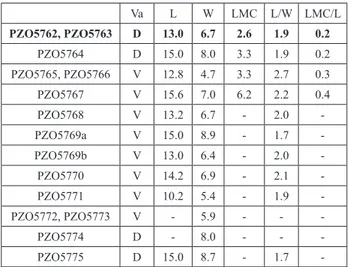
Measurements. (in mm; abbreviations: D,
dorsal valve; L, length, LMC, length of mantle cav-ity; V, ventral valve; Va, type of valve; W, width; in bold the holotype; see the scatter diagram in Fig. 4).
Va L W LMC L/W LMC/L PZO5762, PZO5763 D 13.0 6.7 2.6 1.9 0.2 PZO5764 D 15.0 8.0 3.3 1.9 0.2 PZO5765, PZO5766 V 12.8 4.7 3.3 2.7 0.3 PZO5767 V 15.6 7.0 6.2 2.2 0.4 PZO5768 V 13.2 6.7 - 2.0 -PZO5769a V 15.0 8.9 - 1.7 -PZO5769b V 13.0 6.4 - 2.0 -PZO5770 V 14.2 6.9 - 2.1 -PZO5771 V 10.2 5.4 - 1.9 -PZO5772, PZO5773 V - 5.9 - - -PZO5774 D - 8.0 - - -PZO5775 D 15.0 8.7 - 1.7
-Discussion. Trentingula prinothi n. gen n. sp.
shares with T. lorigae n. gen. n. sp. the shell
micro-structure of virgose type, the disposition of the dorsal umbonal and anterior lateral muscle scars. However, significant differences are also detectable.
Trentingula prinothi has a larger shell with a shorter
mantle cavity, and a shallow median ridge on the dorsal anterior extension of visceral cavity. Besides, the scars of the dorsal and ventral umbonal muscle are larger. The anterior extension of the visceral cav-ity is laterally surrounded by a groove, which con-nects the anterior lateral muscle scar with the central muscle scar. This muscle scar arrangement around the anterior visceral area has been also described in the Cretaceous Credolingula olferievi Smirnova, 2001
(in Smirnova & Ushatinskaya 2001), which shares with the Dolomites species a similar arrangements of the dorsal umbonal scars, shell microstructure
PLATE 4
Shell microstructures of Trentingula prinothi n. gen. n. sp.,
Bellero-phon Formation, upper Permian, Monte Balest, Ortisei (for abbreviations see the caption of Pl. 3).
Figs 1-6 - 1) Oblique fracture of an unknown type of valve; 2) de-tail of the secondary layer; 3, 5) dede-tails of virgose laminae with rods almost parallel to the shell surface; 4, 6) details of compact laminae of the secondary shell layer.
Figs 7-9 - 7) Oblique fracture of a brachial valve; 8) detail of the primary layer; 9) internal part of the secondary layer with a multi-layered structure originated by alternations of densely and loosely packed apatite elements.
Figs 10-12 - 10) Vertical fracture of a dorsal valve showing the se-condary layer with alternations of compact, bored by ca-nals, and virgose laminae; 11) contact between virgose (left) and compact laminae (right); 12) a virgose lamina with lo-osely packed botryoidal and short rods-like aggregates of granular apatite.
and a median slit on the anterior extension of the visceral cavity. However, the Cretaceous species differs from T. prinothi n. gen. n. sp. because of
its radial plications occurring in both valves and an irregular-trapezoidal ventral umbonal muscle scar bisected by a narrow groove produced by the almost conjoined pedicle nerves (Smirnova and Ushatinskaya 2001).
Trentingula prinothi has a shell
microstruc-ture similar to the Carboniferous “Lingula” squa-miformis Phillips, with a secondary layer formed
by a succession of compact and virgose lami-nae respectively constituted by well compacted spherular - botryoidal granules and loose aggre-gates of cylindroid - botryod granules (Cusack & Williams 1996). “Lingula” squamiformis is,
howev-er, characterized by radial folds (Graham 1970). Another similar English Carboniferous species, “Lingula” mytilloides J. Sowerby, differs from T. prinothi n. gen. n. sp. by a more elliptical outline
and fine radial striae. “Lingula” credneri Geinitz,
a widespread Permian species, shows an ellip-tical outline, which also characterizes the most part of Permian Asian and American lingulide species (Archbold 1981). Shell microstructure and internal characters of Geinitz’s species are unknown. “Lingula” occidentaustralis Archbold
has lateral margins almost straight and parallel in mature individuals and a broad median ridge on the dorsal valve (Archbold 1981). T. prinothi
n. gen. n. sp. shares with the Permian “ Lingu-la” arctica Miloradovich a similar outline, muscle
scar arrangement and shell microstructure, but the latter species is characterized by short vascula media which, despite the good preservation of
the types, are not detectable and, therefore, are lacking in the Dolomites shells. The occurrence of vestigial vascula media is the main
taxonomi-cal character of Semilingula (Popov in Egorov &
Popov 1990).
Trentingula mazzinensis n. gen. n. sp. Pl. 2, Figs 1a, b, 2a-c; Pl. 5, Figs 1-12 1980 Lingula sp. - Broglio Loriga et al., pls 1-3.
1990 Lingula sp. - Broglio Loriga et al., pl. 2, fig. 4.
2014 Lingularia yini (Peng and Shi) - Posenato et al., figs 3c-f Diagnosis: Small sized shell with an elongated outline of variable shape, longer than wide; lateral margins slightly arched;
length of ventral visceral area about one-half of valve length; length of dorsal visceral area about 60 per cent of valve length. Anterior extension of dorsal visceral cavity thick, with a forked tongue shape, anteriorly divided by a median slit connected to a very shallow and short drop-like depression. Shell microstructure with a virgose secondary layer originated by an alternation of coarse and fine grained spherules, botryoids and short rods.
Etymology: From the name of the Mazzin Village (Fas-sa Valley, Trento Province, Dolomites) and the homonymous member of Werfen Formation, in which this new species is very common.
Holotype: MPL 5923/3, a dorsal valve and its internal mould (Pl. 2, Figs 2a, c). The holotype is from a sample consi-sting of a bed surfacein which tens of disarticulated valves, of the same species, are preserved. The bed surface was figured by Broglio Loriga et al. (1980, pl. 2). The sample was collected near Malga Panna (Moena, Fassa Valley, Trento Province).
Paratypes: 11 ventral valves and 4 dorsal valves listed in the Tab. 3. They lie on the same bed surface containing the holotype.
Description. The shell has a maximum
length of about 5 mm, and is about two times as long as wide. The length/width ratio ranges from 1.6 to 2.2. The valves are flattened, with the maximum height at valve mid-length, and al-most equivalve, with a variable outline ranging from drop-like to elongated oval. The anterior margin has a rounded outline. The maximum width is located from the anterior quarter to the mid-length. The ornamentation consists of irre-gularly spaced and weak growth lines.
PLATE 5
Shell microstructures of Trentingula mazzinensis n. gen. n. sp., Mazzin
Member, Werfen Formation, Lower Triassic, Malga Panna, Moena (for abbreviations see the caption of Pl. 3).
Figs 1-4 - 1) Vertical fracture of an unknown type of valve with pri-mary and secondary shell layers; 2) detail of the pripri-mary la-yer, with vertical cleavage, made up by spherular apatite and plates, which probably representing recrystallized patches of tension-cracked apatite; 3) detail of the secondary layer with a compact fabric represented by fine grained spheroids; 4) detail of a virgose lamina with short rod-like aggregates. Figs 5-8 - 5) Vertical fracture of an unknown type of valve; 6) detail of the primary layer with a compact fabric; 7, 8) details of the secondary shell layer mainly represented by laminae with a compact fabric originated by a granular fabric of sphe-roids and botryoids.
Figs 9-12 - 9) Vertical fracture of an unknown type of valve; 10) detail of the primary layer with a granular texture affected by vertical cleavages; the plates probably represent recrystal-lized patches of tension-cracked apatite; 11) detail of the secondary layer with alternations of compact and virgose laminae; 12) detail of the compact fabric.
The interior of ventral valve shows a short ventral visceral cavity, generally not exceeding the valve mid-length, with two parallel low me-dian ridges (Pl. 2, Fig. 1a, b), which are anteriorly flanked by the central muscle scars. The mantle cavity is long, occupying about 50 per cent of the total valve length. Transmedian and anterior lateral muscle scars are rarely detectable (Pl. 2, Fig. 1a, b). The ventral pseudointerarea, pedicle valve groove, vascula lateralia and other muscle
scars are not detectable.
The anterior extension of the dorsal visce-ral area is slightly raised and bisected, at the an-terior extremity, by a median furrow (Pl. 2, Fig. 2a, b). The latter is connected to a very shallow drop-like depression, possibly corresponding to the scar of anterior lateral muscles. Central mu-scle scars with an oval outline. The mantle cavi-ty occupies about 40 per cent of the total valve length. Vascula lateralia, umbonal muscle scars
and other muscle scars are not preserved or de-tectable, because a very small shell thickness.
The shell is very thin, ca. 50 μm thick. The primary layer is about 10 μm thick and consists of a granular fabric originated by moderately packed spheroid and botryoid aggregates (Pl. 5, Figs 1, 2). The aggregates show a vertical dispo-sition, probably originated by cleavage (Pl. 5, Fig. 10), as suggested by the occurrence of vertical plates (PL, Pl. 5, Fig. 10), representing recrystal-lized patches of tension-cracked apatite (sensu Cusack & Williams 1996). The secondary layer, about 40 μm thick, is formed by an alternation of coarse and fine grained spherules, botryoids and short rods (Pl. 5, Figs 3, 4). The compact laminae consists of densely packed botryoids and spherules (Pl. 5, Fig. 8), 1- 3 μm thick. The compact laminae are separated by thin virgose laminae (200-400 nm thick) with moderately pa-cked small spherules and short rods (200-300 nm long) (Pl. 5, Fig 4).
Occurrence. Trentingula mazzinensis n. gen.
n. sp. is common throughout the Mazzin
Mem-ber of the Werfen Formation of the Dolomites (Southern Alps, Italy). It is also present in the lowermost Aracs Marl Formation of Transdanu-bian Central Range (Hungary; Broglio Loriga et al. 1990, pl. 6, fig. 1).
Measurements. (in mm; abbreviations: D,
dorsal valve; L, length; LMC, length of mantle
cavity; V, ventral valve; Va, type of valve; W, width; in bold the holotype; see the scatter dia-gram in Fig. 4). Va L W LMC L/W LMC/L MPL 5923/1 V 4.7 2.6 2.4 1.8 0.5 MPL 5923/2 V 5.0 2.8 2.8 1.8 0.6 MPL 5923/3 D 4.2 2.4 1.6 1.8 0.4 MPL 5923/4 D 4.7 2.6 1.8 1.8 0.4 MPL 5923/5 D 4.5 2.5 1.6 1.8 0.4 MPL 5923/6 V 5.2 3.0 2.8 1.7 0.5 MPL 5923/7 V 4.2 2.5 2.2 1.7 0.5 MPL 5923/8 D 3.5 2.2 1.2 1.6 0.3 MPL 5923/9 V 4.7 3.1 2.1 1.5 0.4 MPL 5923/10 V 4.7 2.4 2.6 2.0 0.6 MPL 5923/11 V 4.7 2.4 2.4 2.0 0.5 MPL 5923/12 V 4.7 2.6 2.5 1.8 0.5 MPL 5923/12 V 4.7 2.4 2.4 2.0 0.5 MPL 5923/12 V 4.8 2.8 2.5 1.7 0.5 MPL 5923/12 V 4.7 2.5 2.4 1.9 0.5 MPL 5923/12 D 4.1 2.5 1.6 1.6 0.4
Discussion. Trentingula mazzinensis n. gen. n.
sp. is proposed for the small sized lingulides of the lower Induan Mazzin Member. Their shell minia-turization records the “Lilliput effect”, which re-present the survival strategy of opportunistic bio-ta during the most severe marine environmenbio-tal conditions after the end Permian mass extinction (e.g., Twitchett 2007; Metcalfe et al. 2011; Posenato et al. 2014). The small size and the very thin shell prevent the recognition of many taxonomical cha-racters. However, the detectable features occurring on the inner valve surfaces and shell microstructu-res suggest a strong affinity with T. lorigae n. gen. n.
sp., from which it differs mainly by a smaller shell
size. The size of Trentingula mazzinensis n. gen. n.
sp. is about half of the latter (e.g., Broglio Loriga et al. 1980; Posenato et al. 2014). In the Dolomites, these species are located in different stratigraphic units. Trentingula mazzinensis n. gen. n. sp occurs in
the lower Induan Mazzin Member, while T. lorigae
n. gen. n. sp occurs in younger units, ranging from the upper Induan to Olenekian.
The thick anterior extension of the dorsal vi-sceral area could be interpreted, as sometime sug-gested in literature (e.g. Peng & Shi 2008; Holmer & Bengston 2009), as the scars of the anterior la-teral muscle. Following this interpretation, the an-terior bifurcation of the visceral extension would
indicate that the anterior lateral muscle scars were separated, which represents the most important taxonomical character of Sinolingularia. However,
no trace of muscle scar is detectable on the raised anterior part of dorsal visceral extension, whereas its median anterior furrow is anteriorly connected to a very shallow depression, which is detectable only in few a specimens (e.g., the holotype, Pl. 2, Fig. 2a-c) due to the very thin shell. This slightly perceptible depression has a shape similar to that occurring in the type-species of Trentingula where
it is interpreted as the scar of conjoined anterior lateral muscles (see above). All the other detecta-ble internal morphological features of these small and thin lingulides (e.g., position and shape of the dorsal central and ventral anterior lateral and transmedian muscle scars) are compatible with the taxonomical characters of Trentingula.
c
onclusIvereMarksThe lingulides under examination have been assigned to Trentingula n. gen., which is
characteri-zed by: a) a shell with a virgose fabric of the se-condary layer; b) disjoined and asymmetrical dorsal umbonal muscle scars; c) ventral umbonal muscle scar divided into two parts, separated by subparal-lel pedicle nerve grooves; d) a single dorsal ante-rior lateral muscle scar, without median septum.
Trentingula n. gen. is represented by three different
species, with different stratigraphic setting, ran-ging from the Changhsingian to the Induan (end Permian mass extinction interval), whose main ta-xonomical characters are represented by the shape of the anterior projection of the dorsal visceral cavity, the ventral umbonal muscle scar and the shell size.
On the basis of the shell microstructure,
Trentingula belongs to the virgose lineage, now
represented by Lingula, while the morphology of
the inner shell surface (e.g. the separation of the umbonal muscle scars) would suggest its exclusion from the Lingulidae, as until now defined in the literature. For this reason the diagnosis of that fa-mily has been here emended to avoid taxonomical splitting. The available data on shell microstruc-tures and inner morphology seem to be still frag-mentary and, in many cases, based on species for which integrated information on microstructures
and morphology is not available (Tab. 1).
Biernat & Emig (1993) proposed, on the ba-sis of inner shell morphology, that the extant gene-ra Lingula and Glottidia originated in the Cenozoic
from Lingularia, which differs from the living genera
mainly because of the umbonal muscle scar shape. The ventral umbonal muscle scar of Lingularia is
considered unpaired and described as heart-like in shape (Biernat & Emig 1993; Holmer and Popov 2000), although it is divided into two parts by joi-ned pedicle nerves (Biernat and Emig 1993, fig. 3E). The shape of the dorsal umbonal scar is not clearly detectable in the illustrated types of Lingularia simi-lis Biernat & Emig (type-species of Lingularia). The
dorsal umbonal scar is, however, well detectable in a specimen of the Lower Cretaceous Lingularia smirnovae Biernat & Emig. In this specimen, the scar
is clearly asymmetrical and divided into two parts, with the left part larger than the right one (Biernat & Emig 1993, fig. 7C). A paired disposition of the umbonal muscle scars is also detectable in a dorsal valve of Lingularia similis?, lowermost Cretaceous in
age (Holmer & Nakrem 2012, fig. 4).
The dorsal umbonal muscle scar of upper Pa-leozoic and Mesozoic lingulides (Tab. 1) has a more or less asymmetrical horseshoe-shape or it is paired. Species with the latter disposition are those illustra-ted by excellent photographs, while the former di-sposition is often based on interpretative drawings, which are not clearly detectable in the photographs. The difficulty to detect the horseshoe-shape in the photographs could be related to the high curvature of the umbonal region, which covers the posterior walls. However, when the posterior connection of the two lateral branches is very narrow, it could re-present the perimial line, and an asymmetrical paired disposition of the dorsal umbonal scars seems to be the better definition, as here applied to Trentingula.
The ventral umbonal muscle scar is generally very weakly impressed and its outline is difficult to recognize in the photographed valves. The ventral umbonal scar of almost all lingulides (Tab. 1) has been described as heart-like in shape. However, it appears as a couple of scars, bisected by disjoined or joined pedicle nerve grooves. The groove passes to the right of a single ventral umbonal scar, with a disposition typical of the Cenozoic lingulides, only in the Upper Cretaceous Lingularia? notialis Holmer
and Bengston (Holmer & Bengston 2009). Therefo-re, most Mesozoic lingulides have separate umbonal