Research Article
Type 1 Choroidal Neovascularization Evolution by Optical
Coherence Tomography Angiography: Long-Term Follow-Up
Marco Rispoli,
1Maria Cristina Savastano
,
2,3Bruno Lumbroso,
1Lisa Toto
,
4and Luca Di Antonio
41Centro Italiano Macula, Rome, Italy
2UOC Oftalmologia, Fondazione Policlinico Universitario A. Gemelli IRCCS, Rome, Italy 3Università Cattolica del Sacro Cuore, Rome, Italy
4Ophthalmology Clinic, National High-Tech Center in Ophthalmology, Italian School of Robotics in Ophthalmology, University“G.
d’Annunzio” of Chieti-Pescara, Chieti, Italy
Correspondence should be addressed to Maria Cristina Savastano; [email protected] Received 28 January 2020; Revised 1 April 2020; Accepted 6 April 2020; Published 25 April 2020 Academic Editor: Mitsuru Nakazawa
Copyright © 2020 Marco Rispoli et al. This is an open access article distributed under the Creative Commons Attribution License, which permits unrestricted use, distribution, and reproduction in any medium, provided the original work is properly cited. Purpose. To evaluate structural changes in response to antivascular endothelial growth factor (anti-VEGF) treatment in patients with long-term type 1 choroidal neovascularization (CNV) by optical coherence tomography (OCT) and OCT angiography (OCTA). Method. This is a longitudinal study that involved a total of 51 eyes with type 1 CNV (35 female and 16 male eyes). Structural OCT and OCTA were performed on all the subjects. AngioVue OCTA (XR Avanti, Optovue, Inc., Fremont, CA) was used to obtain qualitative and quantitative information. All eyes were treated with an anti-VEGF ProReNata (PRN) approach and were followed for a mean of 38.9 months (SD ± 7:22). Best-corrected visual acuity (BCVA) was assessed at each follow-up timepoint. Results. We observed two kinds of possible evolution of type 1 CNV:“positive evolution,” including stabilization in 20% of patients and chronicity in 35%, and“negative evolution,” in which fibrosis was shown in 18% of patients, chorioretinal atrophy in 25%, and hemorrhage or RPE tears in 2%. The mean BCVA at baseline was33:67 ± 15:85 ETDRS letters; after 1 and 2 years, it was 31:61 ± 18:04 and 31:18 ± 18:58 ETDRS letters, respectively. The mean BCVA at the end of follow-up was 25:27 ± 20 ETDRS letters. The difference between the values at baseline and at the end of follow-up was not statistically significant (P = 0:06, r2= 0:10). Conclusions. This study describes an in vivo structural long-term evolution of type 1 CNV by OCT and OCTA. Different possible CNV outcomes were observed. This study suggests that new retinal imaging techniques could be useful tools for assessing the potential retinal changes in the evolution of type 1 CNV to develop personalized medicine. Further studies using OCTA in the long term are needed to better understand why similarly treated type 1 CNV cases evolve differently and produce different results.
1. Introduction
Noninvasive dyeless optical coherence tomography angiog-raphy (OCTA) is a clinical technique that is spreading rap-idly all over the world, as it is safer, easier, and faster than fluorescein angiography (FA) and indocyanine green angiog-raphy (ICG) [1, 2]. Structural OCT highlights alterations in the morphology and structure of the retinal layers. OCTA provides images of bloodflow in the retina and choroid with a high level of detail. In contrast, FA cannot show the vascu-lar layers of blood vessels as deep as the capilvascu-lary plexuses,
which are well evidenced by OCTA [3]. This allows for sev-eral potential options of disease analysis, the research of dif-ferent disorders, and the evaluation of new treatments [4].
One of the first pathologies studied by OCTA was wet AMD. The dyeless visualization of new vessels was remark-able for a large number of researchers around the world. OCTA enables the understanding, quantification, and track-ing of the evolution after new vessel (NV) treatment.
CNV treatment should begin early, shortly after symp-toms appear and before the occurrence of extensive struc-tural damage. In the absence of a recognized guideline for
Volume 2020, Article ID 4501395, 8 pages https://doi.org/10.1155/2020/4501395
the treatment and evaluation of the timing of eyes with exuda-tive neovascularization, patients should be closely monitored for treatment and retreatment. Antivascular endothelial growth factor (anti-VEGF) treatment is universally recognized as providing positive results in the reduction of CNV activity and in maintaining good vision for patients for years [5–7].
Several trials (ANCHOR, MARINA, VIEW 1, and VIEW 2) have demonstrated visual improvement of approximately 10 letters at 2 years in eyes with neovascular AMD undergo-ing monthly anti-VEGF therapy [8–10]. Recently, 5-year results from the Comparison of Age-Related Macular Degen-eration Treatment Trial (CATT) study showed long-term visual deterioration with chronic anti-VEGF therapy [11].
Although several factors may play a role in causing vision loss in eyes that undergo long-term anti-VEGF therapy, the mechanism of this process remains poorly understood. One possible reason has been postulated by Dansingani and Freund, in which a mature tangled vascular pattern in type 1 lesions was determined to be a resistance factor to macular atrophy [12].
Recently, Christenbury et al. described a high level of macular atrophy development predominantly eccentric to the PED in long-term anti-VEGF therapy for eyes with type 1 NV secondary to AMD [13]. Despite studies reporting results of chorioretinal atrophy and a decrease in BCVA, sev-eral other studies have reported a maintained or increased BCVA and OCT morphology improvement [14, 15].
The potential chorioretinal involvement after anti-VEGF treatment led us to investigate the evolution of type 1 CNV in exudative AMD eyes, which we analyzed with structural OCT and OCTA in a long-term follow-up study.
This research project is aimed at studying the particular CNV morphological changes seen on OCTA at the end of the observational period.
2. Methods
This study adhered to the Declaration of Helsinki (52nd WMA General Assembly, Edinburgh, Scotland, October 2000), and written informed consent to participate in this study was routinely obtained from all examined patients. The IRB/ethics committee ruled that ethical approval was not required. In this cross-sectional study, fifty-one wet AMD eyes with type 1 CNV (35 female and 16 male eyes) detected by structural OCT according to a previous study [16] were evaluated. The mean age of the patients was 77.41 years, with a standard deviation (SD) of 12.39 years. All eyes were treated by an anti-VEGF ProReNata (PRN) approach and had follow-up every month. The duration of time followed for the entire cohort ranged from 31 months to 58 months, with a mean of 38.9 months (SD 7.22). The pharmacological agents ranibizumab and aflibercept were randomly chosen for administration. The number of intravit-real injections per eye during the course of treatment ranged from 3 to 29 injections, with a mean of 11.86 injections (SD 6.64). The patient demographics are listed in Table 1.
The exclusion criteria included media opacity and con-comitant diseases such as diabetic retinopathy, vein or artery occlusion, glaucoma, any evidence or suspicion of type 2
and/or type 3 CNV, polypoidal choroidal vasculopathy, and any history of photodynamic therapy or macular laser ther-apy. Patients who presented with cataracts were followed without surgery because they did not have a clinically signif-icant increase over time. All patients underwent a baseline ophthalmic examination, including medical and ocular his-tory, family medical hishis-tory, measurement of best-corrected visual acuity (BCVA) expressed in Early Treatment of Dia-betic Retinopathy Study (ETDRS) letters, slit-lamp examina-tion of the anterior and posterior segments, measurement of intraocular pressure, and dilated fundus examination. All eyes were imaged with an AngioVue OCTA (XR Avanti, Optovue, Inc., Fremont, CA) as the collected CNV assess-ment. The structural OCT protocol pattern used centered the B-scan line and crossline onto the fovea. The OCTA pro-tocol used centered3 × 3 mm2and6 × 6 mm2grids onto the fovea. OCTA software programs automatically analyze reti-nal layer scans at different depths, providing images that are rich in details. With OCTA technology, the same tissue area is imaged repeatedly, and the differences between the scans are analyzed, thus allowing one to detect zones with high-flow neovascular rates. In cases of segmentation errors, manual editing of the layers was performed if deemed neces-sary for a correct interpretation. We classified all CNVs as inactive using biomarkers of CNV activity described by Al-Sheikh et al. [17] The OCTA scans that better represented the CNV features were selected and considered for analysis. We have chosen the images that agreed between CNV fea-tures and greater flows in B-scan. If the high flows were observed in the outer retina, the OCTA images in the outer retina were chosen. In case of mainflows shown in the sub-RPE area, the OCTA scans were selected at this level. All images were analyzed by two of the authors (B.L. and M.C.S.) on two separate occasions to ensure accuracy of the grading. In cases of disagreement, both readers reanalyzed the images, and a consensus was obtained. The OCTA images analyzed were taken at the last visit. For statistical analysis, one-way ANOVA followed by a Holm-Sidak multi-ple comparison test was performed using GraphPad Prism (version 6.00 for Windows, GraphPad Software, La Jolla, Cal-ifornia, USA: https://www.graphpad.com). Pearson coe ffi-cient correlation was used to correlate BCVA and the number of injections. Spearman coefficient correlations were calculated between BCVA and morphological CNV details.
P < 0:05 was considered statistically significant.
3. Results
The enrolled eyes included both naïve eyes and those previ-ously treated with anti-VEGF. The mean BCVA at baseline was33:67 ± 15:85 ETDRS letters; at the end of the study, it was25:27 ± 20 ETRDS letters. The difference was not statis-tically significant (P = 0:06, r2= 0:10).
Almost half of the participants had stable BCVA, although there were some eyes in which BCVA decreased dramatically. Two possible visual acuity patterns can be observed in CNV evolution: increased or stable (positive evo-lution) or decreased vision (negative evoevo-lution) (Table 2).
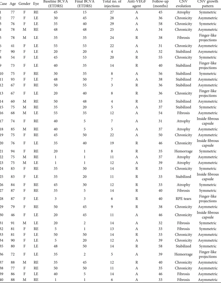
Table 1: Patient demographic characteristics, baseline and final visual acuity, anti-VEGF therapy details, and follow-up interval. Case Age Gender Eye Baseline BCVA
(ETDRS) Final BCVA (ETDRS) Total no. of injections Anti-VEGF agent Follow-up (months) CNV evolution CNV growth pattern 1 77 F RE 45 15 27 A 45 Atrophy Symmetric 2 77 F LE 30 45 28 A 36 Chronicity Asymmetric 3 76 F LE 35 40 29 A 58 Chronicity Symmetric 4 78 M RE 48 48 25 A 34 Chronicity Asymmetric 5 78 M LE 35 35 24 R 38 Fibrosis Finger-like projections 6 41 F LE 55 53 22 A 31 Chronicity Asymmetric 7 90 F LE 20 20 4 A 32 Stabilized Asymmetric 8 54 F LE 45 53 20 R 55 Chronicity Symmetric 9 73 F LE 40 35 14 R 40 Stabilized Finger-like projections 10 75 F RE 30 35 8 A 56 Stabilized Symmetric 11 93 F LE 48 50 7 A 38 Stabilized Asymmetric 12 67 F RE 50 55 3 R 36 Stabilized Asymmetric 13 67 F LE 20 40 8 R 36 Chronicity Finger-like projections 14 60 M RE 50 48 4 R 33 Stabilized Asymmetric 15 75 M RE 35 20 5 A 37 Stabilized Symmetric 16 68 M LE 55 35 12 A 54 Fibrosis Asymmetric 17 74 F RE 40 5 7 A 31 Atrophy Insidecapsulefibrous 18 85 M RE 40 5 5 A 37 Atrophy Asymmetric 19 75 F RE 45 50 22 A 50 Chronicity Asymmetric 20 76 F LE 35 40 19 R 46 Chronicity Insidefibrous
capsule 21 94 F RE 20 1 8 R 35 Hemorrage Symmetric 22 75 M RE 1 1 11 A 37 Atrophy Asymmetric 23 75 M LE 1 1 12 A 39 Atrophy Asymmetric 24 83 F RE 35 30 14 R 33 Chronicity Symmetric 25 83 F LE 35 20 11 R 33 Stabilized Insidefibrous
capsule 26 84 F RE 45 30 12 R 33 Atrophy Symmetric 27 87 F RE 35 5 14 R 40 Fibrosis Symmetric 28 87 F LE 3 3 6 R 40 RPE tears Finger-like projections 29 79 F RE 50 45 9 R 38 Chronicity Asymmetric 30 46 F LE 20 45 11 A 46 Chronicity Insidefibrous
capsule 31 91 M LE 20 2 14 A 32 Fibrosis Symmetric 32 81 F RE 5 1 13 A 33 Fibrosis Symmetric 33 81 F LE 50 50 14 R 33 Chronicity Asymmetric 34 90 F LE 5 20 12 A 39 Chronicity Asymmetric 35 80 F LE 48 50 14 R 58 Stabilized Symmetric 36 72 F LE 35 2 5 A 39 Hemorrage Finger-like projections 37 88 M RE 35 45 12 R 40 Chronicity Asymmetric 38 77 F RE 50 50 11 A 35 Chronicity Asymmetric 39 86 F LE 40 5 14 A 46 Fibrosis Asymmetric 40 88 M RE 1 1 6 A 33 Fibrosis Asymmetric
3.1. Positive Evolution: Stabilization and Chronicity. We observed positive evolution in 55% of the patients, consisting of CNV stabilization (20%) and CNV chronicity (35%). We considered CNV stabilization as long-term remission and an absence offluid or hemorrhaging for more than 6 months; the CNV seemed to stop developing, and no activity signals were seen. The clinical appearance showed no exudation or fluid occurrence (Figure 1). Even if there was no CNV exuda-tion, the CNV area was larger at the end of the observation period, growing from 0.68 mm2to 1.68 mm2.
CNV chronicity was considered when the neovasculari-zation was consistently responsive to anti-VEGF treatment but required repetitive reinjections. In these cases, acute
dis-ease developed into chronic disdis-ease. The CNV was often qui-escent with consistent and frequent recurrences (Figure 2). The CNV area was consistently larger at the end of the obser-vation period, growing from 0.7 mm2to 1.4 mm2.
3.2. Negative Evolution: Fibrosis, Atrophy, Hemorrhage, or RPE Tears. Negative evolution was observed in 45% of the cases, which included fibrosis (18%) (Figure 3), atrophy
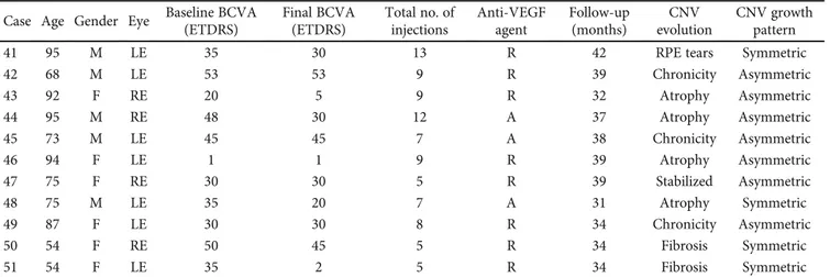
Table 1: Continued. Case Age Gender Eye Baseline BCVA
(ETDRS) Final BCVA (ETDRS) Total no. of injections Anti-VEGF agent Follow-up (months) CNV evolution CNV growth pattern 41 95 M LE 35 30 13 R 42 RPE tears Symmetric 42 68 M LE 53 53 9 R 39 Chronicity Asymmetric 43 92 F RE 20 5 9 R 32 Atrophy Asymmetric 44 95 M RE 48 30 12 A 37 Atrophy Asymmetric 45 73 M LE 45 45 7 A 38 Chronicity Asymmetric 46 94 F LE 1 1 9 R 39 Atrophy Asymmetric 47 75 F RE 30 30 5 R 39 Stabilized Asymmetric 48 75 M LE 35 20 7 A 31 Atrophy Symmetric 49 87 F LE 30 30 8 R 34 Chronicity Asymmetric 50 54 F RE 50 45 5 R 34 Fibrosis Symmetric 51 54 F LE 35 2 5 R 34 Fibrosis Symmetric
RE: right eye; LE: left eye; A: aflibercept; R: ranibizumab; BCVA: best-corrected visual acuity; F: female; M: male.
Table 2: Type 1 CNV long-term evolution.
Evolution of disease NV Evolution % TOT (%) Positive evolution Stabilized NV 20 55
Chronic NV 35
Negative evolution
Fibrosis 18 45 Atrophy 25 Hemorrhage or RPE tears 2
NV: neovascularization; RPE: retinal pigment epithelium. Structural OCT OCT angiography
Figure 1: Stabilized CNV. Structural OCT shows the absence of intraretinal exudative details with an irregular profile of the RPE in the foveal region. OCTA revealed a flow signal with a well-defined outline and the absence of a dark halo. This eye received 3 intravitreal injections and became stable for more than 25 months (Case number 7).
OCT angiography Structural OCT
Figure 2: Chronic CNV. Structural OCT shows the presence of exudations with intraretinal cystic spaces. Stratified hyperreflective material below the RPE can be observed in the foveal region. OCTA revealed the presence of aflow signal with growth of thin capillary leaves and a dark halo around the CNV. The dotted yellow outline highlights the dark halo. These eyes required multiple treatments to remain stable (Case number 13).
Structural OCT OCT angiography
Figure 3: Fibrotic CNV. Structural OCT shows the presence of minimal exudation above the hyperreflective material below the RPE. Bruch’s membrane is well evidenced, as well as the choroidal vessels. OCTA revealed the presence of a large flow signal with a main trunk and a “dead tree” feature. This eye had poor visual acuity, which did not improve after injection (Case number 17).
(25%) (Figure 4), and hemorrhage or RPE tears (2%) (Figure 5).
Long-term monitoring of CNV evolution showed that the new vessels become larger, thicker, and straighter. No thin capillaries orfine loops were visible. For any evolution type, the vessel area was larger after treatment than before treatment (Figure 6).
After each treatment, the same main vessels appeared to return with increasedflow and decreased branch density. It appeared as though some of the main branches were less affected by the treatment. As previously described by Spaide, the onset of a complex pattern after treatment induced a less complex feature of CNV, arterialization, to become detect-able [18]. We defined this morphological pattern as a “matu-ration pattern” (Figure 7).
Similarly, in agreement with the results of the study by Xu et al., we observed 3 CNV growth patterns: symmetric growth, asymmetric growth, and finger-like projections [19]. Furthermore, we observed a new entity of CNV growth, “inside the fibrous capsule.” This CNV grows in vascular density but not in area. In this specific case, the CNV grew in vascular density inside afibrous capsule (Figure 8).
Correlation between BCVA and number of injections was not statistically significant (P = 0:23, r2= 0:02) as well the cor-relation between CNV evolution (P = 0:06, r2= −0:23) and CNV growth pattern (P = 0:69, r2= −0:05).
4. Discussion
Although OCT angiography continues to be developed, it is useful for several visual disorder indications, particularly in the management of AMD. The analysis of neovascularflow without dye injection with OCTA allows detailed monitoring of the different CNV evolutions. Our results show that type 1 CNV has various evolution patterns, which were analyzed over a 4-year observation period.
During long-term type 1 CNV evaluation, recurrence was frequent, and we observed two dissimilar evolutions, positive evolution and negative evolution, occurring independent of the treatment [20].
Type 1 CNV“positive evolution,” manifesting as stabili-zation or improvement, corresponded to 20% of cases, while
that manifesting as chronicity corresponded to 35% of cases. “Negative evolution” included fibrosis, which was observed in 18% of eyes, chorioretinal atrophy, which was observed in 25% of eyes, and hemorrhage or RPE tears, which was observed in 2% of eyes.
In almost all eyes, after the loading phase of the 3 intravit-real anti-VEGF injections, the disappearance of CNV ramifi-cations but not of the main CNV trunk could be observed by OCTA. Thesefindings suggested that it would be very diffi-cult to predict the prognosis from OCTA findings after 3 loading doses.
Most eyes with chronic evolution had periodic reactiva-tion after treatment, with a periodicity of 50 to 60 days after each intravitreal injection. Before the first injection and between recurrences, we observed a dark halo around the CNV of approximately 50 microns in diameter. Although the meaning of the dark halo is still controversial [21–24], in our opinion, it is due to blood sequestering by neovascu-larization reactivation; an increased dark halo means CNV growth [25].
The cycles seemed to be quite regular. After each treat-ment, the same main vessels appeared to return with increased flow and decreased branch density. The normal cyclic recurrence was extensive and generally global, although it could be localized to a segment of the CNV. In a few cases, acute nonperiodic reactivation occurred inde-pendently from treatment. This type of reactivation could take the shape of a shoot, bud, sprout, or outgrowth and may have had a specific location: terminal, axillary, lateral, fingerlike, or adventitious.
The retinal effect of repeated anti-VEGF treatments is still controversial. However, Christenbury et al. recently described that the multilayered PED aspect after chronic VEGF suppres-sion in type 1 CNV may confer a protective effect on the over-lying retinal pigment epithelium and outer retina [13].
The ability of OCTA to assess and quantify CNV may highlight activity biomarkers and guide the evaluation, treat-ment, and monitoring of neovascularization.
According to our previous study and to a recent observa-tion by Al-Sheikh et al., the morphological evaluaobserva-tion of CNV by OCTA can distinguish nonactive CNV lesions from exudative CNV lesions [17, 20].
Structural OCT OCT angiography
Figure 4: CNV associated with atrophy. Structural OCT shows minimal exudation as intraretinal cystic spaces and subretinal fluid above and hyperreflective material below the RPE (yellow asterisks). Foveal backscattering is well observable for the RPE atrophy behind the choroidal vessels. OCTA reveals the presence of a round flow signal with a growth capillary fringe. This eye required injections, but the presence of atrophy compromised visual recovery (Case number 22).
Structural OCT OCT angiography
Figure 5: CNV with RPE tears. Structural OCT shows deconstruction of the neuroretinal tissue and large pigment epithelium detachment associated with RPE tearing. The backscattering close to the RPE indicates the tear margin. Exudation is evident, with intraretinal cystic spaces and subretinal fluid above the RPE tear. OCTA highlights the presence of the 2 main vascular trunks with multiple growth capillary fringes (Case number 28).
Arterialization or Maturation pattern
(a) (b) (c)
Figure 7: Drawing of the CNV evolution after multiple treatments. Before treatment (a), the CNV has a greater proportion of small branching vessels and peripheral arcades, indicating an active lesion. After treatment (b), the same main vessels appear to return with increasedflow and decreased branch density (vessels in red). Some main branches are less affected by the treatment (vessels in black). After several anti-VEGF treatments, the CNV shows fewer complex features. This morphological pattern corresponds to the“maturation pattern” of treatment (c).
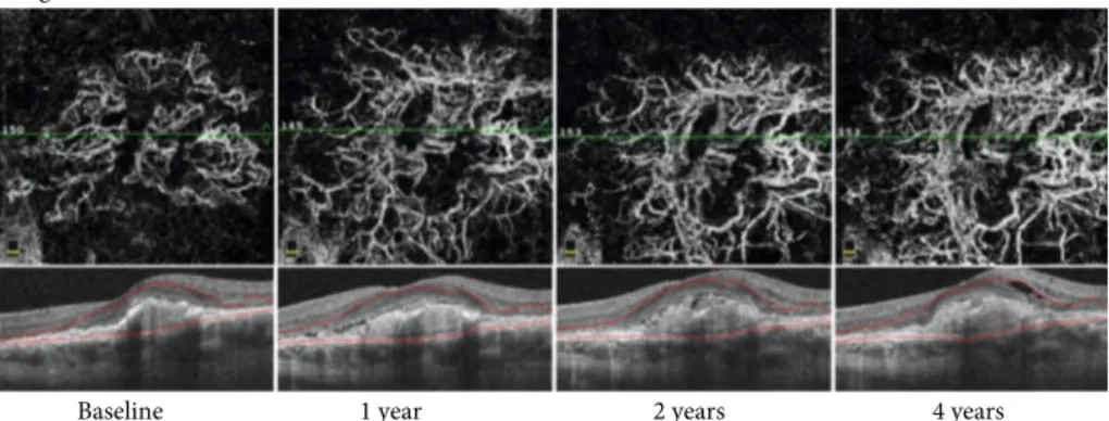
Angio retina multi scans view
Baseline 1 year 2 years 4 years
Figure 6: OCT angiography of type 1 CNV treated with anti-VEGF at different points in the long-term follow-up. The CNV became larger, thicker, and straighter over time. Thefinal NV area was larger than the CNV area before treatment, and the visual acuity worsened (Case number 32).
(a) (b)
(c) (d)
Figure 8: CNV growth patterns. (a) Asymmetric growth, (b) symmetric growth, (c) finger-like projections. (d) We observed a new type of CNV growth,“inside the fibrous capsule,” in which the vascular density increased instead of the vascular area.
In contrast to the results of previous studies [7], in our study, we observed BCVA reduction at the end of follow-up. Although we were unable to determine the real reason for the BCVA decrease, we propose 3 different hypotheses: undertreatment induced by the PRN treatment, the particu-lar aggressiveness of type 1 CNVs, and the lower starting visus compared to that in other studies.
In conclusion, type 1 CNV evolution can progress to dif-ferent outcomes: stabilization, chronicity, fibrosis, atrophy, hemorrhage, or RPE tears. Approximately half of the eyes in this study followed a positive evolution, while the other half became increasingly worse. We do not know why some CNV cases became stable, with the evolution and activity sig-nals coming to a halt; we also do not know why some CNV cases converted to chronicity. Similarly, it is unknown why some of the CNV cases had important growth, while others led to atrophy or severefibrosis. In the future, we hope that the use of OCTA will help to better define the morphological details in the development of type 1 CNV and develop per-sonalized medicine.
Data Availability
The data used to support thefindings of this study are avail-able from the corresponding author upon request and were included within the supplementary informationfile(s).
Additional Points
Précis. The long-term evaluation of type 1 choroidal neovas-cularization (CNV) showed different outcomes for different cases: stabilization, chronicity, fibrosis, atrophy, hemor-rhages, or retinal pigment epithelium (RPE) tears. We do not know why some CNV cases became stable, bringing their evolution and activity indicators to a standstill. Similarly, it is unknown why some CNV cases led to significant growth and others led to hemorrhage, atrophy, or severefibrosis. In the future, we hope that the use of optical coherence tomography angiography (OCTA) will better define the details of the development of type 1 CNV.
Conflicts of Interest
No conflicting relationship exists for any of the authors.
Authors
’ Contributions
Marco Rispoli and Maria Cristina Savastano contributed equally to this work.
References
[1] Y. Jia, O. Tan, J. Tokayer et al.,“Split-spectrum amplitude-decorrelation angiography with optical coherence tomogra-phy,” Optics Express, vol. 20, no. 4, pp. 4710–4725, 2012. [2] R. F. Spaide, J. M. Klancnik Jr., and M. J. Cooney,“Retinal
vas-cular layers imaged by fluorescein angiography and optical coherence tomography angiography,” JAMA Ophthalmology, vol. 133, no. 1, pp. 45–50, 2015.
[3] M. C. Savastano, B. Lumbroso, and M. Rispoli,“In vivo char-acterization of retinal vascularization morphology using opti-cal coherence tomography angiography,” Retina, vol. 35, no. 11, pp. 2196–2203, 2015.
[4] R. F. Spaide, J. G. Fujimoto, N. K. Waheed, S. R. Sadda, and G. Staurenghi,“Optical coherence tomography angiography,” Progress in Retinal and Eye Research, vol. 64, pp. 1–55, 2018. [5] J. Jacob, H. Brié, A. Leys et al.,“Six-year outcomes in
neovas-cular age-related maneovas-cular degeneration with ranibizumab,” International Journal of Ophthalmology, vol. 10, no. 1, pp. 81–90, 2017.
[6] M. G. Maguire, D. F. Martin, G.-s. Ying et al.,“Five-year out-comes with anti–vascular endothelial growth factor treatment of neovascular age-related macular degeneration: the compar-ison of age-related macular degeneration treatments trials,” Ophthalmology, vol. 123, no. 8, pp. 1751–1761, 2016. [7] S. Rofagha, R. B. Bhisitkul, D. S. Boyer, S. R. Sadda, K. Zhang,
and SEVEN-UP Study Group, “Seven-year outcomes in ranibizumab-treated patients in ANCHOR, MARINA, and HORIZON: a multicenter cohort study (SEVEN-UP),” Oph-thalmology, vol. 120, no. 11, pp. 2292–2299, 2013.
[8] D. M. Brown, M. Michels, P. K. Kaiser et al.,“Ranibizumab versus Verteporfin Photodynamic Therapy for Neovascular Age- Related Macular Degeneration: Two-Year Results of the ANCHOR Study,” Ophthalmology, vol. 116, no. 1, pp. 57– 65.e5, 2009.
[9] P. J. Rosenfeld, D. M. Brown, J. S. Heier et al.,“Ranibizumab for neovascular age-related macular degeneration,” The New England Journal of Medicine, vol. 355, no. 14, pp. 1419–1431, 2006.
[10] J. S. Heier, D. M. Brown, V. Chong et al.,“Intravitreal afliber-cept (VEGF trap-eye) in wet age-related macular degenera-tion,” Ophthalmology, vol. 119, pp. 2537–2548, 2006. [11] J. E. Grunwald, M. Pistilli, G. S. Ying et al.,“Growth of
geo-graphic atrophy in the comparison of age-related macular degeneration treatments trials,” Ophthalmology, vol. 122, no. 4, pp. 809–816, 2015.
[12] K. K. Dansingani and K. B. Freund,“Optical coherence tomog-raphy angiogtomog-raphy reveals mature, tangled vascular networks in eyes with neovascular age-related macular degeneration showing resistance to geographic atrophy,” Ophthalmic Sur-gery, Lasers & Imaging Retina, vol. 46, no. 9, pp. 907–912, 2015.
[13] J. G. Christenbury, N. Phasukkijwatana, F. Gilani, K. B. Freund, S. Sadda, and D. Sarraf,“Progression of macular atro-phy in eyes with type 1 neovascularization and age-related macular degeneration receiving long-term intravitreal anti-vascular endothelial growth factor therapy: an optical coher-ence tomographic angiography analysis,” Retina, vol. 38, no. 7, pp. 1276–1288, 2018.
[14] J. G. Garweg, J. J. Zirpel, C. Gerhardt, and I. B. Pfister, “The fate of eyes with wet AMD beyond four years of anti-VEGF therapy,” Graefe's Archive for Clinical and Experimental Oph-thalmology, vol. 256, no. 4, pp. 823–831, 2018.
[15] S. D. Adrean, S. Chaili, H. Ramkumar, A. Pirouz, and S. Grant, “Consistent long-term therapy of Neovascular age-related macular degeneration managed by 50 or more anti-VEGF injections using a treat-extend-stop protocol,” Ophthalmology, vol. 125, no. 7, pp. 1047–1053, 2018.
[16] K. B. Freund, S. A. Zweifel, and M. Engelbert,“Do we need a new classification for choroidal neovascularization in
age-related macular degeneration?,” Retina, vol. 30, no. 9, pp. 1333–1349, 2010.
[17] M. Al-Sheikh, N. A. Iafe, N. Phasukkijwatana, S. R. Sadda, and D. Sarraf,“Biomarkers of neovascular activity in age-related macular degeneration using optical coherence tomography angiography,” Retina, vol. 38, no. 2, pp. 220–230, 2018. [18] R. F. Spaide, “Optical coherence tomography angiography
signs of vascular abnormalization with antiangiogenic therapy for choroidal neovascularization,” American Journal of Oph-thalmology, vol. 160, no. 1, pp. 6–16, 2015.
[19] D. Xu, J. P. Dávila, M. Rahimi et al.,“Long-term progression of type 1 neovascularization in age-related macular degeneration using optical coherence tomography angiography,” American Journal of Ophthalmology, vol. 187, pp. 10–20, 2018. [20] B. Lumbroso, M. Rispoli, and M. C. Savastano,“Longitudinal
optical coherence tomography angiography study of type 2 naive choroidal neovascularization early response after treat-ment,” Retina, vol. 35, no. 11, pp. 2242–2251, 2015.
[21] G. J. Coscas, M. Lupidi, F. Coscas, C. Cagini, and E. H. Souied, “Optical coherence tomography angiography versus tradi-tional multimodal imaging in assessing the activity of exuda-tive age-related macular degeneration: a new diagnostic challenge,” Retina, vol. 35, no. 11, pp. 2219–2228, 2015. [22] Y. Jia, S. T. Bailey, D. J. Wilson et al.,“Quantitative optical
coherence tomography angiography of choroidal neovascular-ization in age-related macular degeneration,” Ophthalmology, vol. 121, no. 7, pp. 1435–1444, 2014.
[23] E. Moult, W. Choi, N. K. Waheed et al., “Ultrahigh-speed swept-source OCT angiography in exudative AMD,” Ophthal-mic Surgery, Lasers & Imaging Retina, vol. 45, no. 6, pp. 496– 505, 2014.
[24] G. Coscas, M. Lupidi, C. Cagini, and F. Coscas,“‘False-friend’ images on optical coherence tomography angiography: early choroidal neovascularization or artefact?,” Acta Ophthalmolo-gica, vol. 96, no. 2, pp. 200–202, 2018.
[25] M. Rispoli, M. C. Savastano, and B. Lumbroso,“Quantitative vascular density changes in choriocapillaris around CNV after anti-VEGF treatment: dark halo,” Ophthalmic Surgery, Lasers & Imaging Retina, vol. 49, no. 12, pp. 918–924, 2018.