3
UNIVERSITA’ DEGLI STUDI DEL PIEMONTE ORIENTALE
“Amedeo Avogadro”
DIPARTIMENTO di SCIENZE ed INNOVAZIONE TECNOLOGICA
Corso di Dottorato di Ricerca in CHEMISTRY & BIOLOGY
Energy, Enviromental and food sciences
XXXI ^ ciclo
“The role of different photoprotection mechanisms in
preventing photoinhibition of Photosystem II”
SSD: BIO/04
PhD student: Dr.ssa Romina Cannata
Supervisor: Prof. R. Barbato
6
TABLE OF CONTENTS
List of abbreviations …9 Abstract …10
1. INTRODUCTION …11
1.1 Oxigenic Photosynthesis: general aspects…12 1.1.2 Photosystem II…15
1.1.3 Photosystem I…16
1.1.4 Linear electron transport chain (LET)…18 1.2 Light and photosynthesis…19
1.3 Photoprotection…20
1.3.1 Light stress and photoprotection…20 1.3.2 Physical Photoprotection…21
1.3.3 Photoprotection mechanisms and mutants…22 1.3.3.1 Non Photochemical Quenching…22
1.3.3.2 Cyclic Electron Flow/Photosynthesis Control…23 1.3.3.3 State Transitions…28
1.3.4 Photohinibition and D1 protein turnover…29
1.3.5 Chl Fluorescence and fluorescence measurements…33
REFERENCE…37
2.OUTLINE OF THE THESIS…55 3. MATERIALS AND METHODS…58
3.1 Mutants…59
3.2 Treatment with lincomycin and light…60
3.3 Isolation of thylakoids…60 3.4 Chlorophyll measurement…62
7
3.5 Polyacrylamide gel electrophoresis and immunoblotting…62
3.6 Fluorescence decay…63
3.7 State transitions measurement…63
3.8 Pulse Amplitude Method measurements…65 3.9 Plant Efficiency Analyzer…66
REFERENCES…67
4.RESULTS…70
4.1 Mutants characterization…71
4.1.1 Mutants lacking CEF/PC are very sensitive to high light…82 4.2 Fluorescence decay measurement…83
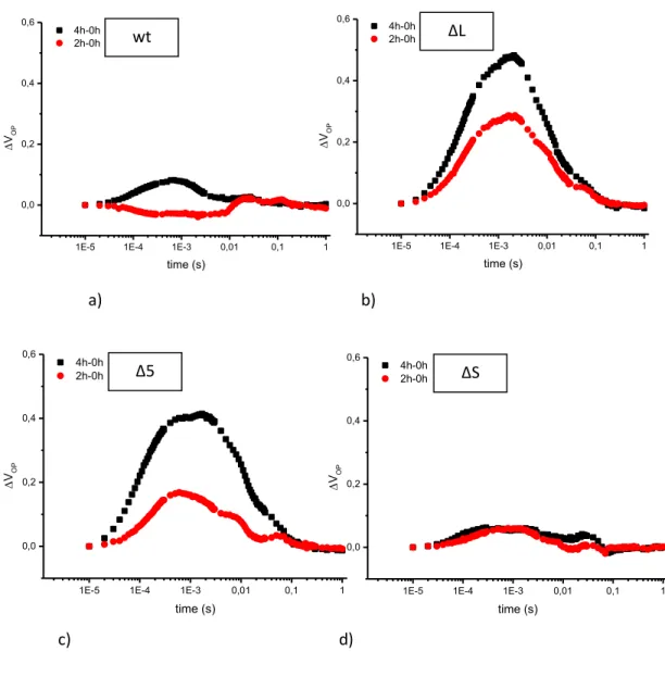
4.3 Prompt fluorescence…90
4.4 NPQ and CEF mutants show an increased turnover of the D1 protein and is paralleled by a loss of Fv/Fm…107
4.5 Preinactivation of PSII prevents damage to PSI in CEF/PS mutants…109 4.6 Phosphorylation studies…115 REFERENCES…118 5. DISCUSSION…120 REFERENCE…124 List of publications…128 Acknowledgement…129
9
LIST OF ABBREVIATIONS
APS Ammonium persulphate ATP Adenosine triphosphate
BCIP 5-bromo-4- chloro-indolyl phosphate Chl Chlorophyll
HEPES: N-2-hydrossiethilpiperazine N’-2-etansulphonic acid LHCII Chl containing light-harvesting complex of PS II
NADP Nicotinamide adenine dinucleotide NBT Nitro blue tetrazolium
OEC Oxygen-evolving complex P680 Primary electron donor in PS II
PQ Plastoquinone
Pheo Pheophytin, localized in D1 protein PS I Photosystem I
PS II Photosystem II
QA The first quinone electron acceptor in PS II
QB The second quinine electron acceptor in PS II
SDS-PAGE Sodium dodecyl sulfate polyacrylamide gel electrophoresis TemedN,N,N- tetramethylethylendiamine
TMBZ Tetramethyl benzidine
Tricine N-[2-hydroxy-1,1- bis(hydroxymethyl) ethyl]glycine Tris Tris-[hydroxymethyl]amino-methane
10
Abstract
Light is fundamental for photosynthesis. However, when in excess, it may produce
reactive oxygen species (ROS) which, in turn, may damage the Photosystem II
reaction center D1 protein. In order to avoid this, photosynthetic organisms have developed different mechanisms able to protect their photosynthetic apparatus. The term photoprotection is used to define all these process. Higher plants have evolved different protective mechanisms which are: the thermal dissipation through NPQ depending on PSBS protein, phosphorylation of LHCII to ensure efficient distribution of light energy between photosystems (state transition) depending on STN7/TAP38 kinase/phosphatase system and Cyclic Electron Flow/
Photosynthesis Control (CEF/PC) around PSI depending on PGR5/PGRL1A/B
proteins. However, how this mechanisms affect D1 turnover is not know. Much of current knowledge about photoprotection comes from studies with knockout mutants of Arabidopsis thaliana in which respective key genes have been inactivated. In this work, this genetic approach was extended by generating, by crossing, high order mutants where two (ΔSL, ΔS5) or all three (ΔSL78) photoprotection mechanisms have been eliminated.
These mutants have been characterized and their light sensibility investigated, by using fluorescence (PSII), absorbance changes (PSI), protein phosphorylation and turnover of D1 protein by immunoblotting. Although all the mechanisms seems to be important in plants photoprotection, the sensibility of PSII to light is more marked in mutants where CEF/PC is absent and this is paralleled by an increase in D1 turnover. PSI is also highly damaged by light, but, if lincomycin is present, it resulted protected. In ΔS mutant the sensibility to light, in terms of PSII efficiency, is not as marked as it could be expected but the D1 turnover is very high.
11
12
1.1 Oxigenic Photosynthesis: general aspects
The overall process whereby plants, algae and prokaryotes use light energy to synthesize organic compounds is called photosynthesis. This process is important because it provides food and biomass as well as fossil fuel; moreover it is the only source of molecular dioxygen required for respiratory activity by aerobic organisms. Molecular dioxygen is released as a by-product of the photosynthetic oxidation of water and, over billions of years, oxygenic photosynthesis has altered the composition of Earth's atmosphere, from anoxygenic to oxygenic enabling the development of aerobic life forms. The present life on Earth is entirely dependent on photosynthesis.
All the reactions that convert solar energy into chemical energy can be represented by the overall reaction:
6 CO2 + 6 H2O → C 6H12O6 + 6 O2
Photosynthesis takes place in chloroplasts, specialized plastids surrounded by a double membrane system called outer and inner envelope. Inside these membranes there is a complex internal membrane system known as thylakoid membrane.
In a typical plant chloroplast the thylakoids are organized in grana (granal thylakoids) a system of appressed membranes, whereas the stromal thylakoids are unstacked. Thylakoids are all connected and the space inside the thylakoid membranes is called lumen whereas the liquid medium surrounding the thylakoids is called stroma.
Introduction
13
light reactions (in thylakoid membranes) which produce O2, ATP and
NADPH;
Calvin cycle which reduces CO2 to carbohydrates and consumes ATP and
NADPH produced by the light reactions: this is located in the soluble compartment of the chloroplast called stroma.
The photosynthetic electron transfer chain, located in the thylakoid membranes, is organized as a number of different multi pigment-protein complexes called
Photosystem II (PSII), Photosystem I (PSI), Cytochrome b6f, and ATP synthase; the water soluble protein plastocyanin links cytochrome b6f to PSI whereas
plastoquinone links PSII to cytochrome b6f.
The electron transfer chain (ETC) takes place in the thylakoidal membranes and the complexes mentioned before play a definite role in this process.
Photosystem complexes contain the antenna proteins to which Chl (a and b) and β-carotene are bound, making the LHC complexes with the function of capturing excitation energy and passing it to the reaction center by a mechanism based on resonance energy transfer (Raven et al., 2002). In the reaction center there is a
chlorophyll a, P680 for the PSII and P700 for PSI, named according to the wavelength of maximum bleaching following their oxidation.
The PSII captures photons and use their energy to extract electrons from water molecules and these can be used in several ways. By removing electrons from water, the water molecules are broken into the dioxygen gas and hydrogen ions, which are used for the ATP synthesis. The remaining electrons are passed down through a chain of electron-carrying proteins and part of the energy is conserved in form of electrochemical gradient, obtained by pumping hydrogen ions across
14
the membrane. Finally, the electrons, which got an additional energy boost at the level of PSI, are placed on NADPH used in all biosynthetic reductive reactions. On the donor side a strong oxidant is generated by the photo-oxidation of P680
that is able, using the Ca, Mn Cluster, to extract electrons from water. On the acceptor side, electrons are collected on a plastoquinole pool.
The Cytochrome b6f Complex is able to re-oxidase the plastoquinoles and then reduces oxidized plastocyanin. The photo-oxidized P700 of the PSI gets electron
from plastocyanin and, once reduced, reduces ferredoxin at its stromal side. Finally, to obtain NADPH, ferredoxin is oxidized by Ferredoxin-NADP+Reductase (FNR). The redox potential relationships between the involved electron acceptors and donors are described by the Z scheme, a schematic representation of the linear electron flow (Fig. 1-2).
This representation is an energy diagram for electron transfer during “light reactions” and shows the pathway of electron transfer from water to NADP+ by which plants transform light energy into chemical energy as reduced NADPH and ATP.
ATP synthesis is due to the proton gradient formed across the thylakoid membrane. This driving force is developed by the proton transfer into lumen by the water splitting process and by plastoquinone that is deprotonated by Cyt b6f on the luminal side.
The distribution of the protein complexes involved in light reaction is not homogeneous in the thylakoid membranes: PSII is localized in the stacked grana regions whereas PSI and ATP synthase in the stroma-exposed thylakoid membranes (Anderson and Andersson, 1982; Chow et al., 1991; Albertsson, 2001; Danielsson et al., 2004; Chow et al., 2005). Cyt b6f complexes are evenly distributed throughout the thylakoid membranes (Albertsson, 2001).
Introduction
15
Fig.1: The Z-Scheme of photosynthesis in plants (Allen J. F. , 2004)
1.1.2 Photosystem II
The solar water-splitting protein complex, photosystem II (PSII), catalizes one of the more energetically demanding reaction in nature by using the light energy to drive a catalyst capable of oxidizing water.
The water oxidation reaction is catalyzed by the tetramanganese-calcium-oxo (Mn2Ca-oxo) cluster in the oxygen evolving complex (OEC) of PSII which cycles
through five light-driven charge-storage or S-state intermediates (S0-S4) as it
accumulates charge equivalents to split water.
The PSII is a multi-protein complex of plants, algae and cyanobacteria embedded in the thylakoid membrane of these organisms.
The detailed structure of PSII has been elucidated, in the past years, by X-ray crystallography (Zouni et al., 2001; Ferreira et al., 2004; Iwata and Barber, 2004; Loll et al. 2005). The structure gives insight into the evolution of photosynthetic
16
organisms showing existence of common ancestors of both photosystems (Grotjohann et al., 2004; Nelson and Ben-Shem, 2005).
This photosystem is composed by 30 protein subunits (Shi and Schröder, 2004; Nelson and Yocum, 2006) that are organized in a reaction center (RC) that contains the D1 and D2: these proteins bind the P680 chlorophyll and all the other
cofactors needed for the PSII electron transport. In addition to the RC there is a light harvesting complex (LHCII) composed by approximately 250 chlorophyll molecules able to absorb light energy and transfer it to the RC by a mechanism called resonance energy transfer (Föster energy transfer).
The electrons from P680 are transferred to the PSII electron transfer components
that are bound by D1 and D2 protein. The primary acceptor is a pheophytin, a
molecule which transfers electrons received from P680 to QA and QB
(plastoquinone molecules); QB is oxidized by an integral membrane complex, the
Cyt b6f, able to transfer electrons to from plastoquinol to plastocyanin and in the meantime protons are translocated from stroma to lumen forming the proton gradient important for the ATP synthesis.
1.1.3 Photosystem I
PSI is one of the most intricate membrane complexes in Nature and is comprised of two complexes: a reaction center (P700) and a light –harvesting complex LHCI,
similar to PSII structure. This photosystem is one of the pigment-protein complexes of photosynthetic electron transfer chain, which is capable of light energy conversion. The primary acceptor in PSI is a special molecule of chlorophyll
Introduction
17
involved in the pathway of electron transfer through PSI are a phylloquinone (A1),
three different Fe-S center (Fx, Fa and Fb), and the Ferredoxin (Fdx), a soluble
iron-sulfur protein.
The last one is a strong reductant (-420 mV) located in the stroma of chloroplast and in its reduced form is able to reduce NADP+ through the aid of an intermediate enzyme, the ferredoxin-NADP+ reductase or FNR (Buchanan et al.,
2003).
18
1.1.4 Linear electron transport chain
The main pathway of the photosynthetic light reactions is represented by the
linear electron transport chain involving the three major thylakoid membrane
protein complexes, PSII, Cytb6f and PSI.
These three cooperate in order to reduce nicotinamide adenine dinucleotide phosphate (NADP+) (Hill et al., 1960) through electrons from water molecules.
The first electron donor of PSII is the chlorophyll P680 which receives the excitation
energy captured by LHCII and the primary charge separation happens between P680 and Pheophytin, the primary electron acceptor.
Then electrons are transferred to a primary and secondary quinone acceptors, QA
and QB.
QB is double-reduced by a subsequent electron transfer (QB2-) and, binding two
proton from the stroma in been converted to plastoquinol. In the next step electrons pass through the Cyt b6f complex and one electron is transported to oxidized P700 via plastocyanin, a small soluble protein and, simultaneously, two
protons are released into the thylakoid lumen.
At the same time the chlorophyll in the reaction center, P700, is excited by light
absorbed by LHCI and the NADP+ is reduced by electrons transferred through several electron carrier via ferredoxin-NADP+-oxidoreductase.
During these processes of linear electron transport a ΔpH is developed through the thylakoid membrane because protons are pumped from the stroma to the lumen and is used to drive the synthesis of ATP by the ATP synthase.
Introduction
19
1.2 Light and Photosynthesis
Under natural condition plants often receive more light, in term of excitation energy than they can use to drive photochemistry.
Once a photon is absorbed by an antenna pigment molecule there are three pathways by which this excited state decays: this energy can be used to drive photochemistry, it can be dissipated as heat through the Xantophylls Cycle or emitted as fluorescence.
In healthy plants only the 2-5% represents Chl fluorescence, about 80% of this energy is used in the photochemical pathway and the rest is dissipated as heat (Niyogi et al., 1998; Govindjee, 2005; Gruszecki et al., 2006; Lazar, 2006; Szabó et al., 2005).
These three process occur in competition , such that any increase in the efficiency of one will result in a decrease in the yields of the other two.
Each of these process has a quantum yield and the sum of quantum yield of fluorescence (ΦF), heat (ΦD) and photochemistry (ΦP) is always one (energy conservation law):
(ΦF)+(ΦD)+(ΦP)=1
It has been demonstrated that to manage the changing in the light environment the percentage wherewith these events occur can change, in particular, an increasing fraction of energy dissipated by heat has been observed when light intensity increases (Külheim et al., 2002; Gruszecki et al., 2006; Lazar, 2006). A non-invasive method to measure the photosynthetic reactions in vivo is represented by the analysis of the yield of fluorescence at different light
20
environment level and this measure may also result in information about the function of the electron transport and heat dissipation.
1.3. Photoprotection
1.3.1 Light stress and photoprotection
Light energy is very important for the photosynthetic process. Absorbed solar energy may be defined as excessive when it exceeds the capacity of photosynthesis to use it for assimilation (Demmig-Adams and Adams, 1992; Niyogi
et al., 1998).
An excess of absorbed light may produce a damage to plants because reactive
oxygen species (ROS) may be generated. These molecules may produce
photodamage to the photosynthetic protein complexes (Barber and Andersson, 1992) and, in order to avoid this, all plants have developed several mechanisms to manage the excess absorbed energy. ROS are important signaling molecules in plants and they can have deleterious effects on photosynthesis and other leaf processes that will reduce growth and plant fitness but they also lead plant cells to death due to interaction of these reactive molecules with the cell membranes (Murchie and Niyogi, 2011).
Photoprotection is the term used to define the mechanisms by which plants
protect themselves from excess of absorbed light; these include the thermal dissipation through NPQ (Holt et al., 2004) mainly depending on PsbS protein, phosphorylation of LHCII to ensure efficient distribution of light energy between
Introduction
21
photosystems (state transition) depending on STN7/TAP38 kinase/phosphatase system and the Cyclic Electron Flow/ Photosynthesis Control (CEF/PC) around PSI depending on PGR5/PGRL1A/B proteins.
1.3.2 Physical Photoprotection
In addition to all the molecular mechanisms trough which plants avoid photodamage, there are a lot of strategy that helps plants to manage an excess of light. These mechanisms includes paraheliotropic leaf orientation and leaf folding, by alteration of whole-leaf light absorption, enhanced reflectance through leaf hairing (Ripley et al., 1999), reflective epicuticular wax layers (Robinson et al., 1993) and a lot of morphological adaptations like small leaf size, thick leaves, etc. Inside cells another process that allowed the physical photoprotection is represented by the chloroplasts movements to reduce light absorption.
Chloroplast movements towards the anticlinal cell walls can occur in sudden high light exposure within minutes (Chow et al., 1988; Park et al., 1996; Briggs and Christie, 2002; Wada et al., 2003). Mutant plants lacking the chloroplast movement response are more susceptible to photoinhibition than wild type plants (Kasahara et al., 2002).
Photosynthetic organisms also have screening compounds like anthocyanins, betalains and rhodoxanthin that are pigments able to decrease the light absorption (Weger et al., 1993; Smillie and Hetherington, 1999; Steyn et al., 2002). Other screening compounds , like flavonoids and hydroxycinnamic acids are situated in the plants cuticle and act against UV radiation (Kolb et al., 2001; Markstädter et al., 2001).
22
1.3.3 Photoprotection mechanisms and mutants
An excess of light could be harmful because plants are unable to manage it when it exceeds the capacity of photosynthesis to use it for the assimilation: the lifetime of the singlet state of the excited chlorophyll is extended and this leads to the formation of a triplet state. In this situation the energy from this triplet state reacts with oxygen generating singlet oxygen, a reactive oxygen species (ROS). The de-excitation of singlet chlorophyll may follow different pathways:
driving photochemistry
return to the ground state by emission of fluorescence by thermal dissipation
1.3.3.1 Non Photochemical Quenching
The major component of photoprotection is the last one, known as NPQ or Non-Photochemical Quenching.
Non-Photochemical Quenching is a regulatory process that maintain the balance between the utilization and the dissipation of the light energy that plants are able to absorb in order to minimize the photo-oxidative damage reducing the generation of reactive oxygen species (Niyogi et al., 2000). This photoprotection mechanism could be described as an increase heat dissipation of absorbed photons when the absorbed light exceeds the capacity of plant to drive photosynthesis.
Introduction
23
High light exposure represents a problem for plant health and NPQ is a molecular adaptation that represent the fastest response of the photosynthetic membrane to excess light (Demming-Adams et al., 2014, Ruban, 2016).
This parameters refers to the ability to protect the PSII reaction centers (RCII) by a rapid regulation of light harvesting complex.
There are different processes that contribute to the induction and relaxation of non photochemical quenching has three components:
- qE energy-dependent quenching - qT state transition quenching;
- qI photoinhibitory quenching (Quick and Stitt, 1989).
The major one in A. thaliana is qE, or NPQ, and it depends on the formation of a transmembrane proton gradient.
qE, the major rapidly reversible NPQ component, reflects a key molecular protective process in the photosynthetic membrane of higher plants and algae, which enables rapid adjustment of light harvesting efficiency to incidental light intensity. (Ruban, 2016)
When the thylakoid lumen pH is low, the violaxanthin-depoxidase enzyme is activated: this lumen- localized enzyme catalyses the conversion of violaxanthin to zeaxanthin. One the other hand, the low thylakoid lumen pH led to the protonation of proteins that are involved in the qE formation like PSBS. (Holt et al., 2004; Cogdell, 2006).
It has been hypothesized that protonation activates a binding site for zeaxanthin (Nyogi et al., 2000).By isolating and characterizing mutants of A. thaliana lacking
24
qE it was shown that qE requires PSBS in addition to a low thylakoid lumen pH and the presence of depoxidazed xanthophylls like zeaxathin.
Recently it has been discovered that PSBS protein is a dimer more stable at low pH and acidification seems to cause a conformational change associated with alteration in luminal intermolecular interactions (Fan et al., 2015).
PSBS acts like a switch that is triggered by ΔpH and not like a quenching site (Ruban et al., 2012).
Recent studies demonstrate that an overexpression of PSBS lead to an increase in qE (Kromdijk et al., 2016).
When a plant is transferred from dark to light NPQ is induced in the time scale of seconds and the accumulation of protons in the lumen of the thylakoid membranes is the initial event in the induction of NPQ (Horton et al., 2005; Szabó
et al., 2005).
The npq1 mutant and npq2 mutant are defective in the Xanthophylls Cycle (npq1 mutant lacks violaxanthin de-epoxidase and npq2 mutant lacks zeaxanthin epoxidase), whereas npq4-1 mutant has a normal xanthophylls cycle but lacks the ΔpH induced conformational change (zeaxanthin-dependent) in thylakoids membrane.
In order for PSBS to function as the site of qE, the presumed pigments bound to PSBS would have to be coupled to one or more chlorophylls in the LHCII system. In this work I used npq4-1 mutant, called ΔS mutant. (Niyogi et al., 2000).
Introduction
25
Fig.3: PSBS protein structure with indicated the two protonable lumenal glutamates (E122/E226) (Li et al., 2004).
1.3.3.2 Cyclic Electron Flow/Photosynthesis Control
As described before, during photosynthesis, two photoreaction centers, located in the chloroplast and more specifically in the thylakoid membrane, PSI and PSII, use light energy to generate ATP and NADPH. There are two modes of electron flow, of which the linear electron flow (LET) is driven by PSI and PSII, generating ATP and NADPH, whereas the Cyclic Electron Flow (CEF)/Photosynthesis Control only generates ATP and is driven by PSI alone (Shikanai et al., 2007; Shikanai T., 2014). The electron distribution between the two photosystems is regulated by different environmental and metabolic conditions that lead to an adjustment of ATP/NADPH ratio: LEF is favored when an highly active Calvin cycle form an efficient sink for electrons from PSI, whereas CEF predominate when the dark reactions of photosynthesis are down regulated, during high light or low CO2
26
conditions (Clarke and Johnson, 2001; Joliot and Joliot, 2002, 2006; Golding and Johnson, 2003; Golding et al., 2004; Dal Corso et al., 2008).
CEF starts with light dependent excitation of P700, the primary electron donor of
PSI, and electron is transferred to Fd. Once oxidized P700+ is reduced by electrons
from PQ pool via Cyt b6f and PC. Electrons from Fd must eventually be donated to PQ to complete the cycle. It can take place by two different pathways: the NDH-dependent pathway plays a major role in cyanobacteria, its contribution in C3 plants is still ambiguous but it has been shown to be essential in photoprotection (Endo et al., 1999), and the Fd-dependent pathway (known as “CEF around PSI) bypasses NADPH and may play a role in the acidification of the C3 thylakoids lumen ( Mi et al., 1995; Ravenel et al., 1994; Asada et al., 1993; Joe῾῾t et al., 2002; Dal Corso et al., 2008).
In the Cyclic Electron Flow ATP is produced via proton-motive force and ΔpH contribute to the downregulation of PSII by NPQ.
Introduction
27
Two components are involved in this alternative pathway: Proton Gradient Regulation (PGR5) and Proton Gradient Regulation Like 1 (PGRL1).
When the activity of Calvin cycle is reduced PGR5 is involved in the generation of ΔpH that induce thermal dissipation through the Xantophylls Cycle. PGR5 is important because through this pathway is able to reduce the photoinibition of PSI by limiting the over-reduction of the acceptor side of PSI.
In pgr5 mutants the absence of PC/CEF leads to an insufficient ΔpH generation that is reflected in a reduced induction of thermal dissipation under excessive light conditions.
The small amount of NPQ in pgr5 may be possibly due to photoinhibition and/or the movement of peripheral light –harvesting antennae from PSII to PSI ( Shikanai
et al., 2002).
The other component of the CEF is the integral thylakoid protein PGRL1 and mutants lack this protein show a malfunction in CEF similar to pgr5 mutants. These two protein interact physically and are associate with PSI (Colombo et al., 2016).
PGRL1 is encoded by two homologous genes, PGRL1A and PGRL1B.
In this mutants the induction of NPQ is reduced like pgr5 mutant and both have a similar behaviour (Dal Corso et al., 2008; Pesaresi et al.,2016).
28 1.3.3.3 State Transitions
In stn7 and stn8 mutants this process is undetectable because they lack these proteins and the dephosphorylation process is not present.
In A. thaliana this process requires a protein kinase called STN7. This is really important because is involved in LHCII reversible phosphorylation associated with the relocation of LHCII and the redistribution of excitation energy between the two photosystems.
STN7 is responsible for the phosphorylation of LHCII in Arabidopsis (Bellafiore et al., 2005), whereas, another one, STN8, is requires for the quantitative phosphorylation of PSII core proteins.
STN7 is activated by the overreduction of PQs and phosphorylates part of LHCII. In the dark, plant thylakoids are in State I, where LHCII is bound to PSII. When the plastoquinone pool becomes reduced in the light, a conformation change in Cyt
b6f activates LHC kinases, and the LHCII of PSII becomes phosphorylated.
Phosphorylated LHCII units have decreased affinity to PSII and they move to PSI resulting in State II (Lunde et al., 2000; Aro and Ohad 2003; Kanervo et al., 2005; Dekker and Boekema 2005).
Under oxidizing conditions the LHCII kinase become inactivated and a phosphatase, TAP38 dephosphorylates LHCII which return to PSII. (Pribil et al., 2010).
State transition is also considered a component of non photochemical quenching (Takahashi, 2009), qT.
Introduction
29
Fig.5: Schematic representation of state transitions (Rochaix, J.D. et al., 2012)
1.3.4 Photohinibition and D1 protein turnover
Light is essential for photosynthesis, but it can also damage PSII in the light induced process called photohinibition. When there is an imbalance between the PSII photodamage and its repair cycle, the term photohinibition is used.
High light condition results in a damage to RC of PSII and this process occurs in any light intensity and the rate constant of photoinhibition is directly proportional to the light intensity (Jones and Kok , 1966; Tyystjärvi and Aro, 1996; Santabarbara et al., 2002; Nishiyama et al., 2005).
30
This term is used to indicates a decrease of the quantum yield of PSII when the rate of damage is faster than the rate of repair (Nishiyama et al., 2006).
The D1 protein of PSII, under natural conditions is the primary target of photodamage but the exact mechanism by which this event occur is still unclear. There different ideas about this process: D1 turnover seems to involve the disintegration of Mn clusters in PSII leading to an energy imbalance and consequently to cleavage of the D1 protein by proteases (Kato et al., 2015).
Other studies argue the hypothesis that this damage is due to an increase proton motive force which leads to an elevated electric field, rather than lumen acidification, which in vivo increased PSII charge recombination rates, producing singlet oxygen and subsequent photodamage (Davis G. A. et al., 2016).
More recent studies have discovered that, in addition to the rapid D1 protein turnover as consequence of photodamage, also subunits of Cyt b6f and NAD(P)H dehydrogenase (NDH) complex are characterized by a fast turnover .
Mutants carried out defects in these two complexes have shown enhanced sensibility linked to an increase photodamage partially correlated to the Cyclic Electron Flow (Li et al., 2018).
Studies demonstrate that during an entire sunny day all the population of the D1 protein is replaced (Ohad et al., 1984; Aro et al., 1993; Melis, 1999; Andersson and Aro, 2001; Chow and Aro, 2005). The repair cycle process occurs in different steps: the phosphorylated PSII dimeric complexes monomerized and monomers migrates to the stroma-exposed thylakoids where damaged D1 proteins are dephosphorylated and then degraded by proteases (Van Wijk et al., 1996; Baena Gonzales et al., 1999; Aro et al., 2005; Aro et al., 2015). Phosphorylation of the PSII core proteins facilitates the migration of the damaged PSII from the grana to the stroma-exposed membranes, where proteolisis of the D1 damaged protein is
Introduction
31
due to the activity of FtsH and Deg proteases (Huesgen et al., 2006; Aro et al., 2015).
The re-synthesis of this protein is regulated at the levels of translation initiation and elongation and after the co- translational insertion in the tylakoid membrane of PSII center (Zhang and Aro, 2002), the protein is then processed by the removal of the C-terminal extension of the pre-D1 protein (Taylor et al., 1988; Diner et al., 1988; Fujita et al., 1995). Finally, the new reassembled PSII migrates back to the grana tylakoids membranes where it forms a functional dimer of PSII. The D1 protein is the only component of the PSII that is not recycled.
It has been demonstrated that the D1 protein damage is proportional to the light intensity (Park et al., 1995; Tyystjarvi et al., 1996) and the inhibition of PSII complexes seems to play a role on the protection of PSI by decreasing the redox pressure.
Photoinhibition does not lead to loss of PSII activity unless the rate of damage is higher than the rate of repair (Tikkanen et al., 2014). To measure photoinhibition one approach is to inhibit the D1 protein turnover using antibiotics like lincomycin (Mulo et al., 2003). This is a translation inhibitor, such as streptomycin or chloramphenicol, able to inhibit the synthesis of plastidial-encoded protein like D1. In the presence of lincomycin, the repair cycle does not take place and after illumination the residual amount of protein can be measured by western blot in order to quantify the extent of damage.
32
Introduction
33
1.3.5 Chl Fluorescence and fluorescence measurements
During the photosynthetic process the excitation energy is transferred to the reaction centers of both photosystems where the photosynthetic energy conversion starts. This energy can follow different pathways: the photochemical pathway is in competition with other two represented by the thermal dissipation and the Chl fluorescence.
The latter refers to a fluorescence emission originated from deexcitation of excited chlorophyll molecules.
In a healthy plant the photochemical pathway is predominant and up to 80% of absorbed energy is used in this route , 2-5% is re- emitted as Chl fluorescence and the remaining faction dissipated as heat.
Fluorescence is a very informative and may be measured in a number of different ways, such as prompt fluorescence (i.e. by PEA), fluorescence decay (i.e. by single turnover flash), PAM fluorimetry and so on, each of which giving information on different aspects of energy transfer, dissipation and photochemistry.
Prompt fluorescence: prompt Fluorescence is measured normally after a dark adaptation. In this condition all the reaction centers are in the reduced state (“open”). Actinic light is then switched on and fluorescence rising directly measured (usually up to 1 sec) during light irradiation.
PAM fluorimetry: in a typical PAM experiment a saturating flash is applied (3000 µmol m-2 sec-1) and the reaction centers are oxidized. A strong fluorescence increase is observed reaching a maximum value called Fm, the reaction center are now in the “closed” form.
34
A non saturating actinic light is then applied to activate photosynthesis afterwards some saturating flashes are fired on the top of this light to achieve full reduction of the primary electron acceptor QA. This technique allows to calculate quenching parameters such as YII and NPQ.
The PAM instrument is not only suited only for evaluation of PSII state via Chl fluorescence, but also to analyze other aspects of photosynthesis, such as energy conversion in PSI (via dual wavelength P700 measurements (Klughammer and Schreiber, 2008).
After a dark adaptation the level of ground fluorescence , indicated as F0 is
measured: a measuring light is applied and this parameter reached. Then a saturating flash is applied which allows to fluorescence to reach its maximum level represented by the Fm parameter: in this case too, it is possible to obtain Fv/Fm, the maximum quantum efficiency of PSII as described before.
Introduction
35
Fig.7: Schematic representation of chlorophyll fluorescence measurement by the saturation pulse method (Van Kooten& Snell, 1990).
Fluorescence decay: fluorescence decay in microsecond to second time scale, allows the evaluation of electron transfer at the acceptor side of PSII and charge recombination with the donor side. In a typical experiment, a short saturating flash (µseconds) is administrated and fluorescence decay measured. The experimental curve may be simulated by a sum of three different components, two exponential and one hyperbolic respectively describing by Vass et al., 1999.
36
Fig. 8 : Example of fluorescence decay experiment in wt
1E-3 0,01 0,1 1 10 100 0,0 0,5 1,0 fluo re scen ce ( ar bitr ar y un its) time (sec) wt control (dark) 2h_high light 4h_high light
37
38
Albertsson, P.-Å. (2001) “A quantitative model of the domain structure of the photosynthetic membrane”
Trends Plant Sci.6: 349-358.
Anderson, J.M. and Andersson, B. (1982) “The architecture of photosynthetic membranes: lateral and transverse organization”.
Trends Biochem. Sci. 7: 288-292.
Andersson, B. and Aro, E.-M. (2001) “Photodamage and D1 protein turnover in Photosystem II. In: Regulation of photosynthesis”
Eds. E.-M. Aro, and B. Andersson. Kluwer Academic Publishers, Dordrecht, pp: 377-393.
Allen, J. F. (2004) “Cytochrome b6f: structure for signalling and vectorial metabolism”
Trends in Plant Science 9 (3), 130-137
Alonso, J. M. et al. (2003) “Genome-wide insertional mutagenesis of Arabidopsis Thaliana”
Science 301, 653–-657
Aro, E.-M. and Ohad, I. (2003) “Redox regulation of thylakoid protein phosphorylation”
Antiox. Redox Sign. 5:55-67
Aro, E.-M., Virgin, I. and Andersson, B. (1993) “Photoinhibition of photosystem II. Inactivation, protein damage and turnover”
39
BBA – Bioenergetics 1143:113-134.
Asada, K., Heber, U., and Schreiber, U. (1993) “Electron flow of the intersystem chain stromal components cyclic electron flow in maize chloroplasts, as detected in intact leaves by monitoring change of P700 and chlorophyll fluorescence” Plant Cell Physiol. 34, 39–50.
Baena-Gonzalez, E., Barbato, R. & Aro, E. M. (1999) “Role of phosphorylation in the repair cycle and oligomeric structure of photosystem II”
Planta 208, 196–-204
Barber, J. and Andersson, B. (1992) “ Too much of a good thing: Light can be bad for photosynthesis”
Trends Biochem. Sci.,17(2), 61-667
Bellafiore, S., Barneche, F., Peltier, G. & Rochaix, J. D. (2005) “State transitions and light adaptation require chloroplast thylakoid protein kinase STN7”
Nature 433, 892–-895 (2005).
Briggs, W.R. and Christie, J.M. (2002) “Phototropins 1 and 2: versatile plant bluelight receptors”
Trends Plant Sci. 7: 204–210.
Buchanan, B., Gruissen, W., Jones, R. L. (2003) “Biochemistry molecular biology of plants”
40
Caffarri, S.,Tibiletti, T., C Jennings, R. & Santabarbara, S. (2014) “A comparison between plant photosystem I and photosystem II architecture and functioning” Curr Protein Pept Sci. 2014 Jun; 15(4): 296–331
Chow, W.S., Qian, L.P., Goodchild, D.J. and Anderson, J.M. (1988) “Photosynthetic acclimation of Alocasia machrorrhiza (L) G Don to growth irradiance - structure, function and composition of chloroplasts”
Aust. J. Plant Physiol. 15: 107-122.
Chow, W.S., Miller, C. and Anderson, J.M. (1991) “Surface-charges, the heterogeneous lateral distribution of the 2 photosystems, and thylakoids stacking”.
BBA – Bioenergetics 1057: 69-77.
Chow, W.S. and Aro E.-M. (2005) “Photoinactivation and mechanism of recovery in Photosystem II – the light driven water:plastoquinone oxidoreductase”
Eds. T.J. Wydrzynski and K. Satoh. Springer, Dordrecht, The Netherlands, p. 627-648.
Chow, W.S., Kim, E.H., Horton, P. and Anderson J.M. (2005) “Granal stacking of thylakoid membranes in higher plant chloroplasts: the physicochemical forces at work and the functional consequences that ensue”
Photochem. Photobiol. Sci. 4:1081-1090.
Clarke, J.E., and Johnson, G.N. (2001) “In vivo temperature dependence of cyclic and pseudocyclic electron transport in barley”
41
Planta 212, 808–816.
Cleland, R.E., and Bendall, D.S. (1992) “Photosystem I cyclic electron transport: measurement of ferredoxin-plastoquinone reductase activity”
Photosynth. Res. 34, 409–418.
Cogdell, R., Gall, A., Köhler, J. (2006) “The architecture and function of the light-harvesting apparatus of purple bacteria: from single molecules to in vivo membranes”
Q Rev Biophys. 2006 Aug;39(3):227-3
Colombo, M., Suorsa, M., Rossi, F., Ferrari, R., Tadini, l., Barbato, R., and Pesaresi, P., (2016) “Photosynthesis Control: An underrated short-term regulatory mechanism essential for plant viability”
PLANT SIGNALING & BEHAVIOR, VOL. 11, NO. 4, e1165382 (6 pages)
Dal Corso, G., Pesaresi, P.,Masiero,S., Aseeva, E., Schunemann, D., Finazzi, G., Joliot, P., Barbato, R., Leister, D., (2008) “A complex containing PGRL1 and PGR5 is involved in the switch between linear and cyclic electron flown in Arabidopsis” Cell 132 , 273-285
Danielsson, R., Albertsson, P.A., Mamedov, F. and Styring, S. (2004) “Quantification of photosystem I and II in different parts of the thylakoid membrane from spinach”
42
Demmig-Adams, B. and Adams, W.W. (1992) “Photoprotection and other responses of plants to high light stress”
Ann. Rev. Plant Physiol. Plant Mol. Biol. 43: 599-626.
Demmig-Adams B, Garab G, Govindgee III WA. (2014) “Non-photochemical quenching and energy dissipation in plants, algae and cyanobacteria”
In Advances in photosynthesis and respiration 40. Dordrecht, The Netherlands: Springer
Diner, B.A., Ries, D.F., Cohen, B.N. and Metz, J.G. (1988) “COOH-terminal processing of polypeptide D1 of the photosystem-II reaction center of Scenedesmus obliquus is necessary for the assembly of the oxygen evolving complex”
J. Biol. Chem 263: 8972-8980.
Dekker JP, Boekema EJ. (2005) “Supramolecular organization of thylakoid membrane proteins in green plants”
Biochim Biophys Acta. 2005 Jan 7;1706(1-2):12-39. Review
Endo, T., Shikanai, T., Takabayashi, A., Asada, K. and Sato, F. (1999) “The role of chloroplastic NAD(P)H dehydrogenase in photoprotection”
FEBS Lett. 457, 5–8.
Fan, X. et al., (2015) “The NdhV subunit is required to stabilize the chloroplast NADHdehydrogenase-like complex in Arabidopsis”
43
Ferreira, K.N., Iverson, T.M., Maghlaoui, K., Barber, J. and Iwata, S. (2004) “Architecture of the photosynthetic oxygen-evolving center”
Science 303: 1831-1838
Fujita, S., Inagaki, N., Yamamoto, Y., Taguchi, F., Matsumoto, A., Satoh, K. (1995) “Identification of the carboxyl-terminal processing protease for the D1 precursor protein of the photosystem II reaction center of spinach”
Plant Cell Physiol. 36: 1169–1177.
Golding, A.J., and Johnson, G.N. (2003) “Down-regulation of linear and activation of cyclic electron transport during drought”
Planta 218, 107–114.
Golding, A.J., Finazzi, G., and Johnson, G.N. (2004) “Reduction of the thylakoid electron transport chain by stromal reductants - evidence for activation of cyclic electron transport upon dark adaptation or under drought”
Planta 220, 356–363.
Govindjee (2005) “Chlorophyll a fluorescence: A bit of basics and history” In: Chlorophyll a fluorescence: A signature of photosynthesis.
Eds. G.C. Papageorgiou and Govindjee.
Kluwer Academic, Dordrecht, The Netherlands, pp. 2-42.
Grotjohann, I., Jolley, C. and Fromme, P. (2004) “Evolution of photosynthesis andoxygen evolution: implications from the structural comparison of photosystems I and II”
44
Phys. Chem. Chem. Phys. 6: 4743–4753
Gruszecki, W.I., Gospodarek, M., Iaskowska, A. and Spiewla, E. (2006)” Adaptation of the photosynthetic apparatus of Nitellopsis obtusa to changing light intensity at the molecular level: different pathways of a singlet excitation quenching”
Acta Phys. Plant. 28: 127-136.
Hill, R. and Bendall, F. (1960) “Function of the two cytocrome components in chloroplasts: A working Hypothesis”
Nature, 186 (4719), 136-137
Holt N. E., Fleming G. R. and Niyogi K. K. (2004) “Toward an understanding of the mechanisms of non photochemical quenching in green plants”.
Biochemistry, 44(26), pp. 8281-8289
Horton, P., Wentworth, M. and Ruban, A. (2005) “Control of the light harvesting function of chloroplast membranes: The LHCII-aggregation model for non photochemical quenching”
FEBS Lett. 597: 4201-4206.
Huesgen, P.F., Schuhmann, H. and Adamska I. (2006) “ Photodamaged D1 protein is degraded in Arabidopsis mutants lacking the Deg2 protease”
FEBS Lett. 580: 6929–6932.
Iwata, S. and Barber, J. (2004) “Structure of photosystem II and molecular architecture of the oxygen-evolving centre”
45
Curr. Opin. Struct. Biol. 14: 447-453.
Järvi, S., Suorsa, M., Aro, E. M. (2015) “ Photosystem II repair in plant chloroplasts — Regulation, assisting proteins and shared components with photosystem II biogenesis”
Biochimica et Biophysica Acta (BBA) - Bioenergetics
Joe¨ t, T., Cournac, L., Peltier, G., and Havaux, M. (2002) “Cyclic electron flow around photosystem I in C3 plants. In vivo control by the redox state of chloroplasts and involvement of the NADH dehydrogenase complex”
Plant Physiol. 125, 1919–1929.
Joliot, P., and Joliot, A. (2002) “Cyclic electron transfer in plant leaf” Proc. Natl. Acad. Sci. USA 99, 10209–10214.
Joliot, P., and Joliot, A. (2006) “Cyclic electron flow in C3 plants” Biochim. Biophys. Acta 1757, 362–368
Jones, L.W. and Kok B. (1966) "Photoinhibition of chloroplast reactions. I. Kinetics and action spectra”
Plant Physiol. 4: 1037–1043.
Kanervo, E., Suorsa, M. and Aro, E.-M. (2005) “Functional flexibility and acclimation of the thylakoid memebrane”
46
Kasahara, M., Kagawa, T., Olkawa, K., Suetsugu, N., Miyao, M. and Wada, M. (2002) “Chloroplast avoidance movement reduces photodamage in plants”
Nature 420: 829–832.
Kato,Y., et al., (2015) “D1 fragmentation in PSII repair caused by photo-damage of two step model”
Photosynth. Res. 126: 409-416
Khương Thị Thu Hương, Robaglia, C., Caffarri, S. (2014) ”The Function of PsbS Protein in Plant Photosynthesis Regulation”
VNU Journal of Science: Natural Sciences and Technology, Vol. 30, No. 2 (201)4 6-22
Klughammer, C., Schreiber, U. (2008) Saturation Pulse method for assessment of energy conversion in PSI”
PAM Application Notes 01-11-14
Koivuniemi, A., Aro, E.-M. & Andersson, B. (199% “Degradation of the D1- and D2-proteins of photosystem II in higher plants is regulated by reversible phosphorylation”
Biochemistry 34, 16022–-16029
Kolb, C.A., Käser, M.A., Kopecký, J., Zotz, G., Riederer, M. and Pfündel, E.E. (2001) “Effects of natural intensities of visible and ultraviolet radiation on epidermal ultraviolet screening and photosynthesis in grape leaves”
47
Kromdijk, j., Głowacka, K., Leonelli, L., Gabilly, S. T., Iwai, M., Niyogi, K. K., Long, S. P., (2016) “Improving photosynthesis and crop productivity by accelerating recovery from photoprotection”
Science Vol. 354, Issue 6314, pp. 857-861
Külheim, C., Ågren, J., Jansson, S. (2002) “Rapid regulation of light harvesting and plant fitness in the field” Science 297: 91-93.
Lazar, D. (2006) “The polyphasic chlorophyll a fluorescence rise measured under high intensity of exciting light”
Funct. Plant Biol. 33: 9-33.
Li, X. P., BjoÈrkman, O., Shih, C., Grossman, A. R., Rosenquist, M., Jansson, S., & Niyogi, K. K., (2000) “A pigment-binding protein essential for regulation of photosynthetic light harvesting”
NATURE |VOL 403 | 27 JANUARY 2000
Li, L., Aro, E. M. and Millar,H., (2018) “Mechanisms of Photodamage and Protein Turnover in Photoinhibition”
Trends in plant science, Vol. 23, No. 8
Loll, B., Kern, J., Saenger, W., Zouni, A. and Biesiadka, J. (2005) “Towards complete cofactor arrangement in the 3.0 Å resolution structure of photosystem II”
48
Lunde C., Jensen P. E., Haldrup A., Knoetzel J. and Scheller H. V. (2000) “The PSI-H subunit of photosystem I is essential for state transitions in plant photosynthesis” Nature, Vol. 408, pp. 613-615
Markstädter, C., Queck, I., Baumeister, J., Riederer, M., Schreiber, U. and Bilger, W. (2001) “Epidermal transmittance of leaves of Vicia faba for UV radiation as determined by two different methods”
Photosynth. Res. 67: 17–25
Melis, A. (1999) “ Photosystem-II damage and repair cycle in chloroplasts: what modulates the rate of photodamage?”
Trends Plant Sci. 4: 130-135.
Mi, H., Endo, T., Ogawa, T., and Asada, K. (1995) “Thylakoid membrane-bound pyridine nucleotide dehydrogenase complex mediates cyclic electron transport in the cyanobacteria Synechocystis PCC6803”
Plant Cell Physiol. 36, 661–668.
Munekage, Y., Hojo, M., Meurer, J., Endo, T., Tasaka, M., Shikanai, T.(2002) “PGR5 is involved in cyclic electron flow around photosystem I and is essential for photoprotection in Arabidopsis”
Cell. 2002 Aug 9;110(3):361-71.
Murchie E. K., Niyogi K. K. (2011) “Manipulation of Photoprotection to Improve Plant Photosynthesis”
49
Mulo, P., Pursiheimo,S., Hou, C. X.,Tyystjärvi, T., and Aro, E. M. (2003) “Multiple effects of antibiotics on chloroplast and nuclear gene expression”
Funct Plant Biol 30:1097–1103
Nelson, N. and Ben-Shem A. (2005) “The structure of photosystem I and evolution of photosynthesis”
Bio Essays 27: 914-922.
Nelson, N., and Yocum, C.F. (2006) “Structure and function of Photosystems I and II”
Annu Rev Plant Biol 57: 521 -565.
Nishiyama, Y., Allakhverdiev, S.I. and Murata, N. (2005) “ Inhibition of the repair of photosystem II by oxidative stress in cyanobacteria”
Photosynth. Res. 84: 1-7.
Nishiyama, Y., Allakhverdiev, S.I. and Murata, N. (2006) “ A new paradigm for the action of reactive oxygen species in the photoinhibition of photosystem II” BBA – Bioenergetics 1757: 742-749.
Niyogi, K.K., Grossman, A.R. and Björkman, O. (1998) “Arabidopsis mutants define a central role for the xanthophyll cycle in the regulation of photosynthetic energy Conversion”
50
Ohad, I., Kyle, D.J. and Arntzen, C.J. (1984) “Membrane-protein damage and repair –removal and replacement of inactivated 32- kilodalton polypeptides in chloroplast membranes”
J. Cell Biol. 99: 481-485.
Park, Y., Chow, W. S., Anderson, J. M. (1995) “Light inactivation of functional photosystem II in leaves of peas grown in moderate light depends on photon exposure”
Planta 196 (1995) 401–411.
Park, Y.I., Anderson, J.M. and Chow, W.S. (1996)”Photoinactivation of functional photosystem II and D1-protein synthesis in vivo are independent of the modulation of the photosynthetic apparatus by growth irradiance”
Planta 198: 300-309.
Pribil, M., Pesaresi, P., Hertle, A., Barbato, R. & Leister, D. (2010) “Role of plastid protein phosphatase TAP38 in LHCII dephosphoryaltion and thyalkoid electron flow”
PLoS Biol. 8, e1000288. (doi:10.1371/journal.pbio.1000288)
Quick, W.P. and Stitt, M. (1989) An examination of factors contributing to nonphotochemical quenching of chlorophyll fluorescence in barley leaves”
BBA - Bioenergetics 977: 287-296.
Raven, H.P., Evert ,F.R., Eichhorn, E.S. (2002) “Biologia delle piante” Zanichelli
51
Ravenel, J., Peltier, G., and Havaux, M. (1994) “The cyclic electron pathways around photosystem I in Chlamydomonas reinhardtii as determined in vivo by photoacoustic measurements of energy storage”
Planta 193, 251–259.
Ripley, B.S., Pammenter, N.W. and Smith, V.R. (1999) “Function of leaf hairs revisited: The hair layer on leaves Arctotheca populifolia reduces photoinhibition, butleads to higher leaf temperatures caused by lower transpiration rates”
J. Plant Physiol. 155: 78-85.
Robinson, S.A., Lovelock, C.E. and Osmond, C.B. (1993) “Wax as a mechanism of protection against photoinhibition – a study of cotyledon-orbiculata”
Bot. Acta 106: 307-312.
Ruban, A. V., Johnson, M. P.,Duffy, C. D. (2012) “ The photoprotective molecular switch in the photosystem II antenna”
Biochim Biophys Acta 1817: 167-181
Ruban, A. V., Murchie, E. H. (2012) “Assessive the photoprotective effectiveness of non-photochemical chlorophyll quenching: a new approach”
Biochim Biophys Acta 1817: 977-982
Ruban, A. V. (2016) “Non-photochemical chlorophyll fluorescence quenching: mechanism and effectiveness in protecting plants from photodamage”
52
Santabarbara, S., Cazzalini, I., Rivadossi, A., Garlaschi, F.M., Zucchelli, G. and Jennings, R.C. (2002) “Photoinhibition in vivo and in vitro involves weakly coupled chlorophyll protein complexes”
Photochem. Photobiol. 75: 613-618.
Shi,L. X. and Schröder, W. P. 2004 “ The low molecular mass subunits of the photosynthetic supracomplex, photosystem II”
Biochim. Biophys. Acta, 1608(2-3), 75-96.
Shikanai, T. (2007) “Cyclic electron transport around Photosystem I: genetic Approaches”
Annu Rev Plant Biol 58: 199 -217.
Shikanai, T., (2014) “Central role of cyclic electron transport around photosystem I in the regulation of photosynthesis”
Curr Opin Biotechnol. 2014 Apr; 26:25-3
Smillie, R.M. and Hetherington, S.E. (1999) “Photoabatement by anthocyanin shields photosynthetic systems from light stress”
Photosynthetica 36: 451-463.
Snyders, S. & Kohorn, B. D. (1999) “TAKs thylakoid membrane protein kinases associated with energy transduction”
53
Steyn, W.J., Wand, S.J.E., Holcroft, D.M. and Jacobs, G. (2002) “Anthocyanins in vegetative tissues: a proposed unified function in photoprotection”
New Phytol. 155: 349-361.
Szabó, I., Bergantino, E. and Giacometti, G.M. (2005) “Light and oxygenic photosynthesis: energy dissipation as a protection mechanism against photooxidation”
EMBO Reports 6: 629-634.
Taylor, M.A., Packer, J.C.L. and Bowye, J.R.n (1988) “Processing of the D1 polypeptide of the photosystem II reaction centre and photoactivation of a low fluorescence mutant (LF-1) of Scenedesmus obliquus “
FEBS Lett. 237: 229-233.
Tikkanen, M., Mekala,N. R., Aro, E. M. (2014) “Photosystem II photoinhibition-repair cycle protects Photosystem I from irreversible damage”
Biochim. Biophys. Acta 1837: 210–215.
Tyystjarvi, E., Aro, E. M. (1996) “The rate constant of photoinhibition, measured in lincomycin-treated leaves, is directly proportional to light intensity”
Proc. Natl. Acad. Sci. U. S. A. 93 (1996) 2213–2218.)
Vass, I., Kirilovsky, D., Etienne, A.-L., (1999) “UV-B Radiation-induced donor- and acceptor- side modifications of Photosystem II in the Cyanobacterium Synechocystis sp. PCC 6803”
54
Wada, M., Kagawa, T. and Sato, Y. (2003) “Chloroplast movement” Annu. Rev. Plant
Biol. 54: 455–468.
Weger, H.G., Silim, S.N. and Guy, R.D. (1993) “Photosynthetic acclimation to low temperature by western red cedar seedlings”
Plant Cell Environ. 16: 711-717.
Zhang, L., Aro, E. M. (2002) Synthesis, membrane insertion and assembly of the chloroplast encoded D1 protein into photosystem II”
FEBS Lett. 512 (2002) 13–18.
Zouni, A., Witt, H.T., Kern, J., Fromme, P., Krauss, N., Saenger, W. and Orth, P. (2001) “Crystal structure of photosystem II from Synechococcus elongatus at 3.8 Å Resolution”
55
56
Although light is essential for photosynthesis, when in excess, it may damage the photosynthetic apparatus, leading to a phenomenon known as photoinhibition. Photoinhibition was thought as a light-induced damage to photosystem II; however it is now clear that even photosystem I may become very vulnerable to light. One main characteristic of light induced damage to photosystem II (PSII) is the increased turnover of the reaction center protein, D1: when rate of degradation exceeds the rate of synthesis, loss of PSII activity is observed. With respect to photosystem I (PSI), an excess of electrons, instead of an excess of light, may be very dangerous. Plants possess a number of mechanisms able to prevent, or limit, such damages by safe thermal dissipation of light energy (energy-dependent quenching, qE), slowing-down of electron transfer through the intersystem transport chain (Photosynthesis Control, PSC) in co-operation with the Proton Gradient Regulation (PGR) proteins, PGR5 and PGRL1 and, at least to some extent, redistribution of light between photosystems (state transition, qT), collectively called as short-term photoprotection mechanisms. In the last years, genes coding for proteins behind these biophysical photoprotection mechanisms have been identified, offering the possibility to study these topics in well-defined genetic materials. In order to extend this genetic approach to the study of photoprotection, a number of higher order mutants were generated during this study by crossing genotypes carrying defects in each of the short-term photoprotection mechanisms. In this way, in a well defined genetical contest we found that mutants carrying a defect in the ΔpH-dependent photosynthesis-control are characterized by photoinhibition of both photosystems, irrespectively of whether qE or state transitions defects were present or not in the same individual, demonstrating the primary role of PSC in photoprotection. Moreover,
Outline of the thesis
57
mutants with a limited capability to develop a strong qE, were characterized by a high turnover of the D1 protein.
The aim of my study consists in the attempt to clarify the role of different photoprotection mechanisms involved in stability of PSII, of PSI and how these mechanisms are correlated with the turnover of D1 protein, the primary target of PSII photodamage.
58
Materials and methods
59
3.1 Mutants
Arabidopsis thaliana mutants were grown in a growth chamber under a photon
flux density of 90-110 µmol m-2 sec-1in 8 h light/ 16 h dark regime at 19-22°C. The control plant was Arabidopsis thaliana wild type ecotype Columbia-0 (Col 0).
A. thaliana mutant lines in the Columbia-0 (Col-0) background were located in the
T-DNA Express Database (http://signal.salk.edu/cgi-bin/tdnaexpress) and obtained from the European Arabidopsis Stock Center.
The pgrl1A/B (ΔL) double mutant is a CEF/PC mutant that was generated by Dal Corso et al (2008). Is an insertion mutant obtained from publicly available T-DNA insertion collection and each lacked the respective transcript. The double mutant was generated by crossing single pgrl1A and pgrl1B knockouts and screening the resulting F2 generation for homozygous double mutants.
By crossing single pgrl1A and pgrl1B knockouts and screening the resulting F2 generation for homozygous double mutants.
The psbS (ΔS) mutant, which is unable to form NPQ, was created by Niyogi et al. (2005) by inducing a short deletion generated by fast-neutron bombardment of seeds which result in an unstable PSBS protein not detected in the thylakoid membrane.
The pgr5 (Δ5) mutant, who lacks the Cyclic Electron Flow/Photosynthesis Control, was described by Shikanai et al (1999) and the mutation consists in a nucleotide transition (from G to A) leading an aminoacidic substitution from glycine to serine (at position 130).
The stn7/8 (Δ78) double mutant lacks two protein kinases : STN7 and STN8 that are involved in state transitions.
60
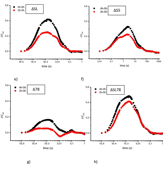
During this work, these mutants were used to generate high order mutants and in particular ΔSL, ΔS5 and ΔSL78. The ΔS5 and ΔSL double and triple mutants were generated by crossing respective mutants defective in one protection mechanism to obtain F1 generation from which genotypes containing both mutations were identified by PCR.
The quintuple mutant ΔSL78 lacks all the photoprotection mechanisms described before: it is unable to develop NPQ, lacks cyclic electron flow around PSI/Photosynthesis Control and also lacks state transition. High order mutants, like ΔSL78, were generated by crossing the ΔSL mutant with the Δ78. Recombinants for all five genes were identified by western blot.
3.2 Treatment with lincomycin and light
The D1 protein turnover was determined by comparing its degradation rate in the presence or absence of lincomycin (1mg/ml). Arabidopsis thaliana mutants leaves were incubated overnight with the antibiotic and then were irradiated with a light intensity of 500 µmol m-2 sec-1 for different periods of time (0, 1, 2 and 4 hours). Samples were collected at the appropriate irradiation time and thylakoids were immediately isolated as described.
3.3 Isolation of thylakoids
In order to isolate thylakoid membranes, samples of A. thaliana mutants leaves were frozen in liquid nitrogen and were homogenized in mortar to obtain a fine powder using a buffer composed of:
Materials and methods 61 - 50 mM HEPES-NaOH pH 7.2 - 5 mM MgCl2 - 15 mM NaCl - 0.4 M sucrose
The homogenate was filtered through eight layers of cotton cloth, then membranes were pelletted by centrifugation for 10 min at 3000 g at 4 °C. Pellets were resuspended in a second buffer composed of:
- 50 mM HEPES-NaOH pH 7.2, - 5 mM MgCl2
- 15 mM NaCl
After this operation the homogenate was spun down at 4500 g for 10 min. Thylakoids were finally re suspended in:
- 50 mM HEPES-NaOH pH 7.2 - 5 mM MgCl2
- 15 mM NaCl - 0.4 M sucrose
The chlorophyll (Chl) concentration was measured with the protocol proposed by Arnon, 1949.
62
3.4 Chlorophyll measurement
The concentration of chlorophyll was estimated by the method of Arnon (1949), by measuring spectrophotometrically the absorbance at 720, 663 and 645 nm of chlorophylls extracted with 80% acetone.
The following formulas were used:
Total chlorophyll [chl (μg / ml)] = 20.2 (A645-A720) + 8.02 (A663-A720) Chlorophyll a [chl (μg / ml)] = 12.7 (A663-A720) – 2.69 (A645-A720) Chlorophyll b [chl (μg / ml)] = 22.9 (A645-A720) – 4.68 (A663-A720)
Thylakoids samples were immediately used or stored at –80 °C after being flash frozen in liquid nitrogen.
3.5 Polyacrylamide gel electrophoresis and immunoblotting
Polyacrylamide gel electrophoresis and immunoblotting SDS–PAGE was as described by Barbato et al (1992).
After electrophoresis, gels were either stained with Coomassie Blue R-250 or blotted to nitrocellulose membranes (Dunn, 1986). For immunodetection, membranes were saturated with 5% (w/v) skimmed milk and probed with antibodies to thylakoid proteins D1, PSBS (Barbato et al., 1992), anti-PGR5 (Munekage et al., 1999), anti-PGRL1A (Dal Corso et al., 2008), anti-STN7 and anti-STN8 (Barbato et al., unpublished). Antibody to Pthr were from Zymed and used as reported by (Rimtamäki et al., 1997).
Materials and methods
63
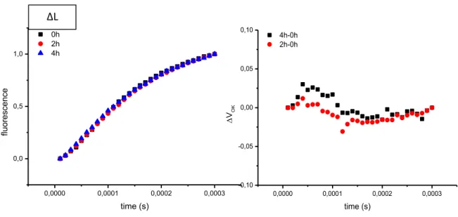
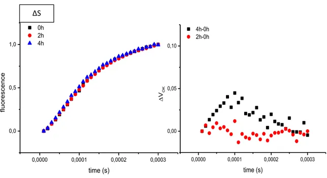
3.6 Fluorescence decay
Decay of flash-induced chlorophyll fluorescence was measured by the double modulation fluorometer FL-3500 (PSI, Brno, Czech Republic) and data were analyzed as described by Vass et al (1999). Multicomponent deconvolution of the measured curves was done by using a fitting function with two exponential components and one hyperbolic component, according to Vass et al (1999):
F(t) – F0 = A1exp(-t/T1) + A2exp(-t/T2) +A3/(1+t/T3) + A0
where F(t) is the variable fluorescence yield, F0 is the basic fluorescence before the
flash, A0 to A3 are the amplitudes and T1 to T3 are the time constants. Very slowly
decaying fluorescence is described by a constant A0 amplitude.
3.7 State transitions measurement
State transition were measured with a pulse amplitude modulated fluorometer (Dual-PAM-100 Walz). Leaves were exposed to a flash of saturating white light (800 ms) in order to measure Fm value. After that were illuminated with 100 µmol
m-2 sec-1 (blue light PSII) for 15min. Far red light is turned on and the maximum fluorescence yield in State 1 was determined. This far red light was then switched off and the fluorescence recorded for 15 min, after which the maximum fluorescence yield in State 2 was determined.
To calculate the relative change in fluorescence the following formula was used : Fr=[(Fi’-Fi)-(Fii’-Fii)]/ (Fi’-Fi) (Jensen et al., 2000)
64
Fig. 9 : Example of state transition experiment in wt and Δ78
0 1000000 2000000 0 2000 4000 stat e t ra nsition s time/s stn7/8 Fii’ Fii Fi’ Fi Δ78 0 1000000 2000000 0 2000 4000 6000 8000 stat e t ra nsition s time/s wt
PSII on PSI on PSI off PSII off
Fm Fm1
Fm2