Contents lists available atScienceDirect
Mechanisms of Development
journal homepage:www.elsevier.com/locate/modReview
Nicole Le Douarin and the use of quail-chick chimeras to study the
developmental fate of neural crest and hematopoietic cells
Domenico Ribatti
⁎Department of Basic Medical Sciences, Neurosciences and Sensory Organs, University of Bari Medical School, Bari, Italy
A R T I C L E I N F O Keywords: Chick embryo Chimeras Hematopoietic system Neural crest Quail embryo A B S T R A C T
The quail-chick chimera marking system, devised in 1969, gave a new impetus to the analysis of cell migrations and interactions in the developing nervous, immune and hematopoietic systems. The method is based on the observation that the constitutive heterochromatin in all embryonic and adult cells of the quail is condensed in one large mass in the centre of the nucleus and is associated with the nucleolus, making this organelle strongly stained with the Feulgen–Rossenbeck reaction. The association of cells or rudiments from two avian species, advocated as a means to identify cells that migrate during embryogenesis, was rapidly recognized in this context as a useful tool for the study of many developmental biology problems. This article summarizes the fundamental contribution of Nicole Le Douarin to the discovery and the application of this technique over the last 40 years.
1. Background
Nicole Marthe Le Douarin (Fig. 1) was born in Lorient, France, on August 20, 1930. In 1954, she graduated from the Sorbonne with a degree in Natural Sciences. In the late 1950s, N. Le Douarin was in-troduced in the laboratory of Etienne Wolff (1904–1996), professor at the Collège de France, and founder of the Institut d'Embryologie Ex-périmentale et de Tératologie du Centre National de la Recherche Sci-entifique (CNRS) et du Collège de France, in Nogent sur Marne on the east side of Paris. In 1964, N. Le Douarin discussed the doctoral thesis entitled“Etude expérimentale de l'organogenèse du tube digestif et du foie chez l'embryon de Poulet”, which was published with the same title in the“Bulletin Biologique de la France et de la Belgique” (Le Douarin, 1964c). In these years, N. Le Douarin studied the morphogenetic movements of the endoderm and mesoderm leading to the ventral closure of the gut (Le Douarin, 1964a, b, c). In 1965, N. Le Douarin was appointed as“Maître de Conférences” at Clermont-Ferrand in the De-partment of Developmental Biology directed by Hubert Lutz. In 1966, N. Le Douarin was appointed at the University of Nantes where she established herfirst independent research group.
2. A Feulgen-Positive nucleolus
N. Le Douarin was interested in the study of the effect of hepatic mesenchyme on the differentiation and growth of anterior intestinal
portal (AIP) endoderm. She associated in organotypic co-culture the hepatic mesenchyme of a quail embryo with the AIP endoderm of a chick (and vice versa). When she observed the microscopic sections of these chimeric liver lobules resulting from the association of chick AIP endoderm and quail (Coturnix japonica) liver mesenchyme, the chick hepatocytes looked the same as in normal liver, whereas the me-senchyme exhibited a large nucleolus. As N. Le Douarin has re-membered:“I noticed the presence of a large nucleolus in all embryonic and adult cell types of this species of bird and further found that the unusual size of this organelle resulted from association of a mass of heterochromatin with the nucleolus proper (essentially made up of RNA). This particularity is of rare instance in the animal kingdom and does not exist in the chick.“(Le Douarin and Dupin, 2018).
N. Le Douarin applied Feulgen-Rossenbeck's, thereafter referred as Feulgen, histological nuclear staining procedure for DNA and the Unna-Pappenheim staining method for RNA component of the nucleoli and demonstrated that the quail nucleolus was mostly composed, not of RNA, but of DNA (Le Douarin, 1969, 1973a, b). She discovered that Feulgen allows to distinguish chick cells from quail cells. Feulgen stained quail cells have interphase nuclei with condensed nucleolar-associated heterochromatin that's not found in chick interphase nuclei (Fig. 2). The intimate association of the heterochromatin and the nu-cleoli in quail cells can be strikingly seen at ultrastructural level. This makes the cells of the two species easy to distinguish on sections stained for DNA with the Feulgen-reaction and suggested to N. Le Douarin the
https://doi.org/10.1016/j.mod.2019.103557
Received 24 April 2019; Received in revised form 15 May 2019; Accepted 22 May 2019
⁎Corresponding author at: Department of Basic Medical Sciences, Neurosciences and Sensory Organs, University of Bari Medical School, Policlinico - Piazza G.
Cesare, 11, 70124 Bari, Italy.
E-mail address:[email protected].
Mechanisms of Development 158 (2019) 103557
Available online 23 May 2019
0925-4773/ © 2019 Elsevier B.V. All rights reserved.
idea of constructing chimeras between these two species of birds, by associating quail and chick cells in the embryo in ovo. As she said: “Quail and chick cells, side by side in chimeric tissues can therefore be easily identified, since the natural nuclear labeling of quail is con-spicuous enough to enable identification of a single quail cell located in chick tissues, provided that the section includes the nucleolus.” (Le Douarin and Dupin, 2018).
3. The construct of chimeras applied to the study of neural crest derivatives
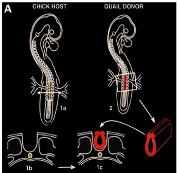
Chimeras resulting from the replacement of definite regions of the embryo of one species by their counterparts from stage-matched em-bryos of the other, develop normally, suggesting that this system can provide reliable information on the behavior and fate of the grafted cells. N. Le Douarin started to construct chimeras by replacing a frag-ment of the neural tube of the chick embryo prior the onset of neural crest cells emigration at this level of the neural axis by that of a stage-matched quail embryo at the same developmental stage. The same type of grafts was performed in both directions, i.e. from quail to chick and from chick to quail (Fig. 3).
When she saws a chimeric embryo whose sections had been treated with the Feulgen-Rossenbeck's staining procedure, she demonstrated that quail cells were present not only in the grafted spinal cord but also were dispersed in other places, along the nerves, as Schwann cells, in the peripheral ganglia, in the suprarenal glands and within the meta-nephritic mesenchyme. This technique, compared to the previous ones used to label the cells (either H3-TdR, vital stains, or carbon nano-particles), was superior due to its being stable and unalterable and also non transmissible to neighboring cells. By constructing quail-chick chimeras, in which part of the neural primordium of the host embryo was substituted by its counterpart taken from a stage-matched donor of the other species, the migration and fate of the neural crest was
followed during the entire embryonic life and could even be pursued after birth thanks to the stability provided by this cell labeling tech-nique. Through this experimental approach, N. Le Douarin established the cell types arising from the neural crest; their level of origin and the pathways they followed to reach their destinations (Fig. 4) (Le Douarin, 1980, 1982).
4. The construct of chimeras applied to the study of the immune system
The quail/chicken labeling system was also applied to the study of the development of the thymus and the bursa of Fabricius and allows through different grafting schemes to identify the origin of every single cell in early rudiments (Le Douarin et al., 1984). As N. Le Douarin said: “With Francine Jotereau, then a young student, we undertook experi-ments which showed that lymphocytes are derived from hemopoietic stem cells (HSC) which invade the thymic epithelial rudiment during cyclic waves. These HSC also give rise to the dendritic cells in the medulla of the thymus. In contrast, the development of the bursa of Fabricius is characterized by a single invasion of HSC which lasts 5 to 6 days and generates B cell precursors for the entire life of the bird (Le Douarin and Joterau, 1973;Le Douarin et al., 1975, 1976a, b, 1977; Jouterau and Le Douarin, 1982;Houssaint et al., 1976)” (Le Douarin, 2005). After grafting the early thymic rudiment from a 3 days quail embryo into the chick somatopleure, N. Le Douarin demonstrated that the thymus developed on this ectopic site and that all the lymphocytes it contained, were derived not from the graft, but from the chick host. In contrast, if the quail thymus was removed from the donor later in de-velopment, then the lymphocytes that differentiated in the graft were of the quail type. This demonstrated that the lymphocytes that developed in the thymus were of extrinsic origin. Furthermore, N. Le Douarin demonstrated that hemopoietic cells entry to the thymus was preceded by successive waves separated by non-receptive periods (Fig. 5) (Jouterau and Le Douarin, 1982).
Furthermore, N. Le Douarin demonstrated that the thymus was colonized by HSCs, non-endothelial cells of blood vessel walls,
Fig. 1. A port trait of Nicole Le Douarin.
Fig. 2. Feulgen staining of DNA shows a large mass of heterochromatin in the centre of the nucleus, which is associated with the nucleolus in quail cells (left). In chick cells, the heterochromatin is evenly distributed (right).
(Modified fromLe Douarin, 2004)
Fig. 3. Quail-chick chimeras for investigating the fate of avian neural crest cells. (A) Schematic of the construction of quail-chick chimeras of the neural tube at“adrenomedullary” trunk level (somites 18–24). The neural tube was removed from chick host (1a, 1b) and replaced by its equivalent (1c), pre-viously taken at the same level from a quail donor of the same developmental stage (2).
including pericytes and smooth muscle cells, derived from the neural crest (Etchevers et al., 2001). Moreover, it was established that en-dothelial cells in the head and neck derived from cephalic paraxial mesoderm (Couly et al., 1995), and that the time when colonization by HSCs begins in the mouse thymus, by associating the cultured early rudiment with fetal liver, as HSCs donor (Le Douarin et al., 1984).
The quail/chick chimeras could hatch and live a few weeks or months indicating that the graft was tolerated. However, between two weeks and three months after birth, xenogeneic tissue grafts were im-mune rejected. In the case of the bursa of Fabricius, signs of rejection can be the immune attack of the stroma occurs within thefirst weeks after birth (Corbel et al., 1987). By using Major Histocompatibility Complex (MHC) matched animals, it was possible to overcome the problem. Within the chicken species, transplantation of limb buds
between MHC-mismatched embryos led to virtually complete tissue tolerance not only of the grafted limb, but also of adult skin from the same MHC type as the limb (Corbel et al., 1990). Embryonic chick wings are well tolerated (with only slight signs of pathology without ultimate rejection) by the quail and as in chick-to-chick MHC mis-matched combinations. It appears from these experiments that the quail immune system consistently has a capacity to tolerate tissues of rela-tively closely related species, a capacity that does not exist in the chick. A further step in the work of N. Le Douarin was the characterization of the monoclonal antibody MB1/QH1, which recognize an antigenic determinant common to the endothelial and white blood cells of the quail at the exclusion of any cell type of the chick (Péault et al., 1983; Pardanaud et al., 1989) (Fig. 6). As N. Le Douarin said:“Our early work on hemopoietic stem cells, carried out by Bruno Péault with the skillful
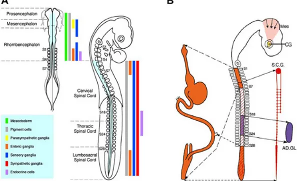
Fig. 4. Fate map of neural crest derivatives in the avian embryo as determined by quail-chick chimeras. (A) Fate map of neural and non-neural derivatives of the neural crest along the neural axis, represented in the cephalic neural crest (left, 7-somite stage embryo) and trunk neural crest (right, 28-somite stage embryo). The rostro-caudal levels of origin of the various neural crest phenotypes are shown with color-coded vertical bars. While the whole (cephalic and trunk) neural crest gives rise to pigment cells (grey bars), the origin of mesenchymal derivatives (including skeletal and connective tissues) (green bar) is confined to the cephalic neural crest from mid-diencephalon down to r8, corresponding to the level of somite 4 (S4). Definite regions of the neural crest yield peripheric nervous system derivatives, including sensory (blue bars), parasympathetic (ciliary ganglion) (yellow bar), sympathetic (red bar) and enteric (orange bars) ganglia. Endocrine (adrenomedullary) cells originate from the trunk neural crest between somites 18 and 24 (purple bar). (B) Schematic representation of a chick embryo of 28 somite-stage, showing various regions of the neural crest along the neural axis and their respective derivatives (same color-coding as in A) in enteric nervous system plexuses (orange), sympathetic ganglia including the superior cervical ganglion (SCG) (red), parasympathetic ciliary ganglion (CG), and in the medulla of the adrenal gland (AD.GL.). (Reproduced fromLe Douarin and Dupin, 2018)
Fig. 5. Successive waves of hematopoietic cell immigration into the thymus in quail and chick embryos and newly hatched birds. (Reproduced fromLe Douarin, 1988)
technical help of Monique Coltey, has attracted our attention to the close relationships between endothelial and blood cells in the yolk sac blood islands. This was made possible by using the MB1-Mab, produced by Bruno, which was thefirst of the numerous species- and cell type-specific antibodies that we further prepared to analyze the chimeras.” (Le Douarin, 2005).
Staining of early quail embryos with the MBl/QHl-Mab revealed that endothelial cells are present in the avian cephalic mesoderm from the second day of the incubation period, prior to the onset of heart beating and the appearance of major blood vessels (Pardanaud et al., 1987;Poole and Coffin, 1988), thus supporting the assumption that the cephalic mesoderm is endowed with angiogenic capacities.
Using the quail-chick chimera system,Noden (1991)had claimed that some of the angiogenic potencies in the mesoderm located from the mesencephalon down to the level of thefifth somite are destined to provide the endothelial wall of the cardiac outflow tract and to form the endocardial cushions. Furthermore, he noted a highly invasive behavior of the grafted endothelial cells (Noden, 1989).
Pardanaud et al. (1989)demonstrated that primordial di fferentia-tion of the endothelial network required two sets of endothelial cells, one associated with the somatopleural (dorsal) mesoderm giving rise only to endothelial cells and one associated with the splanchnopleural (ventral) mesoderm capable of giving rise to endothelial cells and blood. Furthermore, Pardanaud and Dieterlen-Lièvre (1989) grafted quail somites into the chick hosts and followed the progeny of quail endothelial cells. They demonstrated that the body wall was vascular-ized by somite-derived endothelial cells, whereas internal organs
developed vascular networks with angioblasts emerging from the splanchnopleura.
5. Concluding remarks
An embryonic chimera is an animal that has two or more popula-tions of genetically distinct cells that originated in different embryos of the same or different species. Chimeras have been obtained in the avian embryo following the observation of the interphase nucleolus in the Japanese quail. Making quail cells readily distinguishable from those of the chick where the constitutive heterochromatin is evenly dispersed in the nucleus.
These structural differences have been used to device a cell-marking technique through which cell migrations and cell interactions during embryogenesis can be followed in the embryo in ovo by grafting quail cells into chick embryos or vice versa. This method was successfully applied by N. Le Douarin and her co-workers in the study of the on-togeny of the derivatives of the neural crest and of the immune system. References
Corbel, C., Belo, M., Martin, C., Le Douarin, N.M., 1987. A novel method to bursectomize avian embryos and obtain quail-chick bursal chimeras. II. Immune response of bur-sectomized chicks and chimeras and postnatal rejection of the grafted quail bursas. J. Immunol. 138, 2813–2821.
Corbel, C., Martin, C., Ohki, H., Coltey, M., Hlozanek, I., Le Douarin, N.M., 1990. Evidence for peripheral mechanisms inducing tissue tolerance during ontogeny. Int. Immunol. 2, 33–40.
Couly, G., Coltey, P., Eichmann, A., Le Douarin, N.M., 1995. The angiogenic potentials of
Fig. 6. Chick (Donor) to quail (Host) neural graft. MB 1 immunostaining; 24 h after grafting. The im-munostained vascular network of the host tectum (A asterisk) gives rise to a vertical twig (B arrow) pe-netrating the ventricle and reaching the transplant. In the latter (C), donor-native (stars), host-derived (asterisk) and chimera (arrows) microvessels can be seen. Immunostained host-derived macrophage-like cells (arrowheads) are recognizable within and on the host neuroepithelium (A) and in the graft (B), also in a perivascular position. D–F Quail (Donor) to chick (Host) neural graft. MB 1 immunostaining; 24 h after grafting. D A unlabeled host microvessel (star), with a labelled perivascular cell (arrow) is recognizable within the donor tissue; an im-munostained donor microvessel is close to the host tectum (asterisk). E A immunostained microvessel of the graft has a perivascular cell which invades the surrounding tissue (asterisk). F A immunostained donor-native microvessel grown towards the host tectum anastomoses with the chick unlabelled mi-crovasculature, forming a vascular chimera (arrows). Immunostained donor-derived macrophage-like cells are indicated by arrowheads.
the cephalic mesoderm and the origin of brain and head blood vessels. Mech. Dev. 53, 97–112.
Etchevers, H.C., Vincent, C., Le Douarin, N.M., Couly, G.F., 2001. The cephalic neural crest provides pericytes and smooth muscle cells to all blood vessels of the face and forebrain. Development 128, 1059–1068.
Houssaint, E., Belo, M., Le Douarin, N.M., 1976. Investigations on cell lineage and tissue interactions in the developing bursa of Fabricius through interspecific chimeras. Dev. Biol. 53, 250–264.
Jouterau, F.V., Le Douarin, N.M., 1982. Demonstration of a cyclic renewal of the lym-phocyte precursor cells in the quail thymus during embryonic and perinatal life. J. Immunol. 129, 1869–1877.
Le Douarin, N.M., 1964a. Induction de l'endoderme pré-hépatique ar le mésoderme de l'aire cardiaque chez l'embryon de Poulet. J. Embryol. Exp. Morphol. 12, 651–676.
Le Douarin, N.M., 1964b. Isolement expérimental du mésenchyme propre du foie et rôle morphogène de la composante mésodermique dans l'organogenèse hépatique. J. Embryol. Exp. Morphol. 12, 141–160.
Le Douarin, N.M., 1964c. Etude expérimentale de l'organogenèse du tube digestif e du foie chez l'embryon de Poulet. Bull. Biol. Fr. Belg. 98, 544–674.
Le Douarin, N.M., 1969. Particularités du noyau interphasique chez la Caille japonaise (Coturnix coturnix japonica). Utilisation de ces particularités comme‘marquage bio-logique’ dans les recherches sur les interactions tissulaires et les migrations cellulaires au cours de l'ontogenèse. Bull. Biol. Fr. Belg. 103, 435–452.
Le Douarin, N.M., 1973a. A biological cell labeling technique and its use in experimental embryology. Dev. Biol. 30, 217–222.
Le Douarin, N.M., 1973b. A Feulgen-Positive Nucleolus. Exp. Cell Res. 77, 459–468.
Le Douarin, N.M., 1980. The ontogeny of the neural crest in avian embryo chimaeras. Nature 286, 663–669.
Le Douarin, N.M., 1982. The Neural Crest. Cambridge. Cambridge University Press.
Le Douarin, N.M., 1988. The Claude Bernard lecture 1987. Embryonic chimeras: a tool for studying the development of the nervous and immune system. Proc. R. Soc. Lond. B 235, 1–17.
Le Douarin, N.M., 2004. The avian embryo as a model to study the development of the neural crest: a long and still ongoing story. Mech. Dev. 121, 1089–1102.
Le Douarin, N.M., 2005. The Nogent Institute - 50 years of embryology. Int. J. Dev. Biol. 49, 85–103.
Le Douarin, N.M., Dupin, E., 2018. The beginnings of the neural crest. Dev. Biol. 444, S3–S13.
Le Douarin, N.M., Joterau, F., 1973. Origin and renewal of lymphocytes in avian embryo
thymuses through embryonic life in interspecific chimeras. Nat. New Biol. 246, 25–27.
Le Douarin, N.M., Houssaint, E., Joterau, F.V., Belo, M., 1975. Origin of hemopoietic stem cells in embryonic bursa of Fabricius and bone marrow studied through interspecific chimeras. Proc. Natl. Acad. Sci. U. S. A. 72, 2701–2705.
Le Douarin, N.M., Joterau, F.V., Houssaint, E., 1976a. The lymphoid stem cells in the avian embryo. In: Wright, R.K., Cooper, E.L. (Eds.), Phylogeny of Thymus and Bone Marrow - Bursa Cells. Elsevier/North Holland Biomedical Press, Amsterdam, pp. 217–226.
Le Douarin, N.M., Joterau, F.V., Houssaint, E., Belo, M., 1976b. Ontogeny of the avian thymus and bursa of Fabricius studied in interspecific chimeras. Ann. Immunol. 127, 849–856.
Le Douarin, N.M., Houssaint, E., Joterau, F., 1977. Differentiation of the primary lym-phoid organs in avian embryos: origin and homing of the lymlym-phoid stem cells. In: Benedict, A.A. (Ed.), Avian Immunology. Plenum Publishing Corporation, New York, pp. 29–37.
Le Douarin, N.M., Dieterlen-Lièvre, F., Oliver, P.D., 1984. Ontogeny of primary lymphoid organs and lymphoid stem cells. Am. J. Anat. 170, 261–299.
Noden, D.M., 1989. Embryonic origins and assembly of blood vessels. Am. Rev. Respir. Dis. 140, 1097–1103.
Noden, D.M., 1991. Cell movements and control of patterned tissue assembly during craniofacial development. J. Craniofac. Genet. Dev. Biol. 11, 192–213.
Pardanaud, L., Dieterlen-Lièvre, F., 1989. Does the paraxial mesoderm of the avian em-bryo have hemangioblastic capacity? Anat. Emem-bryol. 192, 301–308.
Pardanaud, L., Altman, C., Kttos, P., Dieterelen-Lièvre, F., Buck, C.A., 1987. Vasculogenesis in the early quail blastodisc as studied with a monoclonal antibody recognizing endothelial cells. Development 100, 339–349.
Pardanaud, L., Yassine, F., Dieterlen-Lièvre, F., 1989. Relationship between vasculogen-esis, angiogenesis and haematopoiesis during avian ontogeny. Development 105, 473–485.
Péault, B.M., Thiery, J.P., Le Douarin, N.M., 1983. Surface marker for hematopietic and endothelial cell lineages in quail is defined by a monoclonal antibody. Proc. Natl. Acad. Sci. U. S. A. 80, 2976–2980.
Poole, T.J., Coffin, J.D., 1988. Developmental angiogenesis: quail embryonic vasculature. Scanning Microsc. 2, 443–448.
Roncali, L., Virgintino, L., Coltey, P., Bertossi, M., Errede, M., Ribatti, D., Nico, P., Mancini, L., Sorino, S., Riva, A., 1996. Morphological aspects of the vascularizazion in intraventricular neural transplants. Anat. Embryol. 193, 191–203.