Diagnostic and prognostic value of
18
F-FDG PET/CT
in comparison with morphological imaging
in primary adrenal gland malignancies -
a multicenter experience
Abstract
Objective: To evaluate the diagnostic and prognostic role of fluorine-18 fluoro-2-deoxy-D-glucose positron emission tomography/computed tomography (18F-FDG PET/CT) in comparison to
morpholog-ical imaging such as computed tomography in primary adrenal malignancies. Materials and Methods: In this multicenter retrospective study, 68 patients with adrenal malignancy were included. All patients had histologically proven diagnosis of primary adrenal malignancy (adrenocortical carcinoma, malignant pheochromocytoma, neuroblastoma and lymphoma), one whole body 18F-FDG PET/CT scan and one
whole-body contrast enhancement computed tomography (CECT) scan acquired within one month and were followed clinically and by performing morphological tests for at least 12 months. Results: Overall sensitivity, specificity, accuracy, positive and negative predictive values for CECT and 18F-FDG PET/CT
were respectively, 59%, 100%, 65%, 100%, 27% and 75%, 100%, 82%, 100% and 63%. For adrenocortical carcinomas, 18F-FDG PET/CT showed a better accuracy (93.4%) than CECT (75%). For neuroblastomas 18
F-FDG PET/CT also showed better accuracy (70.4%) than CECT (66.7%). For malignant pheochromocytomas
18F-FDG PET/CT and CECT showed the same accuracy (90%). For primary adrenal lymphomas, 18F-FDG
PET/CT showed better accuracy (100%) than CECT (74.41%). Kaplan-Mayer curves showed that “histo-types” and “metastases at the last follow-up” were similarly detected for both disease free survival (DFS) and overall survival (OS), while “global 18F-FDG PET/CT” and “presence of metastases at diagnosis” were
significant for DFS. Stratifying the sample by the presence or absence of metastases at diagnosis, stan-dardized uptake value (SUVmax) was a significant prognostic factor for DFS when metastases were absent (Wald test=7.035, P=0.008). Conclusion: Our multicenter study demonstrated that 18F-FDG PET/CT better
than CECT diagnosed adrenal malignancies achieving also a good prognostic performance. Therefore management algorithms should include 18F-FDG PET/CT.
Hell J Nucl Med 2015; 18(2): 97-102 Epub ahead of print: 19 July 2015 Published online: 5 August 2015
Introduction
A
drenal gland lesions may frequently be incidentally detected both in patients with a history or without a history of tumors. Only few of these lesions are ma-lignant and can be primary or metastatic. The most common adrenal primitive malignancies are adrenocortical carcinomas, malignant pheochromocytomas, and neu-roblastomas, all characterized by poor prognosis [1-3].Adrenal tumors are usually evaluated by morphological imaging techniques as ul-trasound (US), contrast enhancement computed tomography (CECT) and magnetic res-onance imaging (MRI) [1].
Contrast enhancement computed tomography allows a precise assessment of fea-tures of adrenal tumors as for their size, shape, homogeneity, and calcifications [4, 5]. Dedicated adrenal CECT protocols, which combine non-contrast, early and delayed en-hancement studies were shown to be highly sensitive and specific. Magnetic resonance imaging allows for the detection of adrenal masses with a similar sensitivity as CT but is not considered quite as accurate as CT. Nevertheless, MRI may be useful if CT results are equivocal and the use of a contrast agent is contraindicated [6-8].
Fluorine-18 fluoro-2-deoxy-D-glucose positron emission tomography/computed to-mography (18F-FDG PET/CT) is an important imaging modality in characterizing many malignancies, including adrenal gland lesions [9, 10]. The accuracy of 18F-FDG PET/CT in characterizing adrenal lesions and its impact in clinical staging of patients with ad-renal gland malignancies have been evaluated mostly by studies including a small num-ber of patients and not from a multicenter origin [3, 11, 12].
It is important to identify parameters that can be considered prognostic, like tumor size, which measured by CECT, although in a few studies was prognostic in patients with Angelina Cistaro1,2,3MD, PhD,
Artor Niccoli Asabella4MD, PhD,
Pietro Coppolino5MD, Natale Quartuccio5MD, Corinna Altini4MD, Mariapaola Cucinotta5MD, Pierpaolo Alongi6MD, Michele Balma7MD, Silvia Sanfilippo8MD, Ambra Buschiazzo9MD, Arnoldo Piccardo10MD, Margherita Fanelli4, Gianmario Sambuceti9, MD, PhD, Jamshed Bomanji11MD, Sergio Baldari5MD, PhD, Gianni Bisi7MD, PhD, Stefano Fanti8MD, PhD, Giuseppe Rubini4 MD, PhD
1. Positron Emission Tomography Centre, IRMET S.p.A, Euromedic Inc., Turin, Italy 2. Co-ordinator of PET Pediatric AIMN InterGroup, 10136 Turin, Italy 3. Associate researcher of Institute of Cognitive Sciences and Technologies, National Research Council, Rome, Italy. 4. Nuclear Medicine Unit,
D.I.M. – Diagnostic Imaging –, University of Bari “Aldo Moro”, Bari, Italy 5. Nuclear Medicine Unit,
Department of Biomedical Sciences and of Morphologic and Functional Images, University of Messina, Messina, Italy 6. Department of Nuclear Medicine, San Raffaele Hospital, Milan, Italy 7. Nuclear Medicine,
San Giovanni Battista Hospital and University, Turin, Italy
8. Department of Nuclear Medicine, University Hospital S.
Orsola-Malpighi Bologna, Italy 9. Nuclear Medicine Unit, IRCCS AOU San Martino - IST, Department of Health Sciences, University of Genoa, Genoa, Italy 10. Nuclear Medicine Department, E.O. Galliera Hospital, Genoa, Italy 11. Institute of Nuclear Medicine, University College London Hospitals, NHS Trust, London, UK
Keywords: Primary adrenal gland -malignancies-18F-FDG PET/CT,
-Conventional imaging-Prognosis. Correspondence address: Artor Niccoli Asabella, MD, PhD Piazza G. Cesare 11, 70124 Bari, Italy Phone number: +39 080 5592913 Fax number: +39 080 5593250 [email protected] Received: 24 June 2015 Accepted: 29 June 2015
adrenal gland malignancies [4, 5]. On the other hand, the prognostic value of 18F-FDG PET/CT is still investigated [13]. The purpose of this multicentric study was to evaluate the role of 18F-FDG PET/CT in comparison to CECT in patients with primary adrenal malignancies and to correlate 18F-FDG PET/CT results with prognosis.
Materials and Methods
PatientsA multicentric retrospective study was conducted including 68 patients who referred to 7 Medical Centers located in Turin (2 centers), Genova (2 centers), Bologna, Bari, and London. The including criteria were: a) Histological proven diagnosis of primary adrenal malignancy (adrenocortical carcinoma, malignant pheochromocytoma, neuroblastoma or lym-phoma); b) Medical history negative for other tumors; c) Availability of at least one whole body 18F-FDG PET/CT scan for the purpose of staging; d) Availability of at least one whole-body CECT acquired within one month from the 18 F-FDG PET/CT and e) Availability of a clinical and instrumental follow up of at least 12 months.
Each patient was submitted to a different treatment es-tablished by the clinicians’ team of reference. Patients were given identification numbers and all relevant data were col-lected in a database template, in all centers. All patients gave their informed consent.
Imaging acquisition and data analysis
Examinations by CT were performed with equipment mul-tidetector CT with 16 layers (TSX-101°, Aquilion 16, Toshiba Medical Systems, Tokyo, Japan). Fluorine-18-FDG PET/CT were acquired with a combined modality PET/CT Discovery LSA (GE Healthcare, Waukesha, Wisconsin, USA) that inte-grates a PET (advance nxI) with a 16-slice CT scanner (light speed, plus). The image acquisition was obtained 50min after the intravenous injection of 4.6MBq/kg of 18F-FDG.
Whole body CECT and 18F-FDG PET/CT exams were per-formed using procedures and methods according to the recom-mendation guidelines of the European Association of Radiology and the European Association of Nuclear Medicine [14, 15].
All images were sent as DICOM files and were reviewed ret-rospectively blindly by 2 radiologists and 2 nuclear medicine physicians having at least 8 years of experience in this field. Both maximum intensity projection (MIP) and multiplanar re-construction (MPR) techniques were employed to analyze the acquired images.
Images of CECT were considered positive for malignancies in case of description of adrenal, lymphnodes and/or other organs lesions. Images of 18F-FDG PET/CT were considered positive for malignancies in case of increased 18F-FDG uptake in the adrenal glands, lymph nodes or other sites, excluding sites of normally increased uptake.
Images of 18F-FDG PET/CT were also analyzed semi-quanti-tatively measuring the maximal standardized uptake value-SUVmax) by drawing a region of interest (ROI) around the adrenal lesions. The SUVmax was calculated with the follow-ing formula: activity (MBq/mL)xbody weight/injected dose (MBq/mL) [16].
Statistical analysis
The overall sensitivity (SS), specificity (SP), positive and nega-tive predicnega-tive values (PPV and NPV) for 18F-FDG PET/CT were calculated. The overall accuracy and the histotypes, were sta-tistically studied both for CECT and 18F-FDG PET/CT. The Ka-plan-Meier method was applied to estimate overall survival (OS) and disease free survival (DFS). The Mantel-Cox Log-rank test was used to compare survival curves. The Cox Regression was also performed. P was considered significant if≤0.05. Sta-tistical analysis was carried out using SPSS 20.0 for Mac.
Results
Patients and 18F-FDG PET/CT
Patients’ and lesions’ characteristics are reported in Table 1. The CECT was positive for adrenal involvement in 35/68 pa-tients (51.5%) and negative in 33/68 papa-tients (45.5%). The 18F-FDG PET/CT was positive for adrenal involvement in 36/68 patients (52.9%) and negative in 32/68 patients (47.1%). Mean SUVmax of the adrenal lesion was 3.76 (min=2.8; max=47; SD=7.296).
Global analysis of CECT, regardless of sites involved was positive in 45/68 patients (66.2%) and negative in 23/68 pa-tients (33.8%). Global analysis of 18F-FDG PET/CT was posi-tive in 49/68 patients (72.1%) and negaposi-tive in 19/68 patients (27.9%). In 68 patients with adrenal gland malignancies, 191 lesions were founded.
Diagnostic results
Overall SS, SP, accuracy, PPV and NPV for CECT and also for 18F-FDG PET/CT in characterizing adrenal malignancies were respectively 59%, 100%, 65%, 100%, 27% and 75%, 100%, 82%, 100% and 63%. For adrenocortical carcinomas (Figure 1), a better accuracy was shown by 18F-FDG PET/CT (93.4%) than by CECT (75%).
Furthermore, in neuroblastomas 18F-FDG PET/CT showed a better accuracy (70.4%) than CECT (66.7%). In malignant pheochromocytomas 18F-FDG PET/CT and CECT showed the same accuracy (90%). In evaluation of primary adrenal lym-phomas (Figure 2), 18F-FDG PET/CT showed a better accuracy than CECT (100% vs. 74.41%).
Prognostic results
Mantel Cox Log Rank results for OS and DFS are reported in
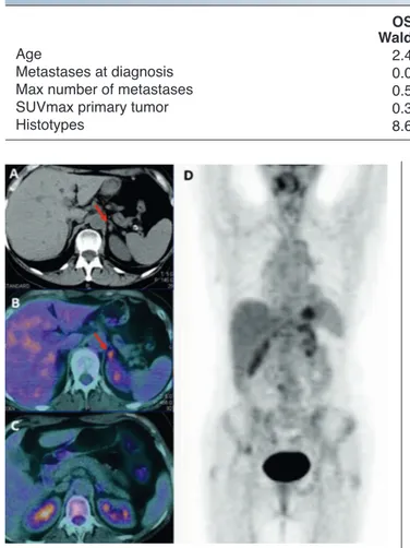
Figure 1. A 74 years old man affected by right adrenocortical carcinoma. Computed tomography transaxial image A) showed a 4.5cm solid adrenal mass characterized by an inhomogeneous enhancement after contrast enhancer injection (green arrow). Fluorine-18-FDG PET/CT axial CT, PET and fused images (B, C and D) con-firmed the presence of the lesion (green arrows) with focal and intense 18F-FDG
cantly related to both DFS and OS (Figure 4). The presence of metastases at diagnosis was a significant factor for DFS, while metastases diagnosed at last follow-up were signifi-Table 2. Kaplan-Mayer curves showed that global 18F-FDG
PET/CT showed significant DFS but not OS (Figure 3). Ka-plan-Mayer curves showed that histotypes were
signifi-Age (years)
Metastases at diagnosis (n) Max number of metastases OS (months) DFS (months) Sex F M Centre Bari Bologna Genova London Turin Histotypes Carcinoma Neuroblastoma Pheocromocytoma Lymphoma 3 months follow-up Disease free Stable disease Progressive disease 6 months follow-up Disease free Stable disease Progressive disease 12 months follow-up Disease free Stable disease Progressive disease Outcome Alive Dead Missed Mean 44.17 3.94 5.16 34.41 16.81 SD 24.00 6.29 7.26 21.04 21.94 Min 1.91 0 0 3 0 Max 78.80 21 21 106 92 N 32 36 8 9 13 9 29 38 16 12 2 30 24 14 32 19 17 36 12 20 46 20 2 % 47.1 52.9 11.8 13.2 19.1 13.2 42.6 55.9 23.5 17.6 3 44.1 35.3 20.6 47 28 25 53 17.6 29.4 67.6 29.4 2.9
Table 1. Characteristics of the 68 patients.
OS= overall survival; DFS= disease free survival
Global 18F-FDG PET/CT (neg/pos)
18F-FDG PET/CT Adrenal localization (neg/pos)
Histotypes
Metastases at diagnosis Metastases at last follow-up
Log Rank test 3.204 0.593 10.971 1.422 3.784 P 0.073 0.441 0.012 0.233 0.052
Table 2. Mantel Cox Log Rank test results for OS and DFS.
OS
Log Rank test 3.204 0.593 10.971 1.422 3.784 P 0.073 0.441 0.012 0.233 0.052 DFS
lignancies. In our study the overall accuracy of CECT was lower (65%) than what is reported in the literature, but the heterogeneity of the sample should be considered in inter-preting these results.
The 18F-FDG PET/CT examination has become a widely used imaging tool in staging adrenal malignancies with high impact on clinical management of the patients [20, 21]. Sev-eral studies, investigating the role of 18F-FDG PET/CT, demonstrated that the whole-body technique has a great potential to characterize malignant lesions and also to eval-uate possible sites of secondary lesions with high detection accuracy [20].
Other studies suggested that SS, SP, PPV, NPV and accu-racy of 18F-FDG PET/CT in detecting adrenal malignancies were: 90%-100%, 78%-100%, 81.8%, 100% and 95.1%, re-spectively [22-25]. In our study, 18F-FDG PET/CT showed a good performance with SS, SP, PPV, NPV and accuracy in line with the above (75.8%, 94.4%, 96.2%, 68% and 83.3% re-spectively). False-positive results of 18F-FDG PET/CT have been reported in a variety of causes including significant 18 F-FDG uptake in some adrenal adenomas, in adrenal endothe-lial cysts, in inflammatory and in infectious lesions. False-negative findings have been seen in the presence of hemorrhage or necrosis and in small lesions [26].
To our knowledge, only a single study has compared CT to 18F-FDG PET/CT for diagnosing adrenal tumors showing better results for CT [19]. The results of our multicentric study revealed a better diagnostic performance of 18F-FDG PET/CT than CECT in the overall evaluation of patients with adrenal malignancies. Dome of differences between our re-sults and those of others [19] may be due to differences in the selection of patients and in their different histotypes.
The contribution of SUVmax in characterizing adrenal le-sions remains open for discussion. Boland et al. (2011) sug-gested that the routine use of SUVmax is problematic because it is subject to many sources of variability, including body habitus and composition, varying times between ra-dionuclide injection and imaging, plasma glucose concen-tration, image reconstruction method, and partial volume effects [25]. To avoid this bias, in our study we considered only examinations performed with the same procedures.
The SUVmax of normal adrenal glands ranged from 0.95 to 2.46, but it is well known that some benign adrenal le-sions can be mildly 18F-FDG avid and the different types of malignancies can have different degrees of 18F-FDG uptake [25, 27, 28].
High SUVmax and large extent of bone and bone marrow disease seemed to correlate with decreased survival and poor prognosis [29].
cantly correlated both for OS and for DFS.
Cox regression analysis results, about the variables ana-lyzed for OS and DFS are reported in Table 3. Histotype was the unique variable predictive for the OS. The variable pre-dictive for DFS was shown to be the “Max Number of Metas-tases”, even if it did not reach statistical significance. Performing survival analysis, stratifying the sample by pres-ence or abspres-ence of metastases at diagnosis, we observed that SUVmax was a significant prognostic factor for DFS when metastases were absent (Wald test=7.035 P=0.008).
Discussion
One of the most difficult goals of imaging is to correctly di-agnose adrenal lesions and avoid unnecessary aggressive procedures. Nowadays, CECT is used to detect large, solid, irregularly shaped lesions frequently associated to hetero-geneous density, due to necrosis, calcifications and hemor-rhages, with accuracy of almost 100% [17-19]. Furthermore CECT provides essential information for staging adrenal ma-Figure 2. A 54 years old female with left primary adrenal non-Hodgkin’s lym-phoma. Computed tomography transaxial image A) showed a not well character-izable left adrenal lesion (red arrow). Fluorine-18-FDG PET/CT fused transaxial image B) showed 18F-FDG uptake (SUVmax 4.8) in the left adrenal gland.
Fluorine-18-FDG PET/CT fused transaxial C) and PET MIP D) images performed after chemotherapy, showed the resolution of the disease.
Age
Metastases at diagnosis Max number of metastases SUVmax primary tumor Histotypes Wald test 2.434 0.031 0.571 0.377 8.686 P 0.119 0.861 0.450 0.539 0.034
Table 3. Cox Regression results for OS and DFS.
OS Wald test 0.048 0.451 4.857 3.432 5.587 P 0.826 0.502 0.028 0.064 0.134 DFS
To the best of our knowledge, only the study of Leboulleux et al. (2006) showed that a high 18F-FDG uptake appears to represent an independent prognostic factor. They reported that 54% of the patients with a SUVmax>10 died within 6 months after 18F-FDG PET/CT examination, whereas none of the patients with a SUVmax<10 died. A high 18F-FDG uptake appears to represent an additional stage-independent prog-nostic factor [30]. In the study of Tessonnier et al (2013) high SUVmax was not significantly associated with shortened OS and DFS [31].
In our study we observed that only when the sample was stratified by the presence/absence of metastases at diagno-sis, SUVmax demonstrated a significant prognostic factor; it was predictive just for DFS, and only when metastases were absent (Wald test=7.035 P=0.008). We acknowledge that the sample size was probably too small to detect subtle differ-ences among patients, and that no statistical adjustment could be made for potential confounding factors.
Only the maximum number of metastases was predictive for DFS. Stratifying the sample by the presence or absence of metastases at the time of diagnosis, SUVmax resulted as a significant prognostic factor for DFS when metastases were absent.
Adrenocortical carcinomas, as well as primary adrenal lym-phomas, usually show moderate to intense 18F-FDG uptake values and reports in the literature showed high 18F-FDG PET/CT diagnostic performance for them, as well as in our study [16, 17, 32]. Neuroblastomas are 18F-FDG avid, and in one study it was proposed that 18F-FDG PET/CT are the only imaging modality to better assess this disease [33]. However, the exact diagnostic accuracy remains to be defined; in our study it resulted 70.4%. Malignant pheochromocytomas are rare tumors often larger than 2cm, and so easily outlined but difficult to differentiate from other adrenal tumors by CT alone, while 18F-FDG PET/CT has high 18F-FDG avidity for this malignancy [34]. In our study both CECT and 18F-FDG PET/CT showed high diagnostic performance for these carcinomas (accuracy of 90%).
The OS and DFS are generally poor in adrenal malignan-cies and are influenced by many factors including histotype, presence of metastases and age. In our study, mean OS and DFS were 34.41 and 16.81 months and all histotypes were accompanied by poor prognosis. Identification of powerful predictors of prognosis could substantially improve the choice of treatment and the clinical outcome.
Our study demonstrated different OS and DFS depending on the histotypes of the tumors. Histotypes greater influ-enced our results. The course of the disease, the incidence of metastases and the treatment modalities in these patients varied greatly depending on the histotypes. Furthermore histotypes influenced both DFS (Log Rank test:12.738, P=0.005) and OS (Log Rank test:10.971, P=0.012). According to the evolution of known diseases, our results also showed the worst DFS and OS in neuroblastoma patients and the better ones in lymphoma patients. Furthermore, from the multivariate evaluation, histotype was the unique variable predictive of OS (Wald test=8.686 P=0.034).
The prognostic value of 18F-FDG PET/CT was established in some other malignancies related with high 18F-FDG
up-take, but was not well established as yet in adrenal malig-nancies [31].
Our study demonstrated that 18F-FDG PET/CT, in terms of positive or negative 18F-FDG uptake in adrenal glands, influ-ences the DFS (Log Rank test:11.743, P=0.001), but did not have any impact on OS (Log Rank test:3.204, P=0.073).
In conclusion, our study suggested that 18F-FDG PET/CT was a powerful imaging tool, offering higher accuracy, in comparison to CECT, for the detection and characterization of the most common histotypes of adrenal carcinomas. Di-agnostic algorithms therefore should include 18F-FDG PET/CT as a step, for the diagnosis of adrenal tumors. Studies with more patients are warranted.
The authors declare that they have no conflicts of interest.
Bibliography
1. Rha SE, Byun JY, Jung SE et al. Neurogenic Tumors in the Ab-domen: Tumor Types and Imaging Characteristics. Radiographics 2003; 23: 29-43.
2. Tsukahara T, Takasawa A, Murata M et al. NK/T-cell lymphoma of bilateral adrenal glands in a patient with pyothorax. Diagn Pathol 2012; 7: 114.
3. Barzon L, Boscaro M. Diagnosis and management of adrenal in cidentalomas. J Urol 2000; 163: 398-407.
4. Keskin S, Taş F, Vatansever S. Adrenocortical carcinoma: clinico-pathological features, prognostic factors and outcome. Urol Int 2013; 90: 435-8.
5. Didolkar MS, Bescher RA, Elias EG et al. Natural history of adrenal cortical carcinoma: A clinicopathologic study of 42 patients. Cancer 1981; 47: 2153-61.
6. Blake A, Kalra MK, Sweeney AT et al. Distinguishing benign from malignant adrenal masses: multi-detector row CT protocol with 10-minute delay. Radiology 2006; 2: 578-85.
7. Szolar DH, Korobkin M, Reittner P et al. Adrenocortical carcinomas and adrenal pheocromocytomas: mass and enhancement loss evaluation at delayed contrast-enhanced CT. Radiology 2005; 234: 479-85.
8. Caoili EM, Korobkin M, Francis IR et al. Adrenal masses: charac-terization with combined unenhanced and delayed enhanced CT. Radiology 2002; 222; 629-33.
9. Niccoli-Asabella A, Altini C, Notaristefano A et al. A retrospective study comparing contrast-enhanced computed tomography with 18F-FDG-PET/CT in the early follow-up of patients with
retroperitoneal sarcomas. Nucl Med Commun 2013; 34(1): 32-9. 10. Rubini G, Altini C, Notaristefano A, et al. Peritoneal carcinomatosis from ovarian cancer: role of 18F-FDG-PET/CT and CA125.
Re-centi Prog Med 2012; 103(11): 510-4.
11. Wong KK, Arabi M, Zerizer I et al. Role of positron emission to mography/computed tomography in adrenal and neuroen-docrine tumors: fluorodeoxyglucose and nonfluorodeoxyglu-cose tracers. Nucl Med Commun 2011; 32: 764-81.
12. Rufini V, Treglia G, Castaldi P et al. Comparison of metaiodoben zylguanidine scintigraphy with positron emission tomography in the diagnostic work-up of pheochromocytoma and paragan glioma: a systematic review. Q J Nucl Med Mol Imaging 2013; 57: 122-33.
13. Gust L, Taieb D, Beliard A et al. Preoperative 18F uptake is strongly
correlated with malignancy, Weiss score, and molecular markers of aggressiveness in adrenal cortical tumors. World J Surg 2012; 36: 1406-10.
14. Choyke PL. ACR Committee on Appropriateness Criteria. ACR Appropriateness Criteria on incidentally discovered adrenal mass. J Am Coll Radiol 2006; 3(7): 498-504.
of Adrenal Masses by Using FDG PET: A Systematic Review and Meta-Analysis of Diagnostic Test Performance. Radiology 2011; 259: 117-26.
26. Chong S, Lee KS, Kim HY et al. Integrated PET-CT for the charac-terization of adrenal gland lesions in cancer patients: diagnostic efficacy and interpretation pitfalls. RadioGraphics 2006; 26: 1811-24.
27. Bagheri B, Maurer AH, Cone L et al. Characterization of the normal adrenal gland with 18F FDG PET/CT. J Nucl Med 2004; 45: 1340-43.
28. Becherer A, Vierhapper H, Pötzi C et al. FDG-PET in adrenocortical carcinoma. Cancer Biother Radiopharm 2001; 16: 289-95. 29. Papathanasiou ND, Gaze MN, Sullivan K et al. 18F-FDG PET/CT
and 123I-metaiodobenzylguanidine imaging in high-risk
neurob-lastoma: diagnostic comparison and survival analysis. J Nucl Med 2011; 52: 519-25.
30. Leboulleux S, Dromain C, Bonniaud G et al. Diagnostic and prog nostic value of 18-fluorodeoxyglucose positron emission to-mography in adrenocortical carcinoma: a prospective compar-ison with computed tomography. J Clin Endocrinol Metab 2006; 91: 920-5.
31. Tessonnier L, Ansquer C, Bournaud C et al. 18F-FDG Uptake at
Initial Staging of the Adrenocortical Cancers: A Diagnostic Tool but Not of Prognostic Value. World J Surg 2013; 37: 107-12. 32. Tessonnier L, Sebag F, Palazzo FF et al. Does 18F-FDG PET/CT
add diagnostic accuracy in incidentally identified non-secreting adrenal tumours? Eur J Nucl Med Mol Imaging 2008; 35: 2018-25. 33. Piccardo A, Lopci E, Conte M et al. PET/CT imaging in neurob
lastoma. Q J Nucl Med Mol Imaging 2013; 57: 29-39.
34. Blake MA, Prakash P, Cronin CG. PET/CT for adrenal assessment. AJR 2010; 195: W91-5.
EANM procedure guidelines for tumour PET imaging: version 1.0. Eur J Nucl Med Mol Imaging 2010; 37(1): 181-200.
16. Wahl RL, Jacene H, Kasamon Y et al. From RECIST to PERCIST: Evolving Considerations for PET response criteria in solid tumors. J Nucl Med 2009; 50(Suppl 1): 122S-50S.
17. Boland GW, Lee MJ, Gazelle GS et al. Characterization of adrenal masses using unenhanced CT: an analysis of the CT literature. AJR 1998; 171: 201-4.
18. Guerrisi A, Marin D, Baski M et al. Adrenal lesions: spectrum of imaging findings with emphasis on multi-detector computed tomography and magnetic resonance imaging. J Clin Imaging Sci 2013; 3: 61.
19. Park BK, Kim CK, Kim B et al. Comparison of delayed enhanced CT and 18F-FDG PET/CT in the evaluation of adrenal masses in
oncology patients. J Comput Assist Tomogr 2007; 31: 550-6. 20. Deandreis D, Leboulleux S, Caramella C et al. FDG PET in the
management of patients with adrenal masses and adrenocortical carcinoma. Horm Cancer 2011; 2: 354-62.
21. Quartuccio N, Cistaro A. Adrenal Gland Cancers. In Atlas of PET/CT in pediatric patients. Cistaro (ed. Springer), Heidelberg, 2014 pp. 147-9.
22. Kumar R, Xiu Y, Yu JQ et al. ISF-FDG PET in evaluation of adrenal lesions in patients with lung cancer. J Nucl Med 2004; 45: 2058-62. 23. Park SY, Park BK, Kim CK. The Value of Adding 18F-FDG PET/CT
to Adrenal Protocol CT for Characterizing Adrenal Metastasis (≥10mm) in Oncologic Patients. AJR 2014; 202: W153-60. 24. Blake MA, Slattery JM, Kalra MK et al. Adrenal lesions: character
ization with fused PET/CT image in patients with proved or sus-pected malignancy–initial experience. Radiology 2006; 238: 970-7.
25. Boland GWL, Dwamena BA, Sangwaiya MJ et al. Characterization