ISSN 0974-8237
Journal of
Craniovertebral
Junction & Spine
www.jcvjs.com
April-June 2018 / Volume 9 / Issue 2
Journal of
Craniovertebral Junction and Spine
Ÿ V olume 9 Ÿ Issue 2 Ÿ April-June 2018 Ÿ Pages 1-*** Spine 5 mm
ABSTRACT
Background: Different surgical techniques have been described for treatment of degenerative lumbar stenosis (DLS). Only postoperative
measures have been identified as predictors of efficacy of decompression. The objective of this study is to assess the role of navigated unilateral laminotomy with crossover to achieve and predict a satisfying decompression and outcome in DLS.
Materials and Methods: We enrolled patients with DLS who underwent navigation‑assisted unilateral laminotomy with crossover. The extent
of decompression was evaluated during surgery using neuronavigation. The outcome was assessed through the Oswestry disability index (ODI) and visual analog scale (VAS) for leg pain. Outcome correlation with the extent of the intraoperative bone decompression was analyzed. Finally, the outcome, surgical time, and in‑hospital length‑of‑stay were compared with a control group treated through standard unilateral laminotomy.
Results: Twenty‑five patients were treated using the navigated technique (Group A), 25 using the standard unilateral laminotomy (Group B).
In Group A, a cut‑off value ≥0.9 cm for bone decompression revealed to be an intraoperative predictor of good outcome, both regarding the ODI and VAS scores (P = 0.0005; P = 0.002). As compared with Group B, patients operated using the navigated technique showed similar operative times, in‑hospital length‑of‑stay, ODI scores, but improved VAS scores for leg pain (P = 0.04).
Conclusions: The intraoperative navigated evaluation of the bone decompression could predict the outcome allowing satisfactory results
in unilateral laminotomy for DLS. The navigated technique also could lead to an improved decompression of lateral recesses resulting in better control of leg pain as compared to standard unilateral laminotomy.
Keywords: Lumbar stenosis, minimally invasive spine surgery, navigation, unilateral laminotomy
INTRODUCTION Background
Simple posterior decompression represents one the most used techniques for surgical treatment of degenerative lumbar spine stenosis.[1‑14] Different surgical techniques have
been described,[4,7‑14] but a clear superiority of one technique
over the others regarding pain and functional outcome has not been identified.[2,10,15,16]
Several attempts to obtain quantitative measurements to assess the degree of decompression achieved and to predict the functional outcome have been made. The dural sac cross‑sectional area (DSCSA) of the lumbar spinal canal, as measured on the postoperative computed
tomography (CT) or magnetic resonance imaging (MRI) scans, turned out to be a reliable predictor of clinical and functional outcome.[2,17‑21] On the other hand, the DCSA
Navigated minimally invasive unilateral laminotomy with
crossover for intraoperative prediction of outcome in
degenerative lumbar stenosis
Access this article online
Website:
www.jcvjs.com
Quick Response Code
DOI:
10.4103/jcvjs.JCVJS_45_18
Salvatore Massimiliano Cardali1, Fabio Cacciola1, Giovanni Raffa1,2, Alfredo Conti1, Maria Caffo1, Antonino Germanò1
1Division of Neurosurgery and 2Department of Clinical and
Experimental Medicine, University of Messina, Messina, Italy
Address for correspondence: Dr. Giovanni Raffa,
Department of Clinical and Experimental Medicine and Division of Neurosurgery, University of Messina, AOU Policlinico "G. Martino", Via Consolare Valeria 1, 98125 Messina, Italy.
E‑mail: [email protected]
How to cite this article: Cardali SM, Cacciola F, Raffa G, Conti A, Caffo M, Germano A. Navigated minimally invasive unilateral laminotomy with crossover for intraoperative prediction of outcome in degenerative lumbar stenosis. J Craniovert Jun Spine 2018;9:107‑15.
This is an open access journal, and articles are distributed under the terms of the Creative Commons Attribution‑NonCommercial‑ShareAlike 4.0 License, which allows others to remix, tweak, and build upon the work non‑commercially, as long as appropriate credit is given and the new creations are licensed under the identical terms.
Cardali, et al.: Navigated decompression predicts outcome in unilateral laminotomy for lumbar stenosis
108 Journal of Craniovertebral Junction and Spine / Volume 9 / Issue 2 / April-June 2018
is a postoperative measurement and cannot be used to change the surgical strategy.[22] Till date, no intraoperative
parameter has been identified as a predictor of good functional outcome.
Objectives
The aim of this study is to describe a new technique for the qualitative and quantitative intraoperative evaluation of the extent of decompression using intraoperative three‑dimensional (3D) fluoroscopy and navigation. These measurements were used to guide the surgical procedure in unilateral laminotomy with bilateral decompression of the spinal canal used as decompressive procedure for the treatment of degenerative lumbar stenosis (DLS). We also analyzed the efficacy of the navigated technique by comparing the outcome with patients treated using standard microsurgical unilateral laminotomy.
MATERIALS AND METHODS Study design
We collected clinical and outcome data from patients operated on for DLS using the navigated minimally invasive posterior decompression. We analyzed the association between the extent of intraoperative decompression, as evaluated through the navigated technique, and the postoperative outcome. Finally, we compared the operative times, in‑hospital length of stay, and 6‑month outcome with that of a historical control group composed of patients operated using the standard microsurgical unilateral laminotomy.
Selection and description of participants
We reviewed and collected the clinical data of patients operated on for single‑level DLS between January and October 2016.
Inclusion criteria were as follows: Age ≥18 years old; the presence of a single‑level lumbar degenerative stenosis; failure of symptoms relief with non‑surgical treatment after a 6 months period. Exclusion criteria were as follows: Age <18 years old; lumbar stenosis associated with spinal instability requiring instrumented fusion; the presence of multilevel stenosis; previous lumbar spine surgery; refuse to sign the informed consent. The historical control group included the last 25 consecutive patients matching the inclusion criteria and operated during the same timeframe without using the navigated technique. The study was conducted according to the Declaration of Helsinki and its later amendments. The informed consent was obtained from all individual participants included in the study.
Data sources
Demographic and clinical data of patients including age, sex, pre‑ and post‑operative oswestry disability index (ODI) and visual analogue scale (VAS) scores, extent of bone decompression, operative time, and in‑hospital length of stay were collected from our Institutional database including: clinical charts, PACS, outpatients’ clinic records.
Measurements
The functional outcome was evaluated by comparing the preoperative versus post‑operative ODI (version 1)[23,24]
and VAS score[25] for leg pain after 6 months from surgery
in both groups. The length of surgery and the in‑hospital staying were also recorded. Finally, 6 months after surgery, patients underwent MRI scan and dynamic X‑rays (during flexion and extension) of the lumbar spine to assess neural decompression or postoperative instability. In Group A patients, a correlation analysis between the extent of bone decompression and the outcome (ODI and VAS scores) was performed. The ability of the quantitative evaluation of the bone decompression to predict the functional outcome was also assessed.
Surgical technique
The navigated minimally invasive posterior decompression consisted of a unilateral laminotomy with crossover. The patient is placed in prone position with flexion of the thigh and legs.[26] The skin incision is performed over the
spinous processes of the interested level. Unilateral incision of the muscular fascia and subperiosteal dissection of paravertebral muscles are performed up to visualize the spinous processes of the two interested vertebrae, the corresponding laminae, and the interlaminar space. The side of the access is defined according to clinical signs/symptoms and/or neuroradiological findings. A Williams retractor is positioned, and a small contralateral incision of the muscular fascia is performed just to visualize the contralateral surface of the spinous process of the upper vertebra. The navigation optical reference is clamped over the spinous process of the upper vertebra and positioned with an inclination toward the head of the patients to avoid interference with the microscopic view and surgical instruments. A baseline intraoperative low‑dose 3D fluoroscopy is performed to acquire imaging for neuronavigation (O‑arm Surgical Imaging, Medtronic Navigation, USA). At this point, the microsurgical technique is employed, and neuronavigation is ready to be used (Spine software, StealthStation S7, Medtronic Navigation, USA) [Figure 1]. A unilateral laminotomy is performed by a partial removing of the lower part of the upper lamina and the upper part of the inferior lamina using Kerrison rongeurs. The medial facet joint is slightly undercut
as well as the basis of the spinous process on the midline. The ipsilateral ligamentum flavum is visualized and removed using rongeurs up to identify the dural sac. The ipsilateral radicular recess is identified with a navigated probe. This helped to qualitatively assess the entity of ipsilateral decompression. In case of non‑satisfying unilateral decompression, a further lamina and ligamentum flavum resection is performed. At this point, the surgical table is tilted contralaterally to gain an appropriate working angle to visualize the contralateral recess. The contralateral ligamentum flavum is then removed with the use of rongeurs. The basis of the spinous process and the medial facet can be further undercut to better visualize the contralateral recess, the contralateral border of the dural sac and the corresponding emerging spinal nerve root. The extent of the contralateral decompression is then qualitatively verified with the navigated probe. If the tip of the probe can successfully reach the contralateral recess, the decompression is considered satisfying. If not, further resection of the ligamentum flavum and undercutting of the medial portion of the contralateral facet are performed. The progressive decompression of the contralateral recess is therefore evaluated under direct view and for the more lateral portion that is not accessible to the sight, through the progression and the degree of mobility of the navigated probe tip along and across the recess and the foramen. The more is the removed ligamentum flavum and the soft tissue contralateral decompression, the easier will be to reach the contralateral recess using the tip of the navigated probe. This will be evaluated also through the visual feedback on the neuronavigation screen [Figure 2]. At the end of the procedure, a second low‑dose 3D fluoroscopy is performed. The entity of decompression is now qualitatively and quantitatively evaluated using the neuronavigation probe on the post‑decompression scan, and the extent of the unilateral bone removal in a lateromedial direction is measured at the level of the larger portion of the laminotomy [Figure 2].
Patients in the control group were operated using the same microsurgical technique but without using intraoperative navigation.
Statistics
The comparison between pre‑ and post‑operative ODI and VAS scores was performed by using the paired Student
t‑test. The correlation between the extent of the bone
decompression and the functional outcome (ODI and VAS scores) was assessed through the Pearson correlation and linear regression analysis. The accuracy of the quantitative measure of the bone decompression in predicting the functional outcome was analyzed using the Fisher’s test and defined through computation of the sensitivity (ST), specificity (SP), positive and negative predictive values (PPV and NPV). The comparison of operative times, in‑hospital length of stay, and outcome between the study group and controls was performed using the unpaired Student t‑test. Statistical significance was defined as a P < 0.05. Data analysis was performed using GraphPad Prism version 6.00 for Windows, GraphPad Software, La Jolla, California, USA, www. graphpad.com.
RESULTS Participants
Fifty patients were enrolled in the study. Twenty‑five patients (14 males, 11 females, mean age 64.2 ± 5.2 years old) were operated using the navigated technique (Group A).
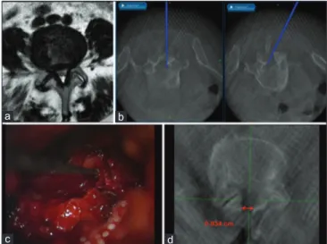
Figure 1: An example of the intraoperative use of neuronavigation for the minimally invasive unilateral laminotomy with crossover in a case of lumbar degenerative stenosis. The blue stick and the green crosshair represent the navigation probe tip visualized in different planes and projections
d c
b a
Figure 2: Case illustration of a patient affected by L4‑L5 degenerative stenosis in Group A. (a) Preoperative axial MRI scan showing a narrowing of the spinal canal. (b) Intraoperative qualitative verification of the decompression of the ipsilateral (on the right) and contralateral (on the left) recesses. (c) Microscopic view of the neuronavigation probe used to verify decompression. (d) Three‑dimensional fluoroscopy after decompression showing the maximal extent of the laminotomy that is intraoperatively quantified as 0.934 cm
Cardali, et al.: Navigated decompression predicts outcome in unilateral laminotomy for lumbar stenosis
110 Journal of Craniovertebral Junction and Spine / Volume 9 / Issue 2 / April-June 2018
The control group included 25 patients operated using the standard non‑navigated unilateral laminotomy (13 males, 12 females, mean age 64.3 ± 3.7) (Group B).
Descriptive data
At the admission, in Group A, the median ODI score was 46% (range 36–50), and the median VAS score for leg pain was 7 (range 5–9). The mean surgical time was 75.88 ± 11.07 min. The mean in‑hospital stay was 2.44 ± 0.71 days.
In Group B, the median preoperative ODI score was 46% (range 38–50), the median VAS score for leg pain was 7 (range 6–9), the mean surgical time 74.52 ± 8.84 min, and the mean in‑hospital stay was 2.48 ± 0.58 days.
No significant differences were observed between the two groups [Table 1].
In Group A, the intraoperative evaluation of decompression was considered satisfying in all cases. The contralateral decompression through crossover (resection of the ligamentum flavum, undercutting of the medial aspect of the facets) was stopped when it was considered satisfying under the direct microscopic view, and for the more lateral portion, when the navigation probe tip could be moved without obstacles along and across the contralateral
recess and foramen on the neuronavigation screen, thus demonstrating a satisfying decompression. The mean bone decompression as measured on the navigation screen was 0.927 ± 0.118 cm.
In Group B, the qualitative evaluation of the decompression was performed under the microscopic view, even tilting the operating table, without any quantitative assessment of the achieved decompression. The decompression was stopped in all cases when the qualitative microscopic assessment of the decompression was considered satisfying.
Outcome data
After 6 months from surgery, we observed an overall improvement of the functional independence (ODI score) and leg pain (VAS score) in both groups.
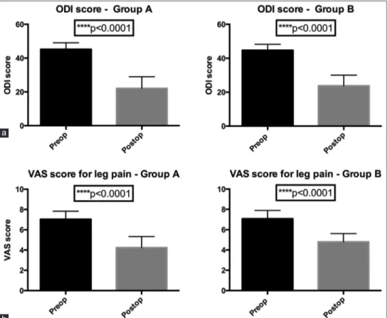
In particular, in Group A, we observed a significant reduction of the median ODI score to 21% (range 8%–40%, P < 0.0001). The median VAS score for leg pain was significantly reduced to 4 (range 2–6, P < 0.0001) [Figure 3].
Interestingly, we observed a significant negative correlation between the entity of the bone decompression and the functional outcome expressed as ODI (r = −0.926, P < 0.0001) and VAS scores (r = −0.885, P < 0.0001). A cutoff value for
Table 1: Clinical characteristics of patients and main findings of the study
Characteristics Measurements Group A
vs. B (P) Group A (Navigated Technique) Group B (standard technique)
n of patients 25 25 NA
Sex
Males 14 13 NA
Females 11 12
Age (mean±SD) 64.2±5.2 years old 64.3±3.7 years old P=0.9 ns
Operative time (mean±SD) 75.88±11.07 min 74.52±8.84 min P=0.6 ns
In-hospital length of stay (mean±SD) 2.44±0.71 days 2.48±0.58 days P=0.8 ns
Extent of bone decompression (mean±SD) 0.927±0.118 cm NA P=NA
Median ODI score (range)
Preoperative 46 (36-50) 46 (38-50) P=0.6 ns
P=0.3 ns
Postoperative 21 (8-40) 23 (14-40)
Pre vs. post P<0.0001 P<0.0001
Median VAS score (range)
Preoperative 7 (5-9) 7 (6-9) P=0.8 ns
P=0.04
Postoperative 4 (2-6). 5 (3-6)
Pre vs. post P<0.0001 P<0.0001
Correlation between outcome and extent of bone resection
ODI score r=-0.926, P<0.0001 NA NA
VAS score r=-0.885, P<0.0001
Accuracy of the extent of bone resection as predictor of outcome
ODI score ≤20 ST=100%, SP=71.4%, PPV=73.3%, NPV=100%, P=0.0005 NA NA
bone decompression ≥ 0.9 cm was identified as a predictor of good outcome both for the ODI and VAS scores [Figure 4]. As well, in Group B, we observed a significant improvement of the ODI score to 23% (range 14%–40%, P < 0.0001) and of the VAS score for leg pain to 5 (range 3–6, P < 0.0001) as compared to the preoperative period.
Interestingly, we observed a significantly better improvement of the median VAS score for leg pain in Group A vs. Group B (P = 0.04) [Figure 5]. No differences were observed for the ODI score.
Other analyses
The best accuracy for intraoperative bone decompression was observed for a cut‑off ≥0.9 cm, with a sensitivity of 100%, a specificity of 71.4%, a PPV of 73.3%, and a NPV of 100% in predicting good outcome expressed as ODI ≤20 (minimal disability) (P = 0.0005) [Figure 4a]. The same cut‑off value revealed a sensitivity of 86.6%, a specificity of 80%, a PPV of 86.6%, and an NPV of 80% in predicting a good relief from leg pain expressed as a VAS <5 (P = 0.002) [Figure 4b]. No case of metameric instability caused by decompression was observed after 6 months from surgery in both groups.
DISCUSSION Key results
In this study, we assessed qualitatively and quantitatively the entity of decompression during surgery using intraoperative 3D fluoroscopy and navigation. Our results suggest that this procedure is associated with a satisfactory outcome. Furthermore, the extent of the bone decompression, as measured intraoperatively, was actually correlated to the improvement in ODI and VAS scores. We also identified a cut‑off value ≥0.9 mm (as measured at the largest portion of the laminotomy) as an intraoperative predictor of a good postoperative functional outcome. Finally, in this series, the use of the navigated technique was associated with a better control of the radicular pain as compared to the standard microsurgical decompression. Nevertheless, the use of the navigated technique did not result in a reduced length of surgery or in‑hospital staying, neither to an improvement of the postoperative ODI as compared to the standard microsurgical laminotomy.
Interpretation
Despite several surgical techniques for decompression of the spinal canal in lumbar degenerative stenosis are well established, routinely used all over the world, and often
Figure 3: Assessment of outcome after 6 months from surgery. (a) The Oswestry disability index score is significantly reduced as well as the (b) visual analog scale score for leg pain compared to the admission in both groups
b a
Cardali, et al.: Navigated decompression predicts outcome in unilateral laminotomy for lumbar stenosis
112 Journal of Craniovertebral Junction and Spine / Volume 9 / Issue 2 / April-June 2018
considered a simple surgical gesture, the postoperative outcome is considered satisfactory by patients in the only 63.4%–81.9% of cases.[27‑29] Different percentages of
satisfaction are reported for different techniques, but the common result is that surgical treatment for spinal stenosis could be considered unsatisfactory in up to 36.6% of patients. This aspect is usually neglected, especially by experienced spine surgeons who consider surgical decompression for lumbar stenosis a straightforward surgical procedure that could not be suitable for/does not need further technical improvement. Another important aspect that is usually neglected is the role that the bone narrowing of the spinal canal plays in the prediction of postoperative outcome. Adamova et al., in a retrospective cohort of 53 patients affected by lumbar stenosis, reported the lowest transverse diameter of spinal canal <13.6 mm measured at the CT scan was the only independent predictor of unsatisfactory clinical outcome;[30] moreover, Choi et al. in a series of 144 patients
with lumbar stenosis treated by unilateral approach for bilateral decompression, reported that the satisfactory rate after surgery was reduced in cases of trefoil stenosis as compared to cases with a round or oval spinal canal.[28]
This could reflect the role of narrowed lateral recesses in radicular compression. Nevertheless, in DLS, it is well known that also soft tissues, such as the ligamentum flavum, play an important role in compression. Such effect can be assessed
through the evaluation of the DSCSA at the MRI scan. The DSCA evaluation has been suggested as a method to assess the compression (before surgery) and decompression (after surgery) of the dural sac by measuring the area of the spinal canal on pre‑operative and post‑operative imaging.[2,17‑21]
Actually, an increase of the DSCSA has been described as positively correlated to a better functional outcome,[31] but
the postoperative nature of this measurements limits its clinical relevance.[22] Collectively, these data suggest that no
other imaging methodologies apart from preoperative and postoperative CT/MRI scans have been identified so far to assess the compression and the decompression of the spinal canal after surgery for lumbar degenerative stenosis. To the best of our knowledge, there are no studies reporting intraoperative imaging data that can positively influence the surgical procedure (i.e., suggesting changes in the size or location of the decompression) and therefore predict patients’ outcome.
Despite the unilateral laminotomy with crossover has been already described as effective to achieve a bilateral decompression,[4,8,32] there are no studies in which the extent
Figure 5: Comparison of the outcome between Group A and B. (a) Patients treated with the navigated technique (Group A) showed a similar postoperative Oswestry disability index score as compared to patients treated through standard microsurgical unilateral laminotomy with crossover (Group B). (b) Conversely, patients in Group A showed a significant improvement of the postoperative visual analog scale score for leg pain as compared to patients in Group B
b a
Figure 4: Analysis of correlation between the extent of bone decompression and the postoperative outcome after 6 months in Group A. (a) The postoperative oswestry disability index and (b) visual analog scale scores show a significant negative correlation with the extent of bone decompression that is quantified during surgery through the neuronavigation. A cut‑off value ≥0.9 cm is significantly associated to a good functional outcome intended as an Oswestry disability index score ≤20 and a visual analog scale score <5
b a
of decompression was intraoperatively quantified. This is a relevant point as the efficacy of unilateral approaches has been questioned because considered inadequate for bilateral decompression of the spinal canal.[12,29,33,34] Actually,
using a standard microsurgical technique, there is no way other than a qualitative visual inspection to achieve a proof that sufficient decompression has been achieved. This does actually lead to a wide intra‑ and inter‑operator variability of decompression results. Nevertheless, this aspect is often neglected. The navigated technique with intraoperative imaging provides objective quantification eventually extending the span of decompression while preserving the facet joints. As expected, larger bone decompression was associated to a better outcome, especially using a cut‑off value ≥0.9 cm. To the best of our knowledge, this is the first intraoperative parameter associated to outcome in posterior decompression for lumbar stenosis.
This value can be measured during surgery, eventually suggesting an extension of bone resection while intraoperative imaging may help to preserve critical structures, such as the facet joints.
It can be argued that bone decompression alone is not able to predict the postoperative functional outcome since it mainly depends on the soft‑tissue decompression (i.e., flavectomy). In the present study, the quantitative evaluation of bone removal was associated to a qualitative evaluation of contralateral soft‑tissue decompression. Although this can be achieved by the direct microscopic view, the visual feedback provided by the navigated probe tip [Figure 3] of the most lateral portion of the recess and the foramen that is not available to the direct view of the surgeon appears to us extremely useful. The free movement without the obstacle of the navigated tip along and across the contralateral recess is perceived through tactile feedback but also by visual feedback on the navigation screen. Moreover, our technique allowed the visualization of the contralateral recess and the navigated probe tip in the three planes of the space including the axial plane.
The improved visualization and decompression of the most lateral portion of the recesses resulted in an improvement of the post‑operative radicular pain. This is witnessed by the significant improvement of the VAS score for leg pain in the group treated by using the navigated technique (Group A) as compared with patients operated using the standard microsurgical unilateral laminotomy (Group B). Nevertheless, the improvement of the radicular pain did not resulted also in an improvement of the overall disability of patients, since no significant differences in the ODI scores between the two
groups were recorded. Moreover, similar findings regarding the operative time and in‑hospital staying were observed using the two different techniques.
Thus, a combination of a qualitative and quantitative intraoperative data represents the strength of this navigated unilateral laminotomy with crossover, especially for the decompression of the most lateral portion of the recesses.
At the same time, the navigated technique prevents from an excessive decompression that could lead to postoperative instability, a possible side effect of standard decompressive surgery,[35] since no cases of postoperative instability
were recorded. Although unilateral laminotomy with crossover is less frequently associated to postoperative instability,[10,33] it could be speculated that the real‑time
visualization of the facet joints obtained by using the navigated technique could better guide the undercutting of the medial facet joints, ensuring an increased but not harmful decompression.
Limitations
This study, as the majority of studies evaluating the outcome after spinal surgery, suffers from bias related to the evaluation of patient‑reported functional outcome. This could lead to potential errors and bias such as the response‑shift.[1]
One important difference with other surgical techniques is the use of X‑ray based intraoperative imaging and the consequent increase of X‑ray exposure for the patients. Nevertheless, we used a low dose 3D fluoroscopy and limited the intraoperative imaging to only two scans (at the beginning and the end of surgery).
Finally, it must be underlined that the retrospective nature of the study limits the strength of our findings.
Nevertheless, the aim of this study was to shed a light on the need to further investigate new operative techniques and strategies for the improvement of the satisfaction rate of patients operated for lumbar degenerative stenosis. Despite all the above‑mentioned limitations, the description of our small experience with the use of the navigated unilateral laminotomy goes in that direction.
CONCLUSIONS
Our results suggest that the 3D fluoroscopy‑based navigation could be a useful intraoperative tool for the surgeon to increase its confidence with the extent of
Cardali, et al.: Navigated decompression predicts outcome in unilateral laminotomy for lumbar stenosis
114 Journal of Craniovertebral Junction and Spine / Volume 9 / Issue 2 / April-June 2018
bone decompression in an already described minimally invasive technique (unilateral laminotomy with crossover) for bilateral decompression in lumbar degenerative stenosis. This intraoperative imaging could reassure the surgeon about an adequate decompression, or suggest to extend bone removal undercutting of the medial facet joint, or even the necessity of instrumentation when an extended decompression is required. The identification of a quantitative cutoff of bone decompression (0.9 cm) predicting the outcome further supports the use of intraoperative image‑guidance. Finally, the navigated technique could be associated with a better decompression of the lateral recesses as compared to the standard microsurgical unilateral laminotomy, thus improving the control of postoperative leg pain. Further prospective studies in larger cohorts of patients are warranted. Financial support and sponsorship
Nil.
Conflicts of interest
There are no conflicts of interest. REFERENCES
1. Crawford CH 3rd, Glassman SD, Mummaneni PV, Knightly JJ,
Asher AL. Back pain improvement after decompression without fusion or stabilization in patients with lumbar spinal stenosis and clinically significant preoperative back pain. J Neurosurg Spine 2016;25:596‑601. 2. Hermansen E, Austevoll IM, Romild UK, Rekeland F, Solberg T,
Storheim K, et al. Study‑protocol for a randomized controlled trial comparing clinical and radiological results after three different posterior decompression techniques for lumbar spinal stenosis: The Spinal Stenosis Trial (SST) (part of the NORDSTEN study). BMC Musculoskelet Disord 2017;18:121.
3. Ciol MA, Deyo RA, Howell E, Kreif S. An assessment of surgery for spinal stenosis: Time trends, geographic variations, complications, and reoperations. J Am Geriatr Soc 1996;44:285‑90.
4. Costa F, Sassi M, Cardia A, Ortolina A, De Santis A, Luccarell G, et al. Degenerative lumbar spinal stenosis: Analysis of results in a series of 374 patients treated with unilateral laminotomy for bilateral microdecompression. J Neurosurg Spine 2007;7:579‑86.
5. Atlas SJ, Keller RB, Wu YA, Deyo RA, Singer DE. Long‑term outcomes of surgical and nonsurgical management of lumbar spinal stenosis: 8 to 10 year results from the Maine Lumbar Spine Study. Spine (Phila Pa 1976) 2005;30:936‑43.
6. Deyo RA, Mirza SK, Martin BI, Kreuter W, Goodman DC, Jarvik JG, et al. Trends, major medical complications, and charges associated with surgery for lumbar spinal stenosis in older adults. JAMA 2010;303:1259‑65.
7. Iguchi T, Kurihara A, Nakayama J, Sato K, Kurosaka M, Yamasaki K, et al. Minimum 10‑year outcome of decompressive laminectomy for degenerative lumbar spinal stenosis. Spine (Phila Pa 1976) 2000;25:1754‑9.
8. Thomé C, Zevgaridis D, Leheta O, Bäzner H, Pöckler‑Schöniger C, Wöhrle J, et al. Outcome after less‑invasive decompression of lumbar spinal stenosis: A randomized comparison of unilateral laminotomy,
bilateral laminotomy, and laminectomy. J Neurosurg Spine 2005;3:129‑41.
9. Katz JN, Lipson SJ, Larson MG, McInnes JM, Fossel AH, Liang MH, et al. The outcome of decompressive laminectomy for degenerative lumbar stenosis. J Bone Joint Surg Am 1991;73:809‑16.
10. Overdevest G, Vleggeert‑Lankamp C, Jacobs W, Thomé C, Gunzburg R, Peul W, et al. Effectiveness of posterior decompression techniques compared with conventional laminectomy for lumbar stenosis. Eur Spine J 2015;24:2244‑63.
11. Komp M, Hahn P, Oezdemir S, Giannakopoulos A, Heikenfeld R, Kasch R, et al. Bilateral spinal decompression of lumbar central stenosis with the full‑endoscopic interlaminar versus microsurgical laminotomy technique: A prospective, randomized, controlled study. Pain Physician 2015;18:61‑70.
12. Mobbs RJ, Li J, Sivabalan P, Raley D, Rao PJ. Outcomes after decompressive laminectomy for lumbar spinal stenosis: Comparison between minimally invasive unilateral laminectomy for bilateral decompression and open laminectomy: Clinical article. J Neurosurg Spine 2014;21:179‑86.
13. Armin SS, Holly LT, Khoo LT. Minimally invasive decompression for lumbar stenosis and disc herniation. Neurosurg Focus 2008;25:E11. 14. Epstein NE. Surgical management of lumbar stenosis: Decompression
and indications for fusion. Neurosurg Focus 1997;3:e1.
15. Gibson JN, Waddell G. Surgery for degenerative lumbar spondylosis: Updated cochrane review. Spine (Phila Pa 1976) 2005;30:2312‑20. 16. Jacobs WC, Rubinstein SM, Willems PC, Moojen WA, Pellisé F, Oner CF,
et al. The evidence on surgical interventions for low back disorders, an overview of systematic reviews. Eur Spine J 2013;22:1936‑49. 17. Ogikubo O, Forsberg L, Hansson T. The relationship between the
cross‑sectional area of the cauda equina and the preoperative symptoms in central lumbar spinal stenosis. Spine (Phila Pa 1976) 2007;32:1423‑8. 18. Schonstrom NS, Bolender NF, Spengler DM. The pathomorphology of
spinal stenosis as seen on CT scans of the lumbar spine. Spine (Phila Pa 1976) 1985;10:806‑11.
19. Hamanishi C, Matukura N, Fujita M, Tomihara M, Tanaka S. Cross‑sectional area of the stenotic lumbar dural tube measured from the transverse views of magnetic resonance imaging. J Spinal Disord 1994;7:388‑93. 20. Sigmundsson FG, Kang XP, Jönsson B, Strömqvist B. Correlation
between disability and MRI findings in lumbar spinal stenosis: A prospective study of 109 patients operated on by decompression. Acta Orthop 2011;82:204‑10.
21. Oba H, Takahashi J, Futatsugi T, Mogami Y, Shibata S, Ohji Y, et al. Study of dural sac cross‑sectional area in early and late phases after lumbar decompression surgery. Spine J 2013;13:1088‑94.
22. Sirvanci M, Bhatia M, Ganiyusufoglu KA, Duran C, Tezer M, Ozturk C, et al. Degenerative lumbar spinal stenosis: Correlation with oswestry disability index and MR imaging. Eur Spine J 2008;17:679‑85. 23. Fairbank JC, Pynsent PB. The oswestry disability index. Spine
(Phila Pa 1976) 2000;25:2940‑52.
24. Fairbank JC, Couper J, Davies JB, O’Brien JP. The oswestry low back pain disability questionnaire. Physiotherapy 1980;66:271‑3.
25. McCormack HM, Horne DJ, Sheather S. Clinical applications of visual analogue scales: A critical review. Psychol Med 1988;18:1007‑19. 26. Schonauer C, Bocchetti A, Barbagallo G, Albanese V, Moraci A.
Positioning on surgical table. Eur Spine J 2004;13 Suppl 1:S50‑5. 27. Aalto T, Sinikallio S, Kröger H, Viinamäki H, Herno A, Leinonen V,
et al. Preoperative predictors for good postoperative satisfaction and functional outcome in lumbar spinal stenosis surgery – A prospective observational study with a two‑year follow‑up. Scand J Surg 2012;101:255‑60.
28. Choi WS, Oh CH, Ji GY, Shin SC, Lee JB, Park DH, et al. Spinal canal morphology and clinical outcomes of microsurgical bilateral decompression via a unilateral approach for lumbar spinal canal stenosis. Eur Spine J 2014;23:991‑8.
29. den Boogert HF, Keers JC, Marinus Oterdoom DL, Kuijlen JM. Bilateral versus unilateral interlaminar approach for bilateral decompression in patients with single‑level degenerative lumbar spinal stenosis: A multicenter retrospective study of 175 patients on postoperative pain, functional disability, and patient satisfaction. J Neurosurg Spine 2015;23:326‑35.
30. Adamova B, Vohanka S, Dusek L, Jarkovsky J, Chaloupka R, Bednarik J, et al. Outcomes and their predictors in lumbar spinal stenosis: A 12‑year follow‑up. Eur Spine J 2015;24:369‑80.
31. Hermansen E, Moen G, Barstad J, Birketvedt R, Indrekvam K. Laminarthrectomy as a surgical approach for decompressing the spinal canal: Assessment of preoperative versus postoperative dural sac cross‑sectional areal (DSCSA). Eur Spine J 2013;22:1913‑9.
32. Moisi M, Fisahn C, Tkachenko L, Tubbs RS, Ginat D, Grunert P, et al.
Unilateral laminotomy with bilateral spinal canal decompression for lumbar stenosis: A technical note. Cureus 2016;8:e623.
33. Overdevest GM, Jacobs W, Vleggeert‑Lankamp C, Thomé C, Gunzburg R, Peul W. Effectiveness of posterior decompression techniques compared with conventional laminectomy for lumbar stenosis. Cochrane Database Syst Rev. 2015 Mar 11;(3):CD010036. doi: 10.1002/14651858. CD010036.pub2. Review.
34. Machado GC, Ferreira PH, Harris IA, Pinheiro MB, Koes BW, van Tulder M, et al. Effectiveness of surgery for lumbar spinal stenosis: A systematic review and meta‑analysis. PLoS One 2015;10:e0122800.
35. Guha D, Heary RF, Shamji MF. Iatrogenic spondylolisthesis following laminectomy for degenerative lumbar stenosis: Systematic review and current concepts. Neurosurg Focus 2015;39:E9.