Tuscia University
Department of Plant Protection
AGR/12
XXIII Cycle of Doctorate in Plant Protection
Curriculum
“Control with low environmental impact”
Thesis:
“PHYTOBACTERIOLOGICAL STUDIES RELATED TO THE NATIVE (Olea cuspidata
Wall.) AND INTRODUCED (Olea europaea L.) OLIVE SPECIES IN NEPAL.”
By
Jay Ram Lamichhane
Thesis submitted in partial fulfilment of the requirements for the degree of Doctor of Philosophy in Plant Protection
Coordinator and Supervisor Assistant supervisor
Prof. Leonardo Varvaro Dr. Giorgio M. Balestra
2
Thesis committee
Thesis assistant supervisor
Dr. Giorgio M. Balestra Researcher of Bacteriology Tuscia University
Other members
Prof. Dr. Lucio Montecchio Professor of Forest Pathology Padova University
Prof. Dr. Nunzio Isidoro Professor of Entomology
Politecnica delle Marche University
This research was conducted under the auspices of the Tuscia University, Department of Plant Protection, within the FAO project entitled as “(GCP/NEP/056/ITA) Promotion of olive production and Consumption in Nepal”.
3
“PHYTOBACTERIOLOGICAL STUDIES RELATED TO THE NATIVE (Olea cuspidata
Wall.) AND INTRODUCED (Olea europaea L.) OLIVE SPECIES IN NEPAL.”
Jay Ram Lamichhane
Thesis
Submitted in partial fulfilment of the requirements for the degree of Doctor of Philosophy in Plant Protection at Tuscia University
by the authority of the Rector Magnificus Prof. Dr. M. Mancini,
In the presence of the
Thesis committee appointed by the Doctorate Board To be defended in public
4 To my wife Claudia and my son Himal
5
CONTENTS
CHAPTER 1. ... 8
GENERAL PART: ... 8
1.2 Importance of olive cultivation in Nepal ... 9
CHAPTER 2. ... 11
BACTERIAL PATHOGEN OF OLIVE ... 11
2.1 Introduction and overview ... 12
2.2 Presence and risk of olive knot in Nepal ... 12
2.3 Pathogen survival and multiplication ... 13
2.4 Possibility of control ... 13
CHAPTER 3. ... 15
REFERENCES ... 15
CHAPTER 4. ... 19
AIM OF THE DOCTORAL THESIS ... 19
CHAPTER 5. ... 21
EXPERIMENTAL PART ... 21
5.1 Preliminary tests ... 22
CHAPTER 6. ... 24
Investigation on the efficiency of methods for sampling, recovery and enumeration of bacteria applied to the phylloplane of Olea spp. ... 24
CHAPTER 7. ... 37
Bacteria colonizing phylloplane of Olea spp. in the fields of Nepal ... 37
CHAPTER 8. ... 57
Survival of Pseudomonas savastanoi pv. savastanoi on phylloplane of two olive species ... 57
CHAPTER 9. ... 67
Reaction of two olive species to different Pseudomonas savastanoi pv. savastanoi strains ... 67
CHAPTER 10. ... 79
In vitro antagonistic action of the bacterial species from Olea spp. phylloplane against Pseudomonas savastanoi pv. savastanoi ... 79 CHAPTER 11. ... 86 GENERAL DISCUSSION ... 86 CHAPTER 12. ... 90 CONCLUSIONS ... 90 CHAPTER 13. ... 92 PHOTOGRAPHS ... 92 ACKNOWLEDGEMENTS ... 99
6
GENERAL ABSTRACT
Two olive species, one introduced from Europe (Olea europaea L.) and cultivated in different Districts and the other one the native species of Nepal (Olea cuspidata Wall.), were studied regarding phytobacteriological aspects. Among the two olive species, the first was widely studied while no study is available on O. cuspidata. In particular, the presence and/or appearance of olive knot disease, the quali-quantitative composition of phylloplane bacterial populations of these species from different Districts, the epiphytic survival of Pseudomonas savastanoi pv. savastanoi (Psav) (the causal agent of olive knot disease) both on O. europaea and O. cuspidata, the susceptibility of these species to Psav and the antagonistic activity of the phylloplane bacteria against Psav in vitro have been studied. Different strains of Psav isolated from different geographic origins were used.
No presence of olive knot disease was found on Olea spp. surveyed during the study in Bajura, Dolpa and Kathmandu Districts. Whereas the disease was found on O. europaea, introduced two decades ago, in a commercial orchard of Makwanpur District. No presence of Psav on Olea phylloplane was found. The size of bacterial epiphytic populations ranged from very low (2.39 x 100 CFU/cm2) to very high (1.09 x 10⁵ CFU/cm2). No quantitative differences statistically significant were found on Olea phylloplane sampled in the three Districts. Fifteen bacterial species were identified as colonizers of Olea phylloplane in Nepal which are so far different than those found in south-east Italy. Both the olive species were dominated by Pantoea agglomerans (former Erwinia herbicola). No qualitative differences statistically significant were seen on Olea phylloplane sampled in three Districts mentioned above.
Olive species showed a significant influence on bacterial epiphytic survival since Psav better multiplies on O. cuspidata compared to O. europaea. Leaf age and bacterial strains did not influence significantly the epiphytic survival of the pathogen. O. cuspidata showed higher susceptibility to Psav strains than O. europaea to artificial inoculations although no difference statistically significant was found. Plant age did not influence significantly host-pathogen interaction. The bacterial strains showed significant differences among them in terms of symptom appearance, knot size and bacterial populations into the knots. None of the phylloplane bacteria inhibited in vitro the growth of P. savastanoi pv. savastanoi.
7
ABBREVIATIONS
ASL Above sea level
ANOVA Analysis of Variance
CFU Colony forming Unit
FAO Food and Agriculture Organization of the United Nations
hrp genes components of type III secretion systems, regulatory proteins
IAA Indole-3-acetic acid
KOH Potassium hydroxide
MOAC Ministry of Agriculture and cooperatives
NA Nutrient agar
NAG Nutrient agar containing 1% glycerol
NAS Nutrient agar supplemented by 5% sucrose
Psav Pseudomonas savastanoi pv. savastanoi
rDNA Ribosomal Deoxyribonucleic acid
RH Relative humidity
RPM Revolutions per minute
8
CHAPTER 1.
GENERAL PART:
9 1.1 State of art
The northwestern part of Nepal, where the altitude ranges from 1400 to 2500m asl, is known for the presence of big natural forests belong to two Olea species identified as as O. cuspidata and O. glandulifera. Secular plants of these species are present mostly in the District of Bajura, Mugu, Humla and Dolpa Districts (Lamichhane et al., 2010a). Thirty five forest types of O. cuspidata were identified and recorded from Nepal under Trans-Himalaya High Alpine Vegetation. The presence of these plants with vigorous growth and their healthy aspects indicate that Olea species find a suitable environmental conditions for growth, development and even for olive production since O. cuspidtata alone produces a remarkable quantity of olive fruit in these areas. Despite this great potentiality, no initiative was taken in the past to study and utilize these species which can give a significant contribution being a main source of income to the local people of these remote areas. Taking into account these factors, an effort was made for olive plantation through a project in collaboration between Italian Ministry of foreign affairs and Nepalese ministry of agriculture under FAO, from 2005.
For the plantation of European olive (Olea europaea L.), two different valleys have been selected, one in Bajura District (Kolti, 1400m asl where the project is in progress) and the other in Dolpa District (Juphal, 2500m where experimental fields are present). Twenty eight Italian olive cultivars (20 plants per variety) have been planted at Kolti in an experimental field together with local species. While at Juphal just a dozen of varieties were introduced. Moreover, a mother orchard has been established at Kirtipur (1300 m asl), in the valley of Kathmandu District.
European olive had already been introduced in 1994 by some private olive growers in Nepal (Chitlang valley, Makwanpur District) for commercial purpose. Olive production in this District was initiated since 2000 (Lamichhane et al., 2010a). This olive farm provides olive oil on the local market although in very small quantities. Since the beneficial properties of olive oil is well-known, its cultivation has continuously been a great concern all over the world.
1.2 Importance of olive cultivation in Nepal
In recent years, Nepal witnessed like most of the south Asian countries a constant growth in demand for edible oils which has essentially been met by imported products. The extent of the demand has induced the authorities, in particular the Government, to look for different alternatives which allow the increasing of the national output of edible oils. Furthermore, the government is committed to launch different projects attempting to introduce new species in
10 order to phase down imports in the coming years. In this regard, olive cultivation for the production of extra virgin olive oil at local level has been launched in collaboration with Italian Ministry of Foreign Affairs and Nepalese Ministry of Agriculture and cooperatives (MOAC) since 2006 and the project is entitled as “Promotion of olive production and Consumption in Nepal (GCP/NEP/056/ITA)”.
Most of the landscapes in Nepal is represented by mountains and hills with almost all terracement and slopes often not good for the cultivation of other plant crops because of low soil fertility and difficulties in practicing crops cultivation. These marginal soils are ideal for olive cultivation since this crop does not require high soil fertility and can give a satisfactory yield also without irrigation since a small quantity of rainfall is enough to obtain a good level of production. Moreover, since olive requires an annual pruning, the residues obtained by pruning, the herbaceous and the woody parts can be used respectively as fodder for animals and firewood at local levels in remote villages. Furthermore, since income source of local farmers is very limited, olive farming can really be a valid economic resource for their livelihood. A fundamental importance of olive cultivation, oil extraction and its use at local level is given by the benefits of olive oil for human health (De La Lastra et al., 2001; Owen et al., 2000). All these factors can contribute with strong and wide impacts on the agronomic, social and economic sectors of the remote and hilly areas of Nepal. Projects on olive cultivation can play a vital role through different way to improve the living and economic standards of farmers and mostly provide opportunities of work to local communities.
11
CHAPTER 2.
12 2.1 Introduction and overview
Like all plant species, also olive is subjected to the attacks of several pathogens. In particular, bacterium known as Pseudomonas savastanoi pv. savastanoi (Psav), the causal agent of olive knot disease, is considered an important problem for olive crops because of its effect on vegetative growth (Quesada et al., 2010., Wilson, 1935), olive yield (Schroth et al., 1973) and even on olive oil quality (Schroth et al., 1968). The disease can cause severe damages in olive groves, mainly when weather conditions favour the survival of epiphytic populations of the pathogen and their entry into the bark.
Olive knot is an endemic disease which occurs in almost all regions of the world where olive is grown. The bacterium produces tumorous outgrowths (knot) occurring on young twigs, branches and stems but occasionally on the leaves and fruits as well (Surico, 1986). For many years, knot development is thought to be the main cause due to the phytohormones indole-3-acetic acid (IAA) and cytokinins produced by the bacterium (Comai and Kosuge 1980; Rodrı´guez-Moreno et al., 2008; Smidt and Kosuge 1978; Surico et al. 1985; Surico and Iacobellis, 1992). Afterward, Psav mutants unable to produce IAA and/or cytokinins multiplied in the host tissues as well as the parental strain confirmed that the phytohormones alone are not responsible of the olive knot disease (Iacobellis et al., 1994). Some authors also proved that the virulence of Psav is due to the capacity of this bacterium to produce hrp genes like that of many other plant pathogenic bacteria (Lindgren, 1997; Penyalver et al. 2000; Sisto et al., 2004). More recently other auxin-producing bacterial species were found to be actively involved in association with Psav in knot development (Cimmino et al., 2006; Marchi et al., 2006; Ouzari et al., 2008).
2.2 Presence and risk of olive knot in Nepal
Although European olive has been introduced in Nepal about two decades ago and cultivated in different parts of the country even in small numbers, no attentions were paid for several years to this species. Recently, phytobacteriological surveys were carried out in different parts of the country both on European and local olive species. The presence of olive knots were not found neither on local olives present in Bajura, Dolpa, Mugu, Humla and Bhajang nor on European olive present in these Districts including the valley of Kathmandu (Lamichhane et al., 2010a). Nevertheless, the presence of olive knot disease were found to be widespread in a private European olive orchard situated in Makwanpur District (Balestra et al., 2009).
13 The presence of the bacterial pathogen in Nepal might be considered seriously and its finding in some Districts but not in others should be a matter of concern to avoid the diffusion of bacterial pathogen from one District to others. In particular, since the pathogen is absent in the Districts where the most widespread natural forests of wild olive species, in particular of O. cuspidata, are present and since this species was found to be highly susceptible to Psav (Lamichhane et al., 2010b), strong control measures must be adopted for the control of this disease.
2.3 Pathogen survival and multiplication
Besides from knots, Psav has been isolated from the phyllosphere of diseased and healthy olive leaves and stems where the pathogen has a resident epiphytic phase (Ercolani, 1978, 1985, 1991). Population sizes were influenced by seasons, reaching a higher level in spring and fall, when wet weather conditions occur. Tissues can be infected through leaf scars, wounds and fissures on stems and twigs, caused by meteorological phenomena and insect miners, as well as by harvest and pruning practices.
Psav can multiply in its saprophytic phase on olive tree phylloplane (Ercolani, 1978; Quesada et al., 2007) and its spread at short distances is caused by splashing rain, windblown aerosols, insects and cultural practices. Wounds caused by harvest and pruning, as well as by hail, frost and leaf scars create niches through which infection occurs (Wilson, 1935). The pathogen could also migrate within the host and develop secondary tumours in new wounds as suggested in oleander (Nerium oleander) (Wilson&Magie, 1964) and olive plants (Penyalver et al., 2006; Marchi et al., 2009). The bacterium can also survive inside knots from one season to the next and if the humidity and Psav populations are high enough, exudates containing the bacteria might be produced providing another source of inoculum (Wilson, 1935).
2.4 Possibility of control
The control measures of Psav is exclusively based on the prevention of the infection since no effective measures exist for an efficient control of this pathogen. Nevertheless, in the recent years, some positive results were obtained by using microbial antagonists able to perform growth inhibition in vitro (Lavermicocca et al., 2001., Rokni Zadeh et al., 2008). Regarding the prevention, the use of resistant cultivars is considered one of the most appropriate methods of control although very little information is available in this regard (Benjama, 1994; Di Rienzo et al., 2002; Marcelo et al., 1999; Varvaro and Surico, 1978). Particular attention must be paid during the
14 propagation of plant materials, pruning and manipulation of infected parts of the plants. In addition, elimination of infected tissues from the plants and their incineration must be done. Disinfection of plants with copper compounds are the essential practices.
The practices recommended for the control of olive knot disease are the following:
- Providing good growing conditions since healthy trees are less susceptible.
- Avoiding excessive fertiliser application and maintain nutrient balance to ensure that trees only produce the leaves necessary for development.
- Avoiding trees wounding.
- Practicing good hygiene by disinfecting tools after pruning suspect trees.
- Pruning of healthy plants before infected ones, not to prune in wet weather or just before wet weather, to prune out shoots and twigs damaged during harvesting since wounds remain susceptible for up to 10 days, to burn pruned branches immediately on site.
Moreover, plants such as Oleander, Ligustrum (Privet), Forsythia, Fraxinus (Ash), Jasmine, Phillyrea and any other plants of the Oleaceae family should not be planted near olives as they are potential hosts of Pseudomonas savastanoi pathovars. Finally, copper sprays should be applied in autumn and spring, before rain and after pruning and leaf fall to protect wound sites.
Psav is included in the list of transmissible agents of olive diseases, and its absence in propagating material is advisable for the certification of olive mother plants.
15
CHAPTER 3.
16 1. Balestra G.M., Lamichhane J.R., Kshetri M.B., Mazzaglia A. and Varvaro L. (2009). First Report of Pseudomonas savastanoi pv. savastanoi on olive in Nepal. Plant Pathology 58, 393.
2. Benjama A. 1994. Étude de la sensibilité variétale de l’olivier au Maroc vis-à-vis de Pseudomonas syringae pv. savastanoi, agent de la tuberculose. Cahiers Agriculture 3, 405-408.
3. Cimmino A., Andolfi A., Marchi G. and Surico G. (2006). Phytohormone production by strains of Pantoea agglomerans from knots on olive plants caused by Pseudomonas savastanoi pv. savastanoi. Phytopathologia Mediterranea 45, 247-252
4. Comai L. and Kosuge T. (1980) Involvement of plasmid deoxyribonucleic acid in indoleacetic acid synthesis in Pseudomonas savastanoi. Journal of Bacteriology 143, 950–957.
5. De La Lastra A., Barranco C., Motilva M.D. and Herrerias J.M. (2001). Mediterranean diet and health: Biological importance of olive oil. Current Pharmaceutical Design 7, 933-950.
6. Di Rienzo J.A., Guzmán A.W. and Casanoves F. (2002). A multiple comparisons method based on the distribution of the root node distance of a binary tree. Journal of Agricultural, Biological, and Environmental Statistics 7, 1-14.
7. Ercolani G.L. (1978) Pseudomonas savastanoi and other bacteria colonizing the surface of olive leaves in the field. Journal of General Microbiology 109, 245 -257.
8. Ercolani G.L. (1985). Factor analysis of fluctuation in populations of Pseudomonas syrinage pv. savastanoi on the phylloplane of olive. Microbial Ecology 11, 41-49.
9. Ercolani G.L. (1991) Distribution of epiphytic bacteria on olive leaves and the influence of leafage and sampling time. Microbial Ecology 21, 35–48.
10. Iacobellis N.S., Sisto A., Surico G., Evidente A. and Di Maio E. (1994). Pathogenicity of Pseudomonas syringae subsp. savastanoi mutants defective in phytohormone production. Journal of Phytopathology 140, 238-48.
11. Lamichhane J.R., Balestra G.M. and Varvaro L. (2010a). Phytobacteriological investigation on Olea spp. In different Districts of Nepal. Petria, 20 (2), 147-148.
12. Lamichhane J.R., Balestra G.M. and Varvaro, L. (2010b). Comparison between two different olive species on their susceptibility towards Pseudomonas savastanoi pv. savastanoi. The 12th International Conference on Plant Pathogenic Bacteria, La Réunion Island 7-11 June. P. 103.
13. Lindgren P.B. (1997). The role of hrp genes during plant-bacterial interactions. Annual Review of Phytopathology 35, 129-152.
17 14. Marcelo A., Fernandes M., Fatima Potes M. and Serrano J.F. (1999). Reactions of some cultivars of Olea europaea L. to experimental inoculation with Pseudomonas syringae pv. savastanoi. Acta Horticulture 474, 581-584.
15. Marchi G.,Mori B., Pollacci P., Mencuccini M. and Surico G. (2009). Systemic spread of Pseudomonas savastanoi pv. savastanoi in olive explants. Plant Pathology 58, 152–8.
16. Marchi G., A. Sisto, A. Cimmino, A. Andolfi, A. Evidente and G. Surico, 2006. Interaction between Pseudomonas savastanoi and Pantoea agglomerans in the olive knots. Plant Pathology 55, 614-624.
17. Ouzari H., Khsairi A., Raddadi N., Jaoua L., Hassen A., Zarrouk M., Daffonchio D. and Boudabous A. (2008). Diversity of auxin-producing bacteria associated to Pseudomonas savastanoi-induced olive knots. Journal of Basic Microbiology, 48, 1-8.
18. Owen R.W., Giacosa A., Hull W.E., Haubner R., Wurtele G. Spiegelhalder B. and Bartsch H. (2000). Olive-oil consumption and health: The possible role of antioxidants. The Lancet Oncology 1, 107-112.
19. Penyalver R., Garcìa A., Ferrer A., Bertolini E. and López M.M. (2000) Detection of Pseudomonas savastanoi pv. phylloplane of the olive. Microbial Ecology 11, 41-49.
20. Penyalver R., García A., Ferrer A., Bertolini E., Quesada J.M., Salcedo C.I., Piquer J., Pérez-Panadés J., Carbonell E.A., del Río C., Caballero J.M. and López M.M. (2006). Factors affecting Pseudomonas savastanoi pv. savastanoi plant inoculations and their use for evaluation of olive cultivar susceptibility. Phytopathology 96, 313–9.
21. Rodríguez-Moreno L., Barceló-Munõz A. and Ramos C. (2008). In vitro analysis of the interaction of Pseudomonas savastanoi pvs. savastanoi and nerii with micropropagated olive plants. Phytopathology 98, 815–22.
22. Quesada J.M., García A., Bertolini E., López M.M. and Penyalver R. (2007). Recovery of Pseudomonas savastanoi pv. savastanoi from symptomless shoots of naturally infected olive trees. International Microbiology 10, 77–84.
23. Quesada J. M., Penyalver R., Pérez-Panadés J., Salcedo C. I., Carbonell E. A. and López M. M. (2010). Dissemination of Pseudomonas savastanoi pv. savastanoi populations and subsequent appearance of olive knot disease. Plant Pathology 59, 262-269.
24. Rokni Zadeh H., Khavazi K., Asgharzadeh A., Hosseini-Mazinani M. and De Mot R. (2008). Biocontrol of Pseudomonas savastanoi, Causative agent of olive knot disease: antagonistic
18 potential of non-pathogenic rhizosphere isolates of fluorescent Pseudomonas. Communications in agricultural and applied biological sciences journal 73, 199-203
25. Schroth M.N., Hildebrand D.C. and O’Reilly H.J. (1968). Off-flavor of olives from trees with olive knot tumors. Phytopathology 58, 524-525.
26. Schroth M.N., Osgood J.W. and Miller T.D. (1973). Quantitative assessment of the effect of the olive knot disease on olive yield and quality. Phytopathology 63, 1064-1065.
27. Sisto A., Cipriani M.G. and Morea M. (2004). Knot formation caused by Pseudomonas syringae subsp. savastanoi on olive plants is hrpdependent. Phytopathology 94, 484–9.
28. Smidt M. and Kosuge T. (1978) The role of indole-3-acetic acid accumulation by alpha-methyl trytophan-resistant mutants of Pseudomonas savastanoi in gall formation on oleanders. Physiology and Plant Pathology13, 203-214.
29. Surico G. (1986). Indoleacetic acid and cytokinins in the olive knot disease. An overwiew of their role and their genetic determinants. In: Biology and Molecular Biology of Plant-Pathogen Interactions, Vol. H1 ed. Bailey, J. pp. 315-329 NATO ASI Series. Germany: Springer-Verlag, Berlin. 30. Surico G. and Iacobellis N.S. (1992). Phytohormones and olive knot disease. Pages 209-229 in: Molecular Signals in Plant-Microbe Communications. D. P. S. Verma, ed. CRC Press, Boca Raton, FL. 31. Surico G., Iacobellis N.S. and Sisto A. (1985). Studies on the role of indole-3-acetic acid and citokinins in the formation of knots on olive and oleander plants by Pseudomonas syringae pv. savastanoi. Physiology and Plant Pathology26, 309-320.
32. Varvaro L. and Surico G. (1978). Comportamento di diverse cultivars di Olivo (Olea europaea L.) alla inoculazione artificiale con Pseudomonas savastanoi (E. F. Smith) Stevens. Phytopathologia Mediterranea 17, 174-178.
33. Wilson E.E. (1935). The olive knot disease: Its inception, development, and control. Hilgardia 9, 231-264.
34. Wilson E.E. and Magie A.R. (1964). Systemic invasion of the host plant by the tumor-inducing bacterium, Pseudomonas savastanoi. Phytopathology 54, 576-9.
19
CHAPTER 4.
20 1. To verify the presence and/or appearance of Pseudomonas savastanoi pv. savastanoi both on native (Olea cuspidata Wall.) and introduced olive (O. europaea L.) species from different geographic regions in Nepal;
2. To study and compare quali-quantitative composition of bacterial epiphytes on those olive species;
3. To evaluate epiphytic survival of Psav on O. europaea and O. cuspidata by artificially contaminated olive phylloplane;
4. To assess susceptibility of two olive species by inoculating artificially with the Psav strains of different geographic origins;
21
CHAPTER 5.
22 5.1 Preliminary tests
Calculation to find out the sample size (n) required for a test (processing of olive leaves to determine the quali-quantitative composition of the phylloplane)
Sample-size determination is an important step in planning a statistical study and it is usually rather complex matter. Among the important hurdles to be surpassed, an estimate of one or more error variances is of fundamental importance for successful and meaningful sample-size determination and specify an effect size of importance.
Sample size is important not only for statistical but also for economic reasons: an under-sized study can be a waste of resources for not having the capability to produce useful results, while an over-sized one uses more resources than are necessary.
The experiment has been carried out in laboratory using 10 replications of cultivar Maurino and the relative data on bacterial recovery has been worked out. Using the two tailed t-table, we found that five is the number of replications necessary to process the olive samples.
The appropriate formula to calculate the number of replication needed to detect is
n 2( / )2 t ν + t2(1-P) ν 2
where, = true standard deviation
= the smallest true difference that is desired to detect (it is necessary to know only the ratio of / , not their actual values), we suppose 20%
ν = degrees of freedom of the sample standard deviation with а groups and n replications per group
= significance level, we suppose 5%
P = desired probability that a difference will be found to be significant (as small as )
t ν and t2(1-P) ν = values from a two-tailed t-table with ν degrees of freedom and corresponding
23
Preliminary analysis to determine the value of true standard deviation:
No. of replications 10
No. leaves/replicate 10
SDW 15 ml.
Average value of bacteria (log UFC/cm2) obtained from the experiments, log X = 3.96 Standard deviation (of 3,96), = 0.24
True = 0.24/3.96 = 6%
We suppose that the difference of the average mean based on the discrimination capacity is of 20% (detection), hence, If we consider n= 10 and = n-1 = 9 from the values of two/tailed table we will obtain n 2( / )2 t v + t2(1-p) v 2 n 2 (6%/20%)2 1.8331+2.2622 2 N 2. 6%/20% .(16,77) N 2. (0,3) 2. 16,77 = N 2. 0,09. 16,77 = N 0,18. 16,77 N 3,01
N 3 (we considered this is too low)
If instead we consider n= 3 and = 3-1 = 2, from the values of two/tailed table we will obtain (2,92+4,30)2 = 52,12
N 2. 6%/20% .(52,12)
N 2. (0,3) 2. 52,12 = N 2. 0,09. 52,12 = N 0,18. 52,12 N 9,39 (we considered this is too high)
If instead we consider n= 4 and = 4-1 = 3, from the values of two/tailed table we will obtain (2,35+3,18)2 = 30,58
N 2. 6%/20% .(30,58)
N 2. (0,3) 2. 30,58 = N 2. 0,09. 30,58 = N 0,18. 30,58
N 5,50 (we considered that this is the right no. of replication to be followed) So, with = 6% and = 20%, N 5 (No. of final replications to be used)
24
CHAPTER 6.
Investigation on the efficiency of methods for sampling, recovery and
enumeration of bacteria applied to the phylloplane of Olea spp.
J. R. Lamichhane1, 21
Dipartimento di Protezione delle Piante, Facoltà di Agraria, Università degli Studi della Tuscia, 01100, Viterbo, Italy;
2
25
ABSTRACT
The development and application of precise and practical methods of assessment and enumeration of harmful bacteria, whether they are plant pathogenic or ice nucleation active bacteria, present on the plants phylloplane, require thorough studies able to provide detailed information. In this regard, olive has continuously been one of the most concerned species although none of the authors considered the aspects concerning the effectiveness of methods used for recovery and enumeration of bacteria. Different experiments were carried out to examine the quantitative recovery methods which are commonly used. In this experiments, Pseudomonas savastanoi pv. savastanoi, causal agent of olive knot disease, was applied to two different olive species. Among the most important features considered, type of sampling indicated that bulk leaf sampling allows a higher bacterial recovery from leaves respect to the single leaf sampling. Recovery of bacteria from leaves showed to be significantly better with lab blender followed by stirrer, stomacher blender and sonicator. The recovery efficiency of lab blender resulted to be constant among the proportions of different contaminated leaves. Storage of samples in a freezer for delay processing demonstrated that short storage of leaves sample without addition of buffer can be practiced but without delaying longer since the survival and recovery of bacteria tend to decline with longer storage. Among the two plating techniques, no difference significantly valid was found between sprayed and drop plating.
INTRODUCTION
Microorganisms of plant phylloplane are widely studied by many researchers for several reasons (Beattie, 2002; Colin et al., 1983; Knudsen et al., 1988; Lindow et al., 1988; Zagory et al., 1983). Aspects regarding survival and recovery of epiphytic microorganisms, in particular of plant pathogenic bacteria, their antagonist and ice nucleation active bacteria have largely been studied in the last years (Lindow et al., 1978; Lindow and Brandl, 2003; Morris et al., 1998). An efficient and accurate detecting method of sampling and recovery of harmful microorganisms is of fundamental importance, since an under-estimation can lead us to the remarkable economic losses, or an over-estimation may translate into several interventions of control and treatments with high cost of production (Donegan et al., 1991).
The supervising of microorganisms of our interest, whether they are genetically engineered microorganisms released into the environment or naturally present in the plant phylloplane, seeks accurate and efficient techniques for sampling, conservation of the samples, recovery and
26 enumeration. Very few researches have been carried out to study applied microorganisms on plant phylloplane (Donegan et al., 1991). Among the most important factors to be taken into account, way of sampling and interval of time from sampling to processing must be considered in developing a sample scheme for recovery of the microorganisms.
European olive (Olea europaea L.) has widely been studied from different reaserchers, mostly in regard to the epiphytic survival of olive knot pathogen (Ercolani, 1971, 1978 and 1979; Quesada et al., 2007; Varvaro and Ferrulli, 1983). No study is available on development and comparison to evaluate the methods for sampling, recovery and enumeration of bacteria. The objective of the study was to compare four commonly used processing methods (washing, blending, stomacher blending and sonication) for the recovery of microorganisms from olive leaves. Besides the influence of processing methods, also the influences of type of sampling, freezed and fresh samples, and plating techniques on bacterial recovery were studied.
Different aspects and methods were compared to address the aforementioned concerns in experiments following application of Pseudomonas savastanoi pv. savastanoi (hereafter Psav) to widely studied European olive (O. europaea L.) and a wild species of olive (O. cuspidata Wall.) which has not been the concern of study up to now.
Among the considered aspects, difference between the single and bulk samples was evaluated to examine sampling methodology. The concentration of microorganisms in the samples, the time between collection and processing of samples and the mechanical tools used for processing were considered in measuring the recovery of microorganisms from samples. In addition, two most widespread methods of plating (drop and spread) were compared to determine the most efficient, accurate and speeding method of plating.
MATERIALS AND METHODS
Olive plants: In case of O. cuspidata, plants were obtained by directly sowing the seeds. Before
sowing, the seeds were put into a wide basin containing vermiculite, wetted and maintained at 4°C for 10 days since this species needs some chill hours for germination because of the seed dormancy. After 10 days of chilling, the basin was kept in a greenhouse and maintained at 20°C for 2 months. Afterwards, the plantlets were transplanted into the 2 litre plastic pots (15 cm wide and 30 cm deep) containing soil, pit and sand (ratio 1:1:1). Whereas for O. europaea, two years old plants (cv. Leccino) obtained from cuttings (vegetative propagation) were used. The plants were
27 kept into the same size plastic pots. All the plants were maintained in greenhouse at 25±1 °C with the relative humidity of 60-80%.
Bacterial strain and culture condition: P. savastanoi pv. savastanoi, (PseNE 107) obtained in Nepal
(Balestra et. al., 2008) was used. It was maintained on nutrient agar or deep frozen in culture medium containing 15% glycerol. It was grown on nutrient agar (NA) medium. After 24 hours, fresh bacterial culture was re-streaked on the same medium to form a dense bacterial culture. Bacterial suspension was prepared taking the bacterial cultures and putting them inside a beaker containing phosphate buffer. The suspension was centrifuged at 15,000 x g for 20 min. The pellet was suspended to obtain an homogeneous bacterial suspension in phosphate buffer. The concentration of the bacterial suspensions was adjusted turbidimetrically to about 104 and 108 CFU/ml by reference to a calibration curve (Varvaro and Surico, 1987).
Contamination of plants: two year old olive plants of both species were sprayed (40
plants/species, total 80 plants) with the bacterial suspension which was distributed homogeneously on leaf surface by using an atomizer. The contamination by spraying was made until both the upper as well as lower leaf surfaces were fully wet. From 2 hours before till 2 hours after the contamination, the relative humidity was maintained at around 90% to slow down the inoculum evaporation. The experiments were carried out in April of 2009.
Statistical analysis: Data from each experiment were separately analyzed for O. europaea and O.
cuspidata by analysis of variance (ANOVA). Duncan’s multiple range test was used to calculate the difference within and among the cultivars.
Sampling experiments: single versus bulk leaf samples and sample size: plants grown as
described above are sprayed with a high concentration of bacterial suspension (108 CFU/ml) by aforementioned method. For single leaf sample, four sets of 25 leaves/species were aseptically collected in sterile lab bags and processed in groups of 5 leaves (total 100 leaves) in 10 ml of phosphate buffer since olive leaves are particularly small to be processed singly. For bulk samples, 5 samples, each consisting of 20 leaves (total 100 leaves), were similarly collected and processed individually in 40 ml of phosphate buffer. Bulk and single leaf samples were collected from both olive species and immediately processed after 3 hours post-spray. All samples were blended for 5 min in a Lab blender. Samples were then serially diluted in phosphate buffer, spread plated in duplicate on nutrient agar medium supplemented by 5% sucrose (NAS) and amended by 800 µl of cycloheximide per liter of NAS. Colony counts of P. savastanoi pv. savastanoi for each single
28 sample were made after plating and incubation at 26±1°C for 48-72 h. APS assess software was used to measure leaf surfaces.
Recovery experiments: comparison of processing methods: five samples per species, each
consisting of 20 leaves, per processing method were aseptically collected after 3 hours post-spray on the same day. Samples were processed by all the four techniques. For each technique, three sets of 20 leaves per sample, were used. (i) In washing (Lab therm shaker), the 20 leaves were aseptically placed in a sterile 250-ml conical flask with 40 ml of 0.01 M Tris buffer (pH 7.5), and shaken for 2h on a stirrer. (ii) In blending (Lab blender 80, PBI), the 20 leaves were aseptically placed in a sterile lab bag with 40 ml of 0.01 M Tris buffer, and blended at high speed for 5 min. (iii) In Stomacher blending (Stomacher® 400 Circulator, PBI), the sample was placed in sterile lab bags with 40 ml of 0.01 M Tris buffer, and processed for 5 min. (iv) For sonication, the sample was placed in a sterile 250-ml conical flask with 40 ml of 0.01 M Tris buffer, and processed in a sonicator (Sonic vibra cell) at amplitude 60 and pulser 0 for 7 min since these parameters resulted to be optimal from preliminary laboratory tests.
Samples from all processing methods were then serially diluted in phosphate buffer, spread plated in duplicate plate. Colony counts of P. savastanoi pv. savastanoi from duplicate plates for each replicate were made after plating and incubation at 26±1°C for 48-72 h. Data from each experiment were averaged, analyzed and converted to CFU/cm2.
Effect of bacterial concentration in sample on recovery efficiency: plants grown as described
above were sprayed with a low concentration of bacterial suspension (104 CFU/ml) and a set of five control plants per species was used where only SDW was sprayed. After 2 hours all leaves were removed and placed in sterile lab bags. Leaves contaminated with bacteria were mixed with leaves sprayed only with phosphate buffer in proportions so that the final sample contained 2g of leaf material and consisted of 5, 10, 25, 50 and 100% leaves contaminated with bacteria. Samples for each proportions were prepared in quadruplicate in sterile lab bags, processed in 40 ml of phosphate buffer in a lab blender for 5 min and serially diluted in phosphate buffer as described above. Dilutions were spread plated in duplicate onto NAS medium. Colony counts of P. savastanoi pv. savastanoi from duplicate plates for each replicate were made after plating and incubation at 26±1°C for 48-72 h. Data from each experiment were averaged, analyzed and converted to CFU/cm2.
29
Effect of freezing samples on bacterial recovery: plants grown as described above are sprayed
with a high concentration of bacteria suspended in phosphate buffer (108 CFU/ml) as mentioned previously. Twenty-four samples per species, each represented by 20 leaves, were aseptically collected and put in sterile lab bags immediately after three hours post-spray. Samples were divided in two groups for each species and freezed at -20°C with and without the addition of 40 ml of phosphate buffer before freezing. After 1, 5, 10, 15, 20 and 30 days, four frozen samples for each species were thawed for 30 min at room temperature and 40 ml of phosphate buffer was added to those sample frozen without buffer. Samples were then processed, plated and colony counts were averaged as described previously.
Comparison between two plating techniques: three sets of eight concentrations, ranging from 101
to 108 CFU/ml, were prepared by serially diluting a 108 CFU/ml concentration of bacterial suspensions of P. savastanoi pv. savastanoi for each strain after adjusting turbidimetrically as described by Varvaro and Surico (1987). Each “drop plate” contained five 10 µl drops of four concentrations, for a total of 20 drops per plate. Whereas, each “spread plate” consisted of 100 µl of each concentration spread onto NAS medium. Platings were made in duplicate for each techniques and incubated at 26±1°C for 48-72 h.
Colony counts were made from both plating techniques in Petri dishes with 30 to 300 colonies (Meynell and Meynell, 1965) and averaged for duplicate plates by adjusting the dilution factor. Data from each experiment were averaged, analyzed and converted to CFU/cm2.
Sampling experiments: single towards bulk leaf sample
Tab. 1. Mean log CFU/cm2 values of Pseudomonas savastanoi pv. savastanoi recovered from single and bulk leaf samples from two olive species
Species Log CFU/cm2
Olea europaea bulk 4.47±0.23* a
Single 3.92±0.03 b
Olea cuspidata bulk 4.69±0.20 a
Single 4.33±0.06 b
Means followed by the same letter are not significantly different at P = 0.05
30 In both experiments, bacterial counts from single leaf and bulk leaf sample were significantly higher for bulk samples.
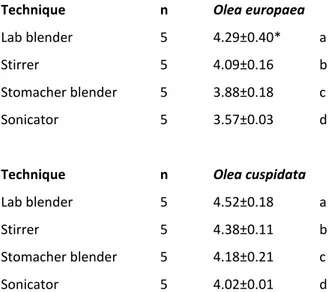
Recovery experiments: comparison between the processing methods
Tab. 2. Comparison on efficiency of processing methods for recovery of Pseudomonas savastanoi
pv. savastanoi
Technique n Olea europaea
Lab blender 5 4.29±0.40* a
Stirrer 5 4.09±0.16 b
Stomacher blender 5 3.88±0.18 c
Sonicator 5 3.57±0.03 d
Technique n Olea cuspidata
Lab blender 5 4.52±0.18 a
Stirrer 5 4.38±0.11 b
Stomacher blender 5 4.18±0.21 c
Sonicator 5 4.02±0.01 d
Means followed by the same letter are not significantly different at P = 0.05
* Standard error
n
Number of replicate
The ANOVA of the log CFU/cm2 values from the four tested processing techniques indicated a significant differences on their efficiency on recovering P. savastanoi pv. savastanoi from leaf surfaces of two assayed olive species (Tab. 2). The bacterial recovery capacity appeared to be different from one method to another between the two assayed Olea species. All methods differ significantly from one another ranking from the most efficient to less efficient as Lab blender, stirrer, stomacher blender and sonicator.
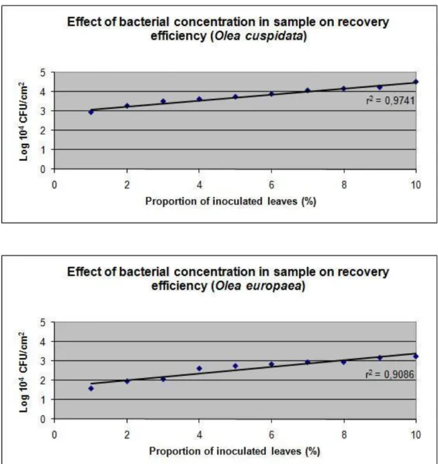
Effect of bacterial concentration in sample on recovery efficiency
Plotting of the CFU/cm2 values produced a straight line with high coefficient of determination value (r2), In both experiments, by putting in relation different proportion of contaminated leaves (Fig. 1 and 2). The linearity of the data and the high value of r2 demonstrate the efficiency, constancy and accuracy of the lab blender in recovering bacteria from leaf samples.
31
Fig. 1. Effect of bacterial concentration in sample of two Olea spp. on recovery efficiency of
Pseudomonas savastanoi pv. savastanoi.
Effect of freezing samples on bacterial recovery
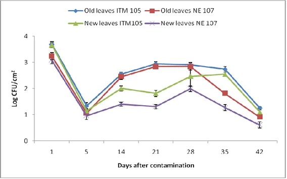
Bacterial numbers decrease more significantly the longer the samples were frozen. In particular, bacterial numbers declined drastically in samples frozen with the addition of phosphate buffer. As shown in Tab. 3, in case of O. europaea the number of bacteria recovered from sample frozen for only 1, 5 or 10 days, without addition of buffer, did not significantly differ. Afterwards, the bacterial numbers tend to decline with longer freezing differing significantly between 10 and 15 days of freezing, with further significant decline after 20 days. Nevertheless, no significant difference was seen on the recovered bacterial number between 20 and 30 days. Whereas for the samples frozen with addition of buffer, the recovered bacterial populations for only 1 and five
32 days did not differ significantly, but subsequently bacterial numbers differ significantly among the samples frozen for 5, 10, 15 days. The populations recovery after the freezing of sample for 15 and 20 days was rather similar with a light decline. No bacterial populations were recovered from the samples frozen with addition of buffer for 30 days.
Bacterial recovery was slightly different in case of O. cuspidata, since the number of bacteria recovered from sample frozen for only 1 and 5 days, without addition of buffer, was not significantly different. Later, the bacterial numbers tend to decline with longer freezing with some similarities among them in their significance from samples processed after 10, 15, 20 and 30 days of freezing without addition of buffer. While for the samples frozen with addition of buffer, the number of bacteria recovered from sample frozen with addition of buffer for 1 day was not significantly different for those frozen for 5 and 10 days but differed significantly to those frozen for 15 and 20 days. No bacteria were recovered from the sample frozen for 30 days with addition of buffer in case of O. cuspidata as well.
Tab. 3. Comparison of immediate and delayed processing of samples collected 3h post-spray on
recovery of P. savastanoi pv. savastanoi from two olive species Days buffer Olea europaea (log CFU/cm2) Olea cuspidata (log CFU/cm2)
1 + 2.24±0.67* c 2.72±0.16 e 5 + 1.78±0.24 c 2.46±0.28 cde 10 + 0.99±0.02 b 1.83±0.27 bcde 15 + 0.29±0.29 ab 1.52±0.47 b 20 + 0.19±0.19 a 0.31±0.31 a 30 + 0.00±0.00 0.00±0.00 1 - 4.94±0.02 e 4.16±0.08 f 5 - 4.91±0.00 e 3.97±0.21 f 10 - 4.80±0.05 e 2.58±0.58 de 15 - 3.95±0.04 d 1.74±0.04 bcd 20 - 2.25±0.24 c 1.60±0.10 bc 30 - 2.10±0.18 c 1.83±0.11 bcde
Means followed by the same letter are not significantly different at P = 0.05
33
Plating experiments: comparison of drop versus spread plating
The average value expressed as CFU/cm2 was higher from those obtained from spread plating respect to drop plating although no significant difference was found among them.
Tab. 4. Difference between the spread and drop plating methods
Plating technique n Mean log CFU/cm2
Spread plating 6 2.61±0.59* a
Drop plating 6 2.49±0.77 a
Means followed by the same letter are not significantly different at P = 0.05
* Standard error
n Number of replicate
DISCUSSION
The results obtained from the sampling, recovery and enumeration of bacteria show considerable variation among the investigated features. Furthermore, the use of two different species belong to Olea showed that the parameters considered during the study differ from one species to another. Concerning the type of sampling, generally, bulk leaf sampling is more quicker and easier at field level and thus it is frequently used respect to single leaf sampling. The latter, often, results unfeasible since it is laborious and time consuming. Furthermore, single leaf sampling, usually is imprecise since single sample could not be representative for quantifying microbial populations respect to the bulk leaf sample collected randomly. Nevertheless, results showed that the recovery of bacterial populations from leaf samples is higher from bulk sample rather than those from single samples from both olive species examined in the study. These results are in agreement with Crosse (1959) since larger bulk samples result in more accurate estimates of bacterial populations.
The recovery of bacterial populations by using the widely used four processing methods and the significant differences among them is of noteworthy importance. Nevertheless, our results suggest that the recovery of microorganisms from leaf surface varies with varying the plant species or at least between herbaceous and woody species. This hypothesis is concordant to the results obtained by Donegan et al., (1991) since in their study stomacher blending allowed the greatest recovery of microorganisms from the leaf surface of oat and bean plants. In our studies, lab blender allowed the greatest recovery of bacteria from leaf surfaces of both olive species. This is probably due to the fact that olive leaves are very particular with waxy and tough leaf surfaces
34 having numerous star hairs on the lower surface of the leaves (Surico, 1993). This finding is of considerable importance since microbial populations of olive is widely studied from several researchers and the advantages of lab blending like quick processing time (5 min. instead of 2h of stirrer and 7 min of sonicator), possibility of sample collection directly in the sterile lab bags suitable for lab blender, time-saving since the bags can be easily disposed without the need of cleaning and autoclaving of containers, like in case of sonication and stirrer, which is very laborious and time-consuming task mostly in experiments with numerous samples. Furthermore, the disadvantage of releasing plant cell contents, caused by beating action of paddles which apply pressure to the bagged sample, do not take place since olive leaves are very resistant. The capacity of lab blender in providing constant recovery confirms its effectiveness which keep our results far from any possible errors.
With regard to sample storage, since sample collections and immediate processing often results unfeasible especially in large scale experiments, an efficient way of storage, which allows on the one hand to ensure the conservation of leaf samples without any alteration and on the other hind the survival of microorganisms of our concern, would be advantageous. Our results show that the freezing of collected samples, with or without the addition of buffer, may negatively influence survival or recovery of microorganisms in the samples. The numbers of bacteria recovered from samples frozen for 1 and 5 days with addition of buffer and 1, 5, and 10 days without addition of buffer did not differ significantly in case of O. europaea and for 1, 5 10 and 15 days with addition of buffer and 1 and 5 days without addition of buffer for O. cuspidata, they indicate that a delay of a few days in sample processing is possible but longer storage should be avoided since the values tend to decline with time with significant differences.
The adverse effect of pre-freezing and the difference between the sample frozen with and without the addition buffer could be because of the bacterial injury caused from freezing and thawing of the buffer (Donegan et al., 1991) and can vary . Probably, also the type of leaf surface may influence since O. cuspidata has tender and less tough leaf surface respect to O. europaea leaves characterized by waxy and tough surface. In fact, different trend of decline in bacterial populations was observed among the two olive species.
No difference significantly valid was seen among the two plating techniques although the colony counts from spread plating were numerically higher than drop plating. Nevertheless, the lack of significant differences suggest several advantageous of drop plating since this technique is more convenient both economically, since the number of plates required is drastically lower given the
35 possibility of plating several concentrations per plate, and also from the point of physical time-saving given the easiness and quickness of the technique and the time time-saving also during the colony counts, since the quantity of bacterial suspension used for this technique is 10 fold less than the possible 100µl used for spread plates.
As conclusion, it is likely that a continuous effort is necessary for improvement and standardization of these techniques. In fact, the recovery of microorganisms probably depends not only from plant species but also from type of bacteria, whether they are plant pathogenic, antagonists or bacteria having ice nucleation activities. This is important in order to make right estimation and to intervene with proper control measures.
REFERENCES
1. Balestra G.M., Lamichhane J.R., Kshetri M.B., Mazzaglia A. and Varvaro L. (2009). First Report of Pseudomonas savastanoi pv. savastanoi on olive in Nepal. Plant Pathology 58, 393.
2. Beattie G.A. (2002). Leaf surface waxes and the process of leaf colonization by microorganisms, p. 3–26. In S. E. Lindow, E. I. Hecht-Poinar, and V. Elliott (ed.), Phyllosphere microbiology. APS Press, St. Paul, Minn.
3. Colin K.C. and McCarter S.M. (1983). Effectiveness of selected chemicals in inhibiting Pseudomonas syringae pv. tomato in vitro and in controlling bacterial speck. Plant Disease 67, 639-644.
4. Donegan K., Matyac C., Seidler R. and Porteous A. (1991). Evaluation of methods for sampling, recovery and enumeration of bacteria applied to the phylloplane. Applied and Environmental Microbiology, 51-56.
5. Ercolani G.L. (1971). Presenza epifitica di Pseudomonas savastanoi Stevens sull’olivo, in Puglia. Phytopathologia Mediterranea 10, 130- 132.
6. Ercolani G.L. (1978). Pseudomonas savastanoi and other bacteria colonizing the surface of olive in the field. Journal of General Microbiology 109, 245 -257.
7. Ercolani G.L. (1979). Distribuzione di Pseudomonas savastanoi sulle foglie dell’olivo. Phytopathologia Mediterranea 18, 85- 88.
8. Knudsen G.R., Walter M.V., Porteous A., Prince V.J., Armstrong J.L. and Seidler R.J. (1988). Predictive model of conjugative plasmid transfer in the rhizosphere and phyllosphere. Applied and Environmental Microbiology 54, 343-347.
36 9. Lindow S.E., Arny D.C. and Upper C.D. (1978). Distribution of ice nucleation active bacteria on plants in nature. Applied and Environmental Microbiology 36, 831-838.
10. Lindow S.E. and Brandl M.T. (2003). Microbiology of the Phyllosphere 69( 4), 1875–1883
11. Lindow S.E., Knudsen R.J., Seidler R.J., Walter M.V., Lambou V.W., Amy P.S., Schmedding D., Pronce V. and Hern S. (1988). Aerial dispersal and epiphytic survival of pseudomonas syringae during a pretest for the release of genetically engineered strains into the environment. Applied and Environmental Microbiology 54, 1557-1563.
12. Meynell G.G. and Meynell E. (1965). Theory and practice in Experimental Bacteriology. Cambridge University Press.
13. Morris C.E., Monier J.M. and Jacques M.A. (1998). A Technique To Quantify the Population Size and Composition of the Biofilm Component in Communities of Bacteria in the Phyllosphere. Applied and Environmental Microbiology 64 (12), 4789–4795.
14. Surico G. (1993). Scanning Electron Microscopy of Olive and Oleander Leaves Colonized by Pseudomonas syringae subsp. savastanoi. Journal of Phytopathology, 138, 31–40.
15. Quesada J.M., García A., Bertolini E., López M.M. and Penyalver R. (2007). Recovery of Pseudomonas savastanoi pv. savastanoi from symptomless shoots of naturally infected olive trees. International Microbiology 10, 77–84.
16. Varvaro L. and Ferrulli M. (1983). Sopravvivenza di Pseudomonas syringae pv. savastanoi (Smith) Young et al. sulle foglie di due varieta’ di olivo (Olea europaea L.). Phytopathologia Mediterranea 22, 1-4.
17. Varvaro L. and Surico G. (1987). Multiplication of wild types of Pseudomonas savastanoi pv. savastanoi (Smith) Young et al. and their indolacetic deficient mutants in olive tissues. In: Plant pathogenic bacteria, (E. Civerolo et al. Editors), Martinus Nijhoff publishers, Dordrecht, 556-565. 18. Zagory D., Lindow S.E. and Parameter J.R. (1983). Toxicity of smoke to epiphytic ice nucleation active bacteria. Applied and Environmental Microbiology 46, 114-119.
37
CHAPTER 7.
Bacteria colonizing phylloplane of Olea spp. in the fields of Nepal
J. R. Lamichhane1, 21
Dipartimento di Protezione delle Piante, Facoltà di Agraria, Università degli Studi della Tuscia, 01100, Viterbo, Italy;
2
38
ABSTRACT
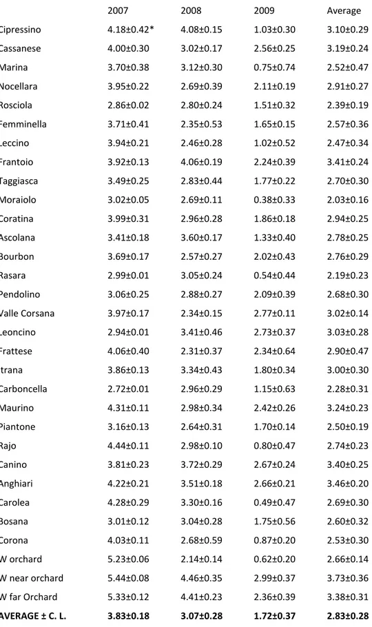
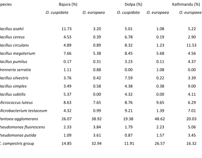
Different bacterial species were isolated from olive leaves sampled in three different districts of Nepal (Bajura, Dolpa and Kathmandu). Almost all of them were aerobic, heterotrophic and mesophilic which were present in plant phylloplane of all the districts although in different percentages. Both of the olive species were dominated by Pantoea agglomerans (former Erwinia herbicola) and Xanthomonas campestris group in all the three districts. The size of the bacterial epiphytic populations ranged from very low (0.38 log CFU/cm2 for cultivar Moraiolo sampled from Bajura district) to very high (5.03 logCFU/cm2 for cv. Marina sampled from Kathmandu district). The isolates were characterized by morphological, physiological, biochemical and molecular assays and they were identified as, Bacillus asahii, Bacillus cereus, Bacillus circulans, Bacillus megaterium, Bacillus pumilus, Bacillus silvestris, Bacillus simplex, Bacillus subtilis, Brenneria serratia, Microbacterium testaceum, Micrococcus luteus, Pantoea agglomerans, Pseudomonas fluorescens, Pseudomonas putida and Xanthomonas campestris group. No presence of Pseudomonas savastanoi pv. savastanoi was found on olive leaves. No significant quantitative differences were found while significant differences in qualitative composition of epiphytic bacterial populations were observed among the three districts and among the two Olea species.
INTRODUCTION
Bacteria are common residents of leaves and there are many bacterial species which colonize plants leaf surfaces that provide physical environments suitable for growth and reproduction of epiphytic bacteria (Ercolani, 1978; Thomson et al., 1976). Almost all of these epiphytic bacteria play a different role which can influence the health of host plants under different conditions suitable for their activities, such as inciting disease in case of plant pathogenic bacteria (PPB) (Beattie and Lindow 1994a; Beattie and Lindow 1994b; Beattie and Lindow 1995; Hirano and Upper 1983; Mew and Kennedy, 1982), causing frost injury in case of ice nucleation active bacteria (INA) (Cambours, 2004; Cambours et al., 2005; Hirano et al., 1981; Lindow et al., 1978b; Lindow, 1986; Nejad et al., 2002, 2004, 2005; Pearce, 2001; Wilson and Lindow, 1994a) and altering plant growth in case of bacteria that produce plant hormons (Brandl and Lindow, 1998; Fett, 1987; Glickmann et al., 1998; Hirano and Upper, 2000; Lindow et al., 1998). In addition, there are also many saprophytes, which are epiphytic inhabitants naturally present on aerial parts of plants, with several beneficial effects to the plants such as growth inhibition of PPB acting as their antagonist with continuous worldwide interest, to attain sustainability in agriculture which is
39 essential to reduce or eliminate the use of chemical pesticides for plant disease control (Andrews, 1992; Blakeman and Fokeman, 1982; Gross, 1991; Ji et al., 1996; Kishore et al., 2005; Thomson et al., 1976). Thus, the knowledge of the bacterial species present on plant phylloplane and their accurate estimates are of essential importance to safely predict phenomena related to them, including disease or frost damage.
Although bacterial phylloplane communities on annual or perennials bearing deciduous leaves have largely been studied by several authors (Hirano and Upper, 1991; Ishimaru et al., 1991; Jacques et al., 1995; Leben and Daft, 1967; Lindemann et al., 1984; Lindow and Andersen, 1996; Malvick and Moore, 1988; Morris and Kinkel, 2002), only few is known of those occurring on the long living leaves of evergreen plants (de Jager et al., 2001; Karamanoli et al., 2000; Périssol et al., 1993; Yadav et al., 2004), including european olive (Olea europaea L.) (Ercolani, 1978, 1991; Lavermicocca et al., 1987). Whereas, no information is available on this issue on African olive (Olea cuspidata Wall.) which is a widespread olive species present mostly in African, Asian and Oceanian continents (Daniel and Boher, 1978; Spennemann and Allen, 2000). It is naturally present in several Himalayan regions of Nepal as well (Lamichhane et al., 2010a), particularly in Dopla, Bajura, Mugu and Humla districts at altitudes ranging from 1300 to 2500m above sea level (asl). Twenty eight Italian olive cultivars belonging to O. europaea are also present in the Himalayan regions of Bajura, Dolpa and Kathmandu districts of Nepal respectively at 1400, 1500 and 2500m (asl).
The aim of this study was to evaluate quali-quantitative composition of the bacterial populations present on olive phylloplane of two different olive species and to ensure whether there was the presence of Pseudomonas savastanoi pv. savastanoi (Psav) on epiphytic surfaces of asymptomatic olive plants present in three different districts of Nepal.
MATERIALS AND METHODS
FIELD
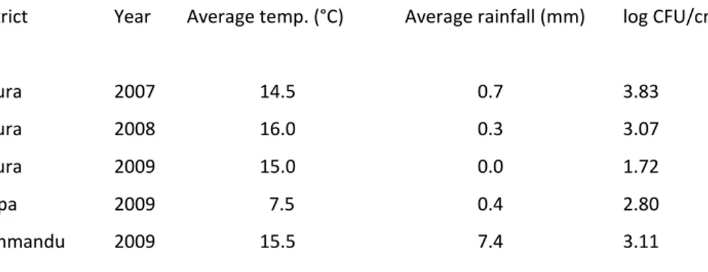
Three fields of european olive were concerned of our study. The fields were situated at Kolti, in the district of Bajura, at 1500m asl, at Juphal, in the district of Dolpa, at 2500m asl and at Kirtipur, Kathmandu district, at 1300m asl. Furthermore, wild olive belonging to O. cuspidata present in Bajura and Dolpa districts as natural forests were studied.
40 OLIVE CULTIVARS
For O. europaea, twenty eight Italian olive cultivars present in three different districts were used. The cultivars were, Anghiari, Ascolana, Bosana, Bourbon, Canino, Carboncella, Carolea, Cassanese, Cipressino, Coratina, Corona, Femminella, Frantoio, Frattese, Itrana, Leccino, Leoncino, Marina, Maurino, Moraiolo, Nocellara del Belice, Pendolino, Piantone, Rajo, Rasara, Rosciola, Taggiasca and Valle Corsana. Whereas, for O. cuspidata, plants naturally present in those area, were used.
SAMPLING OF OLIVE LEAVES
Leaf samples were collected randomly from all the asymptomatic plants during a 3-year period (April 2007- April 2009) in Bajura and only for one year period (April 2009) in Dolpa and Kathmandu districts. Sampling was made early in the morning. In case of O. europaea, leaf samples were randomly collected from 28 Italian cultivars by taking both young and old leaves. 40 leaves from each plant and five plants for each variety for a total of 200 leaves per variety were aseptically collected in sterile lab bags, bulked and put in an icebox. In case of O. cuspidata, the same procedure was used by collecting 10 samples each represented by 200 leaves. Samples were sent to Italy by air express, by air mail or in some cases, hand carried. Climatic data obtained from the meteorological stations installed to our study sites were used.
FREEZING OF THE SAMPLES
Since the volumes of the samples were too large, immediate processing of all the leaves was impossible. Donegan et al., (1991) showed that a delay of a few days in processing is possible since freezing of samples for a short period of time do not influence negatively bacterial recovery. For this reason samples were frozen at -20°C and processed within one week. Lindow et al. (1978a) worked with samples collected from distant locations and sent to laboratory with time between collecting and plating being 1 to 3 days in most cases, without any significant differences with those collected locally, although the effects of that delay have not been determined.
PROCESSING OF LEAF SAMPLES
Each bulk sample was aseptically divided in 10 groups (20 leaves per replicate) and placed in a sterile 250 ml conical flask with 40 ml of phosphate buffer and shaken in the dark by using an orbital stirrer at 26±1 °C and 150 rpm for 2h. Previous studies indicated that over 90% of the
41 phylloplane bacteria passed into suspension and that there was no error due to bacteria multiplying in the washing fluid (Crosse, 1959; Ercolani, 1978). These results were obtained by previous authors by plating 10-fold serial dilutions of homogenates of washed and unwashed leaves. Furthermore, Ercolani (1976) showed that virtually no bacteria passed into suspension from insides the leaves which allow us to make recovery of only phylloplane bacteria.
After washing, the solution was serially diluted in phosphate buffer and spread plated in duplicate onto nutrient agar medium supplemented by 5% (v/v) of sucrose (NAS), amended by 800 µl /l of cycloheximide (previously prepared as stock solution by diluting 1g in 10 ml of ethanol) to prevent fungal contamination. One hundred µl of diluted solution was used for plating. The plates were then incubated at 26±1°C for 48-72 h. APS assess software was used to measure leaf surfaces.
COLONY COUNTS AND THEIR CHARACTERIZATION
Bacterial colonies were first counted with a stereomicroscope to determine the total bacterial population from NAS plates. The colonies were then purified by streaking on the same medium and incubating the plates at 26±1°C. Isolates obtained during the first, second and third year of work from all the districts were screened to determine their reaction towards the different assays. Three hundred and thirty isolates from Bajura district, 170 from Dolpa district and other 140 from Kathmandu district, for a total of 640 isolates collected during the three years of experiments, were tested. Morphological characteristics of the isolates were determined by streaking onto NSA medium followed by the microscopic observation. Gram test was performed by using KOH solution (3%). Levan production was observed after 3 days of incubation on NSA. Cowan and Steel's (1965) methods were used to detect catalase activity (method 1), citrate utilization (method 1), gelatin hydrolysis (method 1), indole production (method 2), urea hydrolysis (method 1), oxidation or fermentation of glucose, oxidase activity, nitrate reduction and starch hydrolysis. Arginine dihydrolase, lysine and ornithine decarboxylase activity (Taylor, 1961) and growth in MacConkey agar (Oxoid CM 7) were all recorded after 5 days of incubation.
IDENTIFICATION OF THE ISOLATES
Isolates were identified to the genus level by using the diagnostic tables of Cowan and Steel (1965) by which they were divided in groups to obtain only those effectively different since bacteria colonizing olive phylloplane are frequently represented by only few species (Ercolani,