University of studies Messina
PhD course in Medical and Surgical Biotechnologies
XXXIII cycle
Coordinator: Prof. Giovanni Squadrito
Analysis of “Streptococcal SUrface REpeat” domain (SSURE)
from Streptococcus surface proteins and investigation of their
binding properties to Extracellular Matrix (ECM) molecules.
PhD student:
Maria Miriam Giardina
Tutor
Prof.ssa Concetta Beninati
Contents
ABBREVIATIONS ... 5
INTRODUCTION ... 7
I. OVERVIEW OF STREPTOCOCCAL PATHOGENESIS ... 7
II. DESCRIPTION OF INTERACTION WITH HOST DURING THE BACTERIAL LIFE. ... 9
III. IS ECMBINDING TO BACTERIAL SURFACE RECEPTORS MEDIATED BY A PROTEIN-PROTEIN INTERACTION? ... 10
III.I CELL WALL ANCHORED AND ANCHORESS ADHESINS ... 13
IV. FOCUS ON ADHESINS:PROTEINS OF STREPTOCOCCUS BINDING ECM ... 14
V. MOLECULAR TECHNIQUES TO STUDY MICROBIAL INTERACTION. ... 17
VI. AFM TO STUDY MICROBIAL ADHESION AT THE SINGLE MOLECULE LEVEL. ... 18
AIM OF THE THESIS ... 20
RESULTS ... 21
I. SEQUENCE ANALYSIS “STREPTOCOCCAL SURFACE REPEAT”DOMAIN. ... 21
II. SSURE ARE VIRULENCE FACTORS? ... 25
III. IMMUNOLOGICAL AND IMMUNOCHEMICAL PROPERTIES. ... 26
III. IIMMUNOLOGICAL RELATIONSHIP BETWEEN SSURE DOMAINS. ... 26
IV. ROLE IN EXTRACELLULAR MATRIX COMPONENTS BINDING SSURE. ... 30
V. APOTENTIAL ADHESIN:FIBRONECTIN AND COLLAGEN TYPE I INCREASES THE STRENGTH OF THE SP6BOND. ... 32
VI. ROLE IN COLLAGEN TYPE I BINDING AND CELL COLONIZATION. ... 35
DISCUSSION ... 37
I. SSUREDOMAINS ARE VIRULENCE FACTORS ... 37
II. BINDINGMECHANISMOFADHESIONS ... 39
CONCLUSION ... 42
MATERIAL AND METHODS ... 44
I. EXPERIMENTS ON THE S. AGALACTIAE,S. GORDONII AND S. PNEUMONIAE ... 44
II.IDNA EXTRACTION ... 44
III. PRODUCTION AND CHARACTERIZATION OF SSURE ... 45
III.ICLONING ... 45
III.IIBACTERIA AND GROWTH CONDITIONS. ... 45
IV. PROTEIN EXPRESSION AND PURIFICATION ... 46
IV.ISDS-PAGE ... 46
IV.I WESTERN BLOT ... 47
V. PRODUCTION OF ANTI-SSURE ANTISERA ... 47
VI. SSUREBINDING TO ECMCOMPONENTS ... 47
VII. SINGLE-MOLECULE FORCE SPECTROSCOPY ... 48
VIII. BIOFILM MODEL ... 48
IX. ANIMAL MODEL OF STREPTOCOCCUS INFECTION ... 49
Abbreviations
Atomic Force Microscope (AFM )
Ammonium persulfate (APS)
Barrier Emato Encephalic (BEE)
Bovine Serum Albumin (BSA)
Collagen Type I (Coll type I)
Collagen type II (Coll type II)
Complement factors (B,H,I C1q)
Extra Cellular Matrix components (ECM)
Enzyme Linked ImmunoSorbent Assay (ELISA)
Fibrinogen (FG)
Fibrinogen-binding protein (FbsA,FbsB and FbsC in general FBsP)
Fibronectin and Plasminogen binding Protein (FbsP o PavB)
Fibronectin (FN)
Group B Streptococcus (GBS)
SSURE 1 and 2 Group B Streptococcus (GBS 1 and GBS2)
Intraperitoneal (i.p)
Intravenous (i.v)
Isopropyl-β-d-thiogalactopyranoside (IPTG)
Lauria Bertani media (LB)
Laminin (Lam)
Plasminogen binding surface protein (PbsP )
Polyethylene Glycol (PEG)
Single Molecule Force Spectroscopy (SMFS)
SSURE 1,2,3 Streptococcus gordonii (SG1, SG2, SG3)
SSURE 1,3,6 Streptococcus pneumoniae (SP1, SP3, SP6)
Streptococcal Surface Repeats (SSURE)
Todd-hewitt broth (THB)
Vitronectin (Vn)
N,N,N',N'-Tetramethylethylenediamine (TEMED)
Worm-Like Chain (WLC)
INTRODUCTION
I. Overview of Streptococcal Pathogenesis
Many humans are healthy carriers of the bacterium and it makes a part of the normal fauna for example bacteria of oral cavity, or bacteria present in the gastrointestinal system, or also bacteria present in one or more zone, like low and high respiratory zone. A significant part of Gram-positive, present in these areas of the human body, are species Streptococcus. It is a name derived from a combination of two Greek words: Strepto meaning twisted and kokkus meaning berry (Watson, D.A, 1993). Currently, there are well over 100 known species of Streptococcus and some of these are commensal bacteria and other pathogenic bacteria.
A bacterium is actually indicated as being able to survive in harmony with its host in an environment that can be advantageous for one or both. These streptococcus
inhabitants are commensal, but they all have the capacity to cause some kind of disease, either superficial or systemic, and can be better described as opportunistic pathogens. A significant share of streptococcus are pathogenic bacteria that differ from their non-pathogenic homologues in their capability to cause disease. In general, the pathogenesis of Streptococcus is mainly based on three mechanisms: (i) ability to colonize and crossing through tissue barriers within the host- tissue; (ii) ability to bypass host defense mechanisms; and (iii) expression of virulence factors causing disease to the host (Mitchell, 2003). For colonization, the streptococcus strain needs several adhesion systems to attach itself to the host surface, which are epithelial cells. Adhesion proteins promote attachment through specific and non-specific
bacterial cells. There are many adhesion proteins, which have important roles in different stages of colonization (Bogaert. Et al 2004).
The characteristic of Streptococcus pathogens is due to the possession of virulence genes which can be transcribed into products, i.e. proteins, which are actually
virulence factors. Furthermore, virulence factors are molecules produced by bacterial organisms, i.e S.pneumoniae, which are essential to cause disease in low and upper tract respiratory. Some of these factors are only part of the virulence of the "lifestyle" and promote colonization; others, the "real" virulence factors, are directly involved in furthering invasion. Most, if not all, proteins exhibited on the surface of bacteria contribute significantly to pathogenesis; such proteins are often implicated in direct interactions with host tissues or in hiding the bacterial surface from the host's defense mechanisms. These proteins are hence virulence factors, and are reasonable
candidates for generating immunogenicity that can be protective, for example, when used in vaccine compositions.
Targeting streptococcal colonization could be of therapeutic benefit in the light of antibiotic resistance and this can be realized through vaccination (active
immunization) or the use of specific ECM-adhesin antibodies (passive
immunization). An effectively adhesin-based vaccine containing a specific surface bacterial protein, or a Recombinant part of it, which, when injected, will induces a specific immune response and protection of humans against subsequent exposure to a pathogen.
Accordingly, to formulate the composition of an effective vaccine against a pathogen, it is essential to select an ECM adhesins that is robustly expressed in vivo and shows high antigenic potential and an effective role in the pathogenesis of the infection.
II. Description of Interaction with Host During the Bacterial
Life.
Studies suggest that streptococci can survive and persist within human cells and remain impenetrable to antibiotic treatment and innate immune defenses. In many cases, these interactions participate efficiently in the infection process; in other cases, they act as warning signals and activate innate immune responses. Host immunity, as well as the flora itself, plays a role in the regulation of niche bacterial populations. These infections vary from small diseases to more invasive ones. One in particular is bacteremia, which is the presence of bacteria in the blood; the origin of systemic bacteria can be localized and cause an infection. The consequences of bacteremia arise when bacteria leave the bloodstream and colonize other tissues such as the kidney or endocardium. In this case the plasma membrane and more precisely its outer face are clearly the first targets of pathogenic bacteria during infection, but the membrane is also the target of bacteria in various tissues and organs.
Another disease affected by streptococcus infection is meningitis, an inflammation of the covering that protects the brain and spinal cord (meningitis; dura mater, arachnoid and Pia mater). During infancy, bacterial meningitis is caused by commonly
pathogens of the respiratory tract such as S. pneumoniae. In general, the basal membranes are located under the bronchial and alveolar epithelial cells, in the
pulmonary interstitium, and form a barrier between the vascular endothelium and the alveolar cell (Dunsmore and Runnels, 1996).
The pathogens penetrate the CNS by interacting with the luminal side of the cerebral endothelium, which is the BBB (Nassif et al., 2002). In meningitis, there are three stages BM rupture and basal lamina is the first step of the infection, ii) by degrading BM, the bacteria will attack the capillary endothelial cells, cross them, enter the bloodstream and continue to multiplicate, iii) the pathogens will break the FCS blood barrier and colonize the underlying tissues.
In addition, interactions with the host and host tissue during bacterial life. Given their spatial position, bacterial surface proteins are naturally involved in complex
interactions of bacterial cells with host tissues. Some of these interactions, and proteins or other molecules involved in these interactions, may only be essential during the colonization phase (e.g. in the nasopharynx for pneumococci), during invasion (e.g. through the epithelial layers of mucous surfaces), or diffusion to other host tissues (e.g. through the bloodstream or nervous system). Others may be crucial during all phases of bacterial life.
In order to colonize their host, streptococci have to overcome a number of barriers: competition with other bacterial members of the normal flora, electrostatic and mechanical forces, and physiological responses that may move or physically remove from the host tissues. This process is aided by bacterial cell-surface components called adhesions, which recognize and bind specific partner molecules presented on the surfaces of host cells and other microbial organisms. (Kline et al.,2009) A well-known condition is that bacterial pathogens must first come into close contact with extracellular host matrix (ECM) proteins on the host cells in order to determine successful infections. That initial contact with ECM proteins or cells is made by highly specific adhesins (Courtney, Hasty & Dale, 2002; Jenkinson & Lamont, 1997; Nobbs, Lamont, & Jenkinson, 2009).
III. Is ECM Binding to Bacterial Surface Receptors Mediated by a
Protein-Protein Interaction?
Obviously, it is essential to examine the dynamic interaction between the pathogen and the host to get an accurate view of the infectious process and to manage bacterial infections. The interaction between pathogenic bacteria and their host structures is a complex picture and results in a multitude of interactions of molecules that are secreted by the bacteria or shown on the bacterial surface with host factors or host cell receptors.
To describe ECM is composed of glycosylated proteins and glycosaminoglycans from a fibrous network. The components of ECM vary between tissues,
developmental phases and pathophysiological states. The matrix components' structural characteristics and content separate the ECM. The individual ECM components include heparin sulphate, chondroitin sulphate, keratin sulphate, hyaluronic acid, collagen, elastin, fibronectin and laminin among others, and circulating plasma proteins which act in the coagulation cascade (fibrinogen,
plasminogen) and proteins involved in the innate immune defense (lactoferrin, CRP, SAP).
Although adhesion to the host surface is initially mediated by interactions with glycoconjugates, a greedy bacterial adhesion is mainly mediated by specific interactions with host proteins. Host ligands include ECM components such as fibronectin, collagen and laminin, as well as molecules that are also present in the blood, including fibrinogen and vitronectin. Proteins of the extracellular matrix are produced and secreted by cells resident inside vivo tissue, the proteins themselves promoting colonization.
A description of adhesives with the ability to bind single or multiple forms of collagen have been described in bacteria. Collagen is the main component of ECM and is the main glycoprotein representing 30% of the total protein content in the human body. Collagen is the main glycoprotein representing 30% of the total protein content in the human body. Its availability is essential for tissue structure, cell
adhesion and many other functions. At first it was believed that all types of collagen were secreted by fibroblasts in connective tissue, but the production of certain types of collagen by epithelial cells shows the high concentration of the molecule in the human body. The most significant structure is the presence of Gly-X-Y, which repeats itself called "non-collagen domains". A triple helix structure is formed by a normal hydrogen bond between proline and glycine residues (Shaw et al., 1991). The presence of collagen-binding proteins (co-binding collagen-BPs) in pathogenic
this ECM protein in organs and tissues. Second most important protein is fibronectin (Fn) is a multi-domain glycoprotein present in body fluids and on cell surfaces with the primary function of connecting the cell to the external ECM. There are two main forms of Fn in the body: soluble (plasma) and insoluble (cellular). The first ECM protein I want to characterize is fibronectin, a large multi-domain dimeric
glycoprotein found in body fluids. (Wing.S et al, 2011).
The second form is that of adhesives, it is a very varied group of proteins with heterologous architecture and domain structure. The fibronectin binding proteins (Fbps) are produced by all streptococci. A binding mechanism of fibronectin a
function of these proteins is the structural alignment of amino acid repetition regions with fibronectin, forming a "hinge" that promotes invasion of host cells (Norris et al.,).Other Fbps show interactions with fibronectin are not yet structurally understood through Streptococcus, but have not been found in oral streptococci, and also other proteins that bind collagen (Cbps). In detail these proteins mediate the adhesion to collagen-rich tissues and carry a tandem region (with collagen binding function) and the residual C-terminal amino acid is repeated. The proteins, as results, bind the collagen through the "embrace" mechanism, in which the initial interaction with the collagen induces the redirection of subdomains to wrap the triple helix of collagen ( Kang et al., 2013).These adhesions can also bind the C1q complement complex recognition proteins in a collagen-like domain and then act as inhibitors of the classic complement path, providing a strategy for immune evasion. This study suggest that are structurally related to the essay of recombinant proteins that we can attach to these two groups of proteins, on which I focused my work. This study of the interactions focuses on a particular type of surface protein, belonging to the streptococcal wall, which are proteins repeated in tandem.
III.I Cell wall anchored and anchoress adhesins
Identification and binding to several ECM proteins is an important precursor for the streptococcal colonization of human tissues. ECM sustains cells and tissues,
maintains the strength and elasticity of the body and is, as a consequence,
omnipresent. Nonetheless, extracellular matrix protein is often exposed to trauma and injury, which creates a primary target for streptococcal adhesion.
Streptococcal adhesives can be clustered into four different families, varying in their relationship to their surfaces: i) first are covalently bonded by their C-terminal to the peptidoglycan cell wall through an LPxTG pattern; ii) second are bonded to the bacterial cell membrane through N-terminal lipid modifications to form lipoproteins; iii) third are bonded to the bacterial surface by non-covalent interactions; or iv) fourth are expressed and preserved on the surface by an as yet undiscovered mechanism. There are several streptococcal proteins located on the cell surface, but they lack the LPxTG peptidoglycan anchoring pattern. In fact, these so-called anchor proteins or "lunar" proteins lack N-terminal signal sequences "Moonlighting" proteins interact with multiple ECM proteins, including fibronectin, fibrinogen, plasmin and
plasminogen, and therefore probably contribute to streptococcal colonization (Esgleas et al.,2008; Henderson & Martin,2011; Siemens Patenge, Otto, Fiedler &
Kreikemeyer, 2011). The description of this category of proteins that bind the proteins of the matrix helps me to introduce the proteins that are the subject of my thesis work, being also proteins without the LPxTG model and N-terminal signal sequences.
In brief, the evidence that proteins mediate adhesion to human cells and tissues is strong, but the differences in specificities reflecting the heterogeneity of this protein and the expression of other superficial adhesions create a complex picture. It is clear,
however, that streptococci do not typically bind directly to host cell surfaces, but actually interact with extracellular matrix proteins that form bridges to host cells.
IV. Focus on Adhesins: Proteins of Streptococcus Binding ECM
These features are in line with the recent observations which suggest that
extracellular matrix protein binding proteins in other streptococci are able to mediate adhesion and are associated with the progression of the pathogenesis of the disease (Courtney et al., 1999).
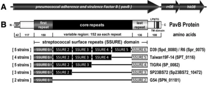
On the basis of the expected function, Fibronectin-binding activity was also reported for PfbA, which also binds plasminogen (Yamaguchi et al.,2008), and a multi domain protein encoded by sp0082 of TIGR4 (Bumbaca et al.,2004). As specified to the TIGR4 genome annotation the PavB protein contains four repeats, designated as SSURE “streptococcal surface repeat” domains.
The genetic organization of the pavB_locus is highly conserved in the pneumococcal strains. Sequence alignments using the JVCI Comprehensive Microbial Resource (CMR) and NCBI BLAST identified SSURE homologous sequences in the three streptococcal species S. pneumoniae (R6: spr0075) S. agalactiae (NEM316: gbs0428) and S. gordonii (NCTC7868: SGO1182) (Jedrzejas,2007) . To demonstrate the
presence of the pavB gene across the pneumococcal strains and serotypes and ssure‐ related sequences in other streptococcal species, we isolated the genomic DNA of streptococcal species, including clinically relevant encapsulated pneumococcal serotypes, and other pathogenic bacteria .The results further confirmed the presence of homologous ssure sequences in other streptococcal species such as S. mitis, S.
agalactiae and S. gordonii and confirmed the absence in S. mutans. In particular
Three different TIGR4 ssure DNA sequences were designed to produce His6‐tagged
able to produce one SSURE domain, representing the second SSURE of TIGR4 (SSURE2) and two SSURE domains, representing the second and third SSURE of
TIGR4 (SSURE2+3). Well, a recombinant PavB protein was produced, termed
SSURE1−5, which represents the mature PavB protein of TIGR4 without the
membrane anchoring domain denominate, SSURE2, SSURE2+3 or SSURE1−5. All three employed SSURE peptides showed fibronectin‐binding activity as shown
previously for one repeat (Bumbaca et al.,2004). As shown by Jensch et al. PavB is a surface exposed, conserved multidomain protein consisting of a signal peptide, cell wall anchoring motif and numerous repetitive SSURE domains that vary from 5 to 9, depending on the pneumococcal isolate tested. Sequence alignments for this projected protein exposed on the surface showed the presence and preservation of a 42 amino acid (SP) signal peptide in the N-terminal region and a 108 amino acid C-terminal part containing the transmembrane hydrophobic domain (TM) with the LPXTG (Figure 1) peptidoglycan anchoring pattern. LPXTG is cleaved from the
transpeptidase species and is assumed to anchor the protein that infects SSURE covalently to the cell wall. LPxTG, which is followed by a tract of hydrophobic amino acids and several positively loaded amino acid residues (rich in proline). Each SSURE domain comprises approximately 150 amino acids, but varies by at least one specific nucleotide, which permits discrimination and accurate DNA sequencing. Bumbaca ’s work used a proteomic approach to identify the protein “Streptococcal Surface Repeats” domain to clear delineation of a repeating domain of 148-152 amino acid residues, which is currently listed as domain Pfam 11966 in the PFAM database (Bumbaca et al.,2004, De Gaetano et al,2018). The putative protein
consist amino acids (aa) and contains an N-terminal region identified by InterPro ( https://www.ebi.ac.uk/interpro/entry/pfam/PF11966/ ) as an N-terminal homologue , followed by an unknown function domain.
We will highlight the difficulty of working with proteins containing multiple
conserved repeats such as the “Streptococcal Surface Repeats” domain for hypothesis tested was that the interactions of SSURE proteins with type I collagen, fibronectin and other extracellular matrix proteins, would have a chemical function similar to that of cross-linking induced by ECM.
Figure 1: SSURE within S. pneumoniae can be grouped in three categories based on homology: N-terminal (first), core and C-terminal (last; Jensch et al 2010)
V. Molecular Techniques to Study Microbial Interaction.
Employing several molecular techniques, including ligand overlay, Jensch et al. assays convincingly demonstrate that PavB binds to fibronectin and, to a lesser extent, plasminogen, like ECM components. Greed for fibronectin and plasminogen has been increased with the number of SSURE domains within the recombinant PavB protein being tested. PavB's surface location, distribution and sequence conservation between strains, and immunogenicity in humans make it a serious antigen candidate vaccine. It is important to note that studies by Papasergi et al. that independently study the same protein according to the nomenclature of plasminogen and
recombinant fibronectin B protein recapitulate the interaction of PavB with fibronectin and plasminogen and its role as an adhesin (Papasergi et al., 2010). Together, these studies explore the initial work of Bumbaca (Bumbaca et al., 2004). In short, matrix proteins play an important role in tissue remodeling, cell
proliferation, apoptosis and host defense (Page-McCaw et al., 2007).
To determine the binding between Fn or Fg or collagen and each of the seven SSURE variants, a microtiter plate assay and a more advanced atomic force microscopy
(AFM) technique have been used. These two techniques allow us to analyze the
mechanical responses of the ligand receptor pairs in the absence (microtiter plate) and presence (AFM) of physical stress. This condition becomes relevant because bacteria attacked on the surface, including streptococcus, are often subjected to physical stress in the human body. Overall, the results presented here show that amino acid
substitutes in the SSURE repetition region formerly associated with infection in humans have a real impact on the binding strength of the host blood protein, Fn and type I collagen. In particular, these amino acid substitutions in the domain repetition region improve binding with Fn and conform to a Bell-type model that describes a single dissociation pathway (24-25). Perhaps more importantly, we have found that some SSURE domains significantly strengthen the interactions between Fn and type I collagen following the application of a tensile force to the bond. This seems that
SSURE interactions with ECM molecules are susceptible to conformation changes under mechanical stress that impact the binding.
VI. AFM to Study Microbial Adhesion at the Single Molecule Level.
To begin with a brief description of the basis of atomic force microscopy (AFM): AFM consists of analyzing the surface of the sample with a nanometric tip mounted on a flexible cantilever on which a laser beam is focused and reflected into a
photodiode that records the deflections of the cantilever when scanning the sample. The accurate positioning of the point on the sample is guaranteed by piezoelectric scanners working in x, y and z direction.
AFM provides the ability to analyze biological samples under physiological
conditions and capture events in real time. Moreover, it is not only an imaging tool, but can also be used to determine intramolecular and intermolecular forces, as they occur in protein folding, receptor -ligand, or pathogen-host interaction. Such
measurements can provide valuable insights into structural biophysical properties of biomolecules and help to decipher the molecular mechanism involved e.g. in
pathogen host recognition. This can be achieved by functionalizing the AFM tip with specific (bio) molecules, such as bacterial adhesins and measuring the unbinding forces between the functionalized AFM tip and a substrate surface where the target molecule has been immobilized (Hinterdorfer and Dufrene,2006, Becke et al,2019). Applied to microbiology, AFM has opened new doors for the description of
topographic characteristics and molecular mechanisms associated with the cell wall as well as adhesion of the extracellular matrix. The adhesion of microbial pathogens to host tissue is often the first step leading to a successful infection. Bacteria have developed a variety of cell surface proteins (adhesins) to bind specifically to host cells, extracellular matrix (ECM)as well as protein-coated biological materials. These interactions allows bacteria to colonize their host and they can activate molecular
signaling pathways in host cells. AFM has been shown to be particularly well adapted to investigate these interactions at the single molecule level (Xiao & Dufrene 2016).
Aim of the Thesis
SSURE are domains found in proteins expressed by various streptococcal species that were initially described by their ability to bind extracellular matrix protein (Bumbaca, 2004, Jedrzajas,2007). The scope of this thesis is to examine the interactions between different SSURE domains and ECM in comparison with other ECM components. In particular, in the course of these studies, I have focused on the use of Atomic Force Microscopy to study interactions between recombinant SSURE domains of S. pneumoniae and S. gordonii .
This study aimed to determine if variation in the streptococcal surface repeat domain is widespread amongst Streptococcus, Two SSURE variants (GBS1 and GBS2), three SSURE variants (SG1, SG2, and SG3) and two SSURE variants (SP1 and SP6) were identified based on SSURE described by Bumbaca in spr0075, which are over 80% identical to one another between species. Moreover, the ligand binding ability of recombinant SSURE, was compared by ELISA-based solid phase binding assays. The results confirm that like SSURE domains is sufficient for ligand-binding with Extracellular Matrix molecule. As well as suggest that these ligand-binding functions are biologically important and are consistent with the predicted location of variant residues on the surface of the protein, thus, also bacteria. (Burke et al., 2010).
RESULTS
I. Sequence Analysis “Streptococcal SUrface REpeat” Domain.
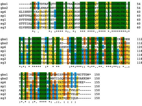
The principle on which this work is based, is the correlation between the structure of the SSURE domains and their probable function, probably given by a point within the structure as one or more amino acids can be, or that they are polar or even only able to interact with other proteins. Research into the structure, function and evolution of proteins has also shown the central importance of an entity unit. This is the protein domain, a substructure produced by any part of a polypeptide chain that can bend independently into a compact and stable structure. Most strikingly, these repetitions (here in after the SSURE domains) showed a wide variation among the Spr0075.
Figure 2: Multiple alignment of the N-terminal part of SSURE domain. The sequences were aligned using Clustlw program and displayed using Multiple Protein Sequence Alignment (MPSA) software. The sequence data represented correspond co to unfinished genome sequences obtained from NCBI:
Streptococcus agalactiae NEM316, Streptococcus gordonii and Streptococcus pneumoniae R6. The color coding for the alignment is as follows: green, single
There are six SSURE domains in this protein. In other streptococci, the number of repeated domains ranged from two in S. agalactiae (which we called GBS1and
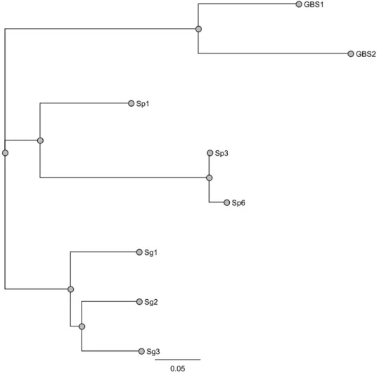
GBS2) to six in the R6 strain of S.pneumoniae (which we called SP1 and SP6). Three SSURE domains were present in S. gordonii protein (which we called SG1, SG2, and SG3).the variation in the number of domains in Spr0075 is due to the similarity of the four central domains. These recombinant proteins can be identified by the alignment created by CLUSTALW, inserting each domain, in which we highlight nine identical epitopes in all domains, parts where one or two amino acids are different (Fig.2). Phylogenetic relationships of genes encoding SSURE in streptococcal species are some genes in the same cluster show high similarity to each other.
Maximum likelihood and Bayesian phylogenetic analyses revealed common clusters between GBS1 and GBS2 shown 76% of identity, respectively to SSURE from
Figure 3: Comparative view of phylogenetic tree (based on a concatenated alignment of 8 recombinant proteins).Trees include Bootstrap are shown on the nodes of the phylogenetic tree.
S.gordonii to more 90% of identity, and in finally we have Sp1 quite different to Sp6
and Sp3.
Bumbaca displayed a structural model of SSURE domains across dependable domain borders obtained from its repetition in streptococcal proteins helped its initial ab modelling in two distinct ways. First, each domain had its own folding, determining the N and C endings of each domain, which also allows each domain to expose itself to the surface. As claimed to this model, the SSURE repetition was to consist of two domains. The first domain of the final model looked like a bundle of four α -helical bundle, preceded by a β -hairpin and with a loop containing β -structure between the two pairs of helixes. The linker region, rich in intrinsic disorder, is shown in the pattern by a long, irregular loop and an isolated β-filament. The second domain continues in which the main constituents are a three thread β-sheet with a long helix α.
The different domains of a protein are often associated with different functions. To determine of all of these interactions, each type of protein has a specific three-dimensional structure, which is established by the order of amino acids in its chain. The final folded structure, also known as conformation, adopted by any polypeptide chain is usually the one in which free energy is minimized. indicating that all the information needed to specify the three-dimensional shape of a protein is contained in its amino acid sequence. As you compare the three-dimensional structures of many different protein molecules, it becomes clear that, even though the entire
conformation of each protein is unique, you often find two regular bending patterns in parts of them. Both patterns were described the first folding pattern, called α helix, and a second folded structure, called β-sheet. These β sheets can be formed either by close polypeptide chains flowing in the same orientation (parallel chains) or by a polypeptide chain that folds on itself, with each section of the chain flowing in the opposite direction to that of its close proximity (antiparallel chains). An α helix is formed when a single polypeptide chain twists on itself to form a rigid cylinder. Short regions of α helix are particularly abundant in proteins found in cell membranes, such
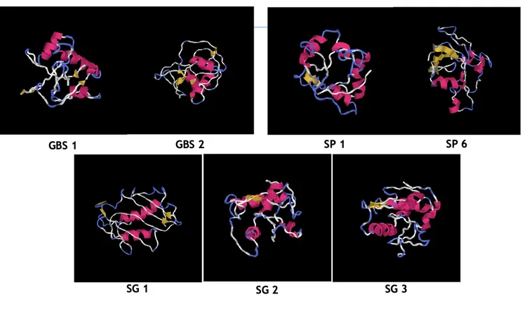
as transport and receptor proteins. In view of understanding the effect of domain repetitions on protein structure. Using computational modeling with I-TASSER, which uses a combination of comparative modeling’s, trampling and ab initio
modeling (Figure 3). The C-score is a confidence score for estimating the quality of I-TASSER models. It is typically in the range of -5 to 2, where a higher C-score
indicates a model with high confidence and vice versa. The modeling’s score (TM-score) and the root value of the mean square root deviation of the predicted model with respect to the native structure are predicted on the basis of the C-score. A TM-score greater than 0.5 indicates a correct topology model.
All the SSURE, which I have produced, have a structure as between five alpha helixes with the first two alpha helixes are followed by four alternating beta sheets with as few alpha helixes.
Streptococcus agalactiae Streptococcus pneumoniae
Streptococcus gordonii
Figure 4 : Bioinformatics description by iTASSER of the folding models of each SSURE; we have 5 alpha helixes shown in yellow and 4 beta leaflets shown in red, folded differently for each streptococcal surface repeat domain. In each domain, we have these structures that form a loop between two different chains.
II. SSURE are Virulence Factors?
In this Study, the functionality of SSURE domain is in the protein’s role in virulence have been researched. Novel virulence genes of Streptococcus agalactiae (GBS1 and GBS2), Streptococcus gordonii (SG1, SG2, and SG3), and Streptococcus pneumoniae
(SP1 AND SP6) were identified using the infection model. Concerning the role of
this domains in virulence, it is of interest that some of this strain, predicted to belong to a high virulence clone. This resolve suggests that the ability to bind Extracellular matrix and promote invasion may be more important than the enzymatic activity. As proposed for some other bacterial surface structures. The discovery of these cluster proteins represent a group of virulence factors with similar function in different species, whereas promotes binding of those pathogens to the extracellular matrix protein. Indeed, studies of some of these proteins may provide information about comparisons are particularly relevant, because little is known concerting the biological function of the different proteins.
To explore the potential role of the recombinant proteins in streptococcal
pathogenesis as well as their possible immune-protective properties. The functional role of SSURE repeats has not been fully explored. Extracellular matrix components make up a diversity of possible receptor structures for these domains. Therefore, it was of interest to explore the ability of SSURE domains from S. gordonii, S.
pneumoniae and S. agalactiae to bind to ECM components. To this end I have
expressed recombinantly various domains with a His 6Tag in an E. coli expression system and purified them by nickel affinity chromatography. Purity of these
preparations was considered satisfactory, based on polyacrylamide gel electrophoresis followed by Coomassie staining.
III. Immunological and Immunochemical Properties.
III. I Immunological Relationship between SSURE domains.
The immunological relationship among different domains has implications for the development of a SSURE vaccine and for the use of antisera for serological typing of strains. Cross reactivities observed among the three domains, analyzed in this
experiment, are summarized in Figure 6. These results were obtained with hyperimmune mouse antisera raised against purified proteins, and they do not necessarily reflect the situation during a natural infection. In general, the cross- reactivities are surprisingly limited, given the extensive residue identity between different domain of the recombinant proteins.
Figure 5 : Measurement of bacterial load and immune responses in mice infected with Streptococcus agalactiae Bm110.comparison of immune responses between different mice immunised with SSURE_GBS1,SSURE _GBS2, and SSURE_SG2 infected with Streptococcusa agalactiae. We use GBS_BM110 , if grown on blood agar, presents a characteristic halo zone around each colony due to β-hemolysis 1.The bacterial load and immune responses can be determined after infection by homogeneous tissue culture on blood agar plates.
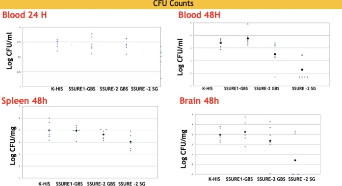
To explore the potential role of the SSURE domains in Streptococcal pathogenesis as well as their possible immune-protective properties, BALB/c mice were immunized with selected SSURE domains (GBS1, GBS2, or SG2) and challenged intravenously with 105 CFU of the serotype III, clonotype 17, S.agalactiae BM110 strain. Mice
were sacrificed at 24 or 48 h after challenge and the numbers of CFU were measured in different organs. BALB/c immunized with GBS2 or SG2 had a significantly lower dose of bacteria in the blood at 48 h post challenge. Moreover, immunization with SG2 also at 48 h post-challenge compared with animals immunized with the HIS 6 TAG. To support the protective effects applied to other models of mice infected with Streptococcus. BALB/c mice were immunized with GBS1, GBS2 and SG2 were protected from lethal streptococcal disease following intranasal challenge (figure 5). Immunization with PbsP has failed to emend survival times compared to fake
immunized controls. We found that immunization of CBA /N mice with GBS1, GBS2 and SG2 also induced protection against intravenous challenge with BM110. While BALB/c immunized with GBS1, GBS2, SG2 had a lower dose of serotype 3 in the brain and kidney following intranasal challenge, only the proteins of the triad histidine and SG2 had a significant protective effect (P < 0.05) against non-lethal intranasal challenge with a strain of serotype 3. BM110 colonies in the blood tend to shrink within 48 hours of the challenge and are significantly reduced when animals are immunized with a particular type of protein fragment called SSURE SG-2.In the kidneys, there is no significant colony reduction compared to control, while the reduction in the brain after immunization with the SSURE SG-2 construct is
encouraging. The data that need to be reconfirmed could be useful in identifying the protein sequence among the protein segments used most useful for bacterial
clearance. GBS has an important in vitro ability to transmigrate vascular endothelial cells and reach the meninges. Overall, therefore, data obtained from the brain after challenge with a hyper-virulent strain of GBS may be useful to better understand aspects of GBS -induced bacterial pathogenesis. These data indicate that
immunizations with the SSURE fragments have an effect on the clearance of the bacteria. sera that were obtained from animals immunized prior to the challenge with
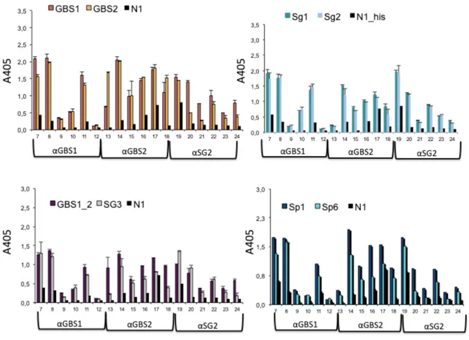
the BM110 strain were then tested in ELISA tests to verify reactivity against the immunizing antigen. Cross-reactivity with fragments derived from proteins of
different species was detected by cross-reactivity ELISA tests. The sera at a dilution of 1:4000 obtained by immunizing with GBS1, GBS2, Streptococcus agalactiae and SG2 fragments from Streptococcus gordonii and taking the initials αGBS1, αGBS2, and αSG2 were tested on antigens other than those used during immunization and on N1, Sp1 and SP6 fragments of Streptococcus pneumoniae, respectively. All animals, except the animal with ID 9 and ID 12 had high titers and the antibodies present in the most reactive sera and obtained against GBS1 and GBS2 antigens specifically recognized fragments obtained from Streptococcus pneumoniae and Streptococcus
gordonii (Figure 6).
Figure 6: We tested in ELISA tests to verify reactivity against the immunising antigen. The sera at a dilution of 1:4000 obtained by immunising with GBS1, GBS2, Streptococcus agalactiae and SG2 fragments from Streptococcus gordonii and taking the initials αGBS1, αGBS2, and αSG2 were tested on antigens other Sp1,Sp6,SG1,SG2,SG3 e N1, which is Positive Control.
Because SSURE domain members hold promise as vaccine components, it is relevance to identify protective epitopes. Only limited information is available concerning the location of such epitopes. However, it is not yet known whether immunization with the repeat region of an SSURE domains protective immunity. Besides its role as an epitope antigen, the functional role of SSURE repeats has not been explored. Extracellular matrix components make up a diversity of possible receptor structures for a variety of bacterial ligands. Thus, as well as this study was to explore if each SSURE domain would binding to extracellular matrix components. The immunological relationship among different domains has implications for the development of an S.agalactiae o S.gordonii vaccine and for the use of antisera for serological typing of strains. Cross reactivities observed among the interested three domains, analyzed in this experiment, are summarized in figure 6. These results were obtained with hyperimmune mouse antisera raised against purifies proteins, and they do not necessarily reflect the situation during a natural infection. In general, the cross-reactivities are surprisingly limited, given the extensive residue identity
between different domain of the streptococcal surface repeat protein. From a vaccine development point of view, it is of relevance that even a limited cross-reactivity may be sufficient to confer cross-protection, as shown by work with GBS1, GBS2, and SG2. Moreover, a very weak cross-reactivity that is hardly detectable by
immunochemical techniques may allow some cross-protection in vivo, as suggested by work with the SSURE in the mouse model. Of note, the cross-reactivities indicate that a vaccine composed of the this recombinant proteins, the sequence variability could represent antigenic variation, allowing bacteria expressing one of this protein, given protection against disease caused from streptococcal infection.
IV. Role in Extracellular Matrix components Binding
SSURE.
Besides Streptococcal Surface Repeat domain members hold promise as vaccine components, it is relevance to identify protective epitopes, the functional role has not clearly been explored, before now. Extracellular Matrix Components make up a diversity of possible receptor structures for a variety of bacterial ligands. Thus, the reason for this study was to explore if each SSURE domain would bind to ECM molecules.
To investigate whether the reactivity was derived from the streptococcal surface repeat domain to extracellular matrix component. An initial standardization of all components was conducted to determine the optimal concentration of recombinant
proteins, sera and conjugate dilutions (data not shown).
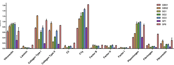
As SSURE proteins are suggested to be surface exposed we questioned whether it could mediate host colonization by adhering to extracellular matrix proteins. Thus, Laminin, Collagen Type I, Collagen type II, Fibrinogen, Vitronectin, Plasminogen
Figure 7 : Enzyme immunoassay test of the seven SSURE in contact with different ECM, such as vitronectin, Collagen Type I and II, Laminin, Plasminogen, Fibrinogen and Fibronectin. SSURE have a high binding affinity for Collagen type I and II, but also for vitronectin, plasminogen and C1q.we indicate the red bar GBS1, the orange bar GBS2, the olive green bar, SG1, the green bar SG2, the blue bar SG3, the purple bar SP1 and the lobster red bar SP6. While on the y-axis we have the individual proteins of the extracellular matrix.
and to the complement components C3,Factor B,H, and I ,were immobilized on micro dilution wells and recombinant protein attachment was assessed by an ELISA-based assay. As shown in figure 7, all SSURE protein exhibited efficient
adhesiveness to Vitronectin and Collagen type I and II, C1q, Plasminogen and Fibronectin. No statistically significant adhesiveness was observed with this protein when wells were coated with to Laminin, C3, Factor B, H and I.
In this assay, the interaction of Sp6 and Sp1 proteins of S. pneumoniae with a number of selected Extracellular Matrix molecules was pursued to explore the possible role of Sp6 in the process of binding S. pneumoniae to host cells through the main ECM components. We investigated the ability of purified SSURE domains to bind to type I Collagen, type II Collagen, Fibronectin, Vitronectin, Plasminogen, C1q and Laminin.
Streptococcal Surface Repeats bind selectively depending on concentration both immobilized and soluble Collagen type I, Fibrinogen, Fibronectin, Plasminogen, and Vitronectin in ELISA experiment. Fibronectin is in fact a multifunctional protein that exhibits different domains, each of which specifically recognizes different types of cells, including bacterial protein. The influence of SP1 domain and Sp6 domain upon the interaction of Fn was analyzed in solid phase ligan binding assays. The observed
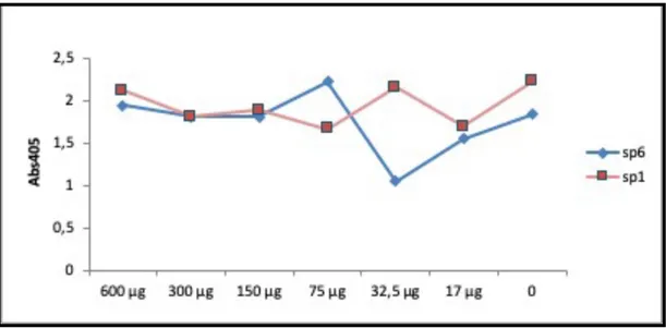
Figure 8: ELISA SSURE with fibronectin: Microtiter plate were coated with Fibronectin (Sigma F0895). Wells were blocked and then incubated with two SSURE at indicated concentration, and then incubated with monoclonal anti fibronectin (Sigma A3648) antibody. Bound antibodies were detected with either Anti Rabbit IgG_AP followed by AP substrate. Graphs are representative in red SP1 and in blue Sp6.
binding affinities of the recombinant proteins were considered in this experiment of inhibition with fibronectin. Comparison of the dose-response curves for Sp6 and Sp1shows a high affinity for fibronectin, but we can add that it has a marked affinity for Sp1 at a concentration of 32.5 µg. this makes us think that although both ssure have an affinity for fibronectin, which affinity we have to the bond under biological mechanical stress.
V. A Potential Adhesin: Fibronectin and Collagen Type
I increases the Strength of the SP6 Bond.
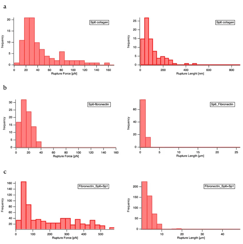
What is the mechanism by which Fibronectin and Collagen type I promotes SSURE binding? Do Fibronectin and Collagen directly activate Binding? To answer these questions and determine the strength of the SP6 Collagen bond, AFM based single-molecule force spectroscopy (SMFS) was conducted.
Sp6 is a putative adhesin that contains 150 aa tandem repeats. The product of this gene is predicted to be expressed on the outer membrane or periplasmic space. Each of these repeats influences the dynamic way in which different strains interact with their hosts, and the flexibility with which they interact with their hosts, and the
flexibility with which a colonizing population can diversify and adapt to the differing and changing environments within a single host.
Recent findings have shown the power of AFM force spectroscopy to decipher the binding mechanisms of microbial adhesins, to unravel the mechanics of cell surface proteins and they role in cellular function, to understand how cell surface proteins assemble into functional nano domains, and to quantify the forces that drive single -cell adhesion. The technique may be used with functionalized tips to probe specific chemical and biological sites. Using tips functionalized with a single type of adhesion protein allows to quantify the adhesion force, bond rupture kinetics, and the free energy landscape of the protein with a target molecule, such as collagen or
fibronectin. A remarkable recent discovery is the ultra-strong forces by which streptococcal surface repeat proteins bind to their ligands. (Valotteau et al 2017) Here single-molecule AFM demonstrated that the interaction of SSURE with human collagen type I and human fibronectin can sustain forces in the range over 20 pN. The
Figura 9: AFM-based single molecule force spectroscopy of Sp6 and Sp1: Histogram of detected rupture forces (left) and rupture length (right) of (a) Sp1 against collagen (a), Sp6 against fibronectin (b), and a combination of Sp6 and Sp1 on the AFM cantilever against fibronectin(c). In approximately 80% of all force curves, no interaction was observed (rupture force = (0 ±10) pN). These curves were not included in the histogram.
AFM results of this thesis have corroborated the finding that Sp6 mediates adhesion to these extracellular matrix proteins. However, the exact binding mechanism could not be completely unraveled by these preliminary experiments. We have determined the binding strength of SP6 with collagen type I, at a constant cantilever retraction velocity of 1000 nm s-1. Figure 9(a) shows histograms of rupture forces (strength) and rupture lengths of Sp6 immobilized on the AFM tip, measured against a glass substrates coated with human collagen I. The binding frequency was ~40%, the average rupture force, was 27- 38pN, which is in the range of weak receptor-ligand or bacterial adhesion-molecule-ECM interactions (Becke, et al 2019). The rupture
length displays a peak around 80-90 nm, which approximately corresponds to two times the length of the used PEG spacer of 40 nm plus the molecule diameter of 5.4 nm. The force curves were well described by the worm chain model (WLC). The SMFS experiments presented in figure 9 (b) and (c) show that the curves on Fibronectin featured adhesion curves of 15 ± 20 pN, 100 ±120 nm for Sp6 and 60 ± 80 pN, 150-300 nm for Sp1 plus Sp6 bonds. The forces measured for Sp6 are in contrast with the much stronger force of the fibronectin interaction with Sp1 plus Sp6.
Taken together, these observations let us to conclude that collagen interactions with Sp6 have moderate strength and that they could be involved in well-defined, linear adhesion with very strong forces consistent with the high affinity collagen hug mechanism. These features are missing in the fibronectin interaction, indicating that their molecular origin is different from the collagen interaction. Moreover, Sp1 plus Sp6 have stronger forces and higher affinity for human fibronectin, and these two SSURE play a role in these interactions. Together, these recombinant proteins show a stronger ligand binding, consistent with the high a fibronectin hinge mechanism (Dufrene et al,2017). We therefore suggest that the function in adhesion of the streptococcal surface domains virulence factor repeats is important for the ability to attach to and colonize host tissue.
VI. Role in Collagen Type I binding and Cell Colonization.
The next experiment we have carried out was to test in vitro the role of Streptococcal surface repeat proteins in the host-pathogenic interaction using these surfaces, which include the surfaces of host cells or components of the extracellular eukaryotic matrix (ECM), are generally soft and sometimes dynamic.
SSURE proteins are a streptococcal colonizing factor and have the ability to interact directly through as yet undiscovered host cell receptor or indirectly through human ECM or serum proteins such as fibronectin, fibrinogen, plasminogen and vitronectin. The structures and functions of mechanical properties have mostly been studied in force-free environments - how force is transmitted to a cell depends on the
mechanical properties of these structures. How do force loading on single or multiple connections influence the adhesive functions? How microorganisms apply and react to force for the adhesion of cells.
0 350 700 1050 1400 1750
S.agalactiae WT S.agalactiae 𝝙 PbsP S.gordonii
OD
59
5
Serum Collagen Type I PBS
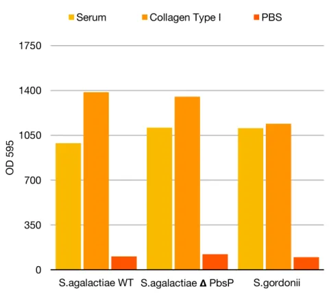
Figure 10: Protective effects of collagen (Yellow bars), serum (orange Bars), as compared with unprotected buffer controls PBS (Red bars) on biofilm cells of S. gordonii strains DL1, S.agalactiae delete PbsP , and Streptococcus agalactiae wild type NEM316. Bars indicate average values standard deviation at 12 hours after antiseptic matched buffer pulse. N = 2 independent experiments.
Biofilms are systems of active substance the bacterial growing produces a mechanical stress within the biofilm by deforming the elastic matrix, strongly influencing its morphology and the orientation of individual cells in the community. As a result, the mechanical properties can be measured with Atomic Force Spectroscopy,
understanding their relevance in the context of the formation and maintenance of the biofilm is due to the matrix formation and interactions established with
microorganisms. To influence signal transport and the mechanical forces generated by the flow or contact can distort or injure the matrix. Within the biofilm there are forces that participate in the organization and morphogenesis of the biofilm. Biofilms from in vivo mucosal surfaces, where they are exhibited to mechanical forces
impacted by their hosts. These surfaces, which include components of the
extracellular eukaryotic matrix (ECM), are generally soft and sometimes dynamic. The mechanosensory convert mechanical forces into biochemical signals, the
adhesion proteins of the cells mediate adhesion through force-induced conformational changes.
To demonstrate that the streptococcal surface repeats itself under an external force, it shows startling mechanical responses that are important for the extracellular
eukaryotic matrix. The modular nature of SSURE proteins could be important for its biological function as it can potentiate bacterial adhesion by increasing the lifetime and energy of the protein-substrate bindings. Thus, the mechanics of proteins makes SSURE proteins optimal to function as a polyvalent bridge protein, enabling
Streptococcus agalactiae, Streptococcus gordonii, Streptococcus pneumoniae to
colonize various surfaces. This approach has allowed key findings in microbiology, such as stress-induced cluster formation of proteins that activate cell adhesion.
DISCUSSION
I. SSURE Domains are Virulence Factors
The innate immune response is the initial defense against microbial invasion. The components of the innate immune pathway are continuing in developing; indeed, Gram-positive bacterial pathogens produce a several virulence factors that interact with the host molecules and modulate specifically response-host.
Virulence factors can be expositing on the surface of the bacterium, which are introduced into the host cell. The phase of bacterial infection on the surface of the cell, is excreted through specialized secretion systems
It is more interesting that pathogens generate a variety of virulence factors that interplay with the host molecules and although are modulated specifically function. Certain the importance of cellular Extracellular matrix adhesion in the adjustment of cellular behaviors have not yet been broadly characterized. We have recently
demonstrated that cellular ECM adhesion is focused on the adhesion of nearly similar structures on the basal surface of cells.
During this research of new virulence genes, we have hypothesized that some
virulence genes are maintained among the various pathogenic bacteria. Recent studies of the Streptococcus strain, whose proteins with the expected domains are
widespread in other streptococcus - These proteins show close homologues found in uncorrelated bacteria and it appears that the intracellular signaling pathways of eukaryotic host cells are adjusted to alter the virulence of the bacteria with
advantages of survival and in vivo . In this case, the virulence and multiple function of this domain particularly in the initial stages of streptococcal protein infection are of crucial importance for the streptococcal colonization of a host.
Cell-ECM relationships are the key determinants of cellular and tissue functions such as growth, proliferation and differentiation. In this respect, the specific character of tissues in extracellular matrix compositions indicates a selected role of adhesive proteins such as collagen or fibronectin, plasmin, fibrinogen, etc. Nevertheless, the complexity of the signal cascades brought on by cell adherence to ECM and the variety of factors concerned will make it more difficult to understand the cell-specific ECM pathway.
Genes or proteins involved in pathogenesis and interaction with the host immune response have been identified in different ways using available technologies. This suggests that different genes of virulence may be involved in different strains of streptococcus. The understanding of genetic diversity is important for comprehension of streptococcal strains of virulence. This research on the streptococcal genome is composed of hypothetical virulence genes that are being slowly clarified, as well as the multi-functionality of proteins not earlier linked directly to virulence. (A.M. Mitchell & T.J.Mitchell, 2010).
The association between the site of the infection and the virulence factors represents for genes encoding virulence proteins currently considered as promising goals for future vaccines. It shows the dynamics of protein interaction changes at the human streptococcal interface.
The correlation between the site of infection and the virulence factors represents a promising target for future vaccines for genes encoding virulence proteins. It shows the pattern of protein interactions at the human streptococcal interface.
Protein interactions in numerous clusters are correlated with increased plasma concentration and include proteins that interact non-specifically or that adhere to different binding pathways.
In this study, we provided an extensive description of the virulence genes carried by the invasion of Streptococcus pneumoniae, S. gordonii and S. agalactiae. We also highlighted how this micro-organism of these genes and consequently their coded
surface proteins could potentially impact the hypothetical efficacy of future SSURE vaccines.
Our study paid particular attention to Sp6, a surface repetition domain of
Streptococcus pneumoniae, due to its high attack level and its ability to cause a wide
variety of invasive diseases. The analysis suggests that there is a common pathogenic mechanism between streptococcus. In addition, a pattern of virulence other than pathogenicity could also contribute to virulence.
The proposals mentioned previously have to be accepted with more care. Some proteins shown on the surface of Gram-positive organisms play a substantial role in the pathogenesis and may be involved in the disease. Although there are no available kinetic data, translocation may be more efficient when the natural substrate
covalently adhered to the passenger is being exported, and the proteins of the alternative carrier may be exposed more slowly.
II. BINDING MECHANISM OF ADHESIONS
The adhesion of microbes to each other and host cells has important associations in microbiology, biotechnology and medicine. Microbial infection is frequently initiated by the specific adhesion of pathogens to host tissues through the adhesions of the cell surface.
A lot is known as to the structure and biosynthesis of microbial adhesions, the molecular details at the basis of their interaction with host receptors are still largely unknown.
This implies the functionalization of the AFM tip with the ligands and the measurement of the specific forces of the transporter-ligand, both on pattern
factor and are likely to play a role in the mechanically strong adhesion of
Streptococcus to host tissues. When trying to understand the interaction forces of pathogens, the use of true clinical isolates of bacteria the compared to laboratory strains should be preferred.
In nature, microbial adhesions are frequently exposed to strength. Studies on a single molecule have found that under an external force, adhesions display remarkable mechanical adhesion patterns that are important for cell adhesion.
Molecular dynamics tests in silico have shown that, in these blocks, three residues in the adhesin form hydrogen bonds with fibronectin, complementing the increased binding force and energy measured by AFM.
The mechanical properties of cell surface proteins play an essential role in
determining cellular functionality. Whereas mechanisms process mechanical forces into biochemical pathways, cell adhesion proteins mediate adherence through force-induced conformal changes. To date, how such cellular proteins react to mechanical inputs to achieve function remains largely unexplained. Through its ability to act as an individual protein adhesion protein, AFM has enabled researchers to address this problem. While the mechanics of proteins have been widely researched in vitro, bringing these nano-mechanical experiments into living cells has long been a challenge (data not shown).
Knowledge of the patterns and dynamics of the surface receptors of cells is
fundamental to our understanding of the surface functions of cells. A key topic is to understand how proteins associated with the surface assemble to form micro and nano-domains.
This method has allowed fundamental advances in microbiology, such as stress-induced cluster formation of proteins that activate signaling and adhesion. In the context of adhesion, a key discovery has been the clustering of microbial adhesions in reaction to mechanical stress. Despite the large potential of SMFS-based imaging for cell surface analysis, the technique has long been limited by its lack of
spatial-temporal resolve. Nonetheless, new multi-parametric imaging methods now allow researchers to visualize the structure and physical characteristics (elasticity and adhesion) of samples at the same time as increased lateral velocity and accuracy. An important issue in surface biology is to characterize simultaneously the structure, physical properties, and interactions of cell surfaces under physiological conditions. Although SMFS spatial resolution allows researchers to correlate structural
visualization of cells with quantitative models of their biophysical properties, the low spatial resolution and velocity of this method has limited its use in microbiology. SMFS maps the binding force and dynamics of individual adhesions. Newly, we have studied SMFS combined SSURE with all-atom steered molecular dynamics
simulations to show that the SP6 and SP1 domains of Streptococcus pneumoniae stabilize fibronectin and type I collagen through an H-binding network between a Fibronectin peptide and the adhesion backbone.
To determine whether a SSURE domain was specifically responsible for the
brokerage of interaction with fibronectin, we used the HIS-6TAG constructions of all seven individual streptococcal surface domain domains for pulldown experiments. This data supported the idea that all domains contributed to binding, but each tandem domain were needed to gain a detectable binding affinity.
In addition, periplasmic binding proteins are initial receptors in the process of active transport through cell membranes and/or chemotaxis. These binding proteins
involved in the transport of a wide variety of substrates constitute a family of proteins that show a significantly large movement of rigid globular domains (Sharff et al., 1992).
In summary, nano-mechanical experiments have provided new insights into how cell surface adhesions and cells respond to mechanical stimuli in relation to function. Note that in addition to protein mechanics, cell mechanics can also be addressed by AFM force spectroscopy, which allows us, for example, to assess the impact of antibiotics on cell stiffness.
CONCLUSION
Streptococcal adherence to host cells has been suggested to bacteria a two -step process. The first step involves targeting an anatomic niche of the host like the
nasopharynx to bind to the host surface glycoconjugates on respiratory epithelial and endothelial cells. These two steps lead to streptococcal invasion of the host and to streptococcal disease. Streptococcal surface repeat protein adherence to the host probably involves an array of adhesin that take part or modulate adhesion molecules expressed on its surface.
The functions of all the above proteins facilitate significant aspects of streptococcal colonization and/or invasion; compromising these function leads to compromised pathogenicity of S. pneumoniae. S. gordonii and S. agalactiae. Therefore, these proteins can serve as targets for the development of novel therapies to treat streptococcal disease. On one hand, the antibodies against three SSURE (GBS1-GBS2-SG2) are protective against the disease, and therefore these antigens can be used as protein-based vaccine candidates. On the other hand, some of these proteins can have their function compromised or totally abolished by the use of small
molecules that most probably bind in their active sites; these small molecules can be used to develop potent drugs.
However, structural information must be accompanied by an increased understanding of the role of SSURE proteins during several stages of pathogenesis in humans.
With regard to surface proteins, the subject of this thesis, the domain is of particular relevance for at least two reasons. First, expression of a given surface protein is often correlated with wall membrane. Second this domain structure help function of
binding extracellular matrix protein for colonize host. In the future I will continue to study not only the interactions of the other proteins that I have not been able to study in depth with the extracellular matrix proteins, but also the in vitro interactions with eukaryotic cells. So that I can design a vaccine against streptococcal diseases.
This situation encourages efforts to develop a protein-based vaccine and therapeutical drugs.
Material and Methods
I. Experiments on the S. agalactiae, S. gordonii and S. pneumoniae
II. Bacteria and growth conditions
The Streptococcus used in these studies was the virulent type 2 R6 strain.
Pneumococci were routinely grown at 37°C with 5% CO2 in air in Todd-Hewitt broth
(Difco) with 0.5% w/v yeast extract when necessary (THY) or on Todd-Hewitt agar (Difco).
II.I DNA extraction
The National Centre for Biotechnology Information website
(http://blast.ncbi.nlm.nih.gov/Blast) was used for DNA and protein BLAST searches. Genomic DNA was isolated from a 5 ml culture of S. Pneumonia R6 grown to optical density (OD) 0.3. The culture was spun down at 13.000 rpm for 10 minutes and the pellet resuspended in 1.000 µL PBS. Then the genomic DNA was extracted using the High Pure PCR Template Isolation kit (Roche) according to the manufacturer’s instructions. The quality and quantity of the DNA extraction was performed using Nano VueTM
III. Production and characterization of SSURE
III.I Cloning
Plasmids were prepared by insertion of DNA fragments obtained from S. agalactiae, S. gordonii and S. pneumoniae (primer Table S2) into pET21b vectors (figure M1).
The fragment was amplified using 5 µl of primers (100µM) and 4µl of 10 mM dNTP (Thermo
Scientific), 10 µl of 10x pff PCR buffer (Invitrogen) and 4 µl of pff polymerase protein (Invitrogen) to a total volume of 25 µl. The PCR program was a follows: 94°C for 2 minutes, then a 30 time repeat of 94°C for 45 seconds, 55°C for 30 seconds (T melting for each fragment SSURE between 53°C -60°C was used for annealing temperature), and 72°C for 2 minutes, after the last cycle at 72°C for 5 minutes and samples were afterwards kept at 4°C. The fragments were subsequently purified in PCR purification kit (Qiagen) and eluted in 50 µl of water. A second PCR was performed using SSURE-F and SSURE-R primers that enabled the insertion of a histidine tag on the C-terminus of each SSURE, using the first PCR fragment as a template. Identical PCR program and PCR purification protocol were used. The pET21b plasmid and SSURE-his fragments were both sequentially digested by first Xho I restriction enzyme (New-England Biolabs) and by NheI restriction enzyme (New-England Biolabs) according to the manufacturer’s instructions. After that, the fragment and the plasmid were ligated using T4 DNA ligase (New-England Biolabs), the ligation reaction was used to transform Bl21 competent bacteria (Novagen). Positive clones were selected by restriction digest and verified by sequencing (Beckman Genomics). The fragment and the plasmid were afterwards ligated as described above as well as bacterial transformation, selection and genetic verification.
III.II Bacteria and growth conditions.
E. coli BL21(DE3) were routinely grown in the Luria Bertani (LB) medium (AthenaES) at
37°C. The transformed bacteria were grown in LB medium with 100 µg/ml ampicillin at 37°C overnight. 10 ml of the pre-culture were inserted into 1L of LB medium with antibiotic and grown shaking at 150 rpm at 37° C for 2 hours. The temperature was then lowered to 30° C until the bacteria grew to mid-log phase (OD600nm ~ 0.5) when they were