UNIVERSITÀ DEGLI STUDI DELLA TUSCIA DI VITERBO DIPARTIMENTO DI SCIENZE ECOLOGICHE E BIOLOGICHE (DEB)
Corso di Dottorato di Ricerca in
GENETICA E BIOLOGIA CELLULARE XXV Ciclo.
TITOLO TESI DI DOTTORATO DI RICERCA: IMMUNE SYSTEM AND CENTRAL NERVOUS SYSTEM CROSSTALK: FOCUS ON IL-18 IN NEURODEGENERATIVE AND PSYCHIATRIC DISEASES
BIO/13
Tesi di dottorato di:
Dott. Ilaria Palladino
Coordinatore del corso Tutore
Prof. Giorgio Pantera Prof. Giuseppe Scapigliati
Firma Firma
Co-tutore
Dott. Paola Bossù Firma………
Data della discussione 31/05/2013
INDEX
1. INTRODUCTION………..………5
1.1. THE IMMUNE SYSTEM: PERIPHERAL AND BRAIN RESPONSES………..7
1.2. BRAIN: ORGANIZATION AND FUNCTIONS……….11
1.3. NEUROINFLAMMATION AND CYTOKINES……….…17
1.3.1 IL-18………20
1.4. FROM NEUROINFLAMMATION TO BRAIN DISEASE: COGNITIVE AND BEHAVIORAL CORRELATES……….23
1.5. NEURODEGENERATIVE DISEASE: ALZHEIMER’S DISESE…..25
1.5.1. MILD COGNITIVE IMPAIRMENT (MCI)………..29
1.5.2. INFLAMMATION IN AD AND MCI……….31
1.6. A FORM OF PSYCHOSIS: SCHIZOPHRENIA………..33
1.6.1. INFLAMMATION IN SCHIZOPHRENIA ………..…35
2. AIM OF THE WORK………..37
3. MATERIALS AND METHODS……….42
3. 1. EXPERIMENTAL STUDY ON RATS………...43
3.1.1. EXPERIMENTAL DESIGN………..43
3.1.2. BEHAVIORAL ASSESSMENT………44
3.1.2.1. ELEVATED PLUS MAZE (EPM)……….44
3.1.2.2. OPEN FIELD WITH OBJECTS (OF)………44
3.1.2.3. MORRIS WATER MAZE (MWM)………45
3.1.3. ASSESSMENT OF PROTEIN LEVELS OF CYTOKINES IN SERUM AND BRAIN TISSUE………...46
3. 2. STUDY ON HUMANS………47
3.2.1. SUBJECTS……….47
3.2.1.1. MCI STUDY………..47
3.2.1.2. SCZ STUDY………47
3.2.2. SERUM AND CYTOKINES MEASUREMENT………..48
3.2.3. STATISTICAL ANALYSIS………..49
4. RESULTS……….50
4.1.1.1. EPM……….51
4.1.1.2. OF………51
4.1.1.3. MWM………..52
4.1.1.4. CYTOKINE ANALYSIS………54
4.1.2. TEN MONTHS FOLLOWING LPS TREATMENT……….…56
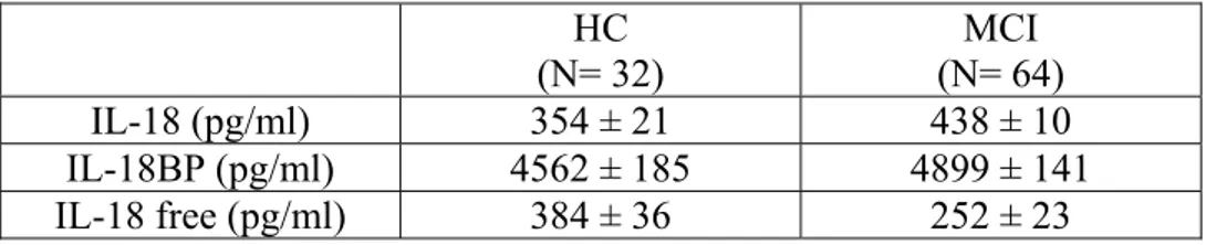
4.1.2.1. EPM……….56 4.1.2.2. OF………56 4.1.2.3. MWM………..56 4.1.2.4. CYTOKINE ANALYSIS………...….59 4.2. MCI STUDY……….61 4.3 SCZ STUDY………...64
4.3.1. SERUM LEVELS OF IL-18/IL-18BP………...64
4.3.2. RELATIONSHIP BETWEEN IL-18, IL-18BP AND AGE………...66
5. DISCUSSION………...67
6. CONCLUSIONS………..…77
1. INTRODUCTION
The immune system (IS) and the central nervous system (CNS) are the two main adaptive systems that provide for the functional integrity and regulate the homeostasis of the whole organism. Convincing experimental evidence, exponentially growing in the recent years, shows that immunity and brain interact to influence health. Indeed, the two systems share many analogies both for the function of their leading cells and for the way of action.
In the past, neuroimmunology was concerned mainly with the manner in which the IS interacts with the CNS in pathological conditions, such as autoimmune disorders. More recently, new levels of intersection and commonalities have emerged: for instance, it is now known that cells within the two systems communicate via morphologically similar physical connections and that they share molecular mediators of communication, such as transmitters and signalling molecules (Kerschensteiner et al., 2009). In addition, cells from both systems show targeted migratory behaviour (neurons mostly but not exclusively during development, immune cells throughout life). Finally, a very interesting functional characteristic that unites the two systems is their ability to connect and carry information from and to distant parts of the body: neurons using their long processes (dendrites and axons) and the immune cells as a result of their mobility. The immune system, through lymphocytes, secretes endocrine substances and neuropeptides so as the central nervous system. The communication of all these molecules produced occurs through receptors that are proteins located on the cell surface that transmit signals within the cell following the binding with the mediators for which have specific affinity. For instance, a list of common surface molecules that act as receptors includes the Toll-like receptors (TRLs), which were originally thought to be primarily functioning in immune cells, but that are now also found in neuronal cells, where they appear to perform a cell-autonomous function (Farina et al., 2007).
Similarities between the two systems are also found in the rules that govern the motility and the direction of movement of the neurons and immune cells. In fact, both types of cells are able to map a precise itinerary in response to attractive or repulsive signals produced by other cells present among their
migratory route or at the final destination. Thus, lymphocyte movement within lymphoid organs appears to be the result of recognition of molecules and structures produced and laid down by stroma cells in precise fashion (Bajenoff et al., 2006). Similarly, neurons migrate and extend their processes also through complex stromal structures after specialized signals that include samaphorins, signalling molecules that are key players in neuronal guidance, and that recently were found to be expressed and used also by lymphocytes (Mizui et al., 2009).
Until a few years ago it was believed that the brain was “an immunological privileged organ” inasmuch isolated from immune and inflammatory response by blood brain-barrier. This view has been drastically changed by a lot of studies that have shown as the brain can exhibit a wide range of immune and inflammatory response, beside being an important regulator of immune system.
A lot of experimental evidences show a crosstalk between the immune system and the nervous system by a bidirectional communication. The efficient coordination of the interaction between these systems guarantees the maintenance of the physiologic state, that if missing, may confer a risk for the onset of pathological processes.
1.1 THE IMMUNE SYSTEM: PERIPHERAL AND BRAIN
RESPONSES
The immune system has the responsibility to defend the body against pathogenic insults, which may be represented not only by exogenous agents of infectious nature (as in the case of pathogenic microorganisms), but also from endogenous substances potentially harmful originated from injury, trauma or altered physiological processes.
Basically, two types of defensive processes are outlined in the immune system: the innate or natural immunity and the acquired or specific immunity. The natural immune response represents the first line of protection of the organism against pathogens and it is mainly based on non-specific defence mechanisms, existing since the birth of the individual. This
is the oldest system of defence against infection from the evolutionary point of view. The main components of innate immunity are the barriers of the organism with peculiar physical or chemical proprieties, such as the skin or the gastric acid contained in the gastric mucosa and different types of cells and soluble molecules. Among the latter, cells with phagocytic activity such as neutrophils, macrophages and dendritic cells and cells with cytotoxic activity or natural killer cells (NK), as well as the molecules belonging to the complement system and cytokines are all important players of the non specific response and immediate attack to pathogens. Following the natural immune response, a sequence of cellular and molecular events takes place which constitutes the acquired immunity, able to enhance the defence mechanisms triggered by the innate immunity. This process is able to activate immune responses highly specific for the different pathogenic macromolecules and to respond in an ever more powerful to repeated exposure to the same antigen, as it is characterized by the onset of a “immunological memory” which allows a persistent protection thanks to the presence of specialized immune cells functionally quiescent but capable of surviving for long periods. The main effector mechanisms of specific immunity are humoral immunity, mediated by antibody producing B lymphocytes and cellular immunity mediated by T lymphocytes. The latter, comprise two distinct subpopulations: cytotoxic and helper T cells, both incapable to recognize soluble antigens, but able to bind processed antigens presented by molecules of the major histocompatibility complex, through specific T cell antigen receptors (TcR). The immunocompetent cells that possess the specialized machinery required to efficiently process and express the antigens in this way are named antigen presenting cells (APC), and include macrophages, B lymphocytes and dendritic cells. Once T cells recognize and bind to the antigen-MHC complex, the APC send out additional co-stimulatory signal to activate T cells, so initiating a new adaptive immune response. While T cytotoxic lymphocytes exert their function directly killing the cells infected by the antigens, T helper cells secrete cytokines to activate the proliferation and the differentiation of other T cells, B lymphocytes and macrophages (Abbas et al., book).
By the anatomical point of view, the immune system consists of a complex network of organs and tissues, connected by blood and lymphatic vessels, that work together to prevent infection or injury-mediated damage throughout the body. The thymus, bone marrow, spleen, lymphoid system including lymph nodes and mucosal associated lymphoid tissue, as well as the circulatory system, are all components of the peripheral IS, as indicated on the basis of the collective definition for all immune responses that take place outside the brain and together are responsible for the creation, transport and function of immunity in mammals. However, also the CNS is under immunological surveillance, and even though the mechanisms are different from the periphery, immune responses occur in the brain (Hickey WF, 1999). Notably, an active interplay exists between brain and IS and increasing evidence points out that circulating immune signals exert an effect on the brain, as well as brain derived signals may influence immune reactions. The interaction element between the blood born cells and molecules of the immune system and CNS components is the blood brain barrier (BBB), which, differently from the past when it was mainly considered a static barrier preventing infiltration of immune elements in the brain, is today seen as an active regulatory interface that controls the exchange between the brain and the periphery and is modified and affected by circulating events and by events on the CNS side (McAllister AK, 2009). A series of different immune cells interact with the BBB to mediate immunity at this side. First of all, perivascular macrophages resides between the astrocytes and the vessel wall and their role is to phagocyte cellular debris and the depletion of these seems to increase meningitis infection (Williams et al., 2001; Polfliet et al., 2001). Moreover, mast cells have been identified to be associated with vessels in specific region of the CNS also if their role in CNS immunity and in BBB regulation has not been clarified (Wilhelm et al., 2005). It has been demonstrated that breakdown of the BBB is a critical component of several disease as multiple scleroses, edema and brain trauma. The breakdown of the BBB accompanies the primary insult in these disorders allows the entry of plasma component, immune molecules and cells, which leads to neural dysfunction and ultimately neural degeneration. New evidence also suggests that BBB dysfunction might be a
widespread component of many different neurodegenerative disorder as Alzhaimer’s disease (Browman et al., 2007) and BBB dysfunction has also been proposed as a cause of this pathology (Pluta et al., 2006; Daneman, 2012).
The brain is protected by damage at several levels: by the interaction with the IS, by the BBB and by several resident cells. The most important resident cells that have the role to defend CNS by damage are some glial cells that constitute the microglia. These brain specific immune cells, are macrophage-like, originate from bone marrow and migrate to the CNS, where develop into glial cells. They are constitutively present in the brain were they actively survey the tissue by monitoring their extracellular space and cellular neighborhood, but are mobilized as a result of injury or infection and form the first line of cerebral defence. Once activated, they have an extension more robust and more branched than their resting counterpart. In CNS parenchyma, resident microglia are monocyte lineage cells, which enter in the CNS during development and are involved in neural development, innate immune response and promoting wound healing, can act as antigen-presenting cells to initiate an adaptative immune response (Ajami et al., 2007; Streit et al., 2005). Moreover, a subset of microglia cells interact with the CNS vessels and have been shown to regulate immune cells passage across the BBB (Persidsky et al., 1999; Hudson et al., 2005). Recent study have shown that other cells over microglia are present in CNS. In fact, as I mentioned earlier, macrophages and mast cells, associated with meninges and perivascular spaces, may be maintained by replacement from blood monocytes (Ajami et al., 2011; Ajami et al., 2007; Ransohoff et al., 2010). Following CNS inflammation the number of myeloid cells increases as a result of infiltration from the blood and the proliferation of the resident microglia cells augments as well. Moreover, albeit the CNS was previously believed to be inaccessible to T cells as an immune privileged site, recent studies have instead proposed that memory T cells can enter the CNS from the blood into the cerebrospinal fluid (CSF) (Ransohoff et al., 2003) by a complex trafficking pattern mediated by chemokines and chemokines receptors.
1.2 BRAIN: ORGANIZATION AND FUNCTIONS
The nervous system is responsible for sending, receiving, and interpreting information from all parts of the body and is coordinated by a complex network of neurons. The brain is the center of the nervous system and it is a highly complex organ both from the anatomic point of view that interaction. During the cerebral development, few weeks after conception, at the level of the cephalic end of the neural tube, begin to be evident three bulges: the forebrain, the midbrain and the hindbrain. Seven weeks after the conception the forebrain and the hindbrain are clearly divided. The forebrain will be divided in telencephalon and in diencephalons: telencephalon will give rise to neocortex, basal ganglia and systemic limbic; diencephalon in thalamus and hypothalamus. The hindbrain will be divided in metencephalon that will give rise to pons and cerebellum and in mielincephalon will give rise to medulla oblongata. (Fig.1) Each part of the brain is connected with the others by various connections to exchange the information and each region underlying a specific behavior.
Fig.1 Subdivisions of the embryonic vertebrate brain. These regions will later differentiate into forebrain, midbrain and hindbrain structures
Begin to know this important system starting from its basic unit: the two principal classes of specific cells called glial cells (o glia) and neuronal cells (or neurons).
The glial cells are 10-50 times more numerous than neurons and are arranged to surround the cellular body, dendrites and axons. These cells perform different functions:
- Act as support to neurons giving shape and structure to the nervous system;
- Act as phagocytic cells by removing the fragments of cells due to injury (microglia);
- Improve the efficiency of nerve signals;
- Two type of glial cells (oligodendrocytes and Schwann cells) give rise to myelin;
- Other glial cells (radial glia) guide the migration of the neurons and their axons during neurogenesis;
- Other glia cells (astrocytes) make up the blood brain-barrier.
Some glia cells, in particular the microglia cells, have a direct modulation on the neurons that are the other cell population of the brain. Microglia cells in fact, as already mentioned before, are activated at cerebral level following a brain injury in an attempt to repair the damage. Microglia produce pro-inflammatory cytokines, which could contribute to augmented local inflammation, release of toxic molecules contributing on neuronal damage. In some situations, the role of microglia has been found to be beneficial, since activated microglia can reduce in Alzheimer’s disease, Aβ accumulation by increasing its phagocytosis, clearance, and degradation (Frautschy et al., 1998; Qiu et al.,1998). Microglia can also secrete a number of soluble factors, such as the glia-derived neurotrophic factor (GDNF), which are potentially beneficial to the survival of neurons (Liu et al., 2003).
The neurons, that are the main brain cells, are principal players of the complex networks that underlie behaviour and cognitive functions. These cells are formed by three areas: the cellular body (where there is the nucleus), dendrites (extension of the cell body) and the axon (single extension of the cell). Dendrites originate from the cell body and branching form arborizations which represent the main equipment to receive messages coming from other nervous cells. Axons extend for long distance from the cellular body transmitting messages to other neurons. Axons can transmit electrical signals called action potential that are nervous impulses generated at the level of the origin of the axon (cone emergency). The potential of the signal does not change along the path and it has the specific characteristic to
be a signal of “all or nothing”. Moreover this signal can regenerate at regular intervals along the axon. The nervous system uses the action potentials to receive, analyzed and disseminate the information. These signals are stereotyped through the nervous system and to increase the conduction velocity, the axons are surrounded by a lipid envelope of myelin. The casing is interrupted at regular intervals by the nodes of Ranvier and in these sites the energy of the action potentials can regenerate. Neurons can also communicate with other neurons at the level of the synapse by the release of chemical molecules called neurotransmitters. Neurotrasmission (or synaptic transmission) is communication between neurons as accomplished by the movement of chemicals or electrical signals across a synapse. For any interneuron, its function is to receive input "information" from other neurons through synapses, to process that information, then to send "information" as output to other neurons through synapses. Consequently, an interneuron cannot fulfil its function if it is not connected to other neurons in a network. A network of neurons (or neural network) is merely a group of neurons through which information flows from one neuron to another. Every neural network headed by a specific neurotransmitter, underlie specific behaviour and cognitive functions. For example, the principal neurotransmitters which are associated specific cognitive functions are the following:
- Serotonin. Serotonin plays a very important role in a range of brain functions and it is synthesised from the aminoacid tryptophan. Within the brain, serotonin is localised mainly in nerve pathways emerging from the raphe nuclei (a group of nuclei at the centre of the reticular formation in the Midbrain) pons and medulla. These serotonergic pathways spread throughout the brainstem, the cerebral cortex and the spinal cord. Serotonin has been linked with a wide variety of functions, including the regulation of sleep, pain perception, body temperature, blood pressure and hormonal activity. This neurotransmitter, moreover seems to be involved in psychotic disease as schizophrenia inasmuch modulates the release of dopamine in several brain region causing the known symptoms of this illness.
- Dopamine. Dopamine is classed as a monoamine neurotransmitter and is concentrated in very specific groups of neurons collectively called the basal
ganglia. Dopaminergic neurons are widely distributed throughout the brain in four important dopamine pathways: mesolimbic, mesocortical, nigrostriatal and tuberoinfundibular. A decreased or an increase of brain dopamine concentration is a contributing factor in several diseases as Parkinson, Huntington and schizophrenia illness.
- Noradrenaline. Noradrenaline is classed as a monoamine neurotransmitter and noradrenergic neurons are found in the locus coeruleus, the pons and the reticular formation in the brain. These neurons provide projections to the cortex, hippocampus, thalamus and midbrain. The release of noradrenaline tends to increase the level of excitatory activity within the brain, and noradrenergic pathways are thought to be particularly involved in the control of functions such as attention and arousal.
- Acetylcholine. Acetylcholine acts in cholinergic pathways that are concentrated mainly in specific regions of the brainstem and are thought to be involved in cognitive functions, especially memory. In fact, severe damage to these pathways is the probable cause of Alzheimer’s disease.
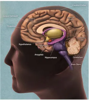
Specific behaviour and cognitive functions can be associated non only with particular neurotransmitter but also with brain regions that underlie specific neural network (Fig.2).
Memory is the principal cognitive function and is particularly correlated with an area that is part of the forebrain and is one of the structure that composes the limbic system: the hippocampus. This area is involved in several function as emotional responces, spatial orientation and memory capacity. In paticular, the hippocampus is the area responsible to acquire new memories are gradually to transferre tham to neocortical stores where the memory are kapt by the process of memory consolidation. Moreover, the hippocamus is the structure througt which the memory can be evoked and than remember. For this, damage to this structures caused for exemple by neuronal death as in case of Alzheimer’s disease, sekes above all severe loss of memory capacity. Another principal cognitive functions are the locomotion and the movement and several brain areas are responsible for this activity as cerebellum and struatum. In particular, the cerebellum is the portion of the brain responsible for the coordination of movement, balance,
equilibrium and muscle tone. It is located beneath the occipital lobe at the base of the skull. This area controls movement by processing and coordinating sensory input. The information is then sent to the motor nerves in order to produce fine motor movements. The cerebellum also calculates and corrects informational discrepancies in order to produce the desired movement. The striatum instead, is the largest nucleus of the basal ganglia. It receives direct input from most regions of the cerebral cortex and limbic and from sensorimotor and motivational regions of the brainstem arrives indirectly via relays in the thalamus. His role in motor skills is bound to his contact with the substantia nigra where by domanine via controls the movement activity. Damage in this neuronal network cause illness as Parkinson’s disesae.
Basic functions necessary for life, such as fluid and electrolyte balance, feeding and energy metabolism, wake-sleep cycles, thermoregulation, stress responses, and sexual behavior and reproduction are regulate and coordinate by the hypothalamus that is a small area at the basis of the brain. This area receives direct sensory inputs from the smell, taste, visual, and somatosensory systems. It also contains within it sensors for such things as blood temperature, blood sugar and mineral levels, and a variety of hormones. Thus the hypothalamus receives sensory inputs necessary to detect challenges in both the internal and external environments. In addition, the hypothalamus receives inputs from forebrain areas including the hippocampus, amygdala, and cingulate cortex. These structures form the limbic lobe of the brain, receives highly processed sensory information from throughout the cerebral cortex. These inputs drive a wide range of emotional responses, and many of the phenomena we associate with emotional expression are mediated by the hypothalamus.
The cerebral cortex is the most highly developed and complex part of the human brain and is responsible for thinking, perceiving, producing and understanding language. The brain is dominates by two hemispheres: left and right hemispheres. Anatomists conventionally divide each hemisphere into four "lobes", the frontal lobe, parietal lobe, temporal lobe, and occipital lobe. The form of the hemispheres is the result of an elaborate folding of tissue where the reliefs are called laps and are separated from each other by
grooves to increase the cerebral surface. Every lobe has a specific function. For example, there are specific areas involved in vision, hearing, touch, movement, and smell. Other areas are critical for thinking and reasoning. Although many functions, such as touch, are found in both the right and left cerebral hemispheres, some functions are found in only one cerebral hemisphere. The parietal lobe is involved in the reception and processing of sensory information from the body; the frontal lobe is involved with decision-making, problem solving, planning, cognition and memory; the occipital lobe is involved with vision and the temporal lobe is involved with memory, emotion, hearing, and language. The informations are sent to the cortex, where are rielaborate, and than they are again again sent to other part of the brain.
Fig. 2 Schematization of the main human brain areas.
A damage to the central nervous system in a brain area or in the production/recapture/action of one of the neurotransmitters, crusading by a pathogen agent or by an uncontrolled inflammatory event, induce several deficit in the all the brain. Injuries to the brain tend to affect large areas of the organ, sometimes causing major deficits in intelligence, memory, personality, and movement. These type of damage can be caused by trauma, edema or stroke events that induce neuronal death or by virus or bacteria leading for example to meningitis. Other damage of the brain can be
classified as neurodegenerative disease such as Alzheimer’s disease characterized by a gradual death of individual neurons leading a progressive cognitive impairment. Mental disorder as psychosis such as schizophrenia may involve particular patterns of neuropsychological functioning related to various aspects of mental and somatic function. Yet is not know how these illness progress and what are the pathogenetic pathway that underlie them but they are shared by a neuro and peripheral inflammatory processes. In the human is difficult to study directly the neuroinflammatory process for practical reason and for this, result very interesting to investigate the peripheral inflammation that characterize the patients affected by psychosis or neurodegenerative disease.
1.3 NEUROINFLAMMATION AND CYTOKINES
Under optimal condition, integrated and concerted activation of the immune mechanisms leads to the elimination of the pathogen or insult and the repair of damage. In particular, the inflammation, mainly mediated by mechanisms of innate immunity and controlled from different processes of the immune response, has a defensive function. However, in case of chronic inflammation, when the regulation of immune processes is not adequate, the pathogen may persist and the mechanisms that normally protect the individual can cause tissue damage and trigger consequential pathologies which is reflected also as cause of damage in CNS. Since CNS has a low capacity for regeneration, it is extremely vulnerable to damage caused by inflammatory processes, which in district often lead to an alteration of the functionally and the irreversible loss of neurons. Neuroinflammation involves resident cells in the CNS (microglia, astrocytes, neurons) and, at peripheral level, different cells of the immune system (macrophages, monocytes, T and B lymphocytes, dendritic cells) and many soluble factors as cytokines. Then, pro-inflammatory cytokines, which have effects on both immune and nervous systems, are central players of inflammation and may contribute to brain changes during both physiological and pathological processes. As key mediators of a chronic neuroinflammation that drives
progressive tissue damage in the brain, cytokines could take part to the pathogenesis and progression of neurodegenerative diseases (Holmes et al., 2003; Mrak et al., 2009; Lampa et al., 2012).
The cytokines are a category of signalling protein molecules locally released by specific cells that carries out their function by binding specific receptors. When cytokines are produced by immune system cells are also called interleukin. These molecules can perform their action in an autocrine manner (acting on the same cell which has produced them) or in paracrine manner (by acting on adjacent cells) or endocrine when produced in large quantities. Cytokines have four main property:
- Pleiotropy: the ability of a cytokine to exert its effects on different cell types;
- Redundancy: more cytokines can mediate the same biological event; - Synergism: the total effect of two or more cytokines is higher than their only addition;
- Antagonism: two or more cytokines are competitors.
With these capacities, they are involved virtually in all aspects of both innate and adaptive immune responses. Cytokines may play a pro-or anti-inflammatory activities depending on how they modulate the anti-inflammatory response, that is by either amplifying or inhibiting it. Nowadays a lot of cytokines have been identified, whose role is under intense study by the researchers as some of them seem to be involved in various immune, neurodegenerative and psychiatric illness. For example, much interest is focusing on TNF- α and IL-1 β, two pro-inflammatory cytokines that are among the first players in the acute inflammatory response that characterizes many CNS diseases as stroke (Lambertsen et al., 2012), obsessive compulsive disorder (Gray et al., 2012), trauma and multiple sclerosis (Arvin et al., 1996).
Tumor necrosis factor (TNF-α) in fact is one of the main mediators of the acute inflammatory response whose produced by phagocytes, T lymphocytes, NK cells, mast cells and macrophages as a result by infectious agents and by gram-negative bacteria. TNF-α plays a central role in
initiating and regulating the cytokine cascade during an inflammatory response. It is produced as a membrane-bound precursor molecule of 26 kDa that is cleaved by the TNF-α converting enzyme to produce a 17 kDa active cytokine (Perry et al., 2001). The levels of TNF-α expression in the healthy brain are low, making it difficult to determine its precise role under physiological conditions. In inflammatory or disease states, TNF-α along with several other proinflammatory mediators and neurotoxic substances are predominantly produced by activated microglia. Neuronal production of TNF-α has been demonstrated (Breder et al., 1993), although brain-derived TNF-α is mostly synthesized by glial cells in response to pathological stimuli. Glial cells secrete both TNF-α and IL-1, which in turn, activate these cells in an autocrine manner to induce further cytokine production and astrogliosis. TNF-α, on the other hand, has been reported to have neuroprotective properties (Akiyama et al., 2000) in the AD brain.
Another pro-inflammatory cytokines closely related to TNF-α is the interleukin- 1 (IL-1), whose main role is to mediate the inflammatory response triggered by bacterial products such as lipopolysaccharide (LPS), a component of cell wall of Gram-Negative Bacteria. IL-1 is an important initiator of the immune response, playing a key role in the onset and development of a complex hormonal and cellular inflammatory cascade. This cytokine is mainly secreted by macrophages and other APC and its production can be further stimulated by the action of other cytokines such as TNF-α. Its mainly role is to promote both adaptive and innate immune response favouring the recall of both macrophages and T cells at sites of infection and stimulating the clonal expansion of B lymphocytes maturation. There are two forms of IL-1 calls IL-1α and IL-1β biochemically distinct but structurally related. IL-1 α / IL-1β exerts its function through the formation of a complex with type I receptor and accessory protein (IL-1 receptor accessory protein) (IL-1 RI and IL-1 RACP) starting the transduction cascade leading to activation of inflammatory genes. IL-1 α / IL-1β has a natural antagonist IL-1 RA functionally inactive, but able to inhibit its function. Both IL-1 α / β are produced as immature form with a signal sequence cleaved by interleukin-1 converting enzyme to produce the biologically active form of 17 kDa. These cytokines are involved in
inducing an acute inflammatory response, their action and production in normal decade then a few hours after stimulation by inflammatory event. In particular, elevated IL-1β has been detected in the CF and brain parenchyma within the early hours after brain injury in both humans and rodents (Winter et al., 2002; Woodroofe et al.,1991).Another pro-inflammatory cytokine, member of the IL-1 family (Ushio et al., 1996; Bazan et al., 1996), is IL-18, which is gaining more and more interest inasmuch it would play a prominent role in neurological and psychiatric disease and for this reason a lot of interest is taken from the entire system of IL-18 composed in the main way from the same IL-18 and from its natural inhibitor (IL-18 binding protein).
1.3.1 IL-18
IL-18 is a pleiotropic cytokine, involved in several pathological conditions and inflammatory diseases. This cytokine, working in collaboration with IL-12, is able to induce cell-mediated immunity response following an inflammatory stimulus. IL-18 induces natural killer (NK) cells and T-cell-helper type 1 to release another cytokine called interferon (INF) that is involved in the activation of macrophages and other immune cells. For this reason IL-18 was originally called INF-inducing factor (Okamura H et al., 1995). IL-18 presents some homologies with IL-1 which is another cytokine member of the IL-1 family. Indeed these molecules are structurally similar among themselves, in fact IL-18 and IL-1 are two of the few cytokines that are all--plated sheet. Moreover IL-18, as well as IL-1, is synthesized in a 24 kD biologically inactive precursor form (proIL-18) which afterward, like proIL-1, is cleaved into an active molecule (18 kD) by an intracellular cysteine protease called IL-1β-converting enzyme (ICE) or caspase-1 (Gu Y,1997). In turn, caspase-1 is produced as an inactive precursor, procaspase-1, that is activated by caspase-11 (Wang S, 1998). Caspase-11 is also induced as an inactive molecule called procaspase-11 which is activated by cathepsin B(Schotte P, 2000) via P38 MAPK
phosphorilation (Vanden Berghe W, 1998) which require NF-kB transactivation into the nucleus (Li X, 2002; Schauvliege R, 2002)
IL-18 is able to perform its biological function by binding to its receptor. 18 receptor (18R) is an heterodimeric complex member of the IL-1/Toll like receptor superfamily that is composed by the IL-18 binding chain IL-18R, member of IL-1 receptor-related protein (IL-1RrP or IL-1R5), and by the nonbinding signalling chain IL-18Rβ or IL-18 accessory protein (IL-18RacP or IL-1R7); both chains are members of the IL-1 receptor super-family (Kato Z et al., 2003) and their peculiarity is to increase the affinity of the heterodimeric complex with the ligand so it is able to initiates signal transduction. Than, IL-18 binds to IL-18R, IL-18Rβ forms a high affinity between heterodimeric complex and the ligand allowing the recruitment of the IL-1R activating kinase (IRAK) via the adapter protein (MyD88). The autophosphorylation of IRAK induces the activation of TNFR-associated factor-6 (TRAF-6) resulting in phosphorylation of NF-kB-induced kinase (NIK) which in turn leads to activation of two IkB kinase (IKK-1 and IKK2) that lead to the phosphorilation of IkB. This latter step induces to the degradation and release of two components of NF-kB: p50 and p65. So, NFkB is free to migrate in the nucleus to induce the gene transcription. IL-18 has the capacity to induce Th1 cells, NK cells, B cells and dendritic cells to produce INF. Moreover IL-18 promotes T cells, NK cells, mast cells and basophils to produce IL-4 and IL-13 (Hoshino T, 1999; Yoshimoto T, 1999), therefore it is able to stimulate both innate immunity and Th1, Th2 mediated responses.
Recent studies have show that IL-18 is present in the central nervous system in addition to periphery where its role in immune response is well known (Alboni et al, 2010). Indeed, it is constitutively present in the brain, it is produced by microglia, astrocytes, ependymal cells and by some habenular neurons (Sugama S, 2006) and its expression is increased in old processes (Dinarello et al., 2006) and in several pathologic condition, associated in psychiatric illness and neuropathological disease. IL-18 has been shown to inhibit the induction of LTP (Long Term Potentiation) in the dentate gyrus, without affecting the baseline neurotransmission (Curran et al., 2001;
Curran et al., 2003; Cumiskey et al., 2007). Recently, IL-18 has been found to play a diverse array of functions in non-immune tissues as the central nervous system (Alboni et al. 2010), and its abnormal expression has been observed both centrally and peripherally in neuropsychiatric disorders such as Alzheimer’s disease and stroke (Bossù et al., 2008; Bossù et al., 2010). Indeed, IL-18 itself is able to stimulate microglia to increase the expression of ICE to produce other pro-inflammatory cytokines amplifying the inflammatory process. Recent studies have highlighted the overproduction of IL-18 in several inflammatory diseases indicating that IL-18 is a pro-inflammatory cytokine which may play an immunopathological role in many pathological conditions. A lot of studies performed both in vivo and in vitro have suggested an up-regulation of IL-18 gene expression in neuropathologic situations such as stroke, brain injury and Alzheimer’s disease, candidating this cytokine as a crucial mediator of neuroinflammatory processes. Further data indicate that also stress contributes to increase IL-18 levels by activation of the hypothalamic-pituitary-adrenal (HPA) axis and suggesting that IL-18 may play also a role in the HPA axis regulation.
The IL-18 activity is modulated by IL-18 binding protein (IL-18BP). IL-18 BP is a 40 kDa protein that is constitutively expressed and appears to be the natural inhibitor of Il-18 activity binding selectively and with high affinity mature form of IL-18 preventing his interaction with IL-18R. Unlike soluble receptors for IL-18, IL-18BP does not have a trasmembrane domain; IL-18BP is a secreted protein with a high-affinity binding and ability to neutralize IL-18. Two human IL-18BP isoforms exhibited the greatest affinity for IL-18 with a rapid on-rate. Moreover, IL-18BP modulates the IL-18’s activity in fact, in addiction to having neutralizing activity, it can induce a negative feedback regulation for IL-18 production to modulate in pathological circumstance the strongly increase of IL-18 (Novick, Schwartsburd, Pinkus et al., 2001). IL-18BP is constitutively present in the serum of healthy humans at molar excess compared with IL-18 (Novick et al., 2001) and it appears to be modulated during the life. In fact, since IL-18 participates in fundamental inflammatory processes that increase during the aging process (Dinarello, 2006) and IL-18BP serum levels tend to increase
in old age with evidence of strong increment in healthy centenarians (Gangemi et al., 2003), it has been suggested that IL-18BP retains a remarkable protective role by acting to limit the impact of age-related inflammation.
1.4 FROM NEUROINFLAMMATION TO BRAIN
DISEASE: COGNITIVE AND BEHAVIORAL
CORRELATES
Growing evidences have highlighted the association between specific mood disorders and altered immune function. More recently, a number of hypotheses have been forwarded to explain how components of the innate immune system can regulate brain function at the cellular and system levels and how these may underlie the pathology of disorders such as depression, Alzheimer’s disease and several psychotic illness. The neuroinflammation seems to have effects on neurodegeneration and depression dysregulating the control and release of pro- and anti-inflammatory cytokines. In particular, pro-inflammatory cytokines, such as IL-1β, TNF-α and IL-18, which have a prevailing role in the immune response to pathogen in the periphery, have also a specific actions on neurons and circuits within the central nervous system. These cytokines, can act on the brain through different channels: or interacting from the periphery to the central, or acting directly in the brain after have been produced at cerebral site. There is in fact good evidence that they can originate both from the peripheral circulation following induction for example of LPS, as well as from local synthesis in neurons and glia (Dantzer et al., 2008; Raison et al., 2006). TNFα, together with IL-1β and IL-6 have well described actions on the hypothalamus including induction of fever, suppression of appetite and stimulation of the hypothalamic pituitary axis (HPA) to release corticotropin-releasing factor (Goshen et al., 2008; Yirminiya et al., 2011; Layé et al., 2000) property that has been viewed belong also to IL-18. Cytokines produced in periphery, particularly IL-1β, TNFα and IL-18, can enter the brain by an active transport system (Skinner et al., 1993; Gutierrez
et al., 2009), by the breakdown of the blood brain barrier and by the cerebrospinal fluid.
Once in the brain, these cytokines induce their own synthesis (Layé et al., 1994; Pitossi et al., 1997) and their expression are also regulated via the centrally generated response to stress by the activation of the HPA. It is the complex interplay between central and peripheral cytokines that ultimately influences the behavioural response to an environmental challenge. Than, during an inflammatory process, the principal events that characterize the damage at CNS levels are: massive breakdown of the blood brain barrier and typical changes in of the cerebrospinal fluid with a massive invasion of activated immune cells accompanied by the activation of resident microglia cells. These cells, when activated, produce several mediator to defeat the damage and in particular the mediators with pro- and anti-inflammatory proprieties that mostly collaborate to immunity response are the cytokines. For exemple, microglia cells are activated in the brain of patients with Alzheimer’s disease and surround amyloid plaques in an attempt to eliminate them. Microglia produce pro-inflammatory cytokines such as IL-1β, IL-6, TNF-α, which could contribute to augmented local inflammation, release of toxic molecules contributing on neuronal damage (Akijama et al., 2000). Moreover, neuroinflammation and cytokines are associate with synaptic protein loss and these changes could contribute to cognitive impairments in neurodegenerative and neuropsychiatric illnesses (Rao et al., 2012).
The neuroinflammation has an effect on neurodegeneration and on specific mood disorders. In this context seems to be very interesting to focus the attention of the research on the crosstalk between inflammatory and central nervous systems and on the possible role as mediator of the cytokines, not only for understanding disease etiology, but also for selecting the best therapeutic approach.
1.5. NEURODEGENERATIVE DISEASE: ALZHEIMER’S
DISESE
The Alzheimer’s disease (AD) was so called after that the German neurologist Alois Alzheimer (1864-1915) described in 1907 the clinical signs of the neuropathology. The doctor in fact, led the autopsy of the brain of a woman who died as a result of an unusual mental illness and he showed the presence of agglomerate protein and neurofibrillary tangles that today we know to characterize the brain of AD patients.
The AD is responsible for two thirds or more of all diagnosed cases of dementia that affects approximately 3%, 19%, 47%, respectively, of the individuals of 65-74, 75-85 and 85 years. Disease are associated with several clinical signs. Among the first symptoms include the inability to create new memories and loss of short term memory. People with cognitive impairment, loss the ability to speak, to think, to remember past events and to develop a spatio-temporal orientation with the progression of the disease. A lot of patients also show delusion and change of personality. In the last stage of illness, patients are mute, immobile, incapable of understanding and the death occurs often due to respiratory failure. There are two types of AD identified by the age of onset of the disease: early AD, if it begins first of 65 years (early-onset) and late AD if it begins after of 65 years (late-onset). In late AD, disease lasts from 8 to 20 years, while in early AD the course of illness is generically more rapid and the death occurs within 5-10 years from the time of diagnosis.
Histological analysis by autopsies of the AD brain, shows the distinctive neuropathological characteristics. It can be observed devastating loss of synapses and neurons at the level of the hippocampus and the enthorinal cortex causing the first symptom of the disease: the mnestid deficit. Other brain areas are also affected by neurodegenerative process as septum, amydgala and several cortical areas with the progression of the disease, causing a huge deficit.
Cytological analysis shows dense spherical structures of 20-200 µm in diameter (senile plaques) outside the neurons and neurofiblillary tangles that accumulate within the cell bodies. It has been shown a strong correlation
between senile plaque in the enthorinal cortex and cognitive dysfunction (Cummings et al., 1996).
Autopsies performed on AD brains have shown that the neurofibrillary tangles appear initially in the limbic system and extend to the cortex with the progression of the disease, while the senile plaque appear first at the level of the frontal cortex and then to the rest of the brain, in parallel with increasing the severity of the pathology (Braak et al., 1998).
The senile plaque (or amyloid bodies) have a filamentary structure, constitute by protein and the main protein molecule present in the plaque is a peptide of 4 kDa (β-amyloid protein or Aβ), consisting of isoforms containing 39-43 aminoacids.
The genomic analysis has revealed that the Aβ isoforms of senile plaques derived by proteolytic cleavage from a protein called amyloid precursor protein or APP. This protein has a single trasmembrane site and it has three isoforms whit 695, 751 and 770 amino acids. Exactly the role of APP molecule is still not clear, but probably the extracellular portion acts for the cell-cell contact or cellular adhesion to the extraneuronal matrix, while the intracellular share seems to be associated whit the cytoskeletron. APP molecules are subjected to the action of different proteolytic enzymes. One of these enzymes is the α-secretase, which cuts the APP protein in site 687 generating two fragments not amyloidogenic. Although the β-secretase, acting at the level of the amino acid site 671, produces two fragments don’t amyloidogenic. Than, a single proteolytic cleavage, by α-secretase or by β-secretase, generates non-pathologic peptides. However, a double proteolytic cleavage by β-secretase and γ-secretase (at the site 711), generates the isoforms Aβ1-40 and Aβ1-42 which, if present in high concentration, are not adequately degraded and they can become amyloidogenic and accumulate outside the neurons they form the senile plaques.
The neurofibrillary tangles inside the neurons of AD patients are constituted by filaments mainly composed by the tau protein. The function of the tau protein under physiological conditions is to assemble the tubulin for the formation of microtubules, a part of the cytoskeleton that allows the axonal transport. Studies in vitro have demonstrated that in pathological condition, such as AD, tau protein is in a state of hyperphosphorylation.
Hyperphosphorylated tau is not able to maintain keep together the fibrils of tubulin, so the microtubule formation is locked and the axonal transporters is consequentially compromised.
Many studies are focusing on the identification of the etiology of Alzheimer’s disease and much interesting is to understand how the genetic can intervene on the onset of the illness. The AD, is characterized by a complex genetic, in fact only the 10% of cases have autosomal dominant inheritance. This 10% is made up of subjects who have in most cases, the presenile dementia (the disease occurs earlier than senile (before 60 years old). The remaining 90% are defined as sporadic cases with an onset of illness after 60 years old. Is interesting to note that 25-40% of late-onset cases have at least one or more kindred with the same disease. This therefore underlines that genetics may play an important role in the etiopathogenesis of AD. Screening tests have allowed us to study families characterized by early-onset AD to identify possible mutations responsible for the disease. Studies carried out both in vivo and in vitro have revealed a missense mutation that alters the amino acid in the side 717 of the APP. Studies carried on cultured cells and on the plasma of individuals which have this mutation in the APP gene, have higher levels of Aβ1-42 compared with health controls, as well as transgenic mice for APP gene produce increased amounts of Aβ1-42 compared to wilde tipe mice.
Studies of genetic association have identified two other mutations for early-onset AD: mutations of the gene for the presenilin 1 (PSEN1) and presenilin 2 (PSEN2). The presenelins are membrane proteins that appear to play a role in cell differentiation during embryogenesis and in the cellular transport of proteins. Even mutations of the genes of these two proteins are associated with an increased production of Aβ1-42. However, the mutations of APP genes, PSEN1 and PSEN2 are responsible for only 50% of cases of early-onset familiar AD and for this reason, remain undiscovered other genes that cause the disease. It is not clear how PSEN1 and PSEN2 mutation interact whit APP but it seems that what unites them in the developed of AD is the formation of Aβ1-42.
Other genetic association studies have shown a relationship between sporadic AD and the gene for apolipoprotein E (APOE). APOE is
synthesized primarily by astrocytes and its function is to sequester cholesterol and triglycerides from the debris of cells and to bring them in neurons where these molecules can be used for the formation of synaptic membranes. The APOE genes is polymorphic for three alleles: APOEε2, APOEε3 and APOEε4 and it would seem that the latter isoform (APOEε4) is associated with susceptibility to AD. This allele, however, represents only 50% of genetic component of sporadic late-onset AD and also this factor is neither sufficient nor necessary for the development of the disease. The APOEε4 is not a cause of AD but it is a risk factor for the disease, in fact, there are individuals that have both the alleles for APOEε4 but they haven’t any symptoms of AD, while there are AD patients that do not show none of these alleles.
AD disease is therefore a very complex neurodegenerative disease characterized by numerous causes of both genetic and non genetic elements. Much researches are being carried out in order to understanding the factors inducing the formation of senile plaques, neurofibrillary tangles and causes of neuronal death. Recently, the more supported hypothesis is the β-amyloid cascade. In this case it is believed that Aβ is the primary cause of AD. Several observations have shown that mutations associated with AD increase the amount of Aβ42 (considereding this a pathogenic peptide), while others don’t give much importance to the amount of Aβ42, but rather the relationship that increasing of Aβ42/Aβ40 could induce to AD. Finally, today the most widely accepted idea is that Aβ is present in soluble oligomeric forms that may represent the major pathogenic factors to disease. Oligomeric forms have been isolated from both brains of animal models of AD (Lesne et al., 2006) that from human brains with AD (Shankar et al., 2008) and parallel studies in vitro have demonstrated the strong cytotoxic power of these oligomers. It is not yet clear how the oligomeric forms are generated in vivo, but suggested two possible hypothesis. One possibility is that Aβ monomers initially form soluble aggregates that will give rise to future oligomers joining in insoluble protofibrils, fibrils and plaques. This hypothesis, therefore, considered pathogenic oligomers both soluble and insoluble (Shankar et al., 2008). An alternative hypothesis configures the existence of two distinct way: a pathogenic way that forms soluble
oligomers responsible for the disease and a not pathogenic way that forms insoluble aggregates and plaque (Catalano et al., 2006; Shanker et al., 2008).
According to the hypothesis of the cascade of β-amyloid, Aβ would seem to be the apex of the cascade triggering a process of neuroinflammation, brinding to the iperphosphorilation of tau protein through the activation of a cyclin-dependent kinases (CDK5) and to a reduction of synapses. This results in synaptic dysfunction, neuronal death and subsequent development of Alzheimer’s disease (Pimplikar et al., 2009)
1.5.1. MILD COGNITIVE IMPAIRMENT (MCI)
Whereas the symptoms of AD performs when the pathology is already overt, and then is difficult to study the early stages of disease, the research is focusing on the study of subjects MCI (Mild Cognitive Impairment) as these individuals have a clinical picture that can be inserted between a normal condition and a very early form of dementia. The first major study focused on the characterization of clinical pathology of MCI was published in 1999 (Peterson et al., 2001). This study showed that MCI subjects are individuals who have a high risk of future cognitive impairment and progressive Alzheimer’s dementia. The criterion adopted to define the state of MCI is based on five parameters (Peterson et al., 2008):
- Mnestic problems;
- Mnestic deficit in relation to the age of subjects; - Cognition generally preserved;
- Daily activities unharmed; - No form of dementia.
In Stockholm in 2003 there was an international conference on diagnostic criteria to include in the disease of MCI other forms of cognitive impairment in addiction to mnestic deficit (Winblad et al., 2004; Peterson et al., 2004). Two subtypes of MCI have emerged: amnesic MCI (characterized only by mnestic deficit) and non-amnestic MCI (characterized by deficit linked to not mnestic domains). It was proposed an
algorithm for diagnosing various type of MCI (Paterson et al., 2008). If the memory is impaired, clinicians considerer the patient as amnesic MCI, and if there is no obvious mnestic deficit but some cognitive impairment in other activities is shown (such language, executive functions and visuospatial tasks), it is believed that the patient is a non-amnesic MCI. Clinicians can also determinate which other cognitive domains may be compromised in MCI using neuropsychological tests. A diagnosis of MCI amnesic single-domain is assumed if the deficit involves only the mnestic activity (pure amnestic), while for amnestic multidomain MCI are considered patients that present in addition to memory impairment also other cognitive deficit. A diagnostic of MCI not amnestic single domain assumes that there is an impairment to only one other domain, while not amnesic multidomin MCI refers to a deficit in different domains that are not related to the function of memory. Following the clinical characterization of patients, the cause of cognitive impairment is determinated by neuroimaging studies and laboratory testes. Clinicians then determinate if the symptoms of MCI are due to a degenerative disease, vascular, psychiatric, or are a secondary consequence of another disease. In general, the type of amnesic MCI have an increased risk of elapse in Alzheimer’s dementia whit a more rapid disease progression for multidomain than single domain, while non amnesic MCI seems to be a greater chance to convert into frontotemporal dementia with Lewy bodies. An extremely interesting observation indicates that the subgroup of amnesic single domain MCI (pure MCI) have a high selectivity, greater than all other subgroup, to develop in AD and not in other forms of dementia (Lopez et al., 2007).
The study of MCI may therefore be used to understand the pathogenetic events that follows in the early stages of AD, not only to investigate the early stage of the disease, but if characterized by longitudinal clinical analysis, it would also help to identify the key processes that lead to the progression of AD in individuals. Identify subjects not yet sick, but with a high risk to develop AD, can be very interesting to understand the inflammatory parameters potentially involved in the disease and how the mediators of immunity may be over or under regulated at this preclinical stage of illness.
1.5.2. INFLAMMATION IN AD AND MCI
Alzheimer’s disease is an example of a pathology characterized by chronic inflammation localized in the brain, where the molecules involved in the modulation of the inflammatory response are produced by cells of microglia and they play a role in the neuroinflammatory process. In particular, the key of the pathogenic events of AD would seem the protein β-amyloyd that seems to be at the basis of specific immune activator that occurs in the brain of AD. β-amyloyd, in fact, actives not only microglia cells (Akiyama et a., 2000) but also other immune cells recruited from the periphery to contribute to the neuroinflammatory process (Ciaramella et al., 2009; Ciaramella et al., 2009).
Numerous studies have attempt to investigate how inflammation can affect the pathogenesis and progression of the disease, through the analysis of the expression of various cytokines. Several studies have shown that IL-1 appears to be expressed in a manner more consistent in the cortical region most rich of plaque in AD patients. (Sheng et al., 1995; Sheng et al., 1998). TNF also appears to play a role in AD, in fact various studies have shown an increase of production of this cytokine in serum (Fillit et al., 1991), in the cerebrospinal fluid (CSF), in the cortex (Tarkowski et al., 1999) and glia cells of AD patients placed in culture after exposure to Aβ (Lue et al., 2001). IL-6 is another pro-inflammatory cytokine that appears to have a role in the pathology of AD (Braida et al., 2004). Maccioni et al., (Maccioni et al., 2009) have recently proposed a hypothesis according to which events of oxidative stress (Koseoglu et al., 2007; Zambian et al., 2004), repair mechanisms in brain damage and folic acid deficiency are sufficient signals to activate innate immunity by modifying the normal microglia activation. The microglia so activated produces an excessive production of IL-6, TNF-α and IL-1, that are cytokines whose levels seem actually elevated in Alzheimer’s disease. The overproduction of these molecules may modulate the hyperphosphorilation of the tau protein and then create cell changes that can lead to neurodegeneration and neuronal progressive cognitive impairment. Moreover, another study seems to confirm the existence of a correlation between TNF-α and cognitive impairment, high serum level of
this cytokine may be associated whit cognitive decline that characterize AD patients (Holmes et al., 2009).
Among pro-inflammatory cytokines, IL-18 may contribute to the chronic neuroinflammation od AD. In fact, IL-18 participates in the initiation of the innate host response and can be normally detected in cortex, cerebellum, hippocampus, hypothalamus and striatum (Chulhaine et al., 1999; Bossù et al., 2012), mainly expressed in response to LPS by microglia and astrocytes (Conti et al., 1999). The importance of this cytokine in other neuroinflammatory and neurodegenerative pathological condition has been demonstrated both clinically and experimentally (Felderhoff-Mueser et al., 2005). Recently, IL-18 has been considered a possible phatogenetic mediator also in Alzheimer’s disease. Several studies have shown a possible involvement of this cytokine in the pathology of AD, since it has been shown increased expression of caspase-1 (enzyme activator of IL-18) in AD patients (Pompl et al., 2003). Moreover it was found that the receptor of IL-18 and in particular the β chain indispensable for signal transduction, is major expressed at the level of lymphocytes of AD subjects. Furthermore, it was observed that polymorphisms of IL-18 gene promoter are associated with the risk of exhibiting Alzheimer’s disease and with the course of the same illness (Bossù et al., 2007). Post-mortem studies carried out on the brains of AD patients, have allowed to quantify directly expression of IL-18 in situ in various brain areas (Ojala et al., 2009). In particular, this study shows a significant increase of IL-18 in the frontal cortex of AD patients compared to controls. Finally, it was shown that the blood cells of AD patients produce a greater amount of IL-18 compared with control subjects and that the high levels of this cytokines are correlated with high levels of cognitive impairment (Bossù et al., 2008).
An altered immunity profile was observed also in MCI. Galimberti et al. (Galimberti et al., 2005) have shown that a beta-chemokine with chemiotactic activity (MCP-1) is mainly produced both in MCI and in people that exhibit an early form of AD, while this molecules don’t appears to be produced significantly during the most severe form of the disease. Other studies have shown that, as well as in patients converted in AD, MCI subjects also exhibit high levels of TNF-α (pro inflammatory cytokine)
compared to TGF-β (anti inflammatory cytokine) in the CSF, indicanding that patients with high risk to develop AD are characterized by a specific pro inflammatory profile (Tarkowski et al., 2003). Other studies have shown that PBMC (Peripheral Blood Mononuclear Cell) of MCI patients produce a greater quantity of pro inflammatory cytokines when stimulated with appropriate inflammatory stimuli such LPS, as IL-6 and IL-8 than anti inflammatory cytokine as IL-10 (Magaki et al., 2007). These results indicate that during the preclinical state of AD (during the stage of MCI), there is an alteration in the production of cytokines and this may suggest that inflammatory events precede the clinical development of the Alzheimer’s disease. Based on all the research carried out it would appear that the inflammation has a significant role both in the pathogenesis and in progression of AD, also if all the processes which lead to the onset of the disease are not been clarified. Research is therefore strongly focused on clarification of molecules involved in the inflammatory process to realize how these act in the early stage of illness. To understand how IL-18 can be involved in AD, it can be helpful to discover new therapeutic procedures for this devastating disease.
1.6. A FORM OF PSYCHOSIS: SCHIZOPHRENIA
Psychosis is a syndrome, or rather a set of symptoms, which may be associated to many different psychiatric disorders. This can be considerate a set of symptoms in which the mental abilities of a person, his actual response and its ability to recognize reality, communicate and relate to others is impaired. Schizophrenia is the most common psychotic illness that affects 1% of the population. By definition, schizophrenia is a disorder that should last at six month or more, including at least one month of delirium, hallucinations, disorganized speech and behaviour. Delirium consist in an erroneous interpretation of reality and perceptions while hallucinations may occurs in any sensory modality. Many studies subdivided this pathology in five sizes: positive, negative, cognitive, aggressive/hostile, depressive/anxious symptoms.
- Positive symptoms: seem to reflect an excess of normal functions and include delirium, hallucinations, exaggeration of language, communication and behaviour.
- Negative symptoms: include five symptoms: affective flattening (limitation in the variety and intensity of emotional expression); alogia (limitation in the fluidity and productivity of speech and think); apathy (limitation in the capacity of action); anhedonia (inability to feel pleasure); deficit of attention.
- Cognitive symptoms: thinking disorder, inappropriate language, incoherence, difficulties of attention and learning often overlap with negative symptoms.
- Aggressive/hostile symptoms: hostility which manifests itself in verbal e physical violence, aggression, problems in impulse control and self-injurious behaviour often overlap with positive symptoms.
- Depressive/anxious symptoms: depressed mood, anxiety, guilt, tension, irritably and anxiety.
Biological bases of schizophrenia are not known. However, dopamine neurotransmitter plays a key role in hypothesis on five sizes symptoms just described. In particular, mesolimbic dopaminergic pathway, mesocortical dopaminergic pathway, nigrostriatal dopaminergic pathway and tuberoinfundibular dopaminergic pathway are the principal cerebral dopaminergic pathway but the first two are responsible of major symptoms that characterized schizophrenia.
- Mesolimbic dopaminergic pathway. This pathway projects from the cell bodies of dopaminergic neurons in the ventral tegmental area of the brain stem to the axonal terminals in limbic areas of the brain, as the nucleus accumbens. It is believed that a specific hyperactivity of this pathway mediate the positive symptoms of the psychosis.
- Mesocortical dopaminergic pathway. The cell bodies of this way are localized in the ventral tegmental area and project to the cerebral cortex. The role of this pathway is likely to mediate the negative and cognitive symptoms of schizophrenia. In fact, many researches believe that a hypoactivity of these neuronal systems is responsible for such behaviour. The dopamine deficiency of mesocortical dopaminergic neurons may be
causes either by a direct dopamine deficiency or by a secondary deficiency due to an excess of serotonin that mediates the activity of the same dopamine.
1.6.1 INFLAMMATION IN SCHIZOPHRENIA
There are many possible hypotheses about the origins of schizophrenia but there is not yet recognized a certain cause, but we can talk about risk factors, or rather conditions that predispose an individual to develop the disease than others. The genetic component is certain the most reliable risk factor in fact much research has been focused on this issue by the twin, family and adoption studies (Riley et al., 2005) and a myriad of geneses would seem to be involved in schizophrenia while the environment seems to play only a minor role (Schwarts and Susser, 2006). Inflammatory system seems to be involved in the schizophrenia pathology and this conception was already supported when researchers began to observe the psychotic disorders in 1845 (Rothermundt et a., 2001). Post-mortem studies have shown an up-regulation of several genes involved in inflammatory response in frontal cortex samples of schizophrenia subjects (Setre et al., 2007). Moreover, the probable involvement of inflammatory process in schizophrenia illness, is causing an increasing attention in pharmacotherapy to adopt anti-inflammatory drugs in schizophrenia treatment (Meyer et al., 2011).
Many evidence suggests that schizophrenia is mostly a neurodevelopmental disorder characterized in many cases by morphologic brain anomalies as ventricular enlargements (Vita et al., 2006) or decrease hippocampal volume (Nelson et al., 1998) and moreover children that develop schizophrenia during their adolescence show a tendency for impaired neurocognitive behaviour (Cannon et al., 2002) and neuromotor function (Murray et al., 2006). These damage could be caused by problems during pregnancy as prenatal exposure to infection (Remington and Klein, 2006). The possible interaction between infection and schizophrenia development is the stimulation of cytokines response (Patterson, 2009), perhaps an excess of
maternal inflammatory cytokines levels could be associated with neurodevelopment disorders (Dammann and Leviton, 1997) and with production of toxic agents by microglia and astroglia in the fetal brain that are harmful to neurons (Patterson, 2009). Recent studies have shown that cytokines play an important role in the development of nervous system (Deverman, 2009) and was seen by post-mortem study that an excess of cytokines levels may annoying the maturation of oligodendrocytes and of the white matter (Davis et al., 2003). Others study have hypothesized that cytokines may play a role non only during embryogenesis but also in the progression of schizophrenia and for this reason many researches are focusing on finding the circulating levels of pro and anti-inflammatory cytokines during schizophrenia illness (Meyer, 2011; Reale et al., 2011; Baker et al., 1996; Potvin et al., 2008). Possible link between infection and schizophrenia development is the stimulation of cytokine response and cytokine-centred hypotheses of schizophrenia pathogenesis have been recently proposed (Watanabe et al., 2010). In particular, IL-18 has been proposed, by recent data, to be connected to schizophrenia. In particular, five polymorphisms in genes related to the IL-18 pathway, including IL-18 receptor genes, have been associated with schizophrenia (Shirts et al., 2008) and some variants of the IL-18 gene have been recently related to the development of schizophrenia symptoms (Liu et al., 2011). Furthermore, the levels of IL-18 circulating protein have been found augmented in affected patients, as compared to controls (Tanaka et al., 2000; Reale et al., 2011).
2. AIM OF THE WORK
It has been widely accepted that immune system and central nervous system are closely interlinked and that they actively communicate especially in case of damage. However, until now the processes that underlie this crosstalk are still not clear and, for this reason it was very interesting to focus my Ph.D study on this issue. In particularly, the attention is focused on the role of inflammatory cytokines, more specifically on IL-18, to investigate the possible role as mediator between peripheral inflammation and brain. This work was conducted along different steps to better focalize my topic of interest through different angles. A first line of research has been developed by an experimental study on rats to investigate how a peripheral inflammatory stimulus can induce several changes in cytokines production in different brain areas also occurring the possible repercussion of these changes in the behaviour. In fact, recently, a great interest has been focused on research addressed to better understand the role of brain cytokines in behavioral and cognitive performance following systemic inflammation, especially in the longer term after inflammatory insults, to realize what happens in the CNS after a peripheral inflammatory event. For this reason, a lot of studies have been conduced by peripheral injection of the bacterial endotoxin component lipopolysaccharide (LPS) of gram negative bacteria that models systemic infection, to induce neuroinflammation in order to analyze at brain level the synthesis of inflammatory cytokines, such as interleukin IL-1β, IL-6, and tumor necrosis factor (TNF)-α (Gatti et al., 1993; Pitossi et al., 1997). These inflammatory mediators, in turn, appear central in driving behavioral modifications, as in the case of the behavioral responses to LPS, known as sickness behavior, which is the acute consequence of cytokine elevation (Dantzer et al., 2001). Notably, endotoxin triggering can also induce protracted behavioral effects, as occurring in aging rats (Barrientos et al., 2006; Barrientos et al., 2009). Differently, investigations in healthy adults aimed at determining whether a single event of systemic inflammation can as such induce long-lasting modifications of behavior and brain cytokine synthesis are still limited. A single LPS administration results in delayed loss of neurons and cholinergic