ORIGINAL ARTICLE
Circulating levels of TNF-related apoptosis inducing-ligand
are decreased in patients with large adult-type granulosa cell
tumors
—implications for therapeutic potential
Anniina Färkkilä1,2&Giorgio Zauli3&Ulla-Maija Haltia1,2&Marjut Pihlajoki1,2&
Leila Unkila-Kallio1&Paola Secchiero4&Markku Heikinheimo2,5
Received: 2 December 2015 / Accepted: 28 March 2016 / Published online: 11 April 2016 # International Society of Oncology and BioMarkers (ISOBM) 2016
Abstract Targeted treatments are needed for advanced adult-type granulosa cell tumors (AGCTs). We set out to assess tumor tissue and circulating levels of TNF-related apoptosis-inducing ligand (TRAIL), a promising anti-cancer cytokine, in patients affected by AGCT. We analyzed tissue expression of TRAIL in 127 AGCTs using immunohistochemistry or RT-PCR. Soluble TRAIL was measured by means of ELISA from 141 AGCT patient serum samples, as well as the conditioned media of 15 AGCT patient-derived primary cell cultures, and the KGN cell line. Tissue and serum TRAIL levels were ana-lyzed in relationship with clinical parameters, and serum es-tradiol, FSH, and LH levels. We found that AGCT samples expressed TRAIL mRNA and protein at levels comparable to normal granulosa cells. AGCT cells did not release soluble TRAIL. TRAIL protein levels were decreased in tumors over
10 cm in diameter (p = 0.04). Consistently, circulating TRAIL levels correlated negatively to tumor dimension (p = 0.01). Circulating TRAIL levels negatively associated with serum estradiol levels. In multiple regression analysis, tumor size was an independent factor contributing to the decreased levels of soluble TRAIL in AGCT patients. AGCTs associate with significantly decreased tumor tissue and serum TRAIL levels in patients with a large tumor mass. These findings encourage further study of agonistic TRAIL treatments in patients with advanced or recurrent AGCT.
Keywords Estradiol . FSH . GATA4 . Granulosa cell tumor . LH . Ovarian cancer . Targeted treatment . TRAIL
Background
Ovarian granulosa cell tumors (GCTs) are hormonally active sex cord-stromal tumors, which represent 5 % of all ovarian malignancies. Adult-type GCTs (AGCTs) are usually diag-nosed at an early stage, but recurrences occur in 20–30 % of the patients after a median time of 4–7 years [1,2]. These recurrences are associated with a high mortality, and early detection and complete tumor removal are key elements in the treatment of recurrent AGCT [3,4]. Adjuvant treatments have limited proven efficacy on recurrent or advanced-stage AGCT [5], and biologically targeted treatment modalities are lacking.
Clinical trials show a promising activity of the TNF-related apoptosis-inducing ligand (TRAIL) cytokine and of agonistic anti-TRAIL receptor antibody against a variety of solid tumors [6–11]. TRAIL is expressed by different cell types belonging predominantly to the immune system [12] either as type II transmembrane homo-trimer or as a soluble protein, detectable in circulation under physiological conditions [12]. The best
Electronic supplementary material The online version of this article (doi:10.1007/s13277-016-5042-x) contains supplementary material, which is available to authorized users.
* Anniina Färkkilä
1
Deparment of Obstetrics and Gynecology, University of Helsinki and Helsinki University Hospital, 00014 Helsinki, Finland
2 Children’s Hospital, Pediatric Research Center, University of
Helsinki and Helsinki University Hospital, 00014 Helsinki, Finland
3
Institute for Maternal and Child Health, IRCCS“Burlo Garofolo”,
Trieste, Italy
4
Department of Morphology, Surgery, Experimental Medicine and LTTA Centre, University of Ferrara, Ferrara, Italy
5 Department of Pediatrics, Washington University School of
characterized role of TRAIL is to act as an anti-cancer molecule [13,14]; this is also clearly demonstrated in TRAIL knockout animal studies showing that lack of TRAIL promotes chronic inflammation, tumorigenesis, and metastasis [15,16]. We and others have previously demonstrated that adult ovaries expressed TRAIL and its receptors TRAIL-R2, TRAIL-R3, and TRAIL-R4 in granulosa cells of small and larger antral follicles [17,18]. Moreover, TRAIL pathway components are active in AGCTs, and recombinant human TRAIL induces ap-optosis in AGCT cells in vitro [17–21]. In addition, clinical response of 23 % reduction in measurable disease was observed in a patient with advanced AGCT, treated with 4 mg/kg of an agonistic anti-TRAIL-receptor-2 antibody [11].
AGCT has a distinct molecular background; most AGCTs harbor a single somatic point mutation in a gene encoding for transcription factor FOXL2 [22,23]. Still, the pathogenesis of AGCTs remains largely unraveled. Originating from the hor-monally active granulosa cells, AGCTs produce, e.g., estradiol, anti-Müllerian hormone, and inhibins, and express receptors for follicle-stimulating hormone (FSH) and luteinizing hor-mone (LH) (reviewed in [24]). Recently, estradiol was found to negatively regulate TRAIL expression in leukocytes [25].
On these bases, we evaluated the tumor tissue expression and the circulating levels of TRAIL in patients affected by AGCT, and evaluated the levels in relation to clinical and hormonal parameters.
Patients and methods
Real-time PCR and primary cell cultures
Total RNA was extracted from 46 primary tumor samples, and one human granulosa-luteal cell sample, three primary AGCT cell samples, and the KGN cell line and using the Qiagen RNeasy Plus mini kit (Qiagen, Hilden, Germany) according to the supplier’s instructions, and as previously described [26]. Once verified the quality of RNA preparation by agarose gel, total RNA was transcribed into cDNA, using the QuantiTect® Reverse Transcription kit (Qiagen). TRAIL gene expression was analyzed using the SYBR Green-based real-time quantitative polymerase chain reaction (RT qPCR) detection method with SA Biosciences RT2Real-TimeTM Gene expression assays, which include specific validated primer sets and PCR master mix (SABiosciences, Frederick, MD). The samples were run in triplicate using the real time thermal analyzer Rotor-GeneTM 6000 (Corbett, Cambridge, UK), or with ABI Prism 7900HT (Thermo Fisher Scientific, MA, USA) as previously described [21,26]. Expression values were normalized to the housekeeping gene POLR2A or Cyclophilin amplified in the same sample and expressed in arbitrary units as described [27].
During 2007–2010, we obtained ten primary and five recur-rent AGCTs for primary cell cultures, which were established as
described [21]. In brief, the fresh tumor tissue was mechanically minced and treated with 0.5 % collagenase (Sigma-Aldrich Corp., St. Louis, MO), filtered through a 140-mm filter mesh, and the single cells were plated in the DMEM/F12 containing 10 % fetal bovine serum. The cells were cultured in 5 % CO2 at +37 °C temperature. All the primary AGCT cells and the KGN cells harbored the c.402C - > G (C134W) mutation in FOXL2 [22]. KGN cells were cultured as described [28]. Cell culture supernatant was collected after 2–5 days depending on cell confluence and analyzed for the release of soluble TRAIL by ELISA (R&D Systems), according to instructions and as pre-viously described [13].
Immunofluorescence
Primary AGCT cells were plated on 4-well chamber glasses on day 6 after primary culture establishment. On day 10, the cells were fixed with 4 % PFA, permeabilized with 0.1 % PBS-Triton-X, blocked with 1 % BSA, and stained with primary antibodies for FOXL2 (sc-25825, Santa-Cruz Biotechnologies, TX, USA) and TRAIL (sc-1891, Santa-Cruz) in 1:100 dilution at +4 °C o/n. Secondary antibody only was utilized as a control (data not shown). Secondary antibody in 1:500 dilution with Alexa fluor 488 (GFP) conjugate was incubated for 1 h at room temperature, and the nuclei were counterstained with DAPI.
Immunohistochemistry
An AGCT tissue microarray of quadruple core samples from 71 primary and 12 recurrent AGCT was used for expression analysis; three normal ovarian tissue samples were used for controls. Clinicopathologic data of the patients and samples in the TMA are presented in Table1. The AGCT diagnoses were verified by FOXL2 mutation and pathological review. Immunohistochemical staining was performed as described [17]. The primary antibody anti-human TRAIL (sc-1891, Santa-Cruz) was used in a 1:50 dilution. Secondary antibody only served as a negative control. The stained tissue sections were examined using a Leica DM RXA microscope (Leica microsystems GmbH, Wetzlar, Germany) ×20 and ×40 objec-tives, and images were acquired using an Olympus DP70 camera and DP-capture software (Olympus, Center Valley, PA, USA). Researchers (AF and UMH) independently ana-lyzed the intensity of staining from four cores of each tumor in relation to granulosa cells in the normal ovary. The expression levels were correlated to clinical data as described previously [1].
Patients and serum samples
We collected 141 serial serum samples from 83 AGCT patients treated at the Department of Obstetrics and Gynecology, Helsinki University Central Hospital during 2007–2012. The
ethical committee of Helsinki University Central Hospital (HUCH) and the National Supervisory Authority for Welfare and Health in Finland approved the study, and all patients gave their informed consent. The patient and sample details are displayed in Table2. The“with disease” (WD) samples were drawn within a month before AGCT treatment, and the “dis-ease-free” (DF) samples were drawn after a minimum of 3 months from the treatment, at the time of a clinical control visit when the patient was disease-free. The samples were classified according to patient’s menopausal status at sample retrieval;“premenopausal” (PreMP) if the patient had one or two ovaries, and menopause was not indicated in the medical records (e.g., cessation of regular bleedings, presence of
menopausal symptoms, use of hormonal replacement thera-py), and“postmenopausal” (PostMP) if both ovaries had been removed independent of age, or the patient was postmeno-pausal according to medical history. The median age was 41 years (range 26–54 years) for PreMP patients and 60 years (range 44–86 years) for postMP patients.
Serum samples were prepared and stored at−80 °C until analyzed for TRAIL with ELISA (R&D Systems, Minneapolis, MN, USA) according to instructions and as previously described [13,29]. Sensitivity of the assay was 2.86 pg/ml, with the intra-and inter-assay variabilites of 3.9 intra-and 6.0 %, respectively. Blood leukocyte counts and serum levels of estradiol (nmol/L), FSH, LH (IU/L) were measured in the hospital routine with
Table 1 Clinicopathologic data of the patients (A) and samples (B) in the
TMA
A Patients N = 79 N (% of total)
Age at diagnosis, yearsa 50 (26–81)
Tumor stage at diagnosis
I 68 (86) II 8 (10) III 3 (4) Initial treatment Surgery only 62 (79) Surg + Ch 12 (15) Surg + Rad 2 (2) Surg + Ch + Rad 3 (4)
Follow up time, yearsa 15.3 (0.7–38.6)
Recurrence Yes 27 (34) No 52 (66) Survival Alive 45 (57) Dead of AGCT 19 (24) Dead of other 15 (19)
B Tumor characteristics n (% of total)
Total n = 81 Primary 69 (86) Recurrent 12 (14) Subtype Sarcomatose Differentiated 31 (38) 50 (62)
Nuclear atypia High 19 (23)
Low 62 (77)
Mitotic Index High 21 (26)
Low 60 (74)
Tumor size ≥10 cm 31 (38)
<10 cm 50 (62)
N number of patients, n number of samples, Surg surgery, Ch chemother-apy, Rad radiotherapy
aMedian (range)
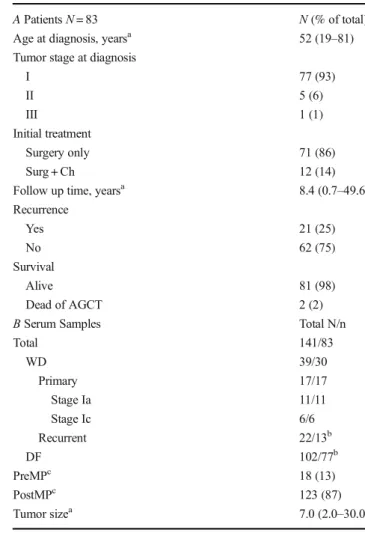
Table 2 Clinicopathologic data of the patients (A) and samples (B) in
the serum analysis
A Patients N = 83 N (% of total)
Age at diagnosis, yearsa 52 (19–81)
Tumor stage at diagnosis
I 77 (93) II 5 (6) III 1 (1) Initial treatment Surgery only 71 (86) Surg + Ch 12 (14)
Follow up time, yearsa 8.4 (0.7–49.6)
Recurrence Yes 21 (25) No 62 (75) Survival Alive 81 (98) Dead of AGCT 2 (2)
B Serum Samples Total N/n
Total 141/83 WD 39/30 Primary 17/17 Stage Ia 11/11 Stage Ic 6/6 Recurrent 22/13b DF 102/77b PreMPc 18 (13) PostMPc 123 (87) Tumor sizea 7.0 (2.0–30.0)
n number of samples, N number of patients, Surg surgery, Ch chemother-apy, WD With Disease, DF Disease-free, MP Menopausal
a
Median (range)
bSerial samples from multiple recurrences
cMP status at sample retrieval;“premenopausal” (PreMP) if the patient
had and one or two ovaries, and menopause was not indicated in the
medical records, and“postmenopausal” (PostMP) if the both ovaries
had been removed, or the patient was postmenopausal according to med-ical history
fluoroimmunometric assays (AutoDELFIA™, Wallac, Turku, Finland). For analysis of continuous data, the values below de-tection limit <0.1 (nmol/L for estradiol, IU/L for FSH and LH) were set to 0.05 (nmol/L or IU/L).
Statistical analysis
TRAIL protein expression levels were correlated to clinical parameters with contingency tabling and Fisher’s exact test. The serum TRAIL data were tested to follow normal distribu-tion with Shapiro-Wilk’s test and serial measurements were analyzed with generalized linear model. Serum FSH, LH, and estradiol levels and tumor size differed from normal distribu-tion even after performing logarithmic transformadistribu-tion and thus Mann–Whitney U test and Spearman’s rho were utilized. For multiple regression, a generalized linear model and max-imum likelihood ratio estimation were utilized for tumor size as a continuous variable (with the most correlations in univariate analysis), menopausal status, estradiol (nmol/L), and FHS and LH (IU/L) levels, with soluble TRAIL (pg/ml) as the dependent variable. p < 0.05 was considered significant. The analyses were performed with JMP version 7.0.1 (SAS Institute Inc., Cary, NC) software.
Results
TRAIL protein expression is reduced in large GCTs
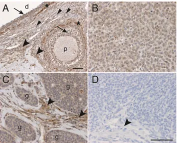
We have previously shown that the TRAIL pathway plays an essential role in granulosa cells [17]. To investigate the role of TRAIL in malignant granulosa cells, we now analyzed the ex-pression of TRAIL in AGCTs at both messenger RNA (mRNA) and protein levels. In an analysis of 46 primary tumor tissues, TRAIL mRNA was expressed at variable amounts, with mean 1.6 ± 1.2 arbitrary units, as compared to 0.08 arbitrary units in normal granulosa-luteal cells (Supplemental Figure1, Supplemental Table1). We found no correlations of TRAIL mRNA levels to tumor size or other clinicopathological char-acteristics (data not shown). In immunohistochemical analysis of normal human ovary, TRAIL protein was expressed in theca and granulosa cells, as well as in the ovarian stroma (Fig.1a). In AGCTs, TRAIL protein was expressed in 48 (59 %) of the tumors at a level comparable to a normal granulosa cell, stain-ing mostly diffusely in the tumor cells (Fig.1b) and more strongly in the tumor stroma (Fig.1c). Thirty (37 %) AGCTs exhibited low-intensity staining, and three (4 %) remained en-tirely negative for TRAIL protein. Upon correlations between clinical and immunohistochemical data, TRAIL protein expres-sion was significantly lower in tumors over 10 cm in diameter when compared to smaller tumors (p = 0.04, OR 0.4 95% CI 0.2–0.97). TRAIL protein expression level was similar in pri-mary (n = 71) and recurrent (n = 12) tumor samples, and the
expression levels did not correlate to tumor subtype, mitotic index, nuclear atypia, tumor stage, or recurrence probability (data not shown). TRAIL expression correlated positively to the expression levels of the TRAIL receptors DR4 (p = 0.006, OR 11.3, 95% CI 1.4–91.9) and DR5 (p = 0.02, OR 8.9, 95% CI 1.1–73.1) previously analyzed in [21]. However, DR4 and DR5 expressions did not correlate to tumor size (data not shown). To further understand the reason for decreased TRAIL protein expression in large AGCTs, we correlated the TRAIL levels to factors linked to granulosa cell proliferation and apoptosis, such as GATA4, Bcl2, and VEGF previously analyzed in [21,30–32]. Interestingly, we found that GATA4 and TRAIL levels positively correlated in the tumor tissue (OR 4.4, 95% CI 1.7–11.6, p = 0.003). However, TRAIL expression did not correlate with the expression of VEGF, VEGF recep-tors, tumor microvessel density, or Bcl2 expression (data not shown).
To investigate whether AGCT cells secrete soluble TRAIL, we next analyzed TRAIL levels from the cell culture superna-tants collected from 15 AGCT primary cell and KGN cell cul-tures. We found that AGCT cells did not secrete measurable amounts of soluble TRAIL into the supernatant (undetectable, data not shown). However, TRAIL mRNA was expressed in three primary AGCT cell preparations (mean = 32.9 ± 25.5 arbi-trary units), at levels comparable to data previously obtained from normal endothelial cells (mean = 35.9 ± 36 arbitrary units), but at lower levels than that in peripheral blood mononuclear cells (mean = 63.4 ± 48.7 arbitrary units) [25]. When studied
Fig. 1 Immunohistochemistry demonstrating TRAIL protein expression in AGCTs. a TRAIL staining in a large dominant follicle (d) and a primary follicle (p) of a normal human ovary. Positive staining is visible in granulosa cells (arrows), thecal cells (asterisks), in endothelial cells (small arrowheads), and in the stroma (big arrowheads). b AGCT of the sarcomatose subtype showing diffuse TRAIL staining. c AGCT with TRAIL staining in the tumor islets (g) and in the tumor stroma (arrowheads). d Secondary antibody only. Tumor cells and tumor stroma (arrowhead) are negative. Original magnification ×20 in a, ×40
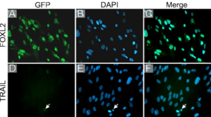
with immunofluorescence staining, the primary tumor cells expressed FOXL2 protein as a marker for AGCT origin (Fig.2a–c) but remained negative for TRAIL expression (Fig.2d–h).
Circulating TRAIL levels are decreased in patients with large AGCTs
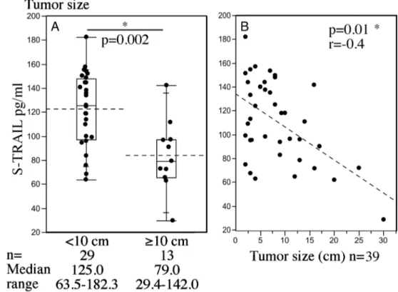
To evaluate the dynamics of circulating TRAIL in AGCT pa-tients, we next analyzed serum TRAIL concentrations from 141 serum samples, before (WD) and after (DF) tumor treatment. We found that the tumor itself had little effect on circulating TRAIL levels, as there was no significant difference in WD compared to DF samples, (data not shown). However, soluble TRAIL levels were significantly lower in patients with larger tumors of over 10 cm in diameter (mean 83.6, SD 29.2, pg/ml), than in patients with tumors less than 10 cm in diameter (mean 122.1, SD 30.2 pg/ml; p = 0.002) (Fig.3a). Consistently, soluble TRAIL levels inversely correlated to the tumor diameter as a continuous variable (r =−0.4, p=0.01) (Fig.3b). Upon correlations to other clinical parameters, we found that TRAIL levels did not correlate to the patient age at sample retrieval, body weight or height, or blood leukocyte counts (data not shown).
Tumor size independently correlates to circulating TRAIL levels
Circulating TRAIL levels have been shown to correlate inverse-ly with patient’s estradiol levels [33], and we next analyzed whether soluble TRAIL levels seen in large AGCTs associate with the patient’s menopausal status, estradiol, and FSH and LH levels. In patients with AGCT (WD), we found that circu-lating TRAIL levels negatively associated with serum estradiol levels (Spearman’s Rho −0.32, p = 0.05), whereas no
correlations were found to menopausal status, FSH, or LH levels (data not shown).
We noted that larger tumors associated with PreMP status, higher estradiol, and lower serum FSH and LH levels (Table3a). Menopausal status, estradiol, FSH and LH levels correlated to each other, as expected (data not shown). In order to verify which factor independently contributed to the decreased TRAIL levels in AGCT patients, we next performed a multiple regression analysis for tumor size, menopausal status, and estra-diol, FSH, and LH levels with serum TRAIL as the dependent variable (Table3b). The analysis indicated that tumor size was the only independent factor influencing circulating TRAIL levels in AGCT patients (p = 0.007).
Discussion
Despite improvements in the treatment modalities over the past decades, ovarian cancer remains the leading cause of death from gynecological cancers. AGCTs generally show an indolent course of disease; however, recurrences occur in 30 % of the patients and the majority of these patients will die of their disease [1,34,35]. Given the fact that current chemo-therapeutics have only limited effect on AGCTs [5,36], new, targeted treatments are critically needed for patients with ad-vanced AGCT.
TRAIL is a pro-apoptotic factor with promising anti-cancer activity in a multitude of solid tumors [6–11]. Previous studies have documented the expression of TRAIL in granulosa cells [17] and TRAIL receptors in normal and malignant granulosa cells [21], suggesting a role for the TRAIL/TRAIL receptor sys-tem in regulating normal and malignant granulosa cell apoptosis. Recombinant human TRAIL (rhTRAIL) induces apoptosis in AGCT cells in vitro [17,21]. Moreover, rhTRAIL was shown to induce the activation of Caspase 3/7 and subsequent cell death with lower concentrations in the primary AGCT tumor cells when compared to the KGN cell line [21]. Preliminary clinical data also suggests that AGCTs are susceptible to agonistic TRAIL therapy also in human AGCT patients [11].
We herein find that AGCTs generally express TRAIL pro-tein at levels comparable to normal granulosa cells. However, tissue expression levels were decreased in large AGCTs. TRAIL protein expression levels positively correlated with those of transcription factor GATA4, which regulates apopto-sis in AGCTs [21], and has been shown to be an important delineator of AGCT prognosis [1,30]. Moreover, GATA4 has been shown to protect AGCT cells from TRAIL-induced apo-ptosis in vitro [21]. Therefore, high expression of GATA4 in AGCTs can be speculated to lead to resistance of TRAIL-mediated apoptosis, and contributing to increased tumor growth. However, we could not detect TRAIL protein in cultured primary AGCT cells. Moreover, in immunohistochemistry, TRAIL pro-tein expression was also detected in the tumor stroma, and
Fig. 2 Immunofluorescent staining of primary cultured AGCT cells. First column; antigen staining visualized with GFP in green, second
column; DAPI showing the nuclei in blue, third column; merge. a–c
FOXL2 protein as a marker for AGCT cells localizes primarily in the
nuclei of tumor cells. d–f AGCT cells are predominantly negative for
TRAIL protein expression. A small amount of TRAIL staining is seen around an apoptotic AGCT cell (arrow)
TRAIL mRNA was present at high levels in the primary AGCT tumor samples, consisting also of tumor stroma. These findings suggest that TRAIL protein is primarily produced by the tumor
stroma and/or infiltrating blood/immune cells. Thus, it can be speculated that the decreased TRAIL production in the stroma of large tumors contribute to AGCT progression via decreasing
Fig. 3 Serum TRAIL levels are decreased in patients with large AGCTs and correlate negatively with tumor size. a S-TRAIL levels in tumors less than 10 cm and in tumors equal to or more than 10 cm in diameter. b S-TRAIL levels in correlation with tumor size as the largest diameter.
r Spearman’s rho. An asterisk
indicates a significant difference with p < 0.05
Table 3 Tumor size
independently correlates to serum TRAIL levels. (A) Univariate analyses to tumor size. (B) Multiple regression with serum TRAIL as the dependent variable
Tumor size (cm) (continuous)
p value
Tumor size <10 cm
(categorical)
≥10 cm p value
A Correlatons to tumor size as continuous and categorical value
PreMP mean ± SD; 14.7 ± 11.0 n = 3 n = 3 PostMP mean ± SD; 7.5 ± 5.0 0.01*a n = 25 n = 8 NSb Estradiol nmol/L r = 0.41 0.01* median 0.07 range 0.01–0.33 median 0.21 range 0.04–0.93 NSc FSH IU/L r =−0.64 <0.0001* median 15.3 range 0.05–101 median 1.2 range 0.05–50.1 0.006*c LH IU/L r =−0.74 <0.0001* median 23.5 range 3–66.9 median 5.9 range 0.05–23.3 0.003*c
B Multiple regression of serum TRAIL as the dependent variable
Estimate 95%CI p value
Intercept 138.4 101.5–175.3 <0.0001* Tumor size −3.0 −5.4– −0.7 0.001* PreMP −15.7 −35.5–4.2 0.1 Estradiol −23.0 −80.5–34.6 0.4 FSH 0.4 −0.2–1.0 1.0 LH −0.9 −1.9–0.2 0.1
PreMP premenopausal, Postmp postmenopausal, SD standard deviation, CI confidence interval, IU international units, L liter, NS not significant
r = Spearman’s rho, *indicates p < 0.05
a
One-way ANOVA
b
Fisher’s exact test
c
apoptosis in the tumor cells expressing the TRAIL receptors DR4 and DR5 [21] and, potentially, also via impairing tumor defense mechanisms [37–39].
Circulating TRAIL levels were similar in patients with or without AGCT; however, the levels were significantly de-creased in AGCT patients with a large tumor mass, consistent with our findings in the tumor tissue. AGCTs are known to produce and release estrogens [24], and recent in vitro data showed that estradiol downregulates the production of TRAIL from leukocytes [25]. We found evidence that serum estradiol levels correlated positively to tumor size, and also negatively to circulating TRAIL levels in patients with AGCT. However, in multivariable analysis, tumor size independently associated with TRAIL levels, and therefore, it can be speculated that other factors, such as the tumor stroma, also contribute to the levels of circulating TRAIL. When designing optimal treatment strate-gies for advanced AGCTs, the targeted treatment preferably antagonizes with inherent pathogenetic factors, such as GATA4, and further studies are warranted to evaluate whether TRAIL agonists have such potential.
Utilizing a unique series of tumor tissue, cell culture, and serum samples, we showed that large AGCTs are associated with significantly decreased levels of tissue and serum TRAIL. Our results support the role of TRAIL in AGCT tu-mor progression, and encourage future studies on agonistic TRAIL treatments for advanced or recurrent AGCT.
AGCT adult-type granulosa cell tumor, DF disease-free, FSH follicle-stimulating hormone, LH luteinizing hormone, PostMP postmenopausal, PreMP premenopausal, TMA tumor tissue microarray, TRAIL TNF-related apoptosis-inducing li-gand, rhTRAIL recombinant human TRAIL, WD with disease
Acknowledgments We thank Dr. Annika Riska and the staff from the
Department of Obstetrics and Gynecology of Helsinki University Central Hospital for patient recruitment, Dr. Noora Andersson for excellent practi-cal assistance, and PhD Emmi Tikkanen for statistipracti-cal advice. This study was supported by grants from from the Academy of Finland, the Clinical Chemistry Research Foundation Finland, the Helsinki University Central Hospital Research Funds, the Maud Kuistila Memorial Foundation, the Sigrid Juselius Foundation, and the Sladjana M. Crosley Foundation for AGCT research.
Author contributions AF collected the samples, produced and analyzed
the data, carried out the statistical analyses and wrote the manuscript; GZ produced and analyzed the data and drafted the manuscript; U-MH and MP performed immunohistochemistry, cell culture studies, qPCR, and analyzed the data; PS, LU-K and MH collected the samples, conceived of the study and interpreted the data; All authors wrote the manuscript, and read and approved this version of the manuscript.
Compliance with ethical standards The ethical committee of Helsinki
University Central Hospital (HUCH) and the National Supervisory Authority for Welfare and Health in Finland approved the study, and all patients gave their informed consent.
Conflicts of Interest None
References
1. Farkkila A, Andersson N, Butzow R, Leminen A, Heikinheimo M,
Anttonen M, et al. HER2 and GATA4 are new prognostic factors for early-stage ovarian granulosa cell tumor-a long-term follow-up
study. Cancer Med. 2014;3:526–36.
2. Mangili G, Ottolina J, Gadducci A, Giorda G, Breda E, Savarese A,
et al. Long-term follow-up is crucial after treatment for granulosa
cell tumours of the ovary. Br J Cancer. 2013;9:29–34.
3. Park JY, Jin KL, Kim DY, Kim JH, Kim YM, Kim KR, et al.
Surgical staging and adjuvant chemotherapy in the management of patients with adult granulosa cell tumors of the ovary. Gynecol
Oncol. 2012;125:80–6.
4. Fotopoulou C, Savvatis K, Braicu EI, Brink-Spalink V,
Darb-Esfahani S, Lichtenegger W, et al. Adult granulosa cell tumors of the ovary: tumor dissemination pattern at primary and recurrent situation, surgical outcome. Gynecol Oncol. 2010;119:285–90.
5. van Meurs HS, Buist MR, Westermann AM, Sonke GS, Kenter
GG, van der Velden J. Effectiveness of chemotherapy in measur-able granulosa cell tumors: a retrospective study and review of
literature. Int J Gynecol Cancer. 2014;24:496–505.
6. Soria JC, Mark Z, Zatloukal P, Szima B, Albert I, Juhasz E, et al.
Randomized phase II study of dulanermin in combination with paclitaxel, carboplatin, and bevacizumab in advanced
non-small-cell lung cancer. J Clin Oncol. 2011;29:4442–51.
7. Pan Y, Xu R, Peach M, Huang CP, Branstetter D, Novotny W, et al.
Evaluation of pharmacodynamic biomarkers in a Phase 1a trial of dulanermin (rhApo2L/TRAIL) in patients with advanced tumours.
Br J Cancer. 2011;105:1830–8.
8. Wainberg ZA, Messersmith WA, Peddi PF, Kapp AV, Ashkenazi A,
Royer-Joo S, et al. A phase 1B study of dulanermin in combination with modified FOLFOX6 plus bevacizumab in patients with meta-static colorectal cancer. Clin Colorectal Cancer. 2013;12:248–54.
9. Lim B, Scicchitano A, Beachler C, Gusani N, Sarwani N, Yang Z, et al.
FOLFIRI plus dulanermin (rhApo2L/TRAIL) in a patient with
BRAF-mutant metastatic colon cancer. Cancer Biol Ther. 2013;14:711–9.
10. Niemoeller OM, Belka C. Radiotherapy and TRAIL for cancer
therapy. Cancer Lett. 2013;332:184–93.
11. Camidge DR, Herbst RS, Gordon MS, Eckhardt SG, Kurzrock R,
Durbin B, et al. A phase I safety and pharmacokinetic study of the death receptor 5 agonistic antibody PRO95780 in patients with
advanced malignancies. Clin Cancer Res. 2010;16:1256–63.
12. Di Pietro R, Zauli G. Emerging non-apoptotic functions of tumor
necrosis factor-related apoptosis-inducing ligand (TRAIL)/Apo2L. J Cell Physiol. 2004;201:331–40.
13. Secchiero P, Zauli G. Tumor-necrosis-factor-related
apoptosis-inducing ligand and the regulation of hematopoiesis. Curr Opin Hematol. 2008;15:42–8.
14. Shirley S, Morizot A, Micheau O. Regulating TRAIL
receptor-induced cell death at the membrane: a deadly discussion. Recent
Pat Anticancer Drug Discov. 2011;6:311–23.
15. Finnberg N, Klein-Szanto AJ, El-Deiry WS. TRAIL-R deficiency
in mice promotes susceptibility to chronic inflammation and
tumor-igenesis. J Clin Invest. 2008;118:111–23.
16. Grosse-Wilde A, Voloshanenko O, Bailey SL, Longton GM,
Schaefer U, Csernok AI, et al. TRAIL-R deficiency in mice en-hances lymph node metastasis without affecting primary tumor
development. J Clin Invest. 2008;118:100–10.
17. Jaaskelainen M, Kyronlahti A, Anttonen M, Nishi Y, Yanase T,
Secchiero P, et al. TRAIL pathway components and their putative role in granulosa cell apoptosis in the human ovary. Differentiation. 2009;77:369–76.
18. Johnson AL, Ratajczak C, Haugen MJ, Liu HK, Woods DC. Tumor
necrosis factor-related apoptosis inducing ligand expression and
19. Woods DC, Alvarez C, Johnson AL. Cisplatin-mediated sensitivity to TRAIL-induced cell death in human granulosa tumor cells.
Gynecol Oncol. 2008;108:632–40.
20. Woods DC, Liu HK, Nishi Y, Yanase T, Johnson AL. Inhibition of
proteasome activity sensitizes human granulosa tumor cells to
TRAIL-induced cell death. Cancer Lett. 2008;260:20–7.
21. Kyronlahti A, Kauppinen M, Lind E, Unkila-Kallio L, Butzow R,
Klefstrom J, et al. GATA4 protects granulosa cell tumors from
TRAIL-induced apoptosis. Endocr Relat Cancer. 2010;17:709–17.
22. Jamieson S, Butzow R, Andersson N, Alexiadis M, Unkila-Kallio
L, Heikinheimo M, et al. The FOXL2 C134W mutation is charac-teristic of adult granulosa cell tumors of the ovary. Mod Pathol.
2010;23:1477–85.
23. Shah SP, Kobel M, Senz J, Morin RD, Clarke BA, Wiegand KC,
et al. Mutation of FOXL2 in granulosa-cell tumors of the ovary. N Engl J Med. 2009;360:2719–29.
24. Jamieson S, Fuller PJ. Molecular pathogenesis of granulosa cell
tumors of the ovary. Endocr Rev. 2012;33:109–44.
25. Zauli G, Tisato V, Melloni E, Volpato S, Cervellati C, Bonaccorsi G,
et al. Inverse correlation between circulating levels of TNF-related apoptosis inducing ligand and 17-beta estradiol. J Clin Endocrinol Metab. 2014;99:E659–64.
26. Gibellini D, Zauli G, Re MC, Milani D, Furlini G, Caramelli E,
et al. Recombinant human immunodeficiency virus type-1 (HIV-1) Tat protein sequentially up-regulates IL-6 and TGF-beta 1 mRNA expression and protein synthesis in peripheral blood monocytes. Br J Haematol. 1994;88:261–7.
27. Livak KJ, Schmittgen TD. Analysis of relative gene expression data
using real-time quantitative PCR and the 2(−Delta Delta C(T)) Method. Methods. 2001;25:402–8.
28. Nishi Y, Yanase T, Mu Y, Oba K, Ichino I, Saito M, et al. Establishment
and characterization of a steroidogenic human granulosa-like tumor cell line, KGN, that expresses functional follicle-stimulating hormone re-ceptor. Endocrinology. 2001;142:437–45.
29. Secchiero P, Gonelli A, Celeghini C, Mirandola P, Guidotti L, Visani
G, et al. Activation of the nitric oxide synthase pathway represents a key component of tumor necrosis factor-related apoptosis-inducing
ligand-mediated cytotoxicity on hematologic malignancies. Blood.
2001;98:2220–8.
30. Anttonen M, Unkila-Kallio L, Leminen A, Butzow R, Heikinheimo
M. High GATA-4 expression associates with aggressive behavior, whereas low anti-Mullerian hormone expression associates with growth potential of ovarian granulosa cell tumors. J Clin
Endocrinol Metab. 2005;90:6529–35.
31. Farkkila A, Anttonen M, Pociuviene J, Leminen A, Butzow R,
Heikinheimo M, et al. Vascular endothelial growth factor (VEGF) and its receptor VEGFR-2 are highly expressed in ovarian granu-losa cell tumors. Eur J Endocrinol. 2011;164:115–22.
32. Farkkila A, Pihlajoki M, Tauriala H, Butzow R, Leminen A,
Unkila-Kallio L, et al. Serum vascular endothelial growth factor A (VEGF) is elevated in patients with ovarian granulosa cell tumor (GCT), and VEGF inhibition by bevacizumab induces apoptosis in GCT in vitro. J Clin Endocrinol Metab. 2011;96:E1973–81.
33. Zauli G. Circulating levels of TNF-related apoptosis inducing
li-gand (TRAIL). Clin Endocrinol (Oxf). 2014;80:182–3.
34. Cronje HS, Niemand I, Bam RH, Woodruff JD. Review of the
granulosa-theca cell tumors from the Emil Novak ovarian tumor registry. Am J Obstet Gynecol. 1999;180:323–7.
35. Evans 3rd AT, Gaffey TA, Malkasian Jr GD, Annegers JF.
Clinicopathologic review of 118 granulosa and 82 theca cell tu-mors. Obstet Gynecol. 1980;55:231–8.
36. van Meurs HS, van Lonkhuijzen LR, Limpens J, van der Velden J,
Buist MR. Hormone therapy in ovarian granulosa cell tumors: a systematic review. Gynecol Oncol. 2014;134:196–205.
37. Kim R, Emi M, Tanabe K. Cancer immunoediting from immune
surveillance to immune escape. Immunology. 2007;121:1–14.
38. Takeda K, Hayakawa Y, Smyth MJ, Kayagaki N, Yamaguchi N,
Kakuta S, et al. Involvement of tumor necrosis factor-related apo-ptosis-inducing ligand in surveillance of tumor metastasis by liver natural killer cells. Nat Med. 2001;7:94–100.
39. Koyama S, Koike N, Adachi S. Expression of TNF-related
apoptosis-inducing ligand (TRAIL) and its receptors in gastric carcinoma and tumor-infiltrating lymphocytes: a possible mechanism of immune