J. Cell. Mol. Med. Vol 10, No 2, 2006 pp. 389-406
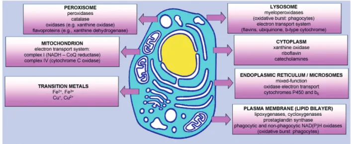
Reactive oxygen species (ROS) are constantly gen-erated within cells at low concentrations under physiological conditions, playing a part in the cel-lular redox regulation. Celcel-lular production of ROS occurs from both enzymatic and non-enzymatic sources (Fig. 1) [for reviews, see 1, 2]. ROS can also occur as the outcome of acute cell stresses and
may result in cell death via apoptosis or necrosis. Cellular oxidative damage develops when the bal-ance between generating systems and ROS-scavenging ones tilts in favour of the former.
The primary cellular target of oxidative stress can vary depending on the cell type, the absolute level and duration of oxidant production, the species
Protein carbonylation,
cellular dysfunction, and disease progression
Isabella Dalle - Donne
a*, Giancarlo Aldini
b, Marina Carini
b, Roberto Colombo
a,
Ranieri Rossi
c, Aldo Milzani
aa Department of Biology, University of Milan, Milan, Italy
b Institute of Pharmaceutical and Toxicological Chemistry, University of Milan, Milan, Italy c Department of Neuroscience, University of Siena, Siena, Italy.
Received: February 16, 2006; Accepted: March 27, 2006
Abstract
Carbonylation of proteins is an irreversible oxidative damage, often leading to a loss of protein function, which is considered a widespread indicator of severe oxidative damage and disease-derived protein dysfunction. Whereas moderately carbonylated proteins are degraded by the proteasomal system, heavily carbonylated proteins tend to form high-molecular-weight aggregates that are resistant to degradation and accumulate as damaged or unfolded pro-teins. Such aggregates of carbonylated proteins can inhibit proteasome activity. A large number of neurodegenerative diseases are directly associated with the accumulation of proteolysis-resistant aggregates of carbonylated proteins in tissues. Identification of specific carbonylated protein(s) functionally impaired and development of selective car-bonyl blockers should lead to the definitive assessment of the causative, correlative or consequential role of protein carbonylation in disease onset and/or progression, possibly providing new therapeutic approaches.
Keywords: protein carbonyls •reactive oxygen species •reactive carbonyl species •
protein unfolding/misfolding •proteasome •aggregation diseases
* Correspondence to: Isabella DALLE-DONNE, Ph.D. Department of Biology, University of Milan, via Celoria 26, I-20133 Milan, Italy.
Tel.: +39 02 50314792 Fax: +39 02 50314781 E-mail: [email protected]
• Introduction
• The impact of carbonylation on protein function • The impact of carbonylation on protein
folding
• The impact of carbonylation on proteolysis • Is protein carbonylation an early event of
cel-lular dysfunction and disease progression? • Conclusion and perspectives
Introduction
www.jcmm.ro www.jcmm.org
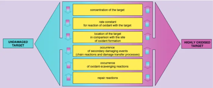
of ROS generated, its site of generation (intra- vs. extra-cellular), and the proximity of the oxidant to a specific cellular substrate. The extent of damage to particular targets depends on a number of factors (Fig. 2). Proteins are major targets for ROS and sec-ondary by-products of oxidative stress when these are formed in vivo either in intra- or extracellular environments, as they are the major component of most biological systems and can scavenge 50–75% of reactive radicals such as .OH [3].
Some ROS-induced protein modifications can result in unfolding or alteration of protein structure, and some are essentially harmless events [4]. For example, protein reversible modifications, such as S-glutathionylation, S-nitrosation, and methionine sulfoxidation, may have a dual role of protection from irreversible oxidation and modulation of pro-tein function (redox regulation) [5–7]. Differently,
irreversible protein modifications can lead to inac-tivation of various proteins and could have lasting detrimental cellular effects.
Although the overall biology of oxidative pro-tein modifications remains complex and ill defined, protein carbonylation is quite well characterised. Carbonylation is an irreversible, non-enzymatic modification of proteins. The chemistry of the reac-tions that give rise to carbonyl groups is discussed in detail in other excellent reviews [8–11].
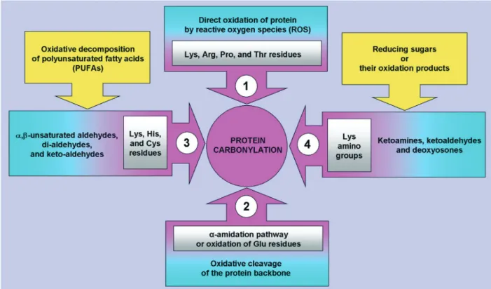
Briefly, carbonyl groups are introduced into pro-teins by a variety of oxidative pathways (Fig. 3). ROS can react directly with the protein or they can react with molecules such as sugars and lipids, generating products (reactive carbonyl species, RCS) that then react with the protein. Direct oxidation of proteins by ROS yields highly reactive carbonyl derivatives resulting either from oxidation of the side chains of
Fig. 1 Cellular sources of reactive oxygen species. Any electron-transferring protein or enzymatic system can
result in the formation of ROS as "by-products" of electron transfer reactions. The mitochondrial electron transport chain is a significant source of ROS. Plasma membrane is a major source of ROS through NAD(P)H oxidases locat-ed on either side. Enzymes of the same class displaying low activity, as well as their components, are also present free in cytoplasm. Smooth endoplasmic reticulum (ER) contains enzymes, including cytochrome P-450 and b5 fam-ilies, which catalyse a series of reactions to detoxify lipid-soluble drugs and other harmful metabolic products. Peroxisomes are an important source of total cellular H2O2production. They contain a number of H2O2-generating enzymes of the oxidase family. Peroxisomal catalase utilises H2O2produced by these oxidases to oxidise a variety of other substrates in peroxidative reactions, particularly in liver and kidney cells in which peroxisomes detoxify a variety of toxic molecules that enter the circulation. Another major function of the oxidative reactions carried out in peroxisomes is β-oxidation of fatty acids. In addition to intracellular membrane-associated oxidases, cytoplasmic soluble enzymes such as xanthine oxidase, aldehyde oxidase, flavoprotein dehydrogenase, and tryptophan dioxyge-nase can generate ROS during catalytic cycling. Autooxidation of small molecules such as dopamine, epinephrine, flavins, and hydroquinones can be an important source of intracellular ROS production. In most cases, the direct product of such autooxidation reactions is O2–..
lysine, arginine, proline, and threonine residues - par-ticularly via metal-catalysed oxidation [8] - or from the cleavage of peptide bonds by the α-amidation pathway or by oxidation of glutamyl residues. Glutamic semialdehyde, a product of oxidation of arginine and proline, and aminoadipic semialdehyde, a product of oxidation of lysine, are the main car-bonyl products of metal-catalysed oxidation of pro-teins, this reaction being a major route leading to the generation of protein carbonyls in biological samples [12]. Carbonyl groups can also be generated by sec-ondary reaction of the primary amino group of lysine residues with reactive carbonyl derivatives (ketoamines, ketoaldehydes, deoxyosones), pro-duced by the reaction of reducing sugars or their oxi-dation products with lysine residues of proteins (gly-cation/glycoxidation reactions), eventually leading to the formation of advanced glycation end-products (AGEs). Finally, carbonyl groups may be introduced into proteins by adduction of carbonyl-containing oxidized lipids derived from the metal-catalysed oxi-dation of polyunsaturated fatty acids [13–15]. These include malondialdehyde (MDA), which reacts with lysine residues, and α,β-unsaturated aldehydes [4-hydroxy-2-nonenal (HNE), acrolein], which can undergo Michael-addition reactions at their C=C double bond with the sulfhydryl group of cysteine, the ε-amino group of lysine or the imidazole group of histidine residues, forming advanced lipoxidation end-products (ALEs).
Protein carbonylation is the most widely used biomarker for oxidative damage to proteins, and reflects cellular damage induced by multiple forms of ROS [7, 10, 16–19].
The impact of carbonylation on
protein function
Increases in carbonylated proteins during ageing and in response to oxidative stress are not random, some proteins being more susceptible than others. However, the set of proteins that become carbony-lated differs in different species. For example, mouse plasma ageing-associated protein carbony-lation was only seen in two proteins, albumin and transferrin [20]. But, in the rat plasma, only albu-min and α1-macroglobulin showed significant age-dependent accrual of carbonylation [20]. Human brain copper-zinc superoxide dismutases (SOD1) is one of the major targets of oxidative damage in brains of subjects afflicted with Alzheimer's dis-ease (AD) and Parkinson's disdis-ease (PD); however, only one out of four human brain SOD1 isoforms is heavily carbonylated [21]. The selectivity of pro-tein carbonylation is clearly demonstrated by the fact that the relative amount of a protein is not a factor in determining the degree of carbonylation [20, 22]. Similar specificity of protein
carbonyla-Fig. 2 Major factors determining the extent of oxidative damage to specific cellular targets. The first two of these
tion was previously noted in the mitochondria of the flight muscles of the flies, where only aconitase [23–25] and adenine nucleotide translocase [24] were found to exhibit an age-associated increase in carbonylation and a corresponding loss in func-tional activity. Cytochrome c, a relatively abundant mitochondrial protein, did not show detectable car-bonylation at any age [26].
An obvious question arising from such studies is why this selectivity? There is not an easy way to
answer this question. It could be merely a conse-quence of protein structure.
Pioneering studies by Stadtman [8, 27] have suggested that the presence of a transition metal-binding site in a protein is a key feature to predict its susceptibility to undergo carbonylation via metal-catalysed oxidation. Protein-bound transition metals are sources of free radicals that initiate a cas-cade of reactions, which result in the addition of carbonyl groups to the side chains of certain
sur-Fig. 3 Origins of carbonylated proteins. Protein carbonyl derivatives can be produced by a variety of oxidative
pathways. Direct oxidation of the Lys, Arg, Pro, and Thr side chains particularly via metalcatalysed oxidation -results in α-aminoadipic semialdehyde from lysyl residue, glutamic semialdehyde from arginyl and prolyl residue, 2-pyrrolidone from prolyl residue, and 2-amino-3-ketobutyric acid from threonyl residue (pathway 1). Direct oxi-dation of proteins by ROS can also yield highly reactive carbonyl derivatives resulting from the cleavage of peptide bonds by the α-amidation pathway or by oxidation of glutamyl residues (pathway 2). Carbonyl groups may be intro-duced into proteins by adduction of reactive aldehydes derived from the metal-catalysed oxidation of polyunsatu-rated fatty acids (pathway 3). These lipoxidation products include di-aldehydes such as malondialdehyde (MDA), which reacts with Lys residues, α,β-unsaturated aldehydes [4-hydroxy-2-nonenal (HNE), acrolein], which can undergo Michael-addition reactions between their electrophilic C=C double bond and the sulfhydryl group of Cys, the ε-amino group of Lys or the imidazole group of His residues, and γ-ketoaldehydes (levuglandins, isoketals, neu-roketals), which react with Lys residues. The chemical modification of protein by reactive carbonyl compounds derived from lipid peroxidation reactions results in the formation of advanced lipoxidation end-products (ALEs) (pathway 3). Finally, carbonyl groups can also be generated by secondary reaction of the primary amino group of Lys residues with reactive carbonyl derivatives (ketoamines, ketoaldehydes, deoxyosones), produced by the tion of reducing sugars or their oxidation products with lysine residues of proteins (glycation/glycoxidation reac-tions), eventually leading to the formation of advanced glycation end-products (AGEs) (pathway 4).
rounding amino acid residues [9]. Other factors, such as molecular conformation, rate of turnover and the relative abundance of amino acid residues susceptible to metal-catalysed oxidation, have also been suggested to be involved in the selectivity of protein carbonylation [28, 29]. In addition, some proteins (e.g., enzymes of Krebs cycle and electron transport chain) may be carbonylated mainly because they are located near sites generating ROS. Concerning selective oxidation of proteins, results obtained by Ros and colleagues [22] indi-cate that prokaryotic and eukaryotic cells display some homologies. These homologies can be a con-sequence of their structural and/or functional rela-tionship. In this context, sequence homologies of pyruvate dehydrogenase and α-ketoglutarate dehy-drogenase from Saccharomyces cerevisiae with respect to those from Escherichia coli were 40 and 59%, respectively. The homologies are even greater when their active sites or lipoic acid bind-ing signatures are compared. This analysis would tend to suggest the importance of the protein struc-ture on the selectivity of oxidative targets. Nevertheless, in the case of heat-shock protein (Hsp) 60 and the Hsp70 chaperone DnaK, they share no significant similarity, indicating that, in this case, chaperoning function could be a reason for their selective damage. The question of whether such proteins have been selected as targets during evolution to better preserve the integrity of the cell after a stress situation remains to be determined. Further studies by the same research group on the two types of yeast ageing models, replicative and chronological, showed that, although in both age-ing models metabolic differences are important, major targets are almost the same [30]. Common targets include stress resistance proteins (Hsp60 and Hsp70) and enzymes involved in glucose metabolism. Interestingly, carbonylated proteins accumulating with replicative age are not inherited by daughter cells during cytokinesis [31].
Nyström and co-workers provided an attractive hypothesis, which supports an increased suscepti-bility of proteins to oxidation [32, 33]. Their elegant studies in E. coli established that transcriptional or translational errors produce aberrant proteins that are more susceptible to carbonylation (see also sec-tion "The impact of carbonylasec-tion on protein fold-ing"). It is not completely clear why aberrant pro-teins are more susceptible to carbonylation, but it is
possible that a slight misfolding of the partly aber-rant polypeptide exposes oxidation-sensitive amino acid residues that are normally hidden during the coupled translation-folding process. Introduction of carbonyl groups on those amino acids may result in further loss of the proteins' integrity. This, in turn, results in an increased exposure of hydrophobic surfaces increasing the target sites for the DnaJ/K/GrpE chaperone system [34].
The introduction of carbonyl derivatives (alde-hydes and ketones) may alter the conformation of the polypeptide chain, thus determining the partial or total inactivation of proteins. A key question when addressing the significance of protein car-bonyls is whether they are simply markers for the presence of oxidative stress, or have some substan-tive consequence on protein function that impacts on cell injury. Protein carbonylation is selective in inactivating particular proteins preferentially and it is likely to be deleterious, since cells are unable to repair protein carbonyls. In the flight muscle mito-chondria of flies, aconitase and adenine nucleotide translocase were found to lose activity in associa-tion with the increase in carbonylaassocia-tion [23–25]. Endoplasmic reticulum (ER) proteins are readily carbonylated in response to peroxide treatment of HL-60 cells [35] and are preferentially carbonylat-ed in the agcarbonylat-ed mouse liver [36]. Carbonylation of specific ER chaperone proteins may induce dys-function of the protein folding processes [35, 36]. Hence, cells that have large amounts of protein car-bonyls may be expected to have proteasomes and chaperones unable to keep up with the rate of duction of unfolded or oxidatively damaged pro-teins and, therefore, an impaired cellular protein turnover, likely resulting in cellular impaired func-tion (see below).
The introduction of carbonyl groups into pro-teins can be triggered by different ROS or sec-ondary by-products of oxidative stress, and can arise at different sites and by different mechanisms (Fig. 3) [10, 11]. Hence, carbonylation can result in several different protein modifications, every one of which may produce (or not) a specific effect on the biological activity of different pro-teins. For example, HOCl-induced in vitro car-bonylation of monomeric actin causes severe inhi-bition of actin filament formation [37], whereas actin carbonylation resulting from the adduction of HNE through Michael addition to Cys374 [38]
negligibly affects actin polymerisation [Dalle-Donne et al., in preparation].
Due to their abundance in mammalian cells, cytoskeletal proteins are common targets for a vari-ety of ROS and low-molecular-weight RCS. For instance, HNE forms Michael adducts with tubulin and disrupts microtubule assembly in neuroblas-toma cells, blocking neurite outgrowth [39]; it also targets neurofilament heavy chains [40]. Actin iso-forms are carbonylated both in vitro [37, 38] and in vivo, e.g., in the skeletal muscles of a diabetes model Otsuka Long-Evans Tokushima Fatty (OLETF) rat [41], in macrophages exposed to hyperoxia [42], in the septic diaphragm [43], and in synaptosomes oxidized by treatment with the 42-amino acid peptide, amyloid β-peptide (1-42) [Aβ(1-42)] [44]. Actin carbonylation has been determined in human intestinal cells exposed to H2O2or HOCl and in colonic mucosa from Crohn's disease patients, where it is associated with the dis-ruption of the actin cytoskeleton and the loss of the monolayer barrier function [45, 46], as well as dur-ing reperfusion of the ischaemic rat heart [47] and in AD subjects [48].
Actin and a number of glycolytic enzymes ( α-enolase, triose phosphate isomerase, phospho-glycerate mutase, and fructose bis-phosphate aldolase) were among the carbonylated proteins detected during etoposide (VP16)-induced apop-tosis of HL60 cells. Consistently, glucose utilisa-tion was reduced dramatically, likely due to car-bonylation-mediated reduction in activity of gly-colytic enzymes [49].
Protein carbonylation has been shown to exert a negative effect on creatine kinase and aldolase activity in the septic rat diaphragm [43]. Varying degrees of activity loss were detected in a number of peroxisomal enzymes following metal-catalysed-induced carbonylation [50]. The formation of pro-tein carbonyls by HNE adducts has been shown to have significant effects on protein function and is frequently associated with their cross-linking. A number of mitochondrial enzymes have been shown to be inactivated following HNE binding, including Na+-K+-ATPase [51], adenine nucleotide translocator [52], and cytochrome c oxidase (com-plex IV of the mitochondrial respiratory chain) in the ischaemic/reperfused rat heart [53], as well as the glial glutamate transporter, GLT-1 (EAAT2), in the brain of AD subjects [54].
Carbonylation and formation of HNE-adducts have been observed, under normal basal conditions, in specific protein subunits of the respiratory chain complexes I–V of adult bovine heart submitochon-drial particles, as well as in two proteins that are part of a complex that forms the mitochondrial per-meability transition pore, heart-specific T1 isotype of adenine nucleotide translocator and voltage-dependent anion channel 1 [55]. However, authors did not investigate the impact of these modifica-tions on mitochondrial complex function. Substantial carbonylation of specific subunits of mitochondrial respiratory complexes I, III, and V has also been shown in chagasic murine hearts infected by Trypanosoma cruzi, which are charac-terised by deficiencies in the activities of the respi-ratory chain complexes and reduced mitochondrial ATP generation capacity during the course of infec-tion and disease development [56]. The extent of protein carbonylation of specific subunits directly correlated with the loss in catalytic activities of the respiratory complexes in the infected myocardium [56]. In addition, the oxidative damage of complex I and III may potentiate oxidative stress in the mito-chondria, as a decline in the activities of these com-plexes is likely to result in the increased release of electrons to molecular oxygen and ROS formation. MDA-induced carbonylation of aconitase, very long chain acyl coenzyme A dehydrogenase, the β-polypeptide of the mitochondrial F1 complex of ATP synthase (or complex V), and the E2 compo-nent of α-ketoglutarate dehydrogenase complex was identified in both heart and skeletal muscle mito-chondria from mice of three different ages [57]. While the amount of MDA-modified proteins did not appear to change during ageing, only aconitase and ATP synthase from heart exhibited an age-relat-ed decrease in activity, whereas very long chain acyl coenzyme A dehydrogenase and α-ketoglutarate dehydrogenase activities remained unchanged dur-ing agedur-ing in both heart and skeletal muscle [57].
Inhibition of mitochondrial complex I reduces profoundly the activity of the proteasome system degrading oxidized proteins by oxidative modifica-tion of the 20S β subunit with acrolein, to which other acrolein-modified proteins were found to bind, resulting in dopamine neuron degeneration [58]. Current evidence resulting from studies in human post-mortem material suggests that mito-chondrial complex I inhibition may be the central
cause of sporadic PD and that derangements in complex I cause α-synuclein aggregation, which binds directly to the proteasome and inhibits ubiq-uitin-dependent proteasomal function. Inhibition of the proteasome would lead to failure to clear pro-tein targeted for degradation by the ubiquitin-pro-teasome system (UPS), ultimately resulting in the demise of dopamine neurons [59].
The impact of carbonylation on
protein folding
Many of the proteins that are synthesised in a cell are destined for secretion to the extracellular envi-ronment. These proteins are translocated into the ER, where folding and post-translational modifica-tions take place before secretion through the Golgi apparatus. Thus, any ROS effect on the structure and function of ER chaperone proteins could affect protein processing efficiency ("quality control") and could result in a decline in cell/tissue function. The ER contains a wide range of molecular chaper-ones and folding catalysts, and the proteins that fold here must satisfy a "quality-control" check before being exported. Such a process is particularly important because there seem to be few molecular chaperones outside the cell, although at least one (clusterin) has been discovered [60]. This quality-control mechanism, involving a remarkable series of glycosylation/deglycosylation reactions, enables correctly folded proteins to be distinguished from misfolded ones: incorrectly folded proteins are detected by the quality-control mechanism and sent along another pathway (the "unfolded protein response") in which they are ubiquitinated and then degraded in the cytoplasm by proteasomes [61]. Failure to fold correctly, or to remain correctly fold-ed, will give rise to the malfunctioning of living systems and hence to disease [62, 63]. Some of these diseases, such as cystic fibrosis and some types of cancer, result from proteins folding incor-rectly and not being able to exercise their proper function, ultimately being degraded by the protea-some. In other cases, proteins with a high propensi-ty to misfold escape all the protective mechanisms and form insoluble aggregates within cells or, more commonly, such as in the amyloidoses, in extracel-lular space. An increasing number of disorders,
including AD, PD, the spongiform encephalopathies, and types II diabetes, are directly associated with the deposition of protein aggregates in tissues [63]. Just as the aberrant behaviour of enzymes can cause metabolic diseases, the aberrant behaviour of the chaperones and other machinery regulating polypeptide conformations can con-tribute to misfolding and aggregation diseases [64–66]. Such a process would explain why most of the amyloid diseases are associated with old age, when there is likely to be an increased tendency for proteins to become misfolded and/or damaged, for instance by increased oxidative stress, coupled with a decreased efficiency of the molecular chaperones and unfolded protein responses.
A number of human diseases are now known to result, directly or indirectly, from aberrant folding [66, 67]. There are various mechanisms by which the accumulation of misfolded proteins may cause cellular dysfunction, and often a combination of these appears to be responsible for the disease. Misfolded proteins not only loose their normal function, they may also form toxic species, includ-ing oligomers or larger aggregates [e.g., amyloid precursor protein in AD and other insoluble fibril-lar aggregates in PD, Huntington's disease, amy-otrophic lateral sclerosis (ALS), and transmissible spongiform encephalopathies], they may be pre-vented from reaching their proper cellular localisa-tion due to retenlocalisa-tion and/or degradalocalisa-tion (e.g., cys-tic fibrosis transmembrane conductance regulator in cystic fibrosis), or they may prevent the function of interacting partners (e.g., myosin in hyper-trophic cardiomyopathy) [67, 68]. Importantly, the association of aggregating disease proteins with the quality-control machinery may itself contribute to cellular toxicity.
Chaperones normally act to render misfolded proteins harmless by shielding interactive surfaces, assisting in refolding or triggering degradation. However, in aggregation diseases, the accumulation of toxic misfolded proteins may overload the cellu-lar chaperone protective capacity, thus giving rise to disease phenotypes, which increase with age. Misfolded proteins may sequestrate components of the chaperone and degradation systems, reducing their activity in the cell. Both these systems are functionally linked [69]; indeed, several compo-nents are known to function in both the folding and the degradation of substrate proteins. Furthermore,
aggregates of misfolded proteins in AD, PD, polyg-lutamine-expansion diseases, such as Huntington's disease and the spinocerebellar ataxias, and prion disease models also include ubiquitin and the 20S and 19S proteasomal components [69], suggesting that the UPS targets disease aggregates for degrada-tion, in an attempt to clear proteins from failing pro-teasomes. However, this attempt is made vain by the degradation resistance of the aggregated disease proteins as compared with their non-aggregated wild-type counterparts [69–71].
Nyström and colleagues have proposed a possi-ble role for protein carbonylation in protein quality control [72]. Studies of protein carbonylation in prokaryotes [32, 33] showed that misfolded pro-teins are more susceptible to carbonylation than native ones, suggesting that carbonylation, being an irreversible protein modification, could signal that a protein is irreparable and, hence, act as a tagging system for the degradation pathway. Protein car-bonylation targets the modified (and generally dys-functional) protein to degradation by the proteaso-mal system in oxidatively stressed mamproteaso-malian cells [70]. The same studies in prokaryotes [32, 33] have raised the possibility that, in AD and other diseases, some proteins are more susceptible to carbonylation because they are misfolded (and, consequently, dys-functional), rather than being dysfunctional because carbonylation has made them misfolded [16].
Actually, some normal, regulated cellular pro-cesses utilise the carbonylation of specific proteins as a mechanism for triggering their degradation. For example, iron regulatory protein 2 (IRP2) is selectively but very rapidly degraded in iron-suffi-cient cells. It is stable and therefore functional only in iron-depleted cells [73, 74]. A series of experi-ments established, both in vitro and in vivo, that IRP2 binds iron and undergoes metal-catalysed oxidative modification in the presence of oxygen, with introduction of carbonyl groups; the carbony-lated IRP2 is ubiquitinycarbony-lated and then degraded by the proteasome [75]. When iron is deficient, IRP2 is stable and active. When iron becomes sufficient, it (or perhaps heme) binds to the protein and catal-yses an oxidative modification that suffices to trig-ger IRP2 degradation by the proteasome. Thus, oxidative modification of proteins need not arise only as an undesirable by-product of an oxygen-based metabolism; it can also function as a mecha-nism for cellular regulation.
The impact of carbonylation on
proteolysis
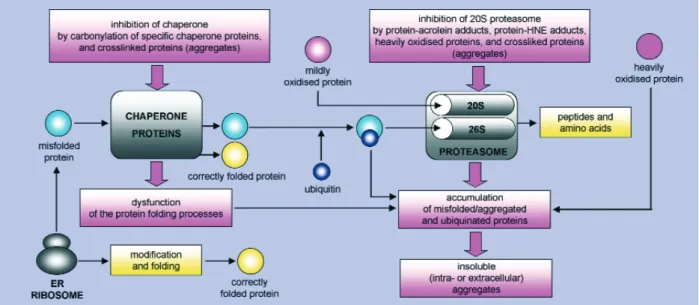
Proteins of reduced or lost function can be harmful to cells if accumulated. Oxidized proteins are either repaired, removed by proteolytic degrada-tion or accumulate as damaged or unfolded pro-teins (Fig. 4). Major intracellular proteolytic sys-tems include lysosomal proteases (cathepsins), cal-cium-dependent proteases (calpains), and multicat-alytic proteases (20S proteasome, which has all the three catalytic activities - chymotrypsin-like, trypsin-like, and peptidyl glutamyl peptide hydro-lase, or caspase-like - and 26S proteasome, with higher catalytic activity than 20S proteasome). Proteasome can degrade proteins by either ubiqui-tin-dependent or ubiquitin-independent non-lyso-somal pathways. In cells, most proteins destined for degradation are labelled first by ubiquitin in an ATP-dependent process and then digested to small peptides by the 26S proteasome. However, oxi-dized proteins are mostly degraded by the 20S pro-teasome that, in contrast to the 26S propro-teasome, does not require tagging by ubiquitin of target pro-teins and ATP for the activity [76, 77]. Davies and colleagues have proposed that the 20S proteasome selectively recognises exposed hydrophobic patch-es of partially unfolded (or denatured) oxidized proteins, since oxidation may cause protein partial unfolding or denaturation with a concomitant increase in surface hydrophobicity [70, 76].
Although mild progressive oxidation of a pro-tein increases its degradation by the proteasome, excessive oxidation and cross-linking of proteins render them resistant to proteolytic degradation by the proteasome, probably because the structural constraint does not allow aggregated or too struc-turally altered proteins to reach the catalytic sites located inside the cylinder of the enzyme complex [71]. Therefore, heavily oxidized and cross-linked protein aggregates accumulate in cells because they inhibit the proteasome and actually cause a progressive further increase in protein aggregation and cross-linking in nondividing (post-mitotic) cells, and may eventually induce apoptosis, as demonstrated in cardiomyocytes [70, 71, 78, 79]. This possibility may have particular importance in post-mitotic tissues such as brain, heart, and skele-tal muscles, where accumulation of oxidized and cross-linked protein aggregates is most marked.
Proteasome activity declines during ageing, as the protease is progressively inhibited by binding to ever increasing levels of oxidized and cross-linked protein aggregates [71, 76]. Conversely, healthy centenaries, and rodents placed on a dietary restriction regiment, exhibit a marked amelioration of age-related increases in protein oxidation and proteasome alterations [80]. On the other hand, direct inhibition of the proteasome in NT-2 (human teratocarcinoma) and SK-N-MC (human neuroblastoma) cells led to increased oxidative damage, NO formation, elevated pro-tein nitration [81], and the formation of propro-tein aggregates [82]. Furthermore, interference with polyubiquitination in the same cell lines led to increased levels of protein carbonylation and nitration, lipid peroxidation, and NO production [83]. Disruption of the UPS may form a common mechanism underlying a number of neurodegen-erative diseases associated with the accumulation of misfolded and/or oxidized proteins.
Proteasomal dysfunction occurs in neurodegen-erative disorders [66, 80, 84–86] and, consistently, the observation of ubiquitinated-protein inclusion bodies in neurons is one of the hallmarks of neu-rodegeneration [66, 85, 87, 88]. In PD, which is currently the only neurodegenerative disease known to be caused by mutations in proteins with-in the UPS such as parkwith-in [66], there is genetic evi-dence for a contribution of UCH-L1 (ubiquitin C-terminal hydrolase-L1), a crucial enzyme for pro-teasomal protein degradation that generates free monomeric ubiquitin [89]. HNE-cross-linked amy-loid β-peptide, which forms the senile plaques in AD, is able to inhibit the proteasome, whereas nei-ther the amyloid β-peptide nor free HNE alone at moderate concentrations inhibit proteasome activi-ty [90]. High concentrations of HNE are unlikely to accumulate in tissues due to very active HNE metabolism. Therefore, a direct inhibition of the proteasome by HNE in vivo seems to be very unlikely, even under pathological conditions.
Fig. 4 Degradation and accumulation of oxidized and/or misfolded proteins. Many newly synthesised proteins are
translocated into the ER, where they fold into their three-dimensional structures with the help of a series of molecular chaperones and folding catalysts. Correctly folded proteins are then transported to the Golgi complex and then delivered to the extracellular environment. However, incorrectly folded proteins are detected by a "quality-control" mechanism and sent along another pathway (the "unfolded protein response") in which they are ubiquitinated and then degraded in the cytoplasm by the 26S proteasome. Proteins can also be mildly or heavily damaged by oxidative stress. Mildly oxi-dized proteins are degraded essentially (if not only) by the 20S proteasome. Cross-linkers like HNE or acrolein further enhance the process of aggregation. The heavily oxidized proteins aggregate and these aggregates cannot be degraded by the 20S proteasome, thus they accumulate and build up larger and larger aggregates. More importantly, they even inhibit the removal of oxidatively damaged proteins by the proteasomal system. Cross-linkers like HNE or acrolein fur-ther enhance the process of aggregation as well as the inhibition of the proteasome.
Much more probable is a decline in the protein turnover or the proteasomal activity due to the for-mation of inhibitory HNE-modified protein aggre-gates. Indeed, an accumulation of proteins modi-fied by HNE occurs in PD and ALS patients [91–93] and both HNE and HNE-modified proteins conjugate with 20S proteasome during oxidative stress, which may contribute to an impairment of proteasomal function [70, 71, 90, 94, 95].
Oxidative modification and inactivation of the 20S proteasome has been demonstrated in the ischaemic heart [96]. Proteasome plays a signifi-cant role in removal of proteins oxidized (car-bonylated) during myocardial ischaemia. A recent study demonstrated an inverse correlation between post-ischaemic proteasome activity and levels of carbonylated and ubiquitinated proteins. In particular, inhibition of the 20S proteasome correlates with increases in protein carbonylation, whereas post-ischaemic inhibition of the 26S pro-teasome leads to accumulation of ubiquitinated proteins [79, 97]. A more recent study [98] has shown that inhibition of the proteasome results in enhanced accumulation of carbonylated proteins in the post-ischaemic rat heart. Furthermore, actin degradation is increased in the post-ischaemic heart, a process that is partially blocked by a pro-teasome inhibitor, and there appears to be no for-mation of ubiquitinated homologues of actin, sug-gesting proteolysis by the 20S proteasome. These observations provide the first evidence that pro-teasome mediates removal of some of the proteins oxidized during myocardial ischaemia/reperfu-sion, and that at least carbonylated actin is removed by the 20S proteasome.
Proteasome subunits may be themselves the target of carbonylation. Treatment with an endogenous inducer of ROS production, a prostaglandin D2 metabolite, 15-deoxy-Δ12,14 -prostaglandin J2 (15d-PGJ2), on human neurob-lastoma SH-SY5Y cells resulted in the accumula-tion of protein carbonyls. Proteomic analysis of oxidation-sensitive proteins showed that the major intracellular target of protein carbonylation was one of the regulatory subunits in 26S protea-some, S6 ATPase, which was associated with (i) a dramatic increase in protein carbonyls within S6 ATPase, (ii) a significant decrease in the S6 ATPase activities, and (iii) a decreased ability of 26S proteasome to degrade substrates [99].
Is protein carbonylation an early
event of cellular dysfunction and
disease progression?
Proteasome inhibitors can induce several hallmarks of apoptosis, including caspase activation, cytochrome C release, elevated p53 expression, chromatin fragmentation, and DNA laddering, in both neuronal and glial cells [80]. It has been sug-gested that protein carbonylation is an early event in NO-induced apoptosis in insulin-secreting RINm5F cells [100, 101]. NO-triggered carbonyla-tion of Bcl-2, adenine nucleotide translocator, and GAPDH precedes DNA fragmentation, and inhibitors of ALEs block NO-dependent carbonyla-tion, prevent NO-induced GAPDH inhibicarbonyla-tion, DNA fragmentation [100], and caspase activation [101]. In addition, NO-induced carbonylation of poly (ADP-ribose) polymerase (PARP) protein precedes its apoptotic degradation and inhibitors of ALE for-mation prevented both events [100], thus suggest-ing that carbonylation of PARP could be mechanis-tically involved in its degradation during apoptosis. Accumulation of aggregates of heavily oxidized proteins (lipofuscin-like materials) induce apopto-sis of cardiomyocytes through inhibition of both 20S- and 26S-proteasome activity, accompanied by large increases in ubiquitinated proteins and dys-regulation of pro-apoptotic proteins [79]. In addi-tion to directly mediating neurotoxicity, proteasome inhibitors increase neural vulnerability to subse-quent oxidative injury [102]. The ability of mild, non-toxic, proteasome inhibition to increase vulner-ability to oxidative stress may be particularly important in ageing, AD, and PD, in which protea-some inhibition would be expected to occur gradu-ally, and not directly induce cell death within the central nervous system. However, once a certain level of proteasome inhibition is achieved, it could then serve as a trigger, and increase the toxicity of subsequent stressors.
A key question is whether protein carbonylation occurs at an early stage of disease, contributing to its development, or whether it is merely a conse-quence of the oxidative tissue damage, reflecting the presence of disease. Basically, the answer to this question requires identification of a specific car-bonylated protein and a positive correlation between altered function of this protein and devel-opment of diseases. Increased levels of protein
car-bonyls have been detected in a large variety of pathological states occurring in humans, thus sug-gesting their potential causative role in disease pathogenesis [7, 18, 19, 89, 103–105].
The plasma protein carbonyl content of children with juvenile rheumatoid arthritis is much higher than in healthy children and, notably, grows with the activity of the inflammatory process [106]. Therefore, carbonyl groups of plasma proteins seem to be a good link of inflammatory process activity and disease progression. Increased protein carbonyls have also been observed in tracheal aspirates from premature infants undergoing ventilation therapy, and a correlation between the protein carbonyl con-tent and myeloperoxidase activity (index of pul-monary inflammation) was established [107]. Severe sepsis and major trauma patients had elevat-ed protein carbonyl concentrations in both plasma and bronchoalveolar lavage fluid, which correlated well with ALE measurements and indices of neu-trophilia and neutrophil activation [108]. Moreover, patients with acute pancreatitis had significantly increased concentration of protein carbonyls in plas-ma, which were related to disease severity [109]. Elevated levels of protein carbonyls were observed in the brain of persons with mild cognitive impair-ment, a condition that often precedes AD, suggest-ing that oxidative damage may be one of the earliest events in the onset and progression of AD [110].
Increased oxidative stress in newly diagnosed child and young diabetic patients with no complica-tions [111, 112] suggests that the increase in oxida-tive stress may not be due to complications, but rather may contribute to their development. Studies on young type 1 diabetic patients showed that the content of carbonyl groups in plasma proteins was much higher than in their healthy peers, and that plasma protein carbonyl levels were even higher in diabetic patients with microvascular complications [113]. Although the primary pathophysiological mechanisms by which diabetic complications develop remain to be conclusively determined, results showing that the increased protein oxidative damage and reduced antioxidative defences were greater in young diabetic patients with microvascu-lar complications than in those without suggest that protein carbonylation could be an important early event in the pathogenesis of complications sec-ondary to diabetes [113]. These findings may also indicate that underlying subclinical pathology
(oxidative stress and vascular dysfunction) may be present despite the apparently good glycemic con-trol and outcome in the majority of these young dia-betic patients [113].
The increase in glycoxidation and lipoxidation products in plasma and tissue proteins suggests that oxidative stress is increased in diabetes. However, some of these products are formed independent on oxidation chemistry and may also result from ele-vated levels of substrates prone to oxidation. Moreover, there is also an increase in products of reaction of proteins with dicarbonyl compounds formed by non-oxidative mechanisms [114]. The increased chemical modification of proteins by car-bohydrates and lipids in diabetes and other diseases such as uremia and atherosclerosis may therefore be viewed as the result of increased "carbonyl stress" (carbonyl overload), which is caused by a gener-alised increase in the steady-state concentration of reactive carbonyl precursors of AGEs/ALEs, gly-coxidation and lipoxidation products. Carbonyl stress may result from an increase in substrate stress and/or a decrease in the efficiency of detoxification of RCS, i.e., an imbalance between the rates of pro-duction and detoxification of reactive carbonyls. Compared with oxidative stress (a condition in which carbonyls are derived exclusively from oxidative reactions), carbonyl stress is a more com-prehensive term, since it includes increases in car-bonyls derived from both oxidative and non-oxida-tive reactions [114–117].
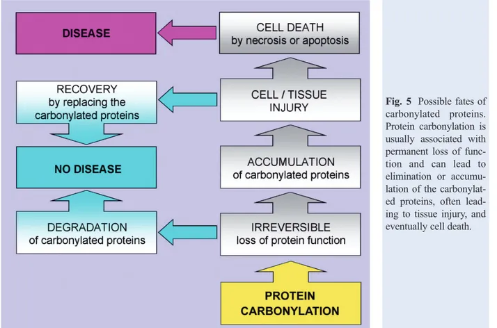
Thus, the consequent loss of function of car-bonylated proteins may be the cause of subsequent cellular dysfunction and tissue damage (Fig. 5). Some studies discussed in this review suggest a positive correlation between increases in protein carbonylation and disease progression. Since car-bonylation can alter protein structure and function and cause the formation of protein aggregates, the "carbonyl stress" hypothesis emphasises the role of RCS, derived from different sources through both oxidative and non-oxidative reactions, and resulting from decreased renal detoxification and/or excre-tion of reactive carbonyl precursors of AGEs/ALEs from plasma, in the induction of pathogenic protein modifications [114, 115, 118, 119].
Although the importance of protein carbonyls in the pathogenic processes responsible for the devel-opment of several diseases remains to be decisively determined, experimental evidences suggest a
causative role of protein carbonylation in the devel-opment of long-term complications of diabetes, as well as in ageing-related diseases. For istance, the increased oxidative/carbonyl stress as well as the accumulation of AGEs/ALEs in tissue proteins are in fact thought to contribute to the development of diabetic complications such as atherosclerosis, vas-cular and neural dysfunction, and retinopathy [114–117, 119–121]. Cumulative modifications by AGEs occur predominantly (but non exclusively) on long-lived proteins such as collagen, neural myelin, and lens crystallins, resulting in insoluble and dysfunctional aggregates that accumulate pro-gressively with time. The formation of inter- and intramolecular cross-links following the glycation of collagen leads to structural alterations, i.e., increased stiffness and resistance to proteolytic digestion [122, 123].
The causative role of protein carbonylation in tis-sue injury in diabetic complications is substantiated by the pharmacological effects elicited by a variety of novel therapeutic agents able to reduce the accumula-tion of AGEs/ALEs in diabetes, which have also gained interest as potential cardioprotective
approaches. These agents include aminoguanidine, AVE7688, pyridoxamine, carnosine, benfotiamine, OPB-9195, LR-90, and the so-called cross-link breakers such as ALT-946 and thiazolium salts (e.g., N-phenacylthiazolium bromide and alagebrium chlo-ride) [116, 117, 124–128]. In addition, it has been demonstrated that a number of established therapies have the ability to reduce the accumulation of AGEs/ALEs in diabetes, including angiotensin con-verting enzyme inhibitors, angiotensin receptor antagonists, metformin, peroxisome proliferators receptor agonists, metal chelators and some antioxi-dants [116, 117, 119, 124–127]. These compounds have been shown to inhibit several diabetic complica-tions such as nephropathy, retinopathy, neuropathy, diabetes-accelerated atherosclerosis, and vascular dis-eases [116, 117, 129–133] as well as the increased blood pressure, decline in glomerular filtration rate, glomerulosclerosis, nephron loss, proteinuria, cardiac hypertrophy, and aorta stiffness in aged rats [129, 134]. The fact that many of these inhibitors of AGEs/ALEs are effective in experimental models, despite their disparate mechanisms of action, supports the keystone role of AGEs/ALEs in diabetic tissue
Fig. 5 Possible fates of
carbonylated proteins. Protein carbonylation is usually associated with permanent loss of func-tion and can lead to elimination or accumu-lation of the carbonylat-ed proteins, often lead-ing to tissue injury, and eventually cell death.
damage. Nonetheless, the clinical utility of AGE/ALE inhibition remains to be firmly established.
Conclusions and perspectives
Most pharmacological approaches of the "anti-AGEs/ALEs strategy" are strictly related to inhibi-tion of protein carbonylainhibi-tion and AGE/ALE forma-tion. AGE/ALE inhibitors, even if belonging to dif-ferent chemical classes, have a common chemical feature: a strong nucleophilic centre able to react with glucose- or lipid-derived RCS at a faster rate than do cell macromolecules. This chemical feature suggests that trapping of RCS and inhibition of pro-tein modification is essential to restrain the patho-logical events. A key role for lipoxidative modifica-tion of proteins in the development of chronic com-plications of diabetes was clearly demonstrated by investigating the mechanism of action of pyridox-amine [116]. As elevated levels of pyridoxpyridox-amine adducts with the intermediates of lipid peroxidation have been determined in the urine of diabetic and hyperlipidemic rats, the protective effect of pyri-doxamine is consistent with its ability to trap RCS. However, it must be highlighted that AGE/ALE inhibitors can not be considered as optimal pharma-cological tools, because all of them have promiscu-ous effects. For example, aminoguanidine is a potent inhibitor of inducible nitric oxide synthase, while carnosine and pyridoxamine are also quenchers of ROS. Thus, the real challenge for future research will be, besides to identify target proteins for RCS and to gain a deeper insight into the molecular mechanisms of carbonylation reactions, to develop more specific pharmacological tools, i.e., selective carbonyl blockers (without any antioxidant/metal chelation effect) for definitive assessment of the possible causative role of protein carbonylation in diseases. Significant advancement on this issue should also contribute to suitable updating of phar-macological intervention in human diseases associ-ated with protein oxidation. To achieve successfully this aim, investigations should focus to the unequiv-ocal identification of specifically carbonylated pro-teins in pathological tissues and fluids.
Proteomic tools now available represent a promising way to elucidate disease mechanism(s) at the protein level [135], because identification of
sites of carbonyl modification should help under-standing the factors affecting protein function. A subsequent goal will be to evaluate the impact of carbonylation on protein function. This point is cru-cial to establish whether carbonylation of specific proteins is causative, correlative or consequential of oxidative stress-associated conditions, because car-bonylation does not necessarily result in protein function alteration. Furthermore, it is essential to compare the in vitro with the in vivo settings when assessing the extent of carbonylation and the conse-quences to protein activity. For instance, it has been demonstrated that the age-related increase in HNE adduction to rat heart α-ketoglutarate dehydroge-nase does not cause loss of its catalytic activity, contrarily to what observed in vitro, suggesting that the extent of HNE binding remains low in vivo, pos-sibly because the cellular HNE concentrations are manyfold lower than those used to inhibit the mito-chondrial enzyme in vitro [136]. Hence, if carbony-lation leads to protein dysfunction, the use of an appropriate pharmacological tool, able to inhibit/prevent protein carbonylation, would unequivocally indicate a causative role of protein carbonylation in disease development and/or pro-gression. Otherwise, if carbonylation does not lead to functional consequences of the oxidized protein, its causative role in disease onset and/or progres-sion would be excluded.
Acknowledgements
This work was supported by grants from FIRST 2004 and FIRST 2005 (Fondo Interno Ricerca Scientifica e Tecnologica), University of Milan, and by Fondazione Monte dei Paschi di Siena.
References
1. Thannickal VJ, Fanburg BL. Reactive oxygen species in
cell signaling. Am J Physiol Lung Cell Mol Physiol. 2000; 279: L1005–28.
2. Moldovan L, Moldovan NI. Oxygen free radicals and
redox biology of organelles. Histochem Cell Biol. 2004; 122: 395–412.
3. Davies MJ, Fu S, Wang H, Dean RT. Stable markers of
of human disease. Free Radic Biol Med. 1999; 27: 1151–61.
4. Cabiscol E, Ros J. Oxidative damage to proteins:
Structural modifications and consequences in cell func-tion. In: Dalle-Donne I, Scaloni A, Butterfield DA, editors. Redox Proteomics: From Protein Modifications to Cellular Dysfunction and Disease. Hoboken: John Wiley & Sons, Inc.; 2006. p. 399–471.
5. Levine RL, Moskovitz J, Stadtman ER. Oxidation of
methionine in proteins: roles in antioxidant defense and cellular regulation. IUBMB Life 2000; 50: 301–7.
6. Dalle-Donne I, Giustarini D, Colombo R, Milzani A,
Rossi R. S-glutathionylation in human platelets by a thiol-disulfide exchange-independent mechanism. Free Radic Biol Med. 2005; 38: 1501–10.
7. Dalle-Donne I, Scaloni A, Giustarini D, Cavarra E, Tell
G, Lungarella G, Colombo R, Rossi R, Milzani A. Proteins as biological markers of oxidative/nitrosative stress in diseases. The contribution of redox-proteomics. Mass Spectrom Rev. 2005; 24: 55–99.
8. Stadtman ER. Metal ion-catalyzed oxidation of proteins:
Biochemical mechanism and biological consequences. Free Radic Biol Med. 1990; 9: 315–25.
9. Stadtman ER, Berlett BS. Fenton chemistry. Amino acid
oxidation. J Biol Chem. 1991; 266: 17201–11.
10. Stadtman ER, Levine RL. Free radical-mediated oxida-tion of free amino acids and amino acid residues in pro-teins. Amino Acids 2003; 25: 207–18.
11. Stadtman ER, Levine RL. Chemical modification of pro-teins by reactive oxygen species. In: Dalle-Donne I, Scaloni A, Butterfield DA, editors. Redox Proteomics: From Protein Modifications to Cellular Dysfunction and Disease. Hoboken: John Wiley & Sons, Inc.; 2006. p. 3–23. 12. Requena J, Chao CC, Stadtman ER. Glutamic and aminoadipic semialdehydes are the main carbonyl prod-ucts of metal-catalyzed oxidation of proteins. Proc Natl Acad Sci USA. 2001; 98: 69–74.
13. Esterbauer H, Schaur RJ, Zollner H. Chemistry and biochemistry of 4-hydroxynonenal, malonaldehyde and related aldehydes. Free Radic Biol Med. 1991; 11: 81–128. 14. Uchida K, Stadtman ER. Modification of histidine residues in proteins by reaction with 4-hydroxynonenal. Proc Natl Acad Sci USA. 1992; 89: 4544–8.
15. Refsgaard HH, Tsai L, Stadtman ER. Modifications of proteins by polyunsaturated fatty acid peroxidation prod-ucts. Proc Natl Acad Sci USA. 2000; 97: 611–6.
16. Dalle-Donne I, Giustarini D, Colombo R, Rossi R, Milzani A. Protein carbonylation in human diseases. Trends Mol Med. 2003; 9: 169–76.
17. Butterfield DA, Castegna A. Proteomic analysis of oxidatively modified proteins in Alzheimer's disease brain: insights into neurodegeneration. Cell Mol Biol. 2003; 49: 747–51.
18. Dalle-Donne I, Rossi R, Colombo R, Giustarini D, Milzani A. Biomarkers of oxidative damage in human dis-ease. Clin Chem. 2006; 52: 601–23.
19. Levine RL, Stadtman ER. Carbonylated proteins and their implication in physiology and pathology. In: Dalle-Donne I, Scaloni A, Butterfield DA, editors. Redox Proteomics: From Protein Modifications to Cellular
Dysfunction and Disease. Hoboken: John Wiley & Sons, Inc.; 2006. p. 563–603.
20. Jana CK, Das N, Sohal RS. Specificity of age-related carbonylation of plasma proteins in the mouse and rat. Arch Biochem Biophys. 2002; 397: 433–9.
21. Choi J, Rees HD, Weintraub ST, Levey AI, Chin LS, Li L. Oxidative modifications and aggregation of Cu/Zn superoxide dismutase associated with Alzheimer's and Parkinson's diseases. J Biol Chem. 2005; 280: 11648–55. 22. Cabiscol E, Piulats E, Echave P, Herrero E, Ros J.
Oxidative stress promotes specific protein damage in Saccharomyces cerevisiae. J Biol Chem. 2000; 275: 27393–8.
23. Yan L-J, Levine RL, Sohal RS. Oxidative damage during aging targets mitochondrial aconitase. Proc Natl Acad Sci. USA. 1997; 94: 11168–72.
24. Yan L-J, Sohal RS. Mitochondrial adenine nucleotide translocase is modified oxidatively during aging. Proc Natl Acad Sci USA. 1998; 95: 12896–901.
25. Das N, Levine RL, Orr WC, Sohal RS. Selectivity of protein oxidative damage during aging in Drosophila melanogaster. Biochem J. 2001; 360: 209–16.
26. Yan L-J, Levine RL, Sohal RS. Effects of aging and hyper-oxia on oxidative damage to cytochrome C in the housefly, Musca domestica. Free Radic Biol Med. 2000; 29: 90–7. 27. Stadtman ER. Protein oxidation and aging. Science 1992;
257: 1220–4.
28. Merker K, Grune T. Proteolysis of oxidized proteins and cellular senescence. Exp Gerontol. 2000; 35: 779–86. 29. Ghezzo-Schöneich E, Esch SW, Sharov VS, Schöneich
C. Biological aging does not lead to the accumulation of oxidized Cu,Zn-superoxide dismutase in the liver of F344 rats. Free Radic Biol Med. 2001; 30: 858–64.
30. Reverter-Branchat G, Cabiscol E, Tamarit J, Ros J. Oxidative damage to specific proteins in replicative and chronological-aged Saccharomyces cerevisiae: common targets and prevention by calorie restriction. J Biol Chem. 2004; 279: 31983–9.
31. Aguilaniu H, Gustafsson L, Rigoulet M, Nyström T. Asymmetric inheritance of oxidatively damaged proteins during cytokinesis. Science 2003; 299: 1751–3.
32. Dukan S, Farewell A, Ballesteros M, Taddei F, Radman M, Nyström T. Protein oxidation in response to increased transcriptional or translational errors. Proc Natl Acad Sci USA. 2000; 97: 5746–9.
33. Ballesteros M, Fredriksson A, Henriksson J, Nyström T. Bacterial senescence: protein oxidation in non-prolifer-ating cells is dictated by the accuracy of the ribosomes. EMBO J. 2001; 20: 5280–9.
34. Fredriksson A, Ballesteros M, Dukan S, Nyström T. Defense against protein carbonylation by DnaK/DnaJ and proteases of the heat shock regulon. J Bacteriol. 2005; 187: 4207–13.
35. England K, Cotter T. Identification of carbonylated pro-teins by MALDI-TOF mass spectroscopy reveals suscep-tibility of ER. Biochem Biophys Res Commun. 2004; 320: 123–30.
36. Rabek JP, Boylston WH, Papaconstantinou J. Carbonylation of ER chaperone proteins in aged mouse liver. Biochem Biophys Res Commun. 2003; 305: 566–72.
37. Dalle-Donne I, Rossi R, Giustarini D, Gagliano N, Lusini L, Milzani A, DiSimplicio P, Colombo R. Actin carbonylation: from a simple marker of protein oxidation to relevant signs of severe functional impairment. Free Radic Biol Med. 2001; 31: 1075–83.
38. Aldini G, Dalle-Donne I, Vistoli G, Maffei Facino R, Carini M. Covalent modification of actin by 4-hydroxy-trans-2-nonenal (HNE): LC-ESI-MS/MS evidence for Cys374 Michael adduction. J Mass Spectrom. 2005; 40: 946–54. 39. Neely MD, Sidell KR, Graham DG, Montine TJ. The
lipid peroxidation product 4-hydroxynonenal inhibits neu-rite outgrowth, disrupts neuronal microtubules, and modi-fies cellular tubulin. J Neurochem. 1999; 72: 2323–33. 40. Wataya T, Nunomura A, Smith MA, Siedlak SL,
Harris PL, Shimohama S, Szweda LI, Kaminski MA, Avila J, Price DL, Cleveland DW, Sayre LM, Perry G. High molecular weight neurofilament proteins are physio-logical substrates of adduction by the lipid peroxidation product hydroxynonenal. J Biol Chem. 2002; 277: 4644–8. 41. Oh-Ishi M, Ueno T, Maeda T. Proteomic method detects oxidatively induced protein carbonyls in muscles of a dia-betes model Otsuka Long-Evans Tokushima Fatty (OLETF) rat. Free Radic Biol Med. 2003; 34: 11–22. 42. O'Reilly PJ, Hickman-Davis JM, Davis IC, Matalon S.
Hyperoxia impairs antibacterial function of macrophages through effects on actin. Am J Respir Cell Mol Biol. 2003; 28: 443–50.
43. Barreiro E, Gea J, Di Falco M, Kriazhev L, James S, Hussain SN. Protein carbonyl formation in the diaphragm. Am J Respir Cell Mol Biol. 2005; 32: 9–17. 44. Boyd-Kimball D, Castegna A, Sultana R, Poon HF,
Petroze R, Lynn BC, Klein JB, Butterfield DA. Proteomic identification of proteins oxidized by Aβ(1-42) in synaptosomes: implications for Alzheimer's disease. Brain Res. 2005; 1044: 206–15.
45. Banan A, Zhang Y, Losurdo J, Keshavarzian A. Carbonylation and disassembly of the F-actin cytoskeleton in oxidant induced barrier dysfunction and its prevention by epidermal growth factor and transforming growth fac-tor alpha in a human colonic cell line. Gut 2000; 46: 830–7.
46. Keshavarzian A, Banan A, Farhadi A, Komanduri S, Mutlu E, Zhang Y, Fields JZ. Increases in free radicals and cytoskeletal protein oxidation and nitration in the colon of patients with inflammatory bowel disease. Gut 2003; 52: 720–8.
47. Powell SR, Gurzenda EM, Wahezi SE. Actin is oxidized during myocardial ischemia. Free Radic Biol Med. 2001; 30: 1171–6.
48. Aksenov MY, Aksenova MV, Butterfield DA, Geddes JW, Markesbery WR. Protein oxidation in the brain in Alzheimer's disease. Neuroscience 2001; 103: 373–83. 49. England K, O'Driscoll C, Cotter TG. Carbonylation of
glycolytic proteins is a key response to drug-induced oxidative stress and apoptosis. Cell Death Differ. 2004; 11: 252–60.
50. Nguyen AT, Donaldson RP. Metal-catalyzed oxidation induces carbonylation of peroxisomal proteins and loss of enzymatic activities. Arch Biochem Biophys. 2005; 439: 25–31.
51. Siems WG, Hapner SJ, van Kujk FJGM. 4-Hydroxynonenal inhibits Na+-K+-ATPase. Free Radic
Biol Med. 1996; 20: 215–23.
52. Chen J, Bertrand AH, Yu BP. Inhibition of mitochondri-al adenine nucleotide translocator by lipid peroxidation products. Free Radic Biol Med. 1995; 19: 583–90. 53. Chen J, Henderson GI, Freeman GL. Role of
4-hydrox-ynonenal in modification of cytochrome c oxidase in ischemia/reperfused rat heart. J Mol Cell Cardiol. 2001; 33: 1919–27.
54. Lauderback CM, Hackett JM, Huang FF, Keller JN, Szweda LI, Markesbery WR, Butterfield DA. The glial glutamate transporter, GLT-1, is oxidatively modified by 4-hydroxy-2-nonenal in the Alzheimer's disease brain: the role of Aβ1-42. J Neurochem. 200l; 78: 413–6.
55. Choksi KB, Boylston WH, Rabek JP, Widger WR, Papaconstantinou J. Oxidatively damaged proteins of heart mitochondrial electron transport complexes. Biochim Biophys Acta 2004; 1688: 95–101.
56. Wen JJ, Garg N. Oxidative modification of mitochondri-al respiratory complexes in response to the stress of Trypanosoma cruzi infection. Free Radic Biol Med. 2004; 37: 2072–81.
57. Yarian CS, Rebrin I, Sohal RS. Aconitase and ATP synthase are targets of malondialdehyde modification and undergo an age-related decrease in activity in mouse heart mitochondria. Biochem Biophys Res Commun. 2005; 330: 151–6. 58. Shamoto-Nagai M, Maruyama W, Kato Y, Isobe K,
Tanaka M, Naoi M, Osawa T. An inhibitor of mitochon-drial complex I, rotenone, inactivates proteasome by oxidative modification and induces aggregation of oxi-dized proteins in SH-SY5Y cells. J Neurosci Res. 2003; 74: 589–97.
59. Dawson TM, Dawson VL. Molecular pathways of neu-rodegeneration in Parkinson's disease. Science 2003; 302: 819–22.
60. Wilson MR, Easterbrook Smith SB. Clusterin is a secreted mammalian chaperone. Trends Biochem Sci. 2000; 25: 95–8.
61. Kaufman RJ, Scheuner D, Schroder M, Shen X, Lee K, Liu CY, Arnold SM. The unfolded protein response in nutrient sensing and differentiation. Nat Rev Mol Cell Biol. 2002; 3: 411–21.
62. Horwich A. Protein aggregation in disease: a role for fold-ing intermediates formfold-ing specific multimeric interac-tions. J Clin Invest. 2002; 110: 1221–32.
63. Dobson CM. Protein folding and misfolding. Nature 2003; 426: 884–90.
64. Bence NF, Sampat RM, Kopito RR. Impairment of the ubiquitin-proteasome system by protein aggregation. Science 2001; 292: 1552–5.
65. Sherman M, Goldberg AL. Cellular defenses against unfolded proteins: a cell biologist thinks about neurode-generative diseases. Neuron 2001; 29: 15–32.
66. Bossy-Wetzel E, Schwarzenbacher R, Lipton SA. Molecular pathways to neurodegeneration. Nature Med. 2004; 10: S2–9.
67. Barral JM, Broadley SA, Schaffar G, Hartl FU. Roles of molecular chaperones in protein misfolding diseases. Semin Cell Dev Biol. 2004; 15: 17–29.
68. Bieschke J, Weber P, Sarafoff N, Beekes M, Giese A, Kretzschmar H. Autocatalytic self-propagation of mis-folded prion protein. Proc Natl Acad Sci USA. 2004; 101: 12207–11.
69. Berke SJ, Paulson HL. Protein aggregation and the ubiq-uitin proteasome pathway: gaining the upper hand on neu-rodegeneration. Curr Opin Genet Dev. 2003; 13: 253–61. 70. Grune T, Jung T, Merker K, Davies KJ. Decreased pro-teolysis caused by protein aggregates, inclusion bodies, plaques, lipofuscin, ceroid, and 'aggresomes' during oxida-tive stress, aging, and disease. Int J Biochem Cell Biol. 2004; 36: 2519–30.
71. Voss P, Grune T. Degradation and accumulation of oxi-dized proteins in age related diseases. In: Dalle-Donne I, Scaloni A, Butterfield DA, editors. Redox Proteomics: From Protein Modifications to Cellular Dysfunction and Disease. Hoboken: John Wiley & Sons, Inc.; 2006. p. 527–562.
72. Nyström T. Role of oxidative carbonylation in protein qual-ity control and senescence. EMBO J. 2005; 24: 1311–7. 73. Guo B, Phillips JD, Yu Y, Leibold EA. Iron regulates the
intracellular degradation of iron regulatory protein 2 by the proteasome. J Biol Chem. 1995; 270: 21645–51. 74. Iwai K, Klausner RD, Rouault TA. Requirements for
iron-regulated degradation of the RNA binding protein, iron regulatory protein 2. EMBO J. 1995; 14: 5350–7. 75. Iwai K, Drake SK, Wehr NB, Weissman AM, LaVaute
T, Minato N, Klausner RD, Levine RL, Rouault TA. Iron-dependent oxidation, ubiquitination, and degradation of iron regulatory protein 2: implications for degradation of oxidized proteins. Proc Natl Acad Sci USA. 1998; 95: 4924–8.
76. Shringarpure R, Davies KJ. Protein turnover by the pro-teasome in aging and disease. Free Radic Biol Med. 2002; 32: 1084–9.
77. Shringarpure R, Grune T, Mehlhase J, Davies KJA. Ubiquitin conjugation is not required for the degradation of oxidized proteins by proteasome. J Biol Chem. 2003; 278: 311–8.
78. Grune T, Merker K, Jung T, Sitte N, Davies KJ. Protein oxidation and degradation during postmitotic senescence. Free Radic Biol Med. 2005; 39: 1208–15.
79. Powell SR, Wang P, Divald A, Teichberg S, Haridas V, McCloskey TW, Davies KJ, Katzeff H. Aggregates of oxidized proteins (lipofuscin) induce apoptosis through proteasome inhibition and dysregulation of proapoptotic proteins. Free Radic Biol Med. 2005; 38: 1093–101. 80. Ding Q, Keller JN. Proteasomes and proteasome
inhibi-tion in the central nervous system. Free Radic Biol Med. 2001; 31: 574–84.
81. Lee M, Hyun DH, Jenner P, Halliwell B. Effect of pro-teasome inhibition on cellular oxidative damage, antioxi-dant defences and nitric oxide production. J Neurochem. 2001; 78: 32–41.
82. Hyun DH, Lee M, Halliwell B, Jenner P. Proteasomal inhibition induces the formation of aggregates containing a wide range of proteins, including nitrated proteins. J Neurochem. 2003; 86: 363–73.
83. Hyun DH, Gray DA, Halliwell B, Jenner P. Interference with ubiquitination causes oxidative damage and
increased protein nitration: implications for neurodegener-ative diseases. J Neurochem. 2004; 90: 422–30.
84. Keller JN, Hanni KB, Markesbery WR. Impaired pro-teasome function in Alzheimer's disease. J Neurochem. 2000; 75: 436–9.
85. Chung KK, Dawson VL, Dawson TM. The role of the ubiquitin-proteasomal pathway in Parkinson's disease and other neurodegenerative disorders. Trends Neurosci. 2001; 24: S7–14.
86. Giasson BI, Ischiropoulos H, Lee VM, Trojanowski JQ. The relationship between oxidative/nitrative stress and pathological inclusions in Alzheimer's and Parkinson's dis-eases. Free Radic Biol Med. 2002; 32: 1264–75.
87. McNaught KS, Olanow CW, Halliwell B, Isacson O, Jenner P. Failure of the ubiquitin-proteasome system in Parkinson's disease. Nat Rev Neurosci. 2001; 2: 589–94. 88. Fortun J, Li J, Go J, Fenstermaker A, Fletcher BS,
Notterpek L. Impaired proteasome activity and accumu-lation of ubiquitinated substrates in a hereditary neuropa-thy model. J Neurochem. 2005; 92: 1531–41.
89. Jenner P. Oxidative stress in Parkinson's disease. Ann Neurol. 2003; 53: S26–38.
90. Shringarpure R, Grune T, Sitte N, Davies KJA. 4-Hydroxynonenal-modified amyloid-β peptide inhibits the proteasome: possible importance in Alzheimer's disease. Cell Mol Life Sci. 2000; 57: 1802–8.
91. Yoritaka A, Hattori N, Uchida K, Tanaka M, Stadtman ER, Mizuno Y. Immunohistochemical detection of 4-hydroxynonenal protein adducts in Parkinson disease. Proc Natl Acad Sci USA. 1996; 93: 2696–701.
92. Pedersen WA, Fu W, Keller JN, Markesbery WR, Appel S, Smith RG, Kasarskis E, Mattson MP. Protein modification by the lipid peroxidation product 4-hydrox-ynonenal in the spinal cords of amyotrophic lateral sclero-sis patients. Ann Neurol. 1998; 44: 819–24.
93. Smith RG, Henry YK, Mattson MP, Appel SH. Presence of 4-hydroxynonenal in cerebrospinal fluid of patients with sporadic amyotrophic lateral sclerosis. Ann Neurol. 1998; 44: 696–9.
94. Friguet B, Szweda LI. Inhibition of multicatalytic pro-teinase (proteasome) by 4-hydroxynonenal cross-linked protein. FEBS Lett. 1997; 405: 21–5.
95. Okada K, Wangpoengtrakul C, Osawa T, Toyokuni S, Tanaka K, Uchida K. 4-Hydroxy-2-nonenal-mediated impairment of intracellular proteolysis during oxidative stress. J Biol Chem. 1999; 274: 23787–93.
96. Bulteau AL, Lundberg KC, Humphries KM, Sadek HA, Szweda PA, Friguet B, Szweda LI. Oxidative modi-fication and inactivation of the proteasome during coronary occlusion/reperfusion. J Biol Chem. 2001; 276: 30057–63. 97. Powell SR, Wang P, Katzeff H, Shringarpure R, Teoh C, Khaliulin I, Das DK, Davies KJ, Schwalb H. Oxidized and ubiquitinated proteins may predict recovery of postischemic cardiac function: essential role of the pro-teasome. Antioxid Redox Signal. 2005; 7: 538–46. 98. Divald A, Powell SR. Proteasome mediates removal of
proteins oxidized during myocardial ischemia. Free Radic Biol Med. 2006; 40: 156–64.
99. Ishii T, Sakurai T, Usami H, Uchida K. Oxidative mod-ification of proteasome: identmod-ification of an