Review Article
Hints for Genetic and Clinical Differentiation of Adult-Onset
Monogenic Autoinflammatory Diseases
Carla Gaggiano,
1Donato Rigante
,
2,3Antonio Vitale
,
4Orso Maria Lucherini
,
4Alessandra Fabbiani,
5Giovanna Capozio,
2Chiara Marzo,
4Viviana Gelardi,
4Salvatore Grosso,
1Bruno Frediani,
4Alessandra Renieri
,
5and Luca Cantarini
41Clinical Pediatrics, Department of Molecular Medicine and Development, University of Siena, Siena, Italy 2Institute of Pediatrics, Fondazione Policlinico A. Gemelli IRCCS, Rome, Italy
3Periodic Fever Research Center, Università Cattolica Sacro Cuore, Rome, Italy
4Research Center of Systemic Autoinflammatory Diseases and Behçet’s Disease Clinic, Department of Medical Sciences,
Surgery and Neurosciences, University of Siena, Siena, Italy
5Medical Genetics, University Hospital of Siena, Siena, Italy
Correspondence should be addressed to Luca Cantarini; [email protected] Received 15 July 2019; Accepted 16 November 2019; Published 31 December 2019 Academic Editor: Maria Rosaria Catania
Copyright © 2019 Carla Gaggiano et al. This is an open access article distributed under the Creative Commons Attribution License, which permits unrestricted use, distribution, and reproduction in any medium, provided the original work is properly cited. Monogenic autoinflammatory diseases (mAIDs) are inherited errors of innate immunity characterized by systemic inflammation recurring with variable frequency and involving the skin, serosal membranes, synovial membranes, joints, the gastrointestinal tube, and/or the central nervous system, with reactive amyloidosis as a potential severe long-term consequence. Although individually uncommon, all mAIDs set up an emerging chapter of internal medicine: recentfindings have modified our knowledge regarding mAID pathophysiology and clarified that protean inflammatory symptoms can be variably associated with periodic fevers, depicting multiple specific conditions which usually start in childhood, such as familial Mediterranean fever, tumor necrosis factor receptor-associated periodic syndrome, cryopyrin-associated periodic syndrome, and mevalonate kinase deficiency. There are no evidence-based studies to establish which potential genotype analysis is the most appropriate in adult patients with clinical phenotypes suggestive of mAIDs. This review discusses genetic and clinical hints for an ideal diagnostic approach to mAIDs in adult patients, as their early identification is essential to prompt effective treatment and improve quality of life, and also highlights the most recent developments in the diagnostic work-up for the most frequent hereditary periodic febrile syndromes worldwide.
1. Introduction
Monogenic autoinflammatory diseases (mAIDs) are clinical
entities characterized by recurrent inflammatory attacks
occurring without any evidence of infections, neoplasms, or
deregulation of the adaptive immune system. This expanding
family of diseases is actually known to be caused by
muta-tions in genes involved in the regulation of innate immunity,
in
flammation, and cell death, including first-line responses to
infectious agents and different tissue injuries [1]. Mutations
in the MEFV gene were
firstly identified for patients with
familial Mediterranean fever (FMF) in 1997 [2, 3]. Few years
later, the genetic basis of three other mAIDs was detected
through candidate gene approach, linkage analysis, and/or
homozygosity mapping. Familial Hibernian fever, commonly
known as tumor necrosis factor receptor-associated periodic
syndrome (TRAPS), was found to be caused by mutations in
the TNFRSF1A gene [4]. In 1999, Drenth et al. identi
fied
mutations in the gene encoding mevalonate kinase (MVK),
the key enzyme in isoprenoid and sterol synthesis, which is
involved in the pathogenesis of mevalonate kinase de
ficiency
(MKD) and hyperimmunoglobulinemia D syndrome (HIDS)
[5]. Furthermore, gain-of-function mutations in the NLRP3
gene (also known as CIAS1) were associated with the disease
spectrum of cryopyrin-associated periodic syndrome (CAPS)
in 2001 [6, 7].
Over the last 20 years, the identification of new putative
genes following the extensive use of next-generation
sequenc-ing technologies and the remarkable progress of molecular
techniques have deepened our knowledge about the
patho-genesis of mAIDs, providing novel insights of mechanisms
involved in innate immunity regulation [8]. In addition, the
discovery of gene modi
fiers and somatic mosaicisms as well
as a better comprehension of the role of epigenetic factors
have clari
fied some aspects of the wide phenotypic
heteroge-neity of these disorders.
Although recurrent high-grade fever represents a
com-mon ground for most mAIDs, the clinical presentation may
be variable, being all organs and tissues potentially targeted
by inflammation. Moreover, atypical or oligosymptomatic
presentations are not rare, especially in patients with
adult-onset disease [9, 10]. In this regard, recent studies have
proved that mAIDs may start off not only in the pediatric
age but also during adulthood [11
–14]. A delayed onset of
mAIDs is often due to low-penetrance mutations which may
be sometimes identi
fied even in healthy carriers [15–18]. For
these reasons, early identi
fication of probands, correct
inter-pretation of low-penetrance mutations, and prevention of
overdiagnosis and overtreatment can be challenging [19, 20].
New evidence-based classification criteria for hereditary
recurrent febrile syndromes have been recently developed
on the basis of international expert consensus and evaluated
in a large cohort of patients from the Eurofever registry in
2019 with the aim of recruiting patients for translational and
clinical studies, and not misused as diagnostic criteria [21].
This review is aimed at describing genetic and clinical
clues of the four historical mAIDs in order to suggest an
empirical
flow chart for diagnosis. Single organ involvement
and systemic features will be examined, with particular
atten-tion to di
fferential diagnosis with multifactorial AIDs. In
addition, the description of the most frequently identi
fied
mutations and an overview of genetic data will be provided
to facilitate physicians unravel among pathogenic variants,
polymorphisms and mosaicisms for genes related to mAIDs.
Genetic and clinical features of the four historical mAIDs are
summarized in the Table 1.
An extensive literature search in the Medline database (via
Pubmed) was performed up to May 2019. We searched for
studies through the following words:
“monogenic
autoinflam-matory disease
”, “familial Mediterranean fever”, “mevalonate
kinase de
ficiency”, “tumor necrosis factor receptor-associated
periodic syndrome
”, “cryopyrin-associated periodic
syn-drome
”, and their synonyms. Papers published in English
language over the last ten years were screened for eligibility,
based on title, abstract, and keywords. Papers were included
if clinical clues to the diagnosis of the four mAIDs, both in
children and in adults, were reported. References in the
rel-evant papers were also reviewed. Main reports published
before the aforementioned period of time were included
as well.
2. Diagnostic and Genetic Overview of mAIDs
2.1. Familial Mediterranean Fever (FMF). FMF is the most
frequent and best characterized autosomal recessive
mono-genic AID. Southern European, Northern African, Turkish,
and Arabic people are more frequently a
ffected (the
prevalence is 1 : 150-1 : 1000 in Turkey) [22]; the Middle
East
and Eastern Europe show a lower prevalence
(1 : 10,000,000). Moreover, a growing number of FMF
diag-nosis were recently established in Japan, USA, and Brazil
[2, 3, 23, 24]. Employing positional cloning, genomic
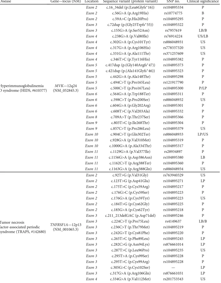
Table 1: Summary of the main genetic and clinical features of FMF, TRAPS, CAPS, and MKD.
Disease Gene locus Inheritance Protein Main clinical features
FMF 16p13.3MEFV AR Pyrin erysipelas-like rash, and systemic AA amyloidosis in untreated patientsFever, serositis, arthritis generally affecting large joints,
TRAPS TNFRSF1A
12p13 AD
Tumor necrosis factor receptor 1
Fever, migrating erythematous skin rash, muscle pain due to monocytic fasciitis, periorbital edema, arthralgia or arthritis, serosal
involvement, and systemic AA amyloidosis in untreated patients FCAS
NLRP3
1q44 AD Cryopyrin
Fever, cold-induced urticaria-like rash, conjunctivitis, and arthralgia
MWS Fever, urticaria-like rash, conjunctivitis, arthralgia, neurosensorial
deafness, and risk of amyloidosis CINCA
Fever, urticaria-like rash, uveitis, papilledema, deforming arthritis mainly involving large joints, chronic aseptic meningopathy, neurosensorial deafness, and risk of amyloidosis HIDS
MVK
12q24 AR Mevalonate kinase
Fever, polymorphous rash, arthralgia, abdominal pain, diarrhea, lymph node enlargement, splenomegaly, and aphthosis
MEVA Psychomotor retardation, growth delay, progressive cerebellar ataxia,
dysmorphisms, and vision deficits in addition to HIDS features
List of abbreviations: AD—autosomal dominant; AR—autosomal recessive; CINCA—chronic infantile neurologic cutaneous and articular syndrome; FCAS—familial cold autoinflammatory syndrome; FMF—familial Mediterranean fever; HIDS—hyperimmunoglobulinemia D syndrome; MEFV— Mediterranean fever; MEVA—mevalonic aciduria; MKD—mevalonate kinase deficiency; MVK—mevalonate kinase; MWS—Muckle-Wells syndrome; NLRP3—NACHT, LRR, and PYD domain-containing protein 3; TNFRSF1A—tumor necrosis factor receptor super family 1A; TRAPS—tumor necrosis factor-associated periodic syndrome.
sequence analysis, and exon trapping in 1997, two
indepen-dent FMF consortia (French FMF Consortium and
Interna-tional FMF Consortium) identified and isolated the MEFV
gene, located on chromosome 16p13.3, as the causative
for FMF [2, 3]. This gene encodes a 781-amino acid protein
known as pyrin/TRIM20/marenostrin, which works as a key
component of the innate immune system and is expressed by
neutrophils, eosinophils, monocytes, and dendritic cells [25].
Although characterized by an autosomal recessive pattern of
inheritance, the FMF phenotype has been observed also in
heterozygous patients, in whom hypothetical modifier genes
and/or environmental factors may play a substantial role in
inducing inflammatory attacks [26, 27]. Disease onset occurs
before the age of 10 in more than 60% of patients and before
the age of 30 in 98% of cases [28]. Acute febrile attacks
usu-ally last a few hours to 3 days; serositis, articular symptoms,
and erysipelas-like erythema in the lower limbs are the most
typical manifestations accompanying fever. Although
adult-onset patients often manifest a milder phenotype, clinical
features are generally similar to those expressed by younger
patients, except for a lower frequency of arthritis and skin
erythema [29]. Systemic reactive AA amyloidosis represents
the most severe long-term complication in untreated FMF
patients [30]. In this regard, three different FMF types have
been suggested: type 1 FMF refers to the presence of overt
clinical in
flammatory disease; type 2 FMF presents with
sys-temic amyloidosis in otherwise asymptomatic subjects; and
type 3 FMF is related to the absence of in
flammatory
mani-festations and systemic amyloidosis in subjects carrying
MEFV mutations [31].
Since the identification of the MEFV gene, more than
340 nucleotide variants have been found, half of them
being clearly associated with FMF. The majority of
FMF-causing mutations are located within exon 10 and are
muta-tional hotspots (including p.M694V/I and p.M680I), which
are associated with a more severe clinical phenotype.
Milder pathogenic variants, such as p.V726A located on
exon 10, have been also reported [23, 32
–34]. Moreover, other
mutations, either of unknown or uncertain signi
ficance
(p.K695R, p.P369S, p.F479L, p.I591T, and p.E148Q) and
path-ogenic variants (p.R761H, p.A744S, p.I692del, p.E167D, and
p.T267I), have been associated with varying degrees of disease
severity [35, 36]. Despite extensive studies over the last
20 years, genotype-phenotype correlations in FMF have not
been fully understood [37]. Many patients with clinical
FMF have no genetic variants or are heterozygous for MEFV,
highlighting the possibility of an autosomal-dominant
trans-mission or of a clinical expression depending on additional
modifying factors as modi
fier genes and environmental or
epigenetic in
fluences. Regarding FMF patients with a single
heterozygous mutation (such as p.H478Y, p.T577S/A/N,
p.M694del/I, p.E148Q, and p.L110P), they were associated
with a wide clinical variability, including an incomplete or
less severe disease phenotype [38–40]. Moreover, modifier
genes encoding components of amyloid A deposits, such as
serum amyloid A1 (SAA1), have been significantly and
independently associated with renal amyloidosis [41]. In
addition, peripheral leukocytes from FMF patients have
shown reduced MEFV transcript expression due to a slightly
increased methylation of exon 2 compared to healthy
con-trols [42]. Among epigenetic modi
fications, a differential
expression of several miRNAs has been demonstrated both
in homozygote and heterozygote quiescent FMF patients,
compared to controls and healthy carriers [43–46].
Pyrin is a member of the TRIM protein family playing a
pivotal role in the inflammatory response against infections
through the regulation of interleukin- (IL-) 1
β production
[47]. The protein is formed by N-terminal pyrin domain
(PYD), zinc
finger domain (bBox), coiled-coil (CC), and
B30.2/SPRY C-terminal domain: the pathogenetic
mecha-nism which links MEFV gene mutations to the development
of the FMF phenotype is not fully clarified. According
to Papin et al., pyrin SPRY domain interacts with
inflamma-some components inhibiting pro-IL-1
β processing; this
C-terminal region of the protein is frequently altered as a
consequence of pathogenic MEFV mutations [48].
Conse-quently, macrophages from pyrin knock-out mice show
enhanced IL-1
β release in response to inflammatory
stim-uli [49]. On the contrary, Chae et al. demonstrated that pyrin
mutations in FMF-knock-in (KI) mice induce
NLRP3-independent IL-1
β activation. In fact, macrophages obtained
from KI FMF-associated B30.2 mutations mice (including
M680I, M694V, and V726A) showed IL-1
β overproduction
after lipopolysaccharide (LPS) stimulation, while no
reduc-tion of IL-1
β production was observed in NLRP3-deficient
KI mice (MEFV
V726A/V726ANLRP3
−/−). These
findings
sug-gested that gain-of-function pyrin mutations induce IL-1
β
activation through an NLRP3-independent manner.
Con-versely, monocytes from patients with FMF had
LPS-induced IL-1
β oversecretion, which was damped after
in vitro NLRP3 downregulation [50]. Interestingly, the same
authors observed that hyperproduction of IL-1
β correlated
with both number and penetrance of MEFV mutations. In
particular, individuals carrying heterozygous p.M694V or
p.K695R mutations have shown higher levels of IL-1
β release
than healthy controls, but lower levels than FMF patients
who were homozygous for the same mutations. In addition,
silencing NLRP3 expression led to the inhibition of IL-1
β
secretion, suggesting that FMF-associated mutations might
also trigger NLRP3-dependent inflammatory response [51].
More recently, the identification of biochemical processes
triggering IL-1
β production in response to bacterial
modifi-cation of the GTPase RhoA shed light on the outstanding role
of pyrin in the inflammatory responses. In this model, pyrin
is kept inactive by serine-threonine kinases PKN1 and PKN2,
through Ser208/Ser242 phosphorylation and subsequent
binding to 14-3-3 proteins, which block the pyrin in
flamma-some. It has been demonstrated that PKN1 and PKN2 are
activated by RhoA GTPase and that Rho-modifying
bacte-rial toxins induce inactivation of these kinases, resulting
in pyrin-inflammasome formation and IL-1β production.
Interestingly, mutated pyrin from FMF patients displayed
a low binding to 14-3-3 proteins and PKN [52]. The latest
research on pyrin functions focused on its interaction with
the cytoskeletal network, demonstrating its pivotal role
in modulating in
flammatory cell polarization and
migra-tion. Supporting these new data, bioinformatics analyses
revealed that mRNAs targeted by miRNAs which are
found overexpressed in homozygous FMF patients (such as
miR-20a-5p and miR-197-3p) cluster in inflammatory
path-ways related to cell migration [53].
FMF diagnosis does not necessarily require genetic
anal-ysis, as it is also driven by clinical evaluation and relies on
diagnostic criteria [54
–56]. Several sets of criteria have been
suggested over time. However, those established at the Tel
Hashomer Medical Center in Israel are currently the most
widely used for adult patients [54]. They include two sets of
diagnostic criteria, consisting of a complete version and a
simplified one: the complete version classifies FMF clinical
manifestations into major and minor items and also includes
ten supportive criteria, as illustrated in Figure 1(a). Both
specificity and sensitivity of these criteria are higher than
95% [54]. The Tel Hashomer group also proposed simpli
fied
criteria that include major and minor criteria, as shown in
Figure 1(b). In this case, FMF diagnosis is allowed when at
least 1 major criterion or at least 2 minor criteria are ful
filled.
The simpli
fied version includes atypical attacks with
abdom-inal involvement as a major criterion. Sensitivity and
speci-ficity are higher than 95% for simplified criteria as well
[54]. More recently, Yalçinkaya et al. have proposed
diag-nostic criteria specifically tailored for children, as described
in Figure 1(c) [55].
2.2. Tumor Necrosis Factor Receptor-Associated Periodic
Syndrome (TRAPS). Originally described in 1982 in a family
of Irish/Scottish descent and previously named
“Hibernian
fever
” from the ancient Latin name of Ireland Hibernia,
TRAPS is caused by mutations in the TNFRSF1A gene,
inher-ited in an autosomal-dominant pattern [57–59]. At the end
of the
‘90s, genome-wide association studies and linkage
analysis in affected families placed the susceptibility locus
on the distal short arm of chromosome 12p13, encoding
tumor necrosis factor (TNF) receptor type 1 (TNFRSF1A/
TNFR1/p55/CD120a) [58]. Afterwards, McDermott et al.
identi
fied germline mutations in the TNFRSF1A gene in
seven families of Irish, Scottish, English, German, Finnish,
and French-Canadian ancestry [4]. Nowadays, TRAPS is
considered a rare disease, with an estimated prevalence of
about 1 : 1,000,000 people. Most patients are of European
ori-gin, though some patients from Africa and Asia have been
reported as well [60].
TRAPS is the most variable and protean among mAIDs
in terms of age at disease onset, disease severity, and clinical
presentation. Nevertheless, the disease is generally
character-ized by long-lasting in
flammatory attacks that may even
reach several weeks of duration. In the clinical practice,
high-grade fever, erythematous migratory skin plaques
associated with underlying myalgia, and joint and ocular
in
flammatory signs are prominent manifestations of TRAPS
[60, 61]. Although most patients experience an early disease
onset, adult-onset TRAPS has been widely reported,
espe-cially in subjects carrying low-penetrance mutations and
pre-senting with atypical features, such as inflammatory
manifestations affecting unusual sites [62–67]. To date, no
clinical diagnostic criteria have been proposed for the
diag-nosis of TRAPS, which is based on the identi
fication of
TNFRSF1A mutations.
TNFRSF1A is a member of the TNF receptor
superfam-ily, constitutively expressed on most cell types, which is
com-posed of an extracellular domain consisting of the tandem
repeat of four cysteine-rich domains (CRD1-4), a
transmem-brane region, and an intracellular death domain (DD)
involved in intracellular signal transduction. The
extracellu-lar domain is characterized by the presence of intramolecuextracellu-lar
disulfide bonds, which bind TNF-α and also mediate the
self-assembly of the receptor itself. Upon activation by a TNF
ligand, TNFR1 signal transduction leads to several
biochem-ical processes, including the release of proin
flammatory
cyto-kines through the nuclear factor-
κB (NF-κB) pathway or,
alternatively, through other intracellular cascades, such as
p38 and c-Jun N-terminal kinase/mitogen-activated protein
kinase (JNK/MAPK) signaling, and programmed cell death
through the activation of cysteine proteases, named caspases.
Moreover, activated TNFR mediates the release of the
extra-cellular domain of the receptor itself into the extraextra-cellular
compartment by means of ADAM17 (TNF-
α converting
enzyme), a key mediator of this
“shedding” process, which
generates a pool of sTNFR1 able to bind circulating TNF-
α,
dampening acute inflammation [62, 68].
Currently, more than 150 TNFRSF1A gene mutations
have been identified. The majority of disease-associated
mutations are missense mutations located in exons 2, 3,
and 4, encoding the extracellular domain of the mature
TNFR protein [69]. As previously written, low-penetrance
variants are associated with milder phenotypes, later onset
of disease, and lower risk of AA amyloidosis, if compared
to high-penetrance mutations. Among low-penetrance
vari-ants, p.R92Q and p.P46L represent the most common
muta-tions found in TRAPS patients; nevertheless, 2% allele
frequency is estimated for p.R92Q in Caucasians, while
about 10% allele frequency is estimated in the African
population for p.P46L [70
–74]. On the contrary,
high-penetrance
“structural” mutations are responsible of severe
phenotypes, characterized by early onset, high frequency of
disabling in
flammatory attacks, and a higher risk of
develop-ing AA amyloidosis over time. Cysteine residue substitutions
(i.e., p.C30R, p.C33Y, p.C43G, p.C52Y, and p.C55Y) and
p.T50M are listed among the structural variants [30, 70–
74]. Moreover, a novel in-frame deletion of 24 nucleotides
(c.255_278del) in exon 3 was recently identified in one
patient [75].
Several pathogenetic mechanisms have been studied
in vitro to demonstrate how TNFRSF1A mutations
con-tribute to the development of the TRAPS phenotype: (i)
impaired TNFRSF1A cell surface expression; (ii) altered
TNF-
α binding; (iii) defective shedding of the receptor; and
(iv) intracellular accumulation of mutated TNFRSF1A
pro-teins [76–81]. The identification of reduced serum levels of
soluble TNFR1 paved the way for the
“defective shedding
hypothesis.” Indeed, activated leukocytes carrying p.C52F,
p.C33Y, p.T50M, and p.C88R mutations showed increased
TNFR1 membrane expression and, on the other hand,
reduced cleavage of the receptor [4]. Interestingly, further
studies have demonstrated that intracellular accumulation
of misfolded TNFRSF1A proteins increases oxidative stress
levels, stimulating proin
flammatory signaling pathways
Typical
Recurrent (3 or more episodes), febrile (RT 38°C or higher) and short (12-72 hours)
Incomplete
Painful and recurrent attacks that differ from typical attacks in 1 or 2 features, as follows: (1) The temperature is normal or lower than 38°C
(2) The attacks are longer or shorter than specified (but not shorter than 6 hours or longer than a week) (3) No signs of peritonitis are recorded during the abdominal attacks
(4) The abdominal attacks are localized
(5) The arthritis is in joints other than those specified
1-3 Incomplete attacks involving 1 or more of the following sites:
1. Abdomen 2. Chest 3. Joint 4. Exertional leg pain
5. Favorable response to colchicine
(1) Family history of FMF (2) Appropriate ethnic origin (3) Age < 20 years at disease onset (4) Severe attacks, requiring bed rest (5) Spontaneous remission of the attacks (6) Symptom-free interval
(7) Transient inflammatory response, with 1 or more abnormal test result(s) for white blood cell count, erythrocyte sedimentation rate, serum amyloid A, and/or fibrinogen
(8) Episodic proteinuria/hematuria
(9) Unproductive laparotomy or removal of white appendix
(10) Consanguinity of parents
Type of attacks
(1) Peritonitis (generalized) (2) Pleuritis (unilateral) or pericarditis (3) Monoarthritis (hip, knee, or ankle) (4) Fever alone
If the patient has incomplete attacks, 2 minor criteria or 1 minor+5 supportive criteria or 1 minor+4 of the first 5 supportive criteria, they are requested to make the diagnosis.
If the patient has typical attacks, 1 of the 4 major criteria is needed to make the diagnosis.
Major criteria
Minor criteria
Supportive criteria
(a) Simplified Tel Hashomer criteria
Major criteria
Minor criteria
(1–4) Typical attacks with (1) Generalized peritonitis
(2) Unilateral pleuritis or pericarditis (3) Monoarthritis (hip, knee, and ankle) (4) Fever alone
(5) Incomplete abdominal attacks
(1–2) Incomplete attacks involving at least one site among
(1) Chest (2) Joint (3) Exertional leg pain
(4) Favorable response to colchicine
At least 1 of the simplified major criteria or 2 simplified minor criteria are requested
(b)
Yalçinkaya pediatric items
At least 2 items are requested in children Fever episodes (axillary body temperature > 38°C)⁎
Abdominal pain⁎ Chest pain⁎ Oligoarthritis⁎
Family history of familial Mediterranean fever ⁎Duration of 6-72 hours, 3 attacks at least (1) (2) (3) (4) (5) (c)
Figure 1: Complete (1a) and simplified (1b) Tel Hashomer criteria adapted by from Livneh et al. [54] for the diagnosis of familial Mediterranean fever (FMF); Yalçinkaya items (1c) for the diagnosis of FMF in childhood, adapted from Yalçinkaya et al. [55].
[82–85]. In support of this suggestion, chronic oxidative
stress as well as enhanced IL-6 and TNF-
α levels were
observed, in response to LPS, in monocytes isolated from
patients with TNFRSF1A structural mutations (i.e., p.C33Y,
p.T50M, p.C33G, p.C52F, and p.C30Y) in comparison with
healthy controls [82, 83]. Moreover, autophagy defects and
endoplasmic reticulum stress with consequent X-box
bind-ing protein 1 (XBP1) hyperactivation were also proposed
as pathogenetic determinants in association with TRAPS
high-penetrance variants [84, 85]. Interestingly, Pucino
et al. observed for the
first time a T cell response pattern of
activation in TRAPS patients carrying high-penetrance
vari-ants compared to healthy controls and, specifically, a lower
frequency of peripheral regulatory T cells, a defective
suppres-sive phenotype associated to ERK1/2, STAT1/3/5,
mamma-lian target of rapamycin, and NF-
κB pathways [86]. Also
epigenetic mechanisms have been recently added to TRAPS
pathophysiology: in fact, TRAPS patients carrying structural
mutations may display a speci
fic serum signature by miRNAs.
Moreover, a different expression of specific miRNAs,
involv-ing miR-92a-3p and miR-150-3p, has been observed between
patients treated with the IL-1 receptor antagonist anakinra
and nontreated patients [87]. More recently, Harrison et al.
reported that primary dermal
fibroblasts from patients
carry-ing the p.T50M, p.C472, and p.C88R mutations showed an
impaired regulation of miR-146a and miR-155, which led to
increased responsiveness to LPS [88].
2.3.
Cryopyrin-Associated
Periodic
Syndrome
(CAPS).
CAPS is a spectrum of diseases caused by mutations in the
NLRP3 gene and including familial cold autoinflammatory
syndrome (FCAS), Muckle-Wells syndrome (MWS), and
chronic infantile neurological cutaneous and articular
syn-drome (CINCA), the latter also known as
“neonatal onset
multisystem inflammatory disease” or NOMID. These three
clinical entities range from the least to the most severe
phe-notype, respectively. In particular, FCAS is characterized by
recurrent attacks typically induced by generalized cold
exposure and consisting of fever, urticaria-like rash,
asthe-nia, conjunctivitis, and arthralgia; in addition to the clinical
picture described for FCAS, MWS is also characterized by
arthritis, optic nerve head swelling, and inflammation of
the vestibule-cochlear nerve, which might lead to progressive
visual loss and bilateral sensorineural hearing loss,
respec-tively [6, 7, 89–91]. Early central nervous system involvement
with chronic aseptic meningitis, increased intracranial
pres-sure, cerebral atrophy, ventriculomegaly, and severe chronic
papilledema are further identi
fied in patients with
CINCA/-NOMID [92, 93]. Moreover, severe chronic arthritis with
conspicuous structural deformities of large joints, bony
over-growth, and loss of articular function are typical
manifesta-tions of CINCA/NOMID [94, 95].
CAPS are rare diseases with a prevalence of 1-2 per 10
6in
the United States and 1/360,000 in France, without any
gen-der predilection [96]. Patients are often of Caucasian origin,
and disease prevalence seems to be higher for FCAS and
MWS in the US and Europe, respectively, while
CINCA/NO-MID is rarer [96]. The gene responsible for these syndromes
was identified at the end of the 20th century after linkage
analysis studies on di
fferent families of patients showing
clin-ical CAPS pictures. Cuisset et al. applied a genome-wide
search strategy to three families, mapping a region closely
related to the MWS phenotype within region 44 of
chromo-some 1 [97]. Afterwards, mutations in the NLRP3 gene
located on chromosome 1q44 were associated to three FCAS
and one MWS families and to several CINCA patients [6, 7].
Diagnosis of CAPS relies on genetic research for NLRP3
mutations, though general clinical diagnostic criteria for
CAPS have been recently developed: these criteria included
the increase of in
flammatory markers as a mandatory item
and six other CAPS typical signs as additional items.
Diagno-sis should require the ful
fillment of the essential item and at
least two out of six additional signs. These criteria, listed in
Table 2, are expected to allow the identification of CAPS
patients regardless of the evidence of NLRP3 mutations, with
a sensitivity of 81% and a specificity of 94% [98]. Genetic
analyses carried out in patients with suspected CAPS allowed
to identify more than 200 NLRP3 mutations. Most of these
are autosomal-dominant or de novo mutations, frequently
localized in the exon 3 [69]. In addition, several studies
described somatic NLRP3 mosaicisms in patients with
early-or late-onset severe clinical features, tested negative fearly-or
germ-line mutations [99, 100]. Genotype-phenotype correlation
studies divided NLRP3 variants into three groups, depending
on the associated clinical picture, which can be (i) at the ends
of the severity spectrum continuum (i.e., FCAS and CINCA);
(ii) contiguous CAPS phenotypes, such as FCAS/MWS or
MWS/CINCA phenotypes; and (iii) related to
polymor-phisms or low-penetrance variants. In this regard, initially
reported FCAS-associated mutations (including p.L305P,
p.L353P, and p.R488K) as well as p.Y570C, p.F309S, or
p.F523L which are commonly associated with the severe
CINCA phenotype [7, 98, 100] belong to group I. Other
mutations instead are common to contiguous CAPS
pheno-types: that is the case of p.R260W found in FCAS and
CINCA patients, and the case of p.T348M and p.G569R,
identified in MWS and CINCA ones [7, 99, 101]. Other
mutations, including those affecting the codons p.A439,
p.R260, and p.D303, are related to different levels of disease
severity, suggesting the involvement of other unknown
genetic factors in the development of the CAPS phenotype
[101
–103]. On the other hand, finding mutations such as
Table 2: Diagnostic criteria for patients with suspected cryopyrin-associated periodic syndrome (CAPS); adapted from Kuemmerle-Deschner et al. [98].
Mandatory item
Increase of inflammatory markers
(C-reactive protein and/or serum amyloid A) Additional items
(i) Urticaria-like rash (ii) Cold-triggered episodes (iii) Sensorineural hearing loss (iv) Musculoskeletal symptoms
(v) Chronic aseptic meningitis (vi) Skeletal abnormalities
p.V198M, p.R488K, and p.Q703K in asymptomatic familial
cases, in CAPS patients with heterogeneous phenotypes,
and even in healthy controls raised the question of whether
these variants might be causative mutations or
low-penetrance variants. Indeed, allele frequencies of p.V198M,
p.R488K, and p.Q703K were 0.7, 1.4, and 5%, respectively,
in healthy Caucasian controls and their clinical signi
ficance
is still under debate. In particular, the most common
low-penetrance variant associated with CAPS milder forms,
p.Q703K, was initially described as a neutral polymorphism,
subsequently considered a gain-of-function mutation and,
according to recent suggestions, redefined as a functional
polymorphism rather than a low-penetrance variant [104–
106]. More recently, Naselli et al. described the clinical
charac-teristics of 57 patients with the p.Q703K variant: the majority
of them displayed a mild CAPS phenotype, characterized by
recurrent episodes of urticaria-like rashes and arthralgia.
Moreover, monocytes isolated from these patients and
stimu-lated with LPS showed a pattern of cytokine secretion
(includ-ing IL-1
β, IL-6, and IL-1 receptor antagonist) similar to that
displayed by healthy controls, suggesting that the variant
p.Q703K has a limited functional and clinical impact [106].
The NLRP3 gene encodes an intracellular sensor protein
known as cryopyrin, NALP3, or PYPAF1, which is expressed
in several cell types, such as monocytes, macrophages,
neutro-phils, and chondrocytes, and is directly involved in the
in
flammatory response, being a key component of the
multi-molecular complex called NLRP3-in
flammasome [107–109].
The identi
fication of the NLRP3 inflammasome was a major
breakthrough in the
field of innate immunity, and research
studies following the identification of the CAPS-associated
gene led to the characterization of the molecular platform
which activates caspase-1, the enzyme required for the
proteolytic cleavage and secretion of IL-1
β. In this context,
Agostini et al. demonstrated that this molecular platform
includes the sensor protein NLRP3, the adaptor protein
ASC, and the cysteine-protease caspase-1 [110], paving the
way for the identi
fication of the molecular basis of
NLRP3 in
flammasome-dependent disorders. Indeed,
CAPS-associated mutations, mostly localized in exon 3 encoding
for the central nucleotide-binding and oligomerization
(NACHT) domain, induce a spontaneous and excessive
pro-duction of active IL-1
β. In this regard, monocytes isolated
from a p.R260W MWS patient displayed higher IL-1
β
secre-tion after LPS-mediated NLRP3-inflammasome activasecre-tion
[110]. Similar results were obtained without ATP stimulation
in monocytes isolated from CINCA patients carrying the
p.N477K, p.D303N, and p.T348M variants [111].
Further-more, studies on monocytes from patients bearing NLRP3
mutations associated with CINCA and MWS suggested that
an intracellular environment characterized by a deranged
redox homeostasis could induce NLRP3-inflammasome
acti-vation and IL-1
β secretion [112]. Interestingly, functional
oligomeric NLRP3-inflammasome particles able to amplify
extracellular and intracellular caspase-1 proinflammatory
activities were detected in the serum of patients with MWS
and severe CINCA phenotypes (carrying the p.R260W,
p.T348M, p.A439T, and p.D303N mutations) and in CAPS
patients with low-grade somatic NLRP3 mosaicism [113].
2.4. Mevalonate Kinase De
ficiency (MKD). MKD
encom-passes two distinct clinical phenotypes: the most severe
mevalonic aciduria (MEVA) and the milder
hyperimmu-noglobulinemia D syndrome (HIDS). This disease was
firstly
described in 1984 by van der Meer et al. in six Dutch patients
with recurrent attacks of fever and elevated IgD levels [114].
Both MKD phenotypes are caused by the deficient activity of
a member of the GHMP (galactokinase, homoserine kinase,
mevalonate kinase, and phosphomevalonate kinase)
super-family, i.e., mevalonate kinase (MVK) [5]. MVK catalyzes
the conversion of mevalonic acid to 5-phosphomevalonic
acid in the second step of the isoprenoid biosynthesis
path-way, which supplies the cell with many bioactive molecules,
such as isoprenyl groups and sterols [115]. The MVK gene,
identified as causative for MKD in 1999, is located in the long
arm of chromosome 12 at position 24.11: its mutations,
inherited in an autosomal recessive manner, lead to
decreased MVK activity, as shown by skin
fibroblasts from
unrelated MKD patients [5]. MKD is a rare condition with
an overall higher prevalence in the Netherland and Western
Europe. Indeed, the founder mutation p.V377I has been
largely reported in many patients from the Netherlands
[116, 117]. HIDS is clinically characterized by fever attacks
lasting 4-7 days and recurring every 4-6 weeks. Fever is often
accompanied by cervical lymphadenopathy, maculopapular
rash, mucosal or genital ulcers, and severe abdominal pain
with diarrhea and/or vomiting. For HIDS patients, MVK
enzyme activity is reduced until 1-10%, while it is reduced
to less than 1% in MEVA, which is characterized by serious
neurological impairment, failure to thrive, and early death,
in addition to HIDS manifestations [118].
Currently, more than 200 MVK variants have been related
to MKD [69]. Most of them are missense mutations linked to
diverse degrees of disease severity [119, 120]. MVK deletions,
insertions, nonsense mutations, and splicing defects have also
been reported. MEVA-associated known mutations are found
in regions encoding the active sites of the MVK enzyme, while
HIDS-associated ones are found throughout the coding
sequence, more often in homozygosity or compound
hetero-zygosity with a second mutation [120]. The most common
HIDS-associated variants are p.V377I and p.I268T: the
first
one is responsible for about 50% of MKD cases and usually
detected in compound heterozygosity, rarely in homozygous
patients, while p.I268T has been reported in association
with both HIDS and MEVA [120]. Other rarer variants
(p.A148T, p.P167L, p.N205D, and p.T209A) can cause the
HIDS phenotype when a second severe mutation such as
p.H20P,
p.Y114fs,
p.L264F,
p.I268T,
p.V310M,
and
p.A334T is present as well [121]. Recently, ter Haar et al.
described genotype-phenotype correlations and response to
treatment in a large international cohort of 114 MKD
patients, observing that the most frequent genotypes are
p.V377I/p.I268T and p.V377I/p.V377I, with a frequency of
22% and 12% respectively. Moreover, AA amyloidosis was
more often associated with the p.V377I/p.I268T genotype,
while patients with variants other than p.V377I showed a
more severe musculoskeletal involvement [122].
Isoprenoids are implicated in several biochemical
path-ways, and one of their functions is the posttranslational
modification of small G proteins involved in the
inflamma-tory response: it has been demonstrated that peripheral blood
mononuclear cells from patients with HIDS have a limited
pool of isoprenoids available and show a decreased Rho
GTPase activity: as a consequence, sustained activation of
Rac1 (a small GTPase) and increased IL-1
β production can
be observed [123]. Recent data suggest that intracellular
oxida-tive stress and impaired autophagosome degradation
stimu-late IL-1
β production [124]. Interestingly, recent discoveries
suggest that IL-1
β release after LPS stimulation of peripheral
blood mononuclear cells isolated from HIDS patients
(includ-ing homozygous and compound heterozygous
p.V377I-posi-tive patients) strictly depends on RhoA inactivation, which
triggers the pyrin in
flammasome [52].
No diagnostic criteria were developed for MKD.
How-ever, van der Hilst et al.
’s guidelines as well as Eurofever
“clinical” classification criteria have been proposed for
select-ing patients with recurrent fevers worthy of beselect-ing addressed
to MVK gene testing [21, 125]. In this respect, it is quite
accepted that high serum IgD level is not specific for the
diag-nosis of HIDS, while intracellular MVK enzyme activity
and/or urinary mevalonic acid measured during a fever
attack are useful supportive data, especially when genetic
analysis is not available or noninformative [21]. Moreover,
Steichen et al. suggested that molecular analysis could be
avoided when in
flammatory attacks arise after the age of 5
years or if febrile
flares persist for more than 14 days or if
articular pain is absent during in
flammatory bouts [126].
3. Multifactorial Autoinflammatory Diseases:
Working for a Proper Differential Diagnosis
In adult patients, periodic fevers may be sustained by
autoin-flammatory conditions not directly related to Mendelian
inheritance. In particular, adult-onset Still
’s disease (AOSD),
Schnitzler
’s disease, and periodic fever, aphthous stomatitis,
pharyngitis, and cervical adenitis (PFAPA) syndrome should
be taken into account for differential diagnosis with mAIDs.
AOSD is a relatively rare condition of unknown origin in
which polygenic predisposition, infectious agents, and other
environmental factors induce an autoin
flammatory systemic
response, which gives rise to daily spiking fevers variably
associated with evanescent salmon-colored maculopapular
rash, arthralgia, myalgia, lymphadenopathy,
hepatospleno-megaly with or without increase in aminotransferases, sore
throat, mono- or polyserositis, pulmonary infiltrates, and
myocarditis. The onset occurs often between 15 and 25
years or between 35 and 45 years. Laboratory assessment
discloses marked leukocytosis with neutrophilia, elevated
inflammatory markers, and highly increased serum ferritin
levels. In particular, ferritin may represent a reliable marker
of disease activity. Indeed, serum ferritin levels above
1000
μg/L combined with a glycosylated fraction < 20% has
shown to be a highly speci
fic clue for the diagnosis of AOSD
[127
–129]. In relationship with disease course, AOSD may
be alternatively characterized by a unique and self-limited
inflammatory bout lasting several weeks to months
(mono-phasic AOSD), but may present a recurrent course with
multiple
flares (intermittent AOSD) or display a chronic
course with persistent polyarthritis and progressive joint
destruction (chronic AOSD) [130, 131].
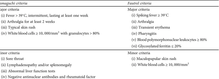
To date, diagnosis of AOSD is based on sets of
classifica-tion criteria proposed during the last decades, as shown in
Table 3 [129, 132]. Those proposed by Yamaguchi et al. are
the most widely used and require the preliminary exclusion
of infections, cancer, and autoimmune diseases [132]; the
cri-teria proposed by Fautrel et al. are more recent and give a
higher weight to ferritin levels and glycosylated ferritin
frac-tion [129]. The Yamaguchi criteria exhibit a sensitivity of
79.2% and specificity of 93.8%; the Fautrel criteria have a
sen-sitivity of 80.6% and specificity of 98.5% [129, 132].
Schnitzler’s disease is a very rare inflammatory condition
presenting with intermittent fever and urticaria-like skin
rashes occurring in subjects with monoclonal gammopathy
(mostly of the IgM type) and a considerable increase of in
flam-matory markers. Arthralgia or arthritis, lymphadenopathy,
Table 3: Clinical criteria for the diagnosis of adult onset Still’s disease (AOSD); adapted from Yamaguchi et al. [132] and Fautrel et al. [129]. Infections, malignancies, and other rheumatologic diseases represent exclusion criteria.
Yamaguchi criteria Fautrel criteria
Major criteria Major criteria
(i) Fever > 39°C, intermittent, lasting at least one week (i) Spiking fever≥ 39°C
(ii) Arthralgia for at least 2 weeks (ii) Arthralgia
(iii) Typical skin rash (iii) Transient erythema
(iv) White blood cells≥ 10, 000/mm3with granulocytes > 80% (iv) Pharyngitis
(v) Blood polymorphonuclear leukocytes≥ 80% (vi) Glycosylated ferritin≤ 20%
Minor criteria Minor criteria
(i) Sore throat (i) Maculopapular skin rash
(ii) Lymphadenopathy and/or splenomegaly (ii) White blood cells≥ 10, 000/mm3
(iii) Abnormal liver function tests
(iv) Negative antinuclear antibodies and rheumatoid factor
and liver and/or spleen enlargement are frequent
manifesta-tions as well. AA amyloidosis represents a potential
long-term consequence of an untreated disease, while monoclonal
IgM gammopathy progresses toward an overt
lymphoprolifer-ative disorder in about 15-20% of patients [133]. Diagnosis is
based on clinical criteria, which have been validated in a cohort
of real-life patients, showing a sensitivity for de
finite and
prob-able diagnosis of 81% and 93%, respectively, with a
corre-sponding specificity of 100% and 97% (see Table 4) [134, 135].
The periodic fever, aphthous stomatitis, pharyngitis, and
adenitis (PFAPA) syndrome is an autoinflammatory
condi-tion characterized by the clinical features described by the
acronym itself. Other various manifestations may occur in
addition to cardinal signs, including skin rash, abdominal
pain, arthralgia, and conjunctivitis [136
–138]. This
syn-drome is currently the most frequent autoin
flammatory
cause of noninfective recurrent fever among children in
the European population. However, although initially
con-fined to the pediatric world, the PFAPA syndrome has been
recently identified as a possible cause of periodic febrile
epi-sodes also among adults [139–143]. In this regard,
diagnos-tic criteria specifically tailored on adult patients have been
described. Table 5 illustrates PFAPA diagnostic criteria for
both children and adults [144–146]. In relationship with adult
patients, the proposed diagnostic criteria identify the PFAPA
syndrome among subjects presenting with an increase of
in
flammatory markers during attacks, symptom-free
inter-vals between in
flammatory bouts, and at least one item
among erythematous pharyngitis and cervical lymphadenitis
during fevers. These criteria should be applied only after
hav-ing excluded infectious, autoimmune, and neoplastic diseases
as well as mAIDs [146].
Behçet’s disease (BD), recently classified at the crossroad
between autoimmune and polygenic AIDs, may also manifest
with otherwise unexplained fever [147, 148]: it is clinically
characterized by a classical
“triad” of recurrent oral
aphtho-sis, genital ulcers, and uveitis, especially in the form of
poste-rior uveitis and panuveitis. However, in
flammation may
involve also (i) the gastrointestinal tract mimicking
inflam-matory bowel diseases, (ii) the vascular tree with multiple
venous thrombosis and arterial aneurysms potentially
affect-ing all body sites, (iii) the nervous system manifestaffect-ing with a
wide array of manifestations, most commonly affecting the
brainstem and diencephalic regions, and (iv) the skin with
erythema nodosum or pseudofolliculitis [149
–151].
Diagno-sis of BD is based on the ful
fillment of international
diagnos-tic criteria and the International Study Group Criteria for
BD, which are reported in Table 6 [152, 153].
4. Specific Organ Involvement in mAIDs
4.1. Constitutional Symptoms. Recurrent fever and asthenia
are the main constitutional symptoms in most mAIDs.
How-ever, while asthenia is a nonspecific manifestation identified
in many systemic disorders, specific inflammatory features
associated with recurrent fever may provide valuable
clin-ical information. In particular, FMF attacks generally last
between 6 and 72 hours, while TRAPS patients are
charac-terized by episodes lasting 1 to 3 weeks; CAPS and HIDS
manifest with fever generally lasting 4-7 days [8, 10, 12,
56, 59]. Of note, also the PFAPA syndrome is
character-ized by fever episodes resolving in 4-7 days and this
disor-der should always be taken into account when considisor-dering
differential diagnosis [136, 137]. Frequency of fever attacks
and body temperature may be extremely variable and are
less useful for diagnostic purposes.
Systemic reactive AA amyloidosis is the most challenging
long-term complication of all mAIDs. Nevertheless, MKD
patients are less frequently involved, with a frequency
rang-ing between 3 and 5% [125, 154, 155]. Similarly, amyloidosis
is infrequent in patients with FCAS, while nontreated FMF,
MWS, and TRAPS may be complicated with systemic
amy-loidosis in up to 50%, 25%, and 20% of the cases over time,
respectively [30].
4.2. Skin Manifestations. The skin is frequently involved in
mAIDs, and some cutaneous
findings may be
pathogno-monic for a rapid diagnosis. This is the case of
erysipelas-like erythema in FMF patients presenting with tender, warm,
and swollen erythematous plaques, generally occurring on
the distal extremities and triggered by physical activity
[10, 12, 156]. Nevertheless, the prevalence of
erysipelas-like erythema varies among populations with FMF and
is quite uncommon in Arabs and Armenians [157]. Skin
biopsy of this lesion might reveal edema with perivascular
and interstitial in
filtrate consisting of neutrophils and
lym-phocytes [158, 159].
A centrifugal migratory erythematous rash is the more
typical skin manifestation of TRAPS: it is generally
accompa-nied by myalgia due to monocytic fasciitis of the underlying
muscles [160, 161]. Although the legs are more frequently
involved, the trunk is not spared [13, 68, 156, 161]. A mild
perivascular and interstitial in
filtrate of mononuclear cells
is usually identi
fied at the skin biopsy [156, 160], even though
a slight deposition of C3 and C4 in the dermis has also been
described [162].
Table 4: Clinical criteria for the diagnosis of Schnitzler’s disease [134]. A definite diagnosis is justified by the fulfillment of the 2 mandatory criteria and at least 2 or 3 minor criteria in patients with IgM or IgG monoclonal gammopathy, respectively. Diagnosis is probable when the 2 mandatory criteria are fulfilled and at least 1 or 2 minor criteria in patients with IgM or IgG monoclonal gammopathy, respectively, are present.
Mandatory criteria
(i) Chronic urticaria-like skin rash (ii) IgM or IgG monoclonal gammopathy Minor criteria
(i) Unexplained recurrent fever > 38°C
(ii) Abnormal bone remodeling assessed by bone scintigraphy or magnetic resonance imaging with abnormal bone alkaline phosphatase level
(iii) Neutrophilic dermal infiltrate at skin biopsy with neither fibrinoid necrosis nor significant dermal edema
(iv) Leukocytosis (neutrophils > 10, 000/mm3) and/or elevated C-reactive protein (>30 mg/l)
Patients with CAPS present with urticaria-like skin
rashes in almost all cases [91]. Indeed, an urticaria-like rash
appears among the diagnostic items recently proposed for
the clinical assessment of CAPS patients [98]. This rash is
typically induced by cold exposure in FCAS, but it may
develop at birth or within a few hours after birth in patients
with MWS or CINCA/NOMID, disregarding environment
temperature. Noteworthy, patients with FCAS are negative
to the ice cube test, unlike those affected by cold physical
urti-caria. Skin biopsy of urticaria-like lesions in CAPS shows a
neutrophilic infiltrate of the dermis, with the absence of
vas-culitis, scarce eosinophils, and sparing of the epidermal layer
[156, 163]. Schnitzler
’s syndrome should be considered for
di
fferential diagnosis in patients with monoclonal
gammopa-thy and skin manifestations [134].
Skin lesions are identified in more than two thirds of
patients with MKD: they are variable and nonspecific,
including maculopapular, morbilliform, erythematous, and
purpuric rashes [156, 163, 164]. Oral and/or genital
aphtho-sis can be seen in about half of MKD patients, requiring
dif-ferential diagnosis with BD, PFAPA syndrome, and FMF
[136, 149, 165, 166].
4.3. Musculoskeletal Manifestations. Most patients with
mAIDs report musculoskeletal manifestations, with FMF
and CINCA/NOMID being the most typically involved.
Nev-ertheless, patients with MKD and TRAPS may refer joint and
muscle symptoms as well [8, 10, 12, 56, 59].
Acute monoarthritis may be the sole disease
manifesta-tion in up to 15% of FMF patients [167
–169]. Nonetheless,
monoarthritis is less frequent when patients are genetically
diagnosed, but do not ful
fill clinical diagnostic criteria for
FMF [169]. In these cases, symmetrical acute arthritis of large
joints of the lower limbs is the most frequent
finding.
Alter-natively, subacute or chronic articular involvement may be
identified [170, 171]. Although the knee and hip are more
frequently involved, FMF-related arthritis may affect all
articular sites, including the elbow, sternoclavicular joints,
and metacarpophalangeal joints [172, 173]. Also sacroiliitis
may be identi
fied in FMF and seems to be closely related to
the M694V MEFV mutation that has been suggested as a
sus-ceptibility factor for enthesitis-related arthritis [174
–178].
While myositis is uncommon in FMF, myalgia is reported in
up to 25% of the patients and may be classified as
spontane-ous, induced by exercise, or protracted febrile myalgia [179,
180]. Specifically, protracted febrile myalgia is a debilitating
manifestation often accompanied by other symptoms such
as abdominal pain, diarrhea, arthritis/arthralgia, and transient
vasculitic lesions mimicking the Henoch-Schönlein purpura.
Electromyography shows only nonspeci
fic changes, while
muscle enzymes are generally within the normal range [181].
There is some evidence that musculoskeletal symptoms may
Table 5: Clinical criteria for the diagnosis of periodic fever, aphthous stomatitis, pharyngitis, and cervical adenitis (PFAPA) syndrome in children and adults; adapted from Thomas et al. [145] and Cantarini et al. [146], respectively. Infections, malignancies, autoimmune and other autoinflammatory diseases should be ruled out before the application of criteria in adult patients.
Diagnostic criteria for children Diagnostic criteria for adults
(1) Regularly recurring fevers with early age of onset (<5 years of age), occurring in the absence of upper respiratory tract infections (2) At least one among the following symptoms:
(i) Aphthous stomatitis (ii) Cervical lymphadenitis (iii) Pharyngitis
(3) Exclusion of cyclic neutropenia
(4) Asymptomatic interval between episodes (5) Normal growth and development
(1) Recurrent fever accompanied by (i) Erythematous pharyngitis and/or (ii) Cervical lymphadenitis
(2) Increased inflammatory markers during febrile attacks (3) Asymptomatic interval between episodes
All the criteria are requested All the criteria are requested
Table 6: Criteria for the diagnosis of Behçet’s disease (BD): International Study Group Criteria (ISGC) [152] and International Criteria for BD (ICBD) [153].
ISGC ICBD
Mandatory item Items Score
(i) Recurrent oral aphthosis (at least 3 episodes per year) (i) Oral aphthosis 2
Additional items (ii) Genital aphthosis 2
(i) Recurrent genital ulcers
(ii) Ocular or retinal inflammatory manifestations
(iii) Skin lesions (papulopustular lesions, erythema nodosum) (iv) Positive pathergy reaction
(iii) Ocular inflammatory lesions 2
(iv) Skin lesions 1
(v) Neurological involvement 1
(vi) Vascular manifestations 1
(vii) Positive pathergy test 1
occur more frequently in patients with late-onset disease than
in those displaying an early onset [182].
Articular involvement in CAPS a
ffects approximately 60%
of patients and ranges from arthralgia with transient joint
swelling during
flares to chronic disabling joint disease [91,
95]. Severe joint manifestations including chronic arthritides
with structural changes and bony overgrowth are described
in patients with CINCA/NOMID and involve especially the
knees. These articular affections bring about severe functional
impairment since early adulthood [91, 183–185].
With regard to TRAPS, myalgia with monocytic fasciitis in
the absence of direct inflammatory involvement of the muscle
is a characteristic feature of the disease. Therefore, serum
levels of muscle enzymes are usually normal [160, 161].
Myal-gia is generally associated with warm and tender patches of the
overlying skin, which are recognized as typical features of
TRAPS [61, 70, 71, 160, 161]. Regarding joint involvement,
TRAPS patients often suffer from arthralgia, while arthritis is
an unusual
finding that eventually affects large joints in a
nonerosive fashion [186]. Despite this, the involvement of
small joints and sacroiliitis have been anecdotally reported in
subjects carrying low-penetrance mutations [187].
Polyarthralgia is observed in approximately 80% of MKD
patients, while nonerosive arthritis of large joints and myalgia
are reported in about half of the cases. Musculoskeletal
mani-festations may be severe and disabling in about 7-8% of
patients, who might develop
flexion contractures, bone
ero-sions, and even deformities. Articular symptoms may persist
also during symptom-free intervals of MKD patients [155].
4.4. Serosal Involvement in mAIDs. Recurrent serosal acute
in
flammation may be a presenting peculiar manifestation of
mAIDs. In particular, recurrent pleuritis and peritonitis
rep-resent typical features of FMF and they are counted among
Recurrent fever with elevatedinflammatory markers and mucocutaneous manifestations
Rule out infections, neoplastic and autoimmune diseases
Aphthosis?
Pseudofollicolitis? Severe acne? Erythema nodosum?
Pyoderma gangrenosum? Maculopapular rash? Migratory erythematous rash?
Urticaria-like rash ? Erysipelas-like rash?
4-7 days
Apply FMF clinical criteria and test
MEFVgene Test MVK gene Apply PFAPA clinical criteria Apply BD clinical criteria Onset <5 years Onset > 5 years or MVK gene tested negative Apply SD clinical criteria Monoclonal gammopathy Apply FMF clinical criteria and test MEFV
gene Test TNFRSF1A gene Salmon-colored rash Morbilliform or purpuric rash Apply AOSD clinical criteria Test MVK gene Apply PFAPA clinical criteria Onset <5 years Onset >5 years or MVK gene tested negative Apply CAPS clinical
criteria and test NLRP3 gene 1-3 days
>7 days
CAPS ruled out Think about
NLRP12-and NLRC4 -related diseases and PLAID
syndrome
Think about PAAND and PSTPiP1-related
disease Think about A20
haploinsufficiency
Figure 2: Diagnostic flow chart for main autoinflammatory diseases displaying recurrent fevers and mucocutaneous manifestations. List of abbreviations: FMF—familial Mediterranean fever; PFAPA—periodic fever, aphthous stomatitis, pharyngitis, and cervical adenitis; BD— Behçet’s disease; CAPS—cryopyrin-associated periodic syndrome; SD—Schnitzler’s disease; AOSD—adult-onset Still’s disease; NLRP12— nucleotide-binding domain and leucine-rich repeat-containing protein 12; NLRC4—nucleotide-binding domain, leucine-rich repeat, and caspase recruitment domain-containing 4; PLAID—phospholipase C gamma 2-associated antibody deficiency and immune dysregulation; PAAND—pyrin-associated autoinflammation with neutrophilic dermatosis; PSTPiP1—proline-serine-threonine phosphatase interacting protein 1.
the major items in the clinical diagnostic criteria [28, 55, 65].
Similarly, chest pain and abdominal pain, which are possibly
due to pericarditis, pleuritis, or peritonitis, appear among the
pediatric FMF criteria [56].
Notably, although the incidence of pericarditis has been
reported to be quite low in FMF [188, 189],
echocardiogra-phy may identify a subclinical involvement in up to 27% of
patients, especially among subjects with a longer disease
duration [190].
Recurrent acute pericarditis has been largely described in
patients with TRAPS. Of note, recurrent acute pericarditis
may be the sole clinical manifestation in subjects carrying
low-penetrance mutations [66, 74, 160, 191, 192]. Therefore,
clinical clues have been proposed to identify among patients
with idiopathic recurrent acute pericarditis those who
poten-tially might carry TNFRSF1A gene mutations: a positive
fam-ily history for either pericarditis or recurrent fevers, a high
rate of recurrences beyond the
first year of disease, colchicine
resistance, and the need for immunosuppressants to achieve
control of the disease should induce a clinical suspicion of
TRAPS [64, 67].
Although not included in the clinical diagnostic criteria,
pleuropericarditis is frequently identified also in patients
with AOSD [193].
4.5. Gastrointestinal Manifestations. Although
gastrointesti-nal symptoms may be found in almost all mAIDs, abdomigastrointesti-nal
pain with or without diarrhea and vomiting are more
fre-quently found in FMF and HIDS [125, 194]. Abdominal pain
in FMF is closely related to aseptic recurrent peritonitis and
usually represents a prevailing clinical manifestation. For this
reason, abdominal pain (with or without peritonitis) appears
in all clinical FMF criteria [55, 56] as well as in the more recent
Eurofever classification criteria [21]. Although less useful for
diagnostic purposes, other gastrointestinal manifestations
have been often reported in FMF patients [195]. Noteworthy,
inflammatory bowel diseases seem to be more frequent and
severe in patients with FMF [196] and, conversely, the rate
of MEFV mutations has been found higher in subjects
diag-nosed with in
flammatory bowel diseases [197].
Abdominal pain, vomiting, and/or diarrhea along with
oral aphthosis are almost constant in HIDS patients during
Recurrent fever with elevatedinflammatory markers and musculoskeletal manifestations
Rule out infections, neoplastic and autoimmune diseases
Myalgia Oligoarthritis Monoarthritis Granulomatous
arthritis Polyarthritis 1-3 days 4-7 days >7 days Test MVK gene Consider Blau syndrome and test
NOD2 gene
Onset <5 years
Apply CAPS clinical criteria and test
NLRP3 gene
Migratory with overlying erythematous
rash Severe febrile myalgia
Apply FMF clinical criteria and test
MEFV gene
Test TNFRSF1A gene clinical criteriaApply AOSD If not fulfilled
Adult onset Early onset, severe, with bony overgrowth
Apply AOSD clinical criteria
Apply BD clinical criteria Apply FMF clinical
criteria and test MEFV gene
Figure 3: Diagnostic flow chart for main autoinflammatory diseases displaying recurrent fevers and musculoskeletal manifestations. List of abbreviations: FMF—familial Mediterranean fever; AOSD—adult-onset Still’s disease; BD—Behçet’s disease; CAPS—cryopyrin-associated periodic syndrome.
fever attacks. Sterile peritonitis mimicking acute appendicitis
can lead to surgical interventions, peritoneal adhesions, and
intestinal obstruction [125]. Based on the higher frequency
of gastrointestinal a
ffections, the occurrence of diarrhea
dur-ing fever attacks has been included in the Eurofever
classifi-cation criteria [21].
Notably, abdominal pain and intestinal inflammation
may be also manifestations of BD, which should be ruled
out during the diagnostic process [152, 153]. Similarly, the
PFAPA syndrome may be associated with abdominal pain
and various intestinal disorders [198].
4.6. Ocular Manifestations. No speci
fic ocular involvement is
reported in FMF. However, fundus abnormalities resembling
colloid bodies in the Bruch membrane have been described
on routine funduscopic examination during FMF fever
attacks [199, 200]. In addition, increased choroidal thickness
has been identified inconsistently during attacks [201].
Ante-rior, posteAnte-rior, and intermediate uveitis have been
occasion-ally described in FMF patients [202]. Conversely, FMF
could represent a predisposing factor for the development
of keratoconus, especially in patients carrying homozygous
mutations [203].
In TRAPS patients, the eye is most commonly a
ffected
with periorbital edema or pain and conjunctivitis [61].
Peri-orbital edema is more common in pediatric patients as well
as in adults with an early disease onset. On the contrary,
eye manifestations are less frequently encountered in patients
carrying the R92Q low-penetrance TNFRSF1A mutation [60].
Nevertheless, bilateral panuveitis has been recently detected in
a child with TRAPS carrying the R92Q variant [204].
Retinitis pigmentosa and cataract may be recognized in
patients with MKD, and in particular retinitis pigmentosa
may occasionally represent the presenting feature of the
dis-ease [205–207].
Optic disc changes including papilledema and optic
atro-phy are the most common ocular
findings in MWS and
CIN-CA/NOMID [91]. In this regard, the gradual loss of nerve
fibers owing to optic disc alterations explains gradual visual
loss in such patients [208]. Of note, retinal vasculitis, focal
choroiditis, vitritis, band keratopathy, nummular stromal
in
filtration, corneal vascularization, and cataract are rare
manifestations occasionally encountered in CINCA/NOMID
[209, 210], while midstromal changes, episcleritis, keratitis,
and uveitis have been reported in MWS [211, 212]. Lastly,
eye involvement is mainly represented by conjunctivitis in
FCAS patients. Keratitis with bilateral corneal scars have
been reported only in severe cases [213].
Ocular inflammatory involvement should also pave the
attention to BD, which may manifest with recurrent, unilateral
or bilateral panuveitis, posterior uveitis, and/or retinal
vasculi-tis; less frequently, anterior uveitis can occur [152, 153].
Recurrent fever with elevatedinflammatory markers and ocular manifestations
Rule out infections, neoplastic and autoimmune diseases
Periorbital
edema or pain Keratoconus
Non-granulomatous Granulomatous Early cataract or retinitis pigmentosa Adult onset Test MVK gene Consider Blau syndrome and test
NOD2 gene Onset <5 years
Apply CAPS clinical criteria and test
NLRP3 gene Fever >7 days Test TNFRSF1A gene Apply BD clinical criteria Uveitis Fever <3 days
Apply FMF clinical criteria and test MEFVgene
Pediatric onset Think about A20
haploinsufficiency
Figure 4: Diagnostic flow chart for main autoinflammatory diseases displaying recurrent fevers and ocular manifestations. List of abbreviations: FMF—familial Mediterranean fever; BD—Behçet’s disease; CAPS—cryopyrin-associated periodic syndrome.

![Table 6: Criteria for the diagnosis of Behçet’s disease (BD): International Study Group Criteria (ISGC) [152] and International Criteria for BD (ICBD) [153].](https://thumb-eu.123doks.com/thumbv2/123dokorg/4658797.42474/10.899.85.827.433.635/criteria-diagnosis-behçet-disease-international-criteria-international-criteria.webp)