UNIVERSITY OF CATANIA
FACULTY OF AGRICULTURE
DEPARTMENT OF AGRI-FOOD AND ENVIRONMENTAL MANAGEMENT SYSTEMS
INTERNATIONAL PHD PROGRAM IN PLANT HEALTH TECHNOLOGY
STUDIES ON THE FUNGAL DISEASES OF MANGO
WITH PARTICULAR REFERENCE TO DISEASES
CAUSED BY BOTRYOSPHAERIA SPECIES
This thesis is presented for the degree of
Doctor of Philosophy by
AHMED MAHMOUD ISMAIL
COORDINATOR
PROF. CARMELO RAPISARDA
CYCLE-XXIV 2008-2011
SUPERVISOR
PROF. GABRIELLA CIRVILLERI
CO-SUPERVISOR
I
DEDICATIONS AND ACKNOWLEDGMENTS
I dedicate this thesis to the following:
My wife thanks for your eternal love, patience and support during the
accomplishment of this thesis.
My dear parents, my great thanks for your pray, eternal love and the endless
support to me in my entire life.
Acknowledgments
I would like to be grateful to my supervisors Prof. Gabriella Cirvilleri and Dr.
Anna Maria D’Onghia for their help to accomplish this thesis.
My sincere thank and gratitude to Prof. Giancarlo Polizzi for his encouragement,
guidance and critical review of this thesis. I really appreciated that for you.
My appreciation, grateful and respect to Prof. Pedro W. Crous, the Director of
the Fungal Biodiversity Center (CBS) Utrecht, The Netherlands, who give me the
opportunity to do the great part of my research project and to learn a lot a bout
mycology in one of the most well-known mycological research institute (CBS).
I would like to express a bout my gratefulness to Dr. Lorenzo Lombard and Dr.
Groenewald for their kind help, guidance, time, directions and efforts during my
stay in the (CBS).
Finally, I would like to thank the Fungal Biodiversity Center (CBS),
Mediterranean Agronomic Institute (IAMB) and the University of Catania for
the financial support.
II
ABSTRACT
Mango (Mangifera indica) is a promising tropical fruit crop in Italy. In the near future, it is expected that commercial and backyard plantings of mango trees will increase being a gainful crop. Cultivation of mango is concentrated in four provinces (Catania, Palermo, Messina and Ragusa) located in the southern Italy (Sicily). Since the introduction of mango cultivation into Italy in 1990, little information is available regarding its problematical diseases. Thus, the present work represents the first attempt to assess the presence and the diversity of fungal species associated with different symptoms patterns observed on mango trees during a survey conducted in 2009-2010. Several fungi have been isolated and tentatively identified up to genus level. The most encountered isolated fungal taxa were Alternaria spp., Botryosphaeria spp., Colletotrichum spp. and Pestalotiopsis spp. All these fungi except Botryosphaeria spp. were identified based on conidia and culture characters and tested for pathogenicity on mango detached leaves cv. Kensington Pride. The isolated fungi showed to be pathogenic with different degrees in virulence. Other fungi were occasionally isolated with low frequency such as Botrytis sp., Macrophoma spp., Phoma spp., and Stemphylium spp.
The present work focused mostly on the Botryosphaeriaceae associated with dieback disease in Italy and in Egypt. In the present thesis, incorporation of morphology and DNA sequence data of ITS and TEF-1α revealed diverse array of
Botryosphaeria spp. associated with mango dieback: Neofusicoccum parvum, N. australe, were the dominant species while, N. vitifusiforme and other two novel
species Neofusicoccum sp. 3 and Neofusicoccum sp. 18 were less frequently isolated and associated with dieback disease in Italy. The two novel species of
Neofusicoccum were phylogenetically and morphologically distinct from the other
species. On the other hand, Lasiodiplodia theobromae and L. pseudotheobromae were the most prevalent species associated with mango dieback in Egypt. The two new described species Lasiodiplodia sp. 8 and Lasiodiplodia sp. 10 were less frequently isolated from mango in Egypt. All tested isolates of Neofusicoccum spp. and
Lasiodiplodia spp. except the new species were pathogenic to apple fruits as well as
mango cv. Kensington Pride seedlings. Additional studies, including, extensive survey and sampling in different geographical are required to understand the ecology of the new species and to determine their role to cause diseases to mango. Furthermore, critical intervention by developing control strategies such as chemical, biological and agricultural methods must be taken in concern.
III
TABLE OF CONTENTS
DEDICATIONS AND ACKNOWLEDGMENTS... I ABSTRACT ... II LIST OF TABLES ... VII LIST OF FIGURES ... VIII LIST OF ABBREVIATIONS ... XIV
CHAPTER .1 ... 1
MANGO AND THE IMPACT OF FUNGAL DISEASES WITH SPECIFIC REFERENCE TO DISEASES CAUSED BY BOTRYOSPHAERIA SPP. ... 1
• INTRODUCTION ... 2
• HISTORICAL REVIEW ... 6
• MANGO DISEASES AND THEIR IMPACT ... 6
• THE MAJOR DISEASES OF MANGO ... 7
• BOTRYOSPHAERIA DISEASES ON MANGO ... 7
• PRE-HARVEST DISEASES ... 7
• TIP DIEBACK ... 7
• BLOSSOM BLIGHT ... 8
• POST-HARVEST DISEASES... 9
• STEM-END ROT ... 9
•TAXONOMY OF BOTRYOSPHAERIA SPECIES ASSOCIATED WITH MANGO DISEASES ... 10 • Botryosphaeria parva ... 10 • Botryosphaeria ribis ... 10 • Botryosphaeria rhodina ... 11 • Botryosphaeria lutea ... 12 • Botryosphaeria australis ... 13 • Neofusicoccum mangiferae ... 13 • Neofusicoccum vitifusiforme ... 14 • EPIDEMIOLOGY... 15 • CONTROL STRATEGIES ... 15 • PRE-HARVEST ... 16 • POST-HARVEST... 16 • ANTHRACNOSE ... 17
IV
• ALTERNARIA BLACK SPOT ... 18
• POWDERY MILDEW ... 18
• MALFORMATION... 19
• GREY LEAF SPOT ... 20
• MACROPHOMA LEAF BLIGHT... 21
• MINOR DISEASES OF MANGO ... 21
• THESIS AIMS... 22
CHAPTER .2 ... 24
ASSESSMENT OF MANGO DISEASES IN ITALY ... 24
• INTRODUCTION ... 25
• MATERIALS AND METHODS ... 26
• Field observations and collection of plant materials ... 26
• Isolation ... 26
• Morphological characterisation ... 28
• Pathogenicity tests ... 28
• RESULTS ... 29
• Field observations and collection of plant materials ... 29
• Incidence and initial identification of isolated fungi ... 34
• Morphological characterisation of the isolated fungi ... 38
• Alternaria spp. ... 38
• Colletotrichum spp. ... 40
• Pestalotiopsis spp. ... 42
• Characterisation of other associated fungi ... 44
• Pathogenicity tests ... 44 • Alternaria spp. ... 44 • Colletotrichum spp. ... 44 • Pestalotiopsis spp. ... 45 •DISCUSSION ... 50 CHAPTER .3 ... 55
MORPHOLOGY AND PHYLOGENYOF BOTRYOSPHAERIA SPP. ASSOCIATED WITH MANGO DIEBACK IN ITALY ... 55
• INTRODUCTION ... 56
• MATERIALS AND METHODS ... 57
V
• Morphological characterisation ... 57
• Temperatures effect on the mycelial growth of Neofusicoccum spp. ... 58
• Data analysis ... 58
• Molecular characterisation ... 58
• DNA isolation ... 58
• PCR amplification ... 59
• DNA sequencing and phylogentic analysis ... 59
• RESULTS ... 71
• Isolation and incidence ... 71
• PCR amplification ... 72
• Phylogenetic analysis of Botryosphaeria spp. ... 73
• TAXONOMY ... 76 • Neofusicoccum parvum ... 76 • Neofusicoccum vitifusiforme ... 78 • Neofusicoccum australe ... 80 • Neofusicoccum sp. 3 ... 82 • Neofusicoccum sp. 18 ... 85 • DISCUSSION ... 89 CHAPTER .4 ... 93
MORPHOLOGY AND PHYLOGENY OF BOTRYOSPHAERIA SPP. ASSOCIATED WITH MANGO DIEBACK IN EGYPT ... 93
• INTRODUCTION ... 94
• MATERIALS AND METHODS ... 95
• Fungal isolation ... 95
• Morphological characterisation ... 95
• Temperatures effect on the mycelial growth of Lasiodiplodia spp. ... 95
• Molecular characterisation ... 95 • RESULTS ... 95 • Isolates ... 95 • Phylogeny of Botryosphaeria spp. ... 95 • PCR amplification ... 95 • Phylogenetic analysis ... 96 • TAXONOMY ... 98 • Lasiodiplodia theobromae ... 98
VI
• Lasiodiplodia pseudotheobromae ... 100 • Lasiodiplodia sp. 10 ... 102 • Lasiodiplodia sp. 8 ... 105 • DISCUSSION ... 109 CHAPTER .5 ... 112PATHOGENICITY TESTS OF BOTRYOSPHAERIA SPP. ASSOCIATED WITH MANGO DIE BACK DISEASE IN ITALY AND IN EGYPT... 112
• INTRODUCTION ... 113
• MATERIALS AND METHODS ... 114
• Apple fruit assay ... 114
• Pathogenicity tests on mango seedlings ... 116
• Studied isolates ... 116
• Inoculation of mango seedlings ... 116
• Data analysis... 117
• RESULTS ... 117
• Apple fruit assay ... 117
• Pathogenicity tests on mango seedlings ... 121
• DISCUSSION ... 126
CHAPTER .6 ... 129
• GENERAL DISCUSSION AND CONCLUSION ... 129
CHAPTER .7 ... 133
VII
LIST OF TABLES
Table 1 The incidence of the isolated fungi from various mango trees organs
recovered from four provinces (Messina, Catania, Palermo and Ragusa) located in south Italy (Sicily) during a survey conducted in 2009 and 2010. --- 35
Table 2 Degenerate Oligonucleotide primers used for PCR amplification and
sequencing --- 61
Table 3 Botryosphaeria isolates retrieved from GenBank used for phylogenetic
analysis. --- 62
Table 4 Neofusicoccum and Lasiodiplodia isolates obtained in this study from
mango in Italy and Egypt --- 67
Table 5 Isolates of Botryosphaeria spp. used for apple fruit assay and
VIII
LIST OF FIGURES
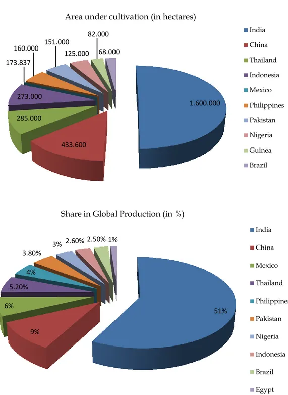
Figure 1 Mango cultivated area in hectares (A), and sharing percentage in the
global production (B) of top 10 countries in the world (Anonymous, 2011). --- 5
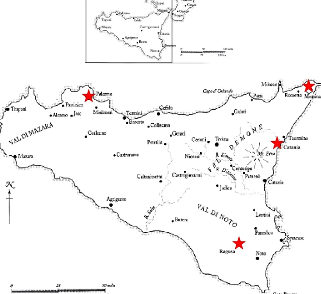
Figure 2 Illustration map of the southern part of Italy (Sicily) showing the
monitored sites (Palermo, Messina, Catania and Ragusa) marked by red stars. --- 27
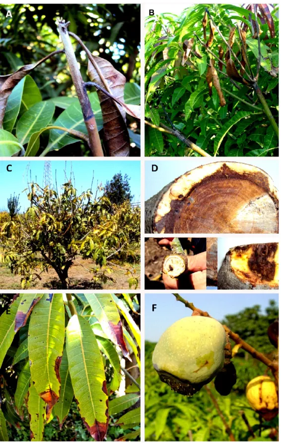
Figure 3 A, B Dieback symptoms on the young twigs and branches starting from
the tip extended downward. C, in severe infection most of the apical parts die and dry leading to death of the whole tree, D, cross sections of stem and branches showing the brown vascular discolouration under cambium tissues, E, brown to black lesions on the leaf margins of the affected leaves. F, mummified small fruits on the early stage and dark brown patches on the big fruit lead to soft rot of the internal pulp. --- 30
Figure 4 Symptoms of Alternaria leaf spot caused by Alternaria spp. in the field. A
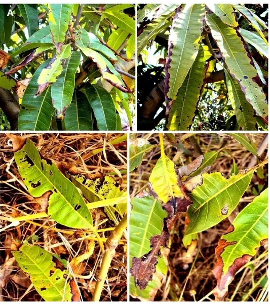
and B, dark brown to black lesions along leaf margins giving the zigzag shape, C and D, coalescing of lesions causing upwards rolling of leaf margins (arrows). -- 31
Figure 5 Grey leaf spot symptoms observed in the field. A, Small to large grey
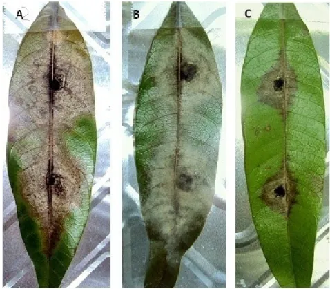
necrotic lesions with black margins B, coalesced to form larger grey patches and on the necrotic tissues abundant black acervuli were formed (see arrow). C, large white to grey specks on the secondary branches (arrow) D, light brown lesions extended to the main and secondary axis of the panicles causing blossom blight symptoms. --- 32
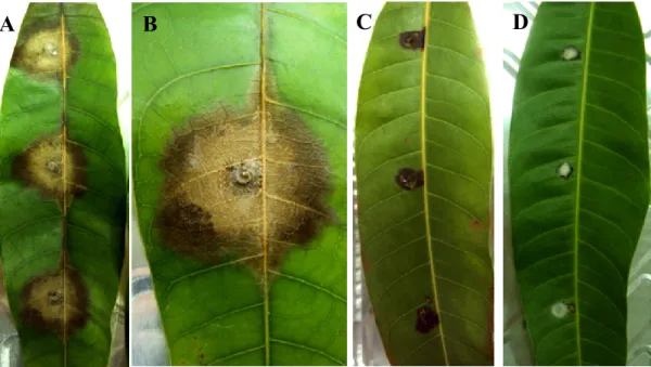
Figure 6 Symptoms of anthracnose disease observed in the surveyed fields. A,B
Tiny black spots scattered on the young leaf blade have irregular margins coalescing to form large necrotic black patches that start from the leaf margins to the apical part covering most of the leaf blade. C, D, blighting of leaves giving the scorching appearance. E, dark necrotic patches lead to dying the apical shoot, orange masses of conidia released from semi-immersed acervuli observed on the necrotic area. F, brown to dark sunken and depressed lesion on the fruit. --- 33
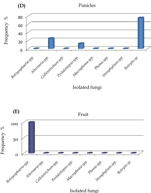
Figure 7 The frequency (%) of isolated fungi according to mango trees organs; A,
Leaves, B, Twigs, C, Branches, D, Panicles and E, Fruit of cv. Kensington Pride manifested different symptoms patterns in the field. Data within columns are the mean percentage (%) calculated from the number of samples yielding the isolated fungi divided on the total number of samples for each plant part. --- 37
Figure 8 Morphological characteristics of colony (front and reverse side), conidia
IX
and H (Al-13). I, J, K and L (Al-17) of Alternaria spp. isolated from mango. Scale bars = 10 µm. --- 39Figure 9 A, B, D, E, G, H, J, K, culture morphology of Colletotrichum spp. on PDA
after 8 days of incubation at 25°C. C, F, I, L, the commonly observed conidia for
Colletotrichum spp. Scale bars = 10 µm. --- 41
Figure 10 Colony and conidia characteristics of representatives Pestalotiopsis
isolates on PDA after 9 days at 25°C. (A, B, D, E, G, H, J, K,), culture morphology of Pestalotiopsis spp. (C, F, I, L), commonly observed conidia from Pestalotiopsis spp. Scale bars = 10 µm. --- 43
Figure 11 Symptoms of Alternaria brown leaf spot developed on the detached
mango leaves after 7 days of inoculation. Different levels of symptoms appeared showing the variability in lesion diameters produced by three isolates A, Al-2 B, Al-29 and C, Al-54 of Alternaria spp. --- 46
Figure 12 Mean lesion diameters (y-axis) induced by eighteen representative
isolates of Alternaria spp. (x-axis) on detached mango leaves cv. Kensington Pride after 7 days of incubation at 25ºC. Values from each isolate are the mean of n=4 replicates ± (SD). Values on the same columns followed by the same letters are not significantly different according to LSD test (P < 0.05).--- 46
Figure 13 Symptoms reproduced by selected isolates of Colletotrichum spp. on
detached mango leaves after 8-days of incubation at 25º C. A, sever symptoms developed by the isolate Co-35. B, magnified photo of the lesion showing orange masses of fungus conidia formed on the necrotic tissues. C, weak symptoms developed by the Co-34 isolate. D, small lesions developed by the Co-4 isolate showing the weakness of the fungus to reproduce the symptoms observed in the field. --- 47
Figure 14 Pathogenicity tests of Colletotrichum spp. isolates. Data represent the
mean lesion diameters (mm) produced by the tested eight isolates on detached mango leaves after 8 days of incubation at 25ºC. Columns bearing the same letters are not significantly different according to the LSD test (P < 0.05). --- 47
Figure 15 Symptoms of grey leaf spot disease on mango detached leaves after 9
days of inoculation with Pestalotiopsis species. Developed symptoms varied from: A, small irregular light to dark brown lesions around inoculation sites. B, enlarged to form larger lesions along the main leaf vein. C, lesions expanded to coalesce forming brown patches along leaf blade giving rise to the scorch appearance. D, brown to black scattered acervuli formed on the center of the necrotic tissues (see arrow). --- 48
X
Figure 16 Lesions diameters (y-axis) developed by 21 isolates of Pestalotiopsis spp.(x-axis) inoculated on detached mango leaves cv. Kensington Pride after 7 days of incubation at 25ºC. Values from each isolate are the mean of n=6 replicates ± (SD). Columns bearing the same letters are not significantly different according to the Least Significant Difference (LSD) test (P < 0.05). --- 49
Figure 17 Incidence of Botryosphaeria spp. recovered from mango in Italy per
locality (A), plant organ (B) and per species (C). --- 71
Figure 18 Amplified PCR products of A, ITS regione using V9G and ITS4 primer
pairs and B, TEF-1α using EF1-728 and EF2 primers set for the first 12 isolates (from left side of gel B), while the other 6 isolates amplified using EF1-688 and EF1-1251 primers set. The selected isolates representing the nine species of
Botryosphaeria are indicated across the top of gel. 1-Kb ladder was used and
molecular weights are shown on the left of the gel. The isolates were in the same order for both ITS and EF-1 α; M, marker; Lan1, NF-22; Lan2, NF-70; Lan3, NF-76; Lan4, NF-71; Lan5, NF-74; Lan6, NF-3; Lan7, NF4; Lan8, NF-5; Lan9, NF-20; Lan10, NF50; Lan11, NF-69; Lan12, NF18; Lan13, BOT-4; Lan14, BOT-5; Lan15, BOT-7; Lan16, BOT-11; Lan17, BOT-12; Lan18, BOT-28; Lan19, BOT-8; Lan20, BOT-10.--- 72
Figure 19 One of the most consensus parsimonious trees obtained from
maximum parsimony analysis through heuristic searches of the combined ITS and TEF-1α sequence data set. A total 87 taxa of Botryospheriaceae of which 43 taxa representative of 78 obtained from mango in this study and the others were obtained from GenBank (TL=670 steps, CI=0.743, RI=0.917, RC= 0.682 and HI=0.257). Branches were supported by bootstrap values (BS %) from 1.000 replicates and Bayesian posterior probabilities (BPP) above branches. The tree was rooted to G.mangiferae CBS115051 and G.citricarpa CBS102374 that treated as out-group taxa. --- 75
Figure 20 Neofusicoccum parvum NF-5 = CBS130995. (a) pycnidia formed on pine
needles on WA; (b) vertical section through pycnidia; (c, d) hyaline conidiogenous cells; (e, f) hyaline conidia with granular contents. Colony morphology: (g) front sides; (h) reverse side. - Scale bars; (b) = 20 µm; (c, d, e, f) = 10 µm. --- 77
Figure 21 Neofusicoccum vitifusiforme NF-74 = CBS 130998; (a) pycnidia formed on
pine needles on WA; (b) vertical section through pycnidia; (c, d) conidiogenous cells; (e,f) hyaline fusiform conidia;. Culture morphology: (g) front sides of colony; (h) reverse side. – Scale bars; (b) =20 µm; (c, d, e, f) = 10 µm.--- 79
Figure 22 Neofusicoccum australe NF-76 = CBS130997. (a) pycnidia formed on pine
needles on WA; (b) vertical section through pycnidia; (c, d) hyaline conidiogenous cells; (e) hyaline conidia with granular contents; (f) light brown 1-3 septate conidia
XI
before germination. Colony morphology: (g) front side; (h) reverse side. Scale bars; (b) = 20 µm; (c, d, e, f) = 10 µm. --- 81Figure 23 Neofusicoccum sp. 3 holotype NF-3 = CBS130993. (a) pycnidia formed
on pine needles on WA; (b) vertical section through pycnidia; (c, d) conidiogenous cells; (e, f) hyaline immature conidia. Culture morphology: (g) front sides of colony; (h) reverse side. – Scale bars; (b) =20 µm; (c, d, e, f) = 10 µm. --- 84
Figure 24 Neofusicoccum sp. 18 holotype, NF-18 = CBS 130994. (a) pycnidia
formed on pine needles on WA; (b) vertical section through pycnidia; (c, d) conidiogenous cells; (e, f) hyaline immature conidia. Culture morphology: (g) front sides of colony; (h) reverse side. – Scale bars; (b) =20 µm; (c, d, e, f) = 10 µm. --- 87
Figure 25 Temperature effect on the growth of Botryosphaeria species
representative of five species isolated from mango in this study. (A) N. parvum, (B) N. vitifusiforme, (C) N. australe, (D) Neofusicoccum sp. 3, (E) Neofusicoccum sp. 18. The dimension of the fungal plugs (6-mm) was subtracted from the final measurements. Means followed by the same letter are not significantly different according to LSD test (P<0.05). --- 88
Figure 26 One of the most parasimonious trees obtained from maximum
parsimony analysis throught heuristic searches of the combined ITS and TEF-1α sequence data set of the 64 taxa of Botryosphaeriaceae of which 27 obtained from mango and the rest were retrived from GenBank (TL= 588 steps, CI= 0.74, RI=0.885, RC= 0.655 and HI=0.260). Branches were supported by bootstrap values (BS %) from 1.000 replicates and Bayesian posterior probabilities (BPP) above branches. The tree was rooted to G. mangiferae CBS115051 and G. citricarpa CBS102374 which treated as outgroup taxa. Branches marked in bold accommodating isolates obtained in this study from M. indica from Egypt. --- 97
Figure 27 Lasiodiplodia theobromae BOT-4 = CBS 130989. (a) pycnidia formed on
pine needles on WA; (b) vertical section through papillate pycnidia; (c) hyaline aseptate with rounded apex paraphyses formed between conidiogenous cells; (d, e) conidiogenous cells; (f) hyaline immature thick-walled conidia; (g, h) and (i, j) dark mature conidia at two different focal planes showing the longitudinal striation. Colony morphology: (k) front side; (l) reverse side. Scale bars: (b) = 20 µm; (c, d, e, f, g, h, I, j) = 10 µm. --- 99
Figure 28 Lasiodiplodia pseudotheobromae BOT-11 = CBS 130990. (a) pycnidia
formed on pine needles on WA; (b) vertical section through papillate pycnidia; (c) hyaline septate paraphyses formed between conidiogenous cells; (d, e) conidiogenous cells; (f) hyaline immature thick-walled conidia; (g, h) and (i, j) dark mature conidia at two different focal planes showing the longitudinal
XII
striation. Colony morphology: (k) front side; (l) reverse side. Scale bars: (b) = 20 µm; (c, d, e, f, g, h, i, j) = 10 µm. --- 101Figure 29 Lasiodiplodia sp. 10, holotype BOT-10 = CBS 130992. (a) pycnidia formed
on pine needles on WA; (b) vertical section through pycnidia; (c) hyaline aseptate paraphyses formed between conidiogenous cells; (d) conidiogenous cells; (e, f) hyaline immature thick-walled conidia; (g, h) and (i, j) dark mature conidia at two different focal planes to show longitudinal striation. Colony morphology: (k) front side; (l) reverse side. Scale bars: (b) = 20 µm; (c, d, e, f, g, h, i, j) = 10 µm. --- 104
Figure 30 Lasiodiplodia sp. 8, holotype BOT-8 = CBS 130991. (a) pycnidia formed
on pine needles on WA; (b) vertical section through pycnidia; (c,d) hyaline aseptate paraphyses formed between conidiogenous cells; (e) conidiogenous cells; (f) hyaline immature thick-walled conidia; (g, h) and (i, j) dark mature conidia at two different focal planes to show longitudinal striation. Colony morphology: (k) front side; (l) reverse side. Scale bars: (b) = 20 µm; (c, d, e, f, g, h, i, j) = 10 µm. -- 107
Figure 31 Temperature effect on the growth of representative four species of
Botryosphaeria isolated from mango in this study. (A) L. theobromae (B) L. pseudotheobromae, (C) Lasiodiplodia sp. 10 and (D) Lasiodiplodia sp. 8. The dimension
of the fungal plugs (6-mm) was subtracted from the final measurements. Means followed by the same letter are not significantly different according to LSD test (P<0.05). --- 108
Figure 32 Development of symptoms on apple fruits inoculated with
Botryosphaeria spp. after incubation at 25°C. A, B, C, D represents the expansion of
the lesions around the inoculation sites over time. --- 118
Figure 33 Mean lesion size-mm (y-axis) on cv. Granny Smith inoculated with 27
isolates of Neofusicoccum parvum, one isolate of N. vitifusiforme and one isolate of
Neofusicoccum sp.3 (x-axis) after 4-days of incubation at 25±2ºC. Data in these
columns are the mean of n=6 lesions developed on three fruits ± (SD).Values within columns followed by the same letter are not significantly different according to least significant difference LSD test (P<0.05). --- 119
Figure 34 Mean lesion size-mm (y-axis) on cv. Granny smith apple fruits
inoculated with 11 isolates (4 of N. australe, 4 of L. theobromae, and 3 of L.
pseudotheobromae) (x-axis) after 4-days of incubation at 25±2ºC. Data in these
columns are the mean of n=6 lesions developed on three fruits ± (SD). The dimension of the inoculated wounds was not subtracted from final measurements. The real values were used for mean separation and analysis of variance. Means in these columns are not significantly different if followed by the same letter according to least significant difference LSD test (P<0.05). --- 120
XIII
Figure 35 A, Symptoms developed around the inoculation sites as dark blacknecrosis and craking of the epidermal tissues in all inoculated plants. B, necrosis and brown discolouration of the cambium tissues extended upward and downward of the inoculation point. C, typical dieback symptoms of mango seedling 4-weeks after stem wound inoculation with Botryosphaeria spp., wilting and death of the apical shoots giving the scorch appearance appeared after rapid upward and downward progressive of the fungus which led to falling of the died leaves but can remain attached to the died twig for some time. D, abundant mycelial growth appeared on the necrotic tissues of died twigs after complete defoliation of the apical leaves. --- 123
Figure 36 Mean lengths (mm) of bark and cambium lesions 6-weeks after
inoculation on mango plants cv. Kensignton Pride with five Botryosphaeria species (A) N. pavum, (B) N. australe, (C) N. vitifusiforme, (D) L. theobromae, (E) L.
pseudotheobromae. Data are the mean of total distance of the bark necrotic tissues
above and below the point of inoculation ± (SD). Data are the mean of total distance of the internal discolouration above and below the point of inoculation ± (SD). The dimension of the inoculated wounds was not subtracted from final measurements.Values were transformed by Log2 for analysis and separation of means. Means within columns followed by the same letter are not significantly different according to LSD test (P<0.05).--- 125
XIV
LIST OF ABBREVIATIONS
Abbreviations AL Alternaria BC Before century bp Bas pairBPP Bayesian posterior probability
BS Bootstrap support
C° Celsius degree
CBS Centraalbureau voor Schimmelcultures
cm Centimeter
Co Colletotrichum
cv Cultivar
DMSO Dimethyl sulfoxide
DNA Deoxyribuneoclic acid
dNTPs Deoxyribonucleotide triphosphate
EtOH Ethanol
gL-1 Gram per liter
HKY+G and GTR+G Models used to calculate the command block by MrModel test program
HWB Hot water brushing
ITS Internal transcript spacer region
Kbp Kilo base pair
KCL Potassium chloride
L/W Length/width ratio
LSD Least significant difference
MCMC Markov Chain Monte Carlo
Mg Magnesium mg Milligram (s) ml Milliliter (s) mM Millimolar mm Millimeter (s) μl Microliter(s) µg Microgram (s) µm Micro millimeter (s) MP Maximum parsimony
NCBI National center for biotechnology information
OA Oat meal agar
PCA Potato carrot agar
PCR Polymerase chain reaction
PDA Potato dextrose agar
XV
ppm Part per million
RAPD Random amplified polymorphic DNA
RH Relative humidity
rRNA Ribosomal RNA
SD Standard deviation
sec Second (s)
TEF-1α Translation elongation factor region
UV Ultra violet light
1
CHAPTER .1
MANGO AND THE IMPACT OF FUNGAL
DISEASES WITH SPECIFIC REFERENCE TO
CHAPTER 1 Mango and the impact of fungal diseases
2
INTRODUCTION
Definition. Mango (Mangifera indica L. family Anacardiaceae) is a tropical
and subtropical fruit tree, belongs to the genus Mangifera that contains several species of tropical fruiting trees (Bally, 2006). The most-cultivated Mangifera species, M. indica (mango) has its origin in India and Myanmar (Litz, 2009). The genus Mangifera contains other species such as, M. caesia, M. foetida, M. kemang, M.
altissima, M. pajang, M. odorata, M. minor, and M. similis that bear edible fruit
however, most of the fruit trees that commonly known as mangos belong to the species Mangifera indica (Litz, 2009). The other edible Mangifera species generally have lower quality fruit and commonly referred to as wild mangos (Bally, 2006). Mango fruit in high demand and fetches a good price all over the world (Prusky, 1996), and has become a major fruit crop particularly in Asia where it has been considered the ‛king of fruit‟ (Bally, 2006).
History. Based on the ancient accounts of travelers and written historical
records, it was believed for several years that mango must have originated in India and spread outwards from there to the South-east Asia and subsequently to the New World and Africa (Litz, 2009). In addition, De Candolle (1884) demonstrated that mango cultivation appeared to have begun at least 4000 years ago and it is impossible to doubt that it is a native of South Asia with the greatest number of species found in Borneo, Java, Sumatra, and the Malay Peninsula. In India, mango is a very important cultural and religious symbol. Among Hindus, mango leaves are ritually used for floral decorations in religious ceremonies and marriages. Most of the mango cultivars such as Alphonso, Dashehari, Langra, Rani Pasand and other cultivars have originated in India and have maintained under cultivation for several years by vegetative propagation (Litz, 2009). In the early period of its cultivation, mango trees produced a low value fruit (small with thin flesh), which after selections for many hundreds years have resulted in great variation in shape and size (Litz, 2009). Ripe mango is well-known for its very sweet and unique taste, and its high water content makes it refreshing to eat (Anonymous, 2009).
Geographical distribution. Since mango seeds are recalcitrant and cannot
survive for more than few days or weeks, mango germplasm in the early days must have been transported as ripe fruit, seedling, or later on as grafted plants.
3
The wide spread of mangoes and their cultivation probably did not occur until the beginning of the European voyages of discovery and colonization in the 15th and16th centuries (Litz, 2009). It was believed that mango has introduced from its
origin (Asia) into Africa (Mozambique and Angola) through Persia and Arabia in the 10th century by Arab traders. Later, mango introduced into Brazil from Africa
by the Portuguese, then mango introduced across the Pacific Ocean into their new world colonies through the pacific trading ports of Mexico and Panama (Litz, 2009). Today mango is cultivated in tropical and warmer subtropical climates in Asia, Africa, Australia, and the Americas with more than 1,000 known cultivars, it‟s been said to be the most commonly eaten fresh fruit worldwide. The earliest recorded introduction into Hawai was prior to 1825; however, the most introductions to the Pacific islands have occurred over the past 100 years. M.
gedebe, M. minor and M. mucronulata are found in the Solomon Islands and M. minor in Micronesia, but these do not fruit or the fruit is inedible. Mango trees are
able to adapt to various environmental conditions that are normally not conductive to growth of other fruit trees (Wolstenholme and Whiley, 1995).
Mango production. Currently mango ranked in the fifth total production
after four major important fruit crops worldwide, after Musa (bananas and plantains) 105,815,354 tons, Citrus 105,440,168 tons, grapes 65,584,233 tons and apples 59,444,377 tons (FAOSTAT, 2006). Mango production has been increased from 16,903,407 ton in 1990 to 28,221,510 ton in 2005. Production of mango from India represent, 51% of the total production of the world‟s mango, however, India‟s production had declined to 38% by 2005, but is still the leader in mango production by 10,800,000 ton, and then China with (3,450,000 ton), Thailand (1,800,000 ton), Pakistan (1,673,900 ton), Mexico (1,600,000 ton), Indonesia (1,478,204 ton), Brazil (1,000,000 ton) and Philippines (950,000 ton). Among these, Mexico, India and Brazil are the major exporting countries with 212,505 ton, 156,222 ton and 11,181 ton respectively (Litz, 2009). The top ten mango producing countries sharing in the current global production (in %) and the area under cultivation (in hectares) are shown in (Fig. 1A, B) (Anonymous, 2011).
CHAPTER 1 Mango and the impact of fungal diseases
4
Diseases influence. During all the stages of its life cycle, mango treeaffected by over 140 pathogens causing different levels of damage from the seedlings in the nursery to the fruit in storage (Prakash, 2004). The majority of plant organs, such as the trunk, branches, twigs, leaves, panicles are affected. These pathogens can express different kinds of diseases symptoms (Haggag, 2010). The changes in the environmental conditions however, often can play an important role either in reducing the tree‟s ability to elicit an active defense response to pathogen infection and invasion (Schoeneweiss, 1984), or in reducing the productivity of the tree in association with the diseases and disorder problems (Prakash, 2004). Mango trees, therefore, can face different levels of stress in different environments, which together with varying levels of pathogen‟s inoculum pressure, can trigger symptoms development and result in disease expression (Finnemore, 2000).
5
Figure 1 Mango cultivated area in hectares (A), and sharing percentage in the global
production (B) of top 10 countries in the world (Anonymous, 2011). 1.600.000 433.600 285.000 273.000 173.837 160.000 151.000 125.000 82.000 68.000
Area under cultivation (in hectares)
India China Thailand Indonesia Mexico Philippines Pakistan Nigeria Guinea Brazil 51% 9% 6% 5.20% 4% 3.80% 3% 2.60% 2.50% 1%
Share in Global Production (in %)
India China Mexico Thailand Philippines Pakistan Nigeria Indonesia Brazil Egypt
CHAPTER 1 Mango and the impact of fungal diseases
6
HISTORICAL REVIEW
MANGO DISEASES AND THEIR IMPACT
Almost every part of mango trees; stem, branch, twig, root, leaf, petiole, flower and fruit are affected by various diseases (Ploetz, 2003). These diseases manifest themselves as several kinds of symptoms such as rots, dieback, mildew, necrosis, scab, blotch, stem bleeding, wilt, spots, canker, sooty mould, malformation, unknown etiology and disorders. Some of these diseases have become limiting factor in mango cultivation (Prakash, 2004).
The diseases of mango crop could play an important role in causing yield losses along with other problems in the agronomic management practices. The occurrence and severity of these diseases were attributed mostly to the environmental conditions (Ploetz, 2004). For example, in the regions where the humidity is high anthracnose is the most threaten along with other diseases, like bacterial black spot, blossom blight and stem-end rot. Whereas, in the arid areas other diseases such as, Alternaria rot (Black spot), malformation, powdery mildew and the decline disease are the most important (Ploetz, 2004). Therefore, mango tree is a host of several pathogens that affect every part of the tree, seedlings and grafted plants in the nursery (Ploetz, 2003). Several fungi have been reported to cause diseases on mango such as; Colletotricum gloeosporioides (Penz.) Penz & Sacc that causes mango anthracnose disease, Alternaria alternata (Fr: Fr.) Keissl and A.
tenuissima (Kunze: Fr.) Wilshire that cause Alternaria leaf spot disease.
Furthermore, Botrydiplodia theobromae Pat, Phoma mangiferae and Dothiorella spp. were reported as responsible for stem-end rot and dieback (Dodd et al, 1997; Okigbo and Osuinde, 2003). Other macroscopic fungi have frequently isolated from mango trees associated with sudden death disease including; Ceratocystis
fimbriata, A. alternata, Cladosporium spp., C. gloeosporioides, D. dominicana, Fusarium
spp., Lasiodiplodia theobromae, Penicillium spp., Pestalotiopsis spp. and Phomopsis spp. (Ploetz, 2004). Therefore, mango as many of fruit trees can be under stress by different threats either biotic or abiotic, which can influence on the healthy status of the tree (Prakash, 2004). In this study, we focused only on the biotic factors, particularly the fungal diseases.
7
THE MAJOR DISEASES OF MANGOBOTRYOSPHAERIA DISEASES ON MANGO
Among a wide diversity of the destructive pathogens that interfere with mango trees are Botryosphaeria spp., which considered as the most threaten of these fungi (Johnson, 1992). Botryosphaeria spp. has been known as saprophytic and successful opportunistic endophytic fungi but occasionally cause extensive disease symptoms on a variety of woody hosts especially when their hosts are under stress or exposing to unfavorable environmental conditions (Schoeneweiss, 1984: Slippers and Wingfiled, 2007). Recently, several studies have demonstrated that such species of Botryosphaeria infect a wide range of hosts (Burgess et al, 2006; Pavlic et al, 2008; Slippers et al, 2004). They can attack different parts of mango tree and fruit, resulting in pre and post-harvest diseases. These opportunistic fungi colonize the twigs and the branches causing twig dieback and extensive cankers of the branches moving down causing canker of the trunk (Slippers and Wingfield, 2007). Infection of the fruit at early stage remains latent until fruit ripen, in that case the development of the pathogen in the fruit lead to a soft brown rot giving the typical symptoms of stem-end rot (Johnson, 1992; Lonsdale, 1993).
PRE-HARVEST DISEASES TIP DIEBACK
A twig dieback or decline disease of mango is a complex phenomenon and considered as a serious problem in various mango-producing countries (Jacobs, 2002). Symptoms associated with mango decline are diverse and include: dieback of terminal shoots with or without defoliation, gummosis on branches and trunk, vascular discolouration, chlorosis and necrosis of leaves margins (Ploetz et al, 1996; Ploetz, 2004). From the most encountered Botryosphaeria anamorphs, that cause significant losses annually and considered, as an important and primary cause of mango twig dieback in Australia, is D. dominicana (Dravas, 1991; Johnson et al, 1991). In addition, Dravas (1993) demonstrated that D. dominicana was associated with branch dieback in South Africa and has reported in the United States as a pathogen of mango for the first time (Ploetz et al, 1996). Whereas, D.
mangiferae found on mango trees worldwide especially in Australia and Thailand
CHAPTER 1 Mango and the impact of fungal diseases
8
it is known as a pathogen of avocado (Johnson, 1992). Furthermore, L. theobromae is a common and wide spread pathogen of decline symptoms on mango; it has reported to cause a serious dieback of mango in the Jaipur district of India (Verma and Singh, 1970), the Sonsonate area of El Salvador (Acuna and Waite, 1977) and in Egypt (Ragab et al, 1071). L. theobromae was also associated with a trunk canker disease of mango in Indonesia (Muller, 1940), and causes a gummosis and dieback of mango in Puerto Rico (Alvarez-García and López-García, 1971). Recently,Neofusicoccum parvum = (D. dominicana) has been reported as mango dieback
pathogen in Peru (Javier-Alva et al, 2009) and in Brazil (de Olivera Costa et al, 2010).
The etiology of this disease remained confused for several years due to the different causal agents that have been associated with this disease. Ramos et al, (1991) have isolated fungi from mango trees that showed tip die back symptoms, such as B. ribis Gross & Duggar (anamorph: Fusicoccum sp. Corda) and less commonly a Diplodia sp. which caused tip die back after artificial inoculation. Furthermore, Smith and Scudder (1951) found that Diplodia sp. associated with dieback of mango but did not confirm the pathogenicity of the fungus experimentally. Whereas, in Florida Diplodia theobromae and F. aesculi were responsible for symptoms associated with mango decline on „Keit‟ and „Tommy Atkins‟ trees in association with other isolated fungi (Ploetz et al, 1996). The authors demonstrated that both D. theobromae and F. aesculi were most virulent and manifested significant necrosis, gummosis and vascular discolouration. Other anamorphs of Botryosphaeria species Diplodia or Fusicoccum and B. dothidea (anamorph: F. aesculi) have been reported also as a causal agents of fruit rot and decline of mango (Ploetz, 2004).
BLOSSOM BLIGHT
The whole tree parts can be infected with Botryosphaeria species. Whereas, the inflorescences are the most extensively colonized by Botryosphaeria species causing blossom blight especially during the rainy seasons (Darvas, 1991a). Only
F. aesculi and F. parvum have reported as blossom blight agents in South Africa
and they were associated with mango decline and stem-end rot in several different areas (Ploetz, 2004). The symptoms of blossom blight starts as a minute black spots resulting in wilting of the inflorescences, which later enlarge and
9
coalesce to cause shrinking and drying of the axes (Lonsdale, 1992; Lonsdale, 1993). The disease severity depends on environmental conditions in combination with any stress on the tree during inflorescence growth (Lonsdale, 1993).POST-HARVEST DISEASES STEM-END ROT
The marketability of mango fruit has been depended mainly on the fruit quality of which fruit must be free from diseases (stem-end rot and other diseases) and physiological disorders which all together lead to losses in the quantity (yield) and market value (quality) of mango fruit (Kapse et al, 2009). The losses of post-harvest diseases can be attributed to several reasons, including physiological changes, physical damage, chemical injury or residues and pathological effect (Swart, 1999). The most economically important post-harvest decay of mango in various countries is stem-end rot (Johnson 1992; Johnson and Sangchote, 1994). Stem-end rot has reported from all major mango-growing regions of the world (Jacobs, 2002). The „stem-end rot‟ term has been used to describe lesions that develop at the pedicle end of the fruit after harvest and lead to complete fruit decay (Johnson et al, 1991). The lesions on the rotted fruit appear as water socked tissue start from the stem ends or infected areas on the fruit body that rapidly darken and coalesce to form irregular lesions. Later, on the surface of the lesions white mycelium of the pathogen may be seen protruding from the pedicle end of fruit. Sometimes, water drops can release from the necrotic area as well as the raised fruiting bodies of the pathogen can be observed on the surface (Darvas, 1991b; Johnson 1992; Lonsdale, 1993).
CHAPTER 1 Mango and the impact of fungal diseases
10
TAXONOMY OF BOTRYOSPHAERIA SPECIES ASSOCIATED WITH MANGO DISEASES Kingdom: Fungi Division: Ascomycota Class: Dothideomycetes Order: Botryosphaeriales Family: BotryosphaeriaceaeGenus: Botryosphaeria (Crous et al, 2009)
Botryosphaeria parva Pennycook & Samuels, Mycotaxon 24: 455. 1985.
Anamorph: Fusicoccum parvum Crous, Slippers & A.J.L. Phillips, Studies in Mycology, 55: 248. 2006.
Synonym: Neofusicoccum parvum (Pennycook & Samuels) Crous, Slippers & A.J.L. Phillips, Studies in Mycology, 55: 248. 2006.
This anamorph (F. parvum) has been identified previously as D. dominicana and is one of the most common pathogens of mango causing fruit stem-end rot, dieback and blossom blight in Australia (Slippers et al, 2005). This anamorph isolated from mango in Australia was identical to those reported from the type F.
parvum by Slippers et al, (2004). This specie, produce on PDA initially fluffy
intense white aerial mycelium, later become dark olivaceaous grey to black, on the reverse side display olivaceaous to iron grey colour (Jacobs, 2002; Phillips et al, 2002), and can be differentiated from other Botryosphaeria fungi on mango by conidial characteristics. The conidia have typical characteristics: they are aseptate, hyaline, granular, broadly ellipsoid to fusoid with average 17-19 × 5-6 µm. The older discharged conidia sometimes become 1-2 septate and light brown with darker middle cell (Slippers et al, 2004). However, some isolates of both F. parvum and F. luteum occasionally produce micro-conidia; confusion can be exist between them, but F. parvum can be distinguished easily from F. luteum by the absence of yellow pigment colour in culture. Moreover, can be confused also with F. ribis, but the conidia of F. parvum frequently become brown and develop 1-2 septate as they mature and the middle cell is sometimes light to dark brown (Phillips, 2007).
Botryosphaeria ribis Grossenb. & Dugg., Tech. Bull. N.Y. Agric. Exp. St. 18: 128.
1911.
11
Synonym: Neofusicoccum ribis (Grossenb. & Dugg.) Crous, Slippers & A.J.L Phillips, Studies in Mycology 55: 249. 2006.N. ribis was isolated and described for the first time from its original host Ribes spp. in Florida (Grossenbacher and Dugger, 1911) and has reported for the
first time as a causal agent of tip dieback of mango (Ramos et al, 1991). The fungus initially produce on PDA white cottony mycelium later turn grey to black with age and produce pigmented hyphae with swollen hyphal cells or chlamydospores. The dark mycelium is septate with segments of about 20 µm long, and sometimes has hyphal knots. Pycnidia form on sterilized poplar twigs on water agar, they are superficial globose, mostly solitary or in aggregates covered with pale olivaceous appendage-like hyphae (Phillips, 2007). Conidia are hyaline, aseptate and rarely septet with age, fusiform, have truncate to rounded base, with average size 20.8 × 5.5 µm. N. ribis and N. parvum are closely related species that belong to the Botryosphaeriaceae, Ascomycetes, Botryosphaeriales (Crous et al., 2006). Differentiation between N. parvum and N. ribis is difficult and unreliable and confusion can exist due to the overlap in the morphological characteristics of their teleomorphs and anamorphs (Pennycook and Samuels, 1985; Phillips, 2007). These difficulties were resolved in a study of Slippers et al, (2004) who relied on the combined multiple gene sequence data to delimite the anamorph F. ribis from N. parvum. Therefore, the ideal differentiation among the closely related species should base on the using of multiple gene region sequence (Slippers et al, 2004).
Botryosphaeria rhodina Botryosphaeria rhodina Berk. & M.A. Curtis) von Arx, Gen.
Fungi Sporulating in Culture (Lehr.): 143. 1970.
=Physalospora rhodina Berk. & M.A. Curtis, Grevillea 17: 92. 1889.
Anamorphs: Botryodiplodia theobromae Pat. 1892. = Lasiodiplodia tubericola Ell. & Everh. 1896.
Synonym: Lasiodiplodia theobromae (Pat.) Griff. & Maubl., Bull. Trimest. Soc. Mycol. Fr. 25: 57. 1909.
B. rhodina (anamorph L. theobromae) is a common and cosmopolitan
endophytic pathogen that has a worldwide distribution and can be found on more than 500 tree species in tropical and subtropical regions (Burgess et al, 2006; Mohali et al, 2005). L. theobromae can be distinguished by producing very fluffy
CHAPTER 1 Mango and the impact of fungal diseases
12
white aerial mycelium that grows very fast to cover the entire medium surface within two days. The mycelium rapidly turns pale to dark olivaceaous on the front side and on the reverse side of Petri dishes; the colour becomes iron to very dark grey colour with age. Pycnidia simple or in aggregates appeared scattered and immersed in the mycelium mat, and covered with smooth hyphae (Jacobs, 2002). Conidia ooze from pycnidia as cirri, they are initially hyaline aseptate, with granular contents, ovoid to ellipsoid, thick-walled, and when mature becomes brown septate with one septum measuring 20-30 µm length × 10-15 µm width (Jacobs, 2002; Khanzada et al, 2004; Ploetz, 2004). The distinct criterion of this genus is the vertical striations on the mature conidia surface (Alves et al, 2008; Burgess et al, 2006; Punithalingam, 1976). Recently, several studies have revealed cryptic species in the Lasiodiplodia genus complex based on multiple gene sequences along with the morphological characters (Abdollahzaedh et al, 2010; Alves et al, 2008; Damm et al, 2007; Pavlic et al, 2004).Botryosphaeria lutea A.J.L. Phillips, Sydowia, 54: 59. 2002.
Anamorph: Fusicoccum luteum Pennycook & Samuels, Mycotaxon, 24: 456. 1985. Synonym: Neofusicoccum luteum (Pennycook & Samuels) Crous, Slippers & A.J.L. Phillips, Studies in Mycology, 55: 248. 2006.
F. luteum (teleomorph B. lutea) occurs on mango and seemed to be
common in Australia (Johnson, 1992), and it was isolated from Kiwifruit in New Zeeland (Pennycook and Samuels, 1985). The main distinct feature used for separation of this species from other Botryosphaeria species like, F. aesculi and N.
parvum is the production of yellow pigment colour on PDA, Oat Meal Agar (OA)
and Potato Carrot Agar (PCA) media. This colour becomes intense after 3 days at 25°C and by 6-7 days could no longer seen in the media. The colonies by time turn grey to dark grey colour and on the reverse side display dark grey. On OA, the fungus produces unilocular pycnidia, partially immersed in the medium, globose, usually papillate and covered with olive green appendage-like hyphae. Conidia oozing from the ostioles between 5-7 days at 23°C, they are hyaline, thin-walled, aseptate fusiform to elliptical with a truncate or rounded base. Conidia size average 19.7 ± 1.8 × 5.6 ± 0.6 µm. Some isolates produce micro-conidia that are rod-shaped have either truncate or rounded end with size average 3-5 × 1-2 µm.
13
At germination, conidia develop one or two septa but remain hyaline (Phillips et al, 2002).Botryosphaeria australis Slippers, Crous, & M.J. Wingf., Mycologia, 96:
1035. 2004.
Anamorph: Fusicoccum australe, Slippers, Crous, & M.J. Wingf, Mycologia, 96: 1035. 2004.
Synonym: Neofusicoccum australe (Slippers, Crous, & M.J. Wingf.) Crous, Slippers & A.J.L Phillips, Studies in Mycology, 55: 248. 2006.
N. australe was firstly isolated from Acacia spp. in Australia and described
as new species by Slippers et al, (2004) based on the multiple gene sequences and morphological data. It produces yellow pigment colour in the media as also was reported for N. luteum. However, N. asutrale can be distinguished from N. luteum by its longer conidia. Moreover, N. luteum produces much lighter yellow pigment in culture than N. australe at 25ºC. Unlike N. luteum, no yellow colour was produced between 25-30ºC (Slippers et al, 2004). The discrimination among the closely related species based on single gene genealogies is insufficient, especially to distinguish cryptic species of Botryosphaeria (Slippers et al, 2005). For example, ITS sequence data alone was unable to give a distinct discrimination in some species complexes in previous studies like, B. ribis/B. parva (Slippers et al, 2004) and between B. lutea/B. australis (Deneman et al, 2003; Smith and Stanosz, 2001). Recently, phylogenetic inference study by Slippers et al, (2004) have demonstrated that N. asutrale is phylogenetically distinct from N. luteum based on the multiple gene (ITS, EF-1α and β-tubulin) sequences data.
Neofusicoccum mangiferae (Syd. & P. Syd.) Crous, Slippers & A.J.L. Phillips,
Studies in Mycology, 55: 248. 2006.
Anamorph: Fusicoccum mangiferae (Syd. & P. Syd.) Johnson, Slippers & M.J. Wingf., Mycologia 97: 106. 2005.
Basionym: Dothiorella mangiferae Syd. & P. Syd., Ann. Mycol. 14:192. 1916.
≡ Nattrassia mangiferae (Syd. & P. Syd.) B. Sutton & Dyko, Mycol. Res. 93: 484. 1989.
CHAPTER 1 Mango and the impact of fungal diseases
14
This species was isolated from mango in Australia and previously identified as D. mangifera (= Nattrassia mangiferae) by Johnson (1992). Sydow et al, (1916) described D. mangifera from mango and observed only the aseptate conidia. Sutton and Dyko (1989) have re-examined the type material and reported the presence of the 1-2 septate pigmented conidia. The confusion due to the misidentifications of Botryosphaeriaceae still exist and this confusion attributes mainly to overlaping of the morphological characters (e.g. conidial septation and colour) from nature as well as from culture (Johnson, 1992; Slippers et al, 2004; Slippers et al, 2005). Now these names has adopted to F. mangiferae a new epithet was proposed by Slippers et al, (2005) who re-examined these isolates and found that they are identical to those of F. mangiferae. Recently, Crous et al, (2006) reduced Fusicoccum to synonym Neofusicoccum based on the DNA sequence data of 28S rDNA. This species can be confused with N. parvum due to the obvious similarities of septation and pigmentation of the conidia when becomes septate with 1-2 septum. The most distinct features can discriminate N. mangiferae from N.parvum is the conidia of N. mangiferae are shorter in length average (~13-14 µm)
and small length/width ratio (2-2.5). N. mangiferae produces vegetative, toruloid cells in culture as well as in nature and occasionally fluffy grey coloured aerial mycelium (Slippers et al, 2005).
Neofusicoccum vitifusiforme (Niekerk & Crous) Crous, Slippers & AJL Phillips,
Studies in Mycology, 55: 249. 2006.
Synonym: Fusicoccum vitifusiforme Niekerk & Crous, Mycologia, 96: 793. 2004.
F. vitifusiforme was reported for the first time on grapevine (Vitis vinifera
L.) in South Africa and described as new species on the basis of morphological and DNA sequence data (van Niekerk, et al, 2004). This species is closely related to F. australe and F. luteum since it has the fusiform shape of conidia. It can be discriminated easily from the later two species basing on the production of pigment colour in culture and conidia length. F. vitifusiforme does not produce pigment colour whereas F. australe and F. luteum produce pigment colour with a little difference in the degree of the colour (Slippers et al, 2004). The conidia of F.
vitifusiforme are shorter (up to 22 µm in length) than those of F. australe (18-30 µm)
and F. luteum (15-30 µm) (van Niekerk, et al, 2004). The fungus has been reported as a pathogen on olive fruit in Italy (Lazzizera et al, 2008), on grapevine in
15
Arkansas and Missouri (Urbez-Torres et al, 2011) and on Prunus spp. in South Africa (Damm et al, 2007).EPIDEMIOLOGY
Studying and understanding the infection processes as well as the epidemiology of the pathogens can allow us to establish an effective control approaches (Johnson and Sangchote, 1994) and to uphold quarantine regulations (Slippers et al, 2005). The mode of Botryosphaeria entry into mango tissues is unknown well. Natural openings and wounds caused by pruning and insects considered the most methods of infection (Johnson, 1992; Mass and Uecker, 1984).
Botrysophaeria spp. can occur endophytically in healthy plant tissue, plant debris
and soil (Mass and Uecker, 1984). Invasion through natural openings lead to localized infections that appear as sunken to necrotic lesions and occasionally gum exudations release from the trunks and limbs (Jacobs, 2002). The pathogen is also able to invade the vascular system moving down the stem but with slow lateral movement (Ramos et al, 1991). Under stress conditions such as, mineral deficiency, hail, drought, freezing and other environmental factors, mango trees usually have no ability to resiste or tolerate the infection with Botryosphaeria spp. and the disease symptoms develop rapidly (Ramos et al, 1991; Schoeneweiss, 1984). During later stages of infection, fruiting structures of Botryosphaeria spp. are often produced on affected mango organs (Jacobs, 2002), and once the ostioles open, conidia are easily discharge and spread by raindrops splashing, wind, pruning tools and become in direct contact with the healthy host tissue (Mass and Uecker, 1984; Sutton, 1981).
CONTROL STRATEGIES
Once Botryosphaeriaceae have introduced into a new area, they are difficult to control (Swart and Wingfield, 1991). They are able to infect a wide range of plants. Chemical control of such fungi is extremely difficult on a large scale, whereas removal or treatment of diseased parts of trees is possible in intensively managed orchards (Flowers et al, 2001). This together with the orchard sanitation can help to reduce disease incidence by reducing the inoculum density of the pathogen (Brown-Rytlewski and McManus, 2000; Stanosz et al, 2005). Over time, new symptoms are likely to continue appearing from other endophytic
CHAPTER 1 Mango and the impact of fungal diseases
16
infections so the control is not absolute. Breeding for resistance might be possible, but resistance is likely easily to overcome by high gene flow and sexual reproduction. Therefore, prevention of the entry of such pathogenic species and genotypes is the best approach to control Botryosphaeriaceae pathogens. The development of control approach for the economically important pre- and post-harvest diseases caused by these fungi need a focus on the pathogen epidemiology. These fungi exist endophytically in mango tree and spread systemically through the vascular system expressing pre and post-harvest symptoms. Therefore, controlling of such pathogens could be achieved during two stages:PRE-HARVEST
Quiescent infection of Botryosphaeriaceae fungi can be influenced by fungicides treatments; orchard sanitation, cultivar resistance, climate and tree age (Cooke et al, 1998; Johnson et al, 1992). Therefore, some pre-harvest control measures for example: using resistant or tolerant cultivars, reducing wounds as well as fungal inoculum density needed and aim in reducing such infections (Jacobs, 2002). Chemical fungicides such as Iprodione, Imazalil, Prochloraz, Manganese chloride were proven to have sufficient level of effectiveness against
Botryosphaeria spp. (Johnson, 1992). In addition, Khanzada et al, (2005) have found
that the three treatments with 15 days intervals with Carbendazim (2 gL-1),
Thiophanate-methyl (1.43 gL-1) and Fosetyl aluminium (1.25 gL-1) fungicides were
effective in reducing the infection of mango trees with L. theobromae and the treated trees showed increasing in the vegetative growth after each treatment.
POST-HARVEST
The combination between physical (hot water) and chemical (fungicides) methods in controlling post-harvest diseases of mango is still widely used and proven to be effective against pre and post-harvest diseases of mango (Kapse et al, 2009; Swart et al, 2009a; Swart et al, 2009b). In a study conducted by Swart et al, (2009a) on evaluating the effect of Fludioxonil and Prochloraz on soft brown rot and stem-end rot of mango; heated solutions (50°C) of Fludioxonil at (450 and 300 ppm) and Prochloraz (405 ppm) gave the best control of stem end rot. In addition, Sangchote (1991) has found that treatment of mango fruit with 500 ppm of
17
Benomyl at 52°C for 5 minutes provided effective control of stem end rot caused by B. theobromae, and to ensure the high effectiveness, fruit must be treated immediately after harvest.ANTHRACNOSE
Anthracnose is a serious fungal disease of flowers, fruit, and leaves, and considered from the devastating post-harvest diseases to mango in humid growing areas. The disease incidence can reach almost 100% in fruit produced under wet or very humid conditions (Arauz, 2000). Wet conditions during flowering promote anthracnose development after the fruit reaches approximately 4 cm in diameter; the fruit‟s natural defense mechanisms protect it from anthracnose by inducing the fungus into a quiescent period (Johnson, 1994). Mango anthracnose is caused by Colletotrichum spp. teleomoph, Glomerella
cingulata (Stoneman) Spauld & H. Schrenk (Fitzell and Peak, 1984). This genus
includes the most important two species (C. acutatum and C. gloeosporioides) that are considered as threatens for wide range of hosts (Martinez et al, 2009), and they can cause losses on mango up to 35% of the harvested fruit (Páez, 1995). C.
gloeosporioides is responsible for many diseases, also referred to as “anthracnose,”
on many tropical fruits including banana, avocado, papaya, coffee, passion fruit and others (Nelson, 2008). Management of mango anthracnose can be achieved either in the field or after harvest by different means; I) cultivar resistance can be considered: however, the majority of mango commercial cultivars are susceptible to anthracnose pathogens, but some of them are less susceptible than others such as Keitt less than Kent while, Kensington Pride is considered as moderately resistant to the disease. II) Different fungicides can apply under field conditions such as, Mancozeb, Ferbam and Copper fungicides are well known and recommended for controlling the disease, but less effective than dithiocarbamate fungicides. III) Chemical and thermal combination has been widely proven to be effective together; Imazalil and Prochloraz in combination with hot water 50 to 55°C for 3-15 min have been recommended (Akem, 2006). This combination is still the most effective for post-harvest diseases of mango (Fivaz, 2009; Kapse et al, 2009; Swart et al, 2009a; Swart et al, 2009b).
CHAPTER 1 Mango and the impact of fungal diseases
18
ALTERNARIA BLACK SPOTAmong several post-harvest diseases Alternaria rot or black spot, is mostly significant in arid environmental conditions (Prakash, 2004). The disease caused by A. alternata, and several commercial mango verities are susceptible to this pathogen (Ploetz, 2004). It was reported that the pathogen infect fruit mainly thought lenticels before harvest, and the infection remain quiescent until fruit ripening to resume again during prolonged storage (Prusky et al, 2002). A.
alternata is frequently isolated from affected mango fruits as well as leaves and
panicles and reported to cause diseases to wide range of hosts such as, apple, tomato, blueberry, mango and others (Hu et al., 1995; Prusky et al., 1983). Although, this fungus is cosmopolitan and has been reported from numerous host plants, the disease on mango appears to be limited to arid environments, for example it is recognized as a cause of heavy losses in South Africa (Ploetz, 2004), and considered as greater pre and post-harvest problem than anthracnose in Israel (Prakash, 2004). It would seem that environmental conditions in these areas favor
A. alternata over other pathogens. In Senegal Diedhiou et al, (2007) demonstrated
that Alternaria spp. B. theobromae, Dothiorella sp., Aspergillus niger and other unidentified fungi were responsible for mango rotting during first harvesting. Moreover, Abdalla et al, (2003) found that the most encoutered fungi recovered from Egyptian mango fruits during storage were B. theobromae, C. gloeosporioides,
A. alternata and Botrytis cinerea. Most of mango commercial cultivars are
susceptible. Therefore, the effective control of such disease can be done before harvest by three applications of some fungicides (Maneb) after fruit set with 2 weeks (Ploetz, 2003). Whereas, after harvest the disease can be effectively controlled with combined hot water spray and fruit brushing (hot water brushing-HWB) treatment for 15–20 sec with 225 µg ml−1 Prochloraz with a high relative
quiescent infection, while at low incidence of quiescent infection treatment with low efficacy fungicides like Chlorine might be sufficient (Prusky et al, 2002).
POWDERY MILDEW
Mango powdery mildew caused by Oidium mangiferae Berthet was reported for the first time in Brazil by (Berthet, 1914) and in Hawai (1983). It was present in India before 1874; but Wagle reported it in 1928. The fungus has minor importance when recorded earlier but now has become increasingly a critical
19
threat to mango cultivation in India (Prakash, 2004). The disease has reported also in other countries: Israel, Lebanon (Asia), New South Wales (Zaire), Queensland and New Caledonia (Australia), Congo, Egypt, Ethiopia, Kenya, Malawi, Mozambique, Mauritius, Reunion, Tanzania, Zambia, Zimbabwe, South Africa (Africa), USA (California and Florida), Mexico, Jamaica, Costa Rica, Guatemala (Central America), Brazil Venezuela, Colombia, Peru (South America) (Prakash et al, 1996).The fungus is a sporadic but very severe disease on mango leaves, panicles, and young fruits (Ploetz, 2004). Mango powdery mildew is an easily recognizable problem; the symptoms are very apparent and are diagnostic. Most of plant parts can be affected, infected panicles may set few or no fruit and the infected small fruit may abort. Young leaves are more susceptible than older one, symptoms on leaves appeared as white powdery coating, (fungal structure) form on both sides of leaf blade, underside infection mostly restricted to the midrib, the affected parts become purplish and necrotic (Ploetz, 2004). Up to 90 percent of losses can occur due to its effect on fruit set and development. The most critical phase of the disease during flowering however, infection can occurs before flowering or fruit set. Therefore, its recommended that growers monitor the development of the inflorescences and to apply fungicides when panicles begin to change colour and continue every 3 weeks until panicle susceptibility decreased at the end of fruit set (Ploetz, 2003; Prakash, 2004). Removing out of the old inflorescences of the previous seasons could reduce the initial inoculum (Schoeman et al, 1995).
MALFORMATION
Malformation is one of the most devastating diseases to mango cultivations in many countries especially India and Egypt (Plotez, 2003). This name of the disease refers to the abnormal growth of inflorescences as well as vegetative buds that occurs on the affected trees, other names also used for description of the symptoms of the disease such as, „bunchy top‟ and „witches broom‟ (Ploetz, 2004). The disease can lead to high losses since the inflorescences do not set fruit (Ploetz, 2004; Youssef et al, 2007). The malformed panicles produce up to three times the normal number of flowers giving them the abnormal appearance. Moreover, the number of male flowers increase and the