UNIVERSITY OF CATANIA
DEPARTMENT OF AGRI-FOOD AND ENVIRONMENTAL SYSTEMS
MANAGEMENT
SECTION OF PLANT PATHOLOGY
INTERNATIONAL Ph.D. PROGRAM IN PLANT HEALTH TECHNOLOGIES
AND PROTECTION OF AGRO-ECOSYSTEMS
XXV cycle 2009-2012
STUDIES ON MANGO SOILBORNE DISEASES WITH
SPECIAL REFERENCE TO PHYTOPHTHORA ROOT ROT
This thesis is presented for the degree of
Doctor of Philosophy by
YOSRA MAHMOUD MOHAMED AHMED
COORDINATOR:
Prof. Carmelo RAPISARDA
SUPERVISORS:
Prof. Gabriella CIRVILLERI
Dr. Anna Maria D’ONGHIA
Dr. Thaer YASEEN
I
Dedication
I would like to dedicate my thesis
To all those people who were my sun and my stars
To those who were my lights and my inspiration.
To those who parted the clouds for me
To those who believed that I could fly.
To those who encouraged me along in bad weather.
To all those who believe in the richness of learning
To all those who believe in the value of the science
II
I feel unrealistically lucky to have been able to do something every day that I
enjoyed so much, for so many years. Praise and thanks be to Allah, for directing me
to the right way.
My sincere thanks and gratitude to my supervisor Prof. Gabriella CIRVILLERI
for her kind supervision, precious advices and her guidance to achieve this work.
I wish to extend my profound gratitude to Dr. Anna Maria D’ONGHIA for
blessing me with her kind guidance during Five years of studies in Italy, and for her
great advices and encouragement throughout my Ph.D. program. I really appreciate
her untiring efforts in reading my manuscript to attain a professional standard.
My special recognitions to Dr. Thaer YASEEN, for his generous guidance,
continuous encouragement and valuable help. Without him, this work would not
have been accomplished. My deep appreciation for his worth advices, friendly
supervision and his contribution towards the success of my research.
This work would not have been possible without the continuous support of
IAM-BARI. My special thanks to all the staff of IPM sector, Dr. K. Djelouah, Dr. M.
Digiaro, Dr. D. Frasheri, Dr. T. Elbeaino, Dr. S. Gualano, Dr. F. Santoro, Dr F.
Valentini, Dr G. Santoro, J. Cavallo and A. Fanelli for their generous support and
kindness. As well as my friends D.Yahiaoui, M. Afechtal, for their sustained help
and respectable motivation.
AHMED HUSSEIN has been my lifeline in Italy. His constant support,
encouragement and occasional nudge have kept me going, without his help, my
manuscript would not have been possible to accomplish.
III
I would like to thank Dr. M. COFFEY for accepting me into his Lab., at
UCR-USA, for encouraging me to seek opportunities I had not previously considered and
for introducing me to Phytophthora world.
I’m so grateful to Prof. ROISTACHER, and his wonderful wife JEAN, for
showing me an excellent care and kindness all the time.
This project would not have been possible without the participation of all
colleagues at University of Catania, specially, the support and coordination of Prof.
C. RAPISARDA, the guidance and motivations of Prof. G. POLIZZI. My sincere
thanks go to Prof. A. PANE for her pain-staking effort in identifying my
Phytophthora isolates, for R. FAEDDA for his precious time spending to explain
the molecular identification tools, and for all my colleagues.
I would like to thank Prof. EBTISAM, Prof. A. MOSA and Prof. A. IPPOLITO
for guiding me in my educational and professional development in the field of plant
pathology. I have grown tremendously under their directions.
I wish to express my great recognitions to Dr. F. NEGM, who was a great source
of motivation and inspiration for me, whose insight, knowledge, wisdom and
patience were invaluable to me as I faced the challenges of my studies.
Grateful appreciation for all colleagues in the Dept. of Mycology Res. & Dis.
Survey, ARC. for their caring and continuous support. I give my most sincere
thanks to all my friends and colleagues in Egypt, Italy and California Riverside for
their priceless friendship.
Finally, I thank my family for their loving care, patience and support. They always
encouraged me to work hard and continuously reminded me that I was investing in
my future. They were right; all my hard work did pay off.
IV subtropical countries. Despite the importance of the crop worldwide, the literature review highlighted the lack of knowledge about the impact of soilborne diseases on mango expansion and productivity. Recently, mango has been introduced into Italy mainly in some provinces of Sicily. However, its future as approaching commodity in Sicilian agriculture is threatened by diverse biotic and abiotic threats. This study aimed to assess the occurrence of the fungal soilborne diseases and their causal agents in the island. Special reference was provided to Phytophthora species, oomycetes -like fungi that cause Phytophthora root and crown rot on mango.
Surveys were conducted over summer and spring (2010- 2011) in different mango orchards located in five provinces (Palermo, Messina, Catania, Agrigento and Ragusa) in Sicily. Several diseases induced by soilborne pathogens were reported in all the investigated orchards. Typical symptoms of damping off, root rot, crown rot, wilt, Armillaria root rot and wood decays diseases were observed. Morphological and molecular identification of the isolated fungi and oomycetes showed that they belong to different genera: Rhizoctonia, Fusarium, Pythium and Armillaria. The percentage of disease incidence and fungal frequency were recorded.
Verticillium wilt, a vascular disease caused by Verticillium dahliae, was reported for the first time in a new mango grove in Catania province. Typical symptoms of the disease were observed. The pathogen identity was initially made based on colony morphology and formation of microsclerotia and further confirmed by molecular method. Greenhouse inoculation trial, performed on young Kensington Pride cv. mango plants, fulfilled its pathogenicity.
Phytophthora root and crown rot disease was reported in provinces of Messina and Palermo. P. cryptogea was consistently isolated from diseased tissues taken from the crown and necrotic roots of mango. The fungus was identified on the basis of colony morphology, characterization of the sexual and asexual reproductive structures, and temperature range. In addition, DNA sequence data of ITS, COI, LSU and 60S loci were
V used for phylogenetic inferences. Pathogenicity tests conducted to assess its ability to cause disease revealed that the fungus is a possible pathogen of this crop. This study showed that P. cryptogea is the cause of crown and root rot on mango in Italy and represented its first occurrence on mango worldwide.
The diversity of P. cryptogea, the causal pathogen of mango root rot, within
Phytophthora worldwide populations was assessed. In this study, re-evaluation of global
collection of 140 isolates assigned to P. cryptogea, P. drechsleri and P. erythroseptica was carried out. Single and multiple gene phylogenetic analyses were performed on DNA sequences of nuclear (Internal Transcribed Spacers, ITS) and mitochondrial (Cytochrome c Oxidase subunit I, COI) genes. Both markers provided an acceptable resolution for these species. High levels of intraspecific variation were found within P. cryptogea population in which two different clades were inferred. P. cryptogea isolates recovered from mango in Italy were fall all together in P. cryptogea group II (GII), along with other 20 isolates collected from different woody trees and diverse origins.
The possibility to set up a molecular approach to provide an accurate detection of
P. cryptogea was investigated. Species-specific primer pairs for P. cryptogea, and two other
species (P. megasperma and P. citrophthora), were designed from the most variable fraction of IGS regions. P. cryptogea specific primer (Cry5F/Cry5R) amplified 79 bp short fragment, while the primers Cit3F/Cit3R and Mega10F/Mega10R amplified 144 and 121bp fragments in P. citrophthora and P. megasperma, respectively. The above three sets of species specific primers pair were chosen to develop specific probes for the detection of the three
Phytophthora species in the Real-time PCR (TaqMan) assay.
An extensive number of potentially pathogenic fungi including species of
Phytophthora, Verticillium and Armillaria were found associated with mango in Italy. As
some of these fungal species could serve as sources of inoculum onto economically important crops, the present research provides basis for understanding phytosanitary issues in mango. Hopefully results of this study will serve as valuable tools in mango integrated crop management.
VI OBJECTIVES 1 General Introduction ... 2 1. Literature Review ... 8 2.
Scope of the thesis ... 27 3.
Literature Cited ... 29 4.
CHAPTER 2. SURVEY OF SOILBORNE DISEASES OF MANGO IN SICILY (SOUTHERN ITALY) ... 34
Abstract ... 35 1.
Introduction ... 36 2.
Materials and methods ... 37 3. Results ... 41 4. Discussion ... 54 5. Conclusion ... 57 6. References ... 58 7.
CHAPTER 3. VERTICILLIUM WILT DISEASE OF MANGO IN SICILY (SOUTHERN ITALY) 63
Abstract ... 64 1.
Introduction ... 65 2.
Materials and methods ... 66 3. Results ... 68 4. Discussion ... 76 5. Conclusion ... 78 6. References ... 79 7.
CHAPTER 4. PHYTOPHTHORA ROOT AND CROWN ROT OF MANGO IN SICILY (SOUTHERN ITALY) ... 80
Abstract ... 81 1.
Introduction ... 82 2.
Materials and Methods ... 83 3.
Results ... 91 4.
Discussion ... 108 5.
VII Conclusion ... 113 6.
References ... 114 7.
CHAPTER 5 PHYLOGENETIC ANALYSIS OF PHYTOPHTHORA CRYPTOGEA, P. .
DRECHSLERI AND ASSOCIATED SPECIES BASED ON MITOCHONDRIAL AND
NUCLEAR DNA SEQUENCES ... 117 Abstract ... 118 1.
Introduction ... 119 2.
Materials and methods ... 139 3. Results ... 155 4. Discussion ... 161 5. Conclusion ... 171 6. References ... 172 7.
CHAPTER QUANTITATIVE DETECTION OF P. CRYPTOGEA, P. MEGASPERMA AND P. 6.
CITROPHTHORA, USING MULTIPLEX REAL-TIME PCR ASSAY; BASED ON
INTERGENIC SPACER REGION (IGS) SEQUENCES. ... 179 Abstract ... 180 1.
Introduction ... 181 2.
Material and methods ... 184 3. Results ... 194 4. Discussion ... 200 5. Conclusion ... 202 6. References ... 203 7.
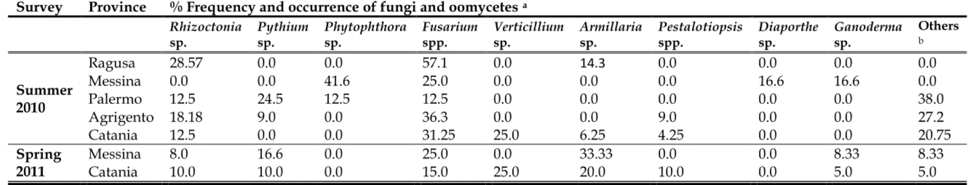
VIII Table 2.1 Primers used for PCR amplification and sequencing ... 40 Table 2.2 Disease incidence (%) of soilborne diseases occurred in mango orchards located in five Sicilian provinces during two survey periods ... 48 Table 2.3 Frequency (%) of fungi and oomycetes occurred in mango orchards located in five provinces in Sicily during two survey periods ... 48 Table 3.1 Primers used in the molecular characterization of Verticillium isolates. ... 67 Table 4.1 Phytophthora species used as reference isolates for mating type study. ... 85 Table 4.2. PCR primer sequences, reaction mixtures composition, and cycling conditions for the ITS, COI, LSU, and 60 ribosomal loci. ... 87 Table 4.3 Phytophthora species and origins of their isolates studied and their GenBank sequence accession numbers... 88 Table 4.4 Sexual compatibility among isolates of P. cryptogea infecting mango in Italy ... 96 Table 4.5 Pathogenicity tests of five isolates of Phytophthora cryptogea on mango plants, 10 months after inoculation. ... 107 Table 5.1 PCR primer sequences, reaction mixtures composition, and cycling conditions for the ITS and COI loci used in this study. ... 142 Table 5.2 List of Phytophthora isolates used in this study ... 143 Table 6.1 Phytophthora species with used in the study. ... Error! Bookmark not defined. Table 6.2 Universal primers designed and used for the amplification of IGS region within the rDNA Gene of Phytophthora sp. ... 187 Table 6.3 Specific primers designed in IGS regions of P. cryptogea, P. citrophthora and P. megasperma and amplification conditions. ... 190 Table 6.4 Specific primers and fluorescent probe sequences used to develop species-specific assays for Phytophthora cryptogea, P. citrophthora and P. megasperma... 193
IX
LIST OF FIGURES
CHAPTER 1………
Figure 1.1 The major countries share in global mango exports % (A) and global imports %(B). ... 4
Figure 1.2 Disease cycle of Pythium damping off and root rot ... 10
Figure 1.3 Disease cycle of Phytophthora root rot ... 14
Figure 1.4 Disease cycle of Verticillium wilt ... 16
Figure 1.5 Disease cycle of Armillaria rot ... 18
Figure 1.6 Typical disease cycle of wood rot and decay fungi ... 19
CHAPTER 2………
Figure 2.1 Map of Sicily (Southern Italy) showing the geographical locations of the surveyed orchards (Palermo, Messina, Ragusa, Agrigento and Catania). ... 37Figure 2.2 Damping off and root rot symptoms observed in the mango nursery. A and B, general wilt, collapse and death symptoms on young seedlings. C, destruction of young seedlings roots by root rot fungi. ... 42
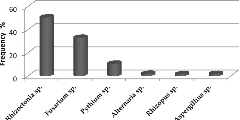
Figure 2.3 The frequency (%) of fungi recovered from mango roots in the nursery ... 43
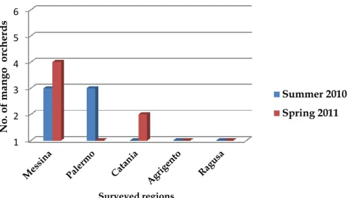
Figure 2.4 Number of mango surveyed orchards surveyed during summer 2010 and spring 2011 in five regions in Sicily. ... 44
Figure 2.5 Symptoms of black root diseases observed on mango trees in the field. A, symptoms of stunting and foliage yellowing appear on the canopy. B, symptoms of root rot and decay on mango roots ... 45
Figure 2.6 Symptoms of wood rot and decay diseases observed on mango trees in the fields. A, a limb extensively invaded with wood rotting fungi. B and C, wood discoloration. ... 45
Figure 2.7 Symptoms of Armillaria root diseases observed on mango trees in fields. A, overall appearance of the tree infected with Armillaria rot. B, symptoms of yellowing and wilted foliage associated with rot on the trunk. C, mortality and death of the entire mango tree. D, rot and decay on mango trunk. E, outer bark removed from tissues of an infected mango tree to reveal the presence of mycelial sheets of Armillaria. F, severe infection by Armillaria with symptoms extended to mango limbs. G, H and I, stumps and roots of mango tree showing mycelial sheets with typical Armillaria infection. ... 46
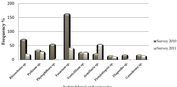
Figure 2.8 The frequency (%) of fungi and oomycetes isolated from mango tissues. ... 47
Figure 2.9 Colony morphology of Pythium sp. on PDA after 7 days at 25°C. A, colony from the front side; B, reverse colony ... 49
Figure 2.10 Morphological characteristics of R. solani. A, C, colony morphology of two R. solani isolates on PDA after 7 days at 25°C.; B and D, typical Rhizoctonia mycelium showing its branching at a right angle and septa close to the branching point. ... 49
X Figure 2.12 Morphological characteristics of F. oxysporum. A, colony morphology from the front side; B, reverse side; C, short conidiophore; E, macroconidia; F, microconidia and F, chlamydospores. ... 51 Figure 2.13 Morphological characteristics of Armillaria mellea. A. colonies recovered from mango wood tissues. B, colony morphology on MEA media after 21 days at 25°C. The long spidery out growths are the rhizomorphs. ... 52 Figure 2.14 Morphological characteristics of Ganoderma sp. A, colony morphology from the front side; B, reverse side on MEA media after 15 days at 25°C. ... 52 Figure 2.15 Figure 21 Agarose gel electrophoresis of amplified PCR products from genomic DNA of Pythium and Fusarium isolates collected from mango plants in Sicily. Lane 1, amplified PCR product of ITS using ITS4 and ITS6 primer pairs from Pythium
vexans. Lanes 2 and 6, amplified PCR products of β-tubulin using T1-T2 from F. oxysporum
isolates. Lanes 3, 4, 5, 7 and 8 amplified PCR products of β-tubulin using T1-T2 from F.
solani isolates. ... 53
CHAPTER 3………
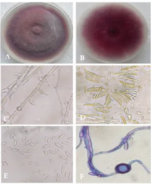
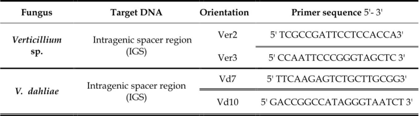
Figure 3.1 Symptoms of Verticillium wilt on mango trees in the field. A, yellowing, wilting and dieback on one side of mango tree. B, dead leaves attached to infected branches giving the tree fired appearance. C and D, cross sections of mango trunk showing vascular discoloration. E and F, characteristic vascular streaking caused by Verticillium wilt. Arrows in A and B, showing mango trees closely planted with olive trees. ... 69 Figure 3.2 Colony morphology of two different isolates of V. dahliae on PDA after 30 days at 25°C. (Vd-1 on the left side and Vd-2 on the right one). ... 70 Figure 3.3 Morphological characteristics of V. dahliae recovered from mango trees. A and B, erect, hyaline conidiophores; C, typical verticillate branches of Verticillium; D, hyaline, ovoid conidia; E, microsclerotia formed on hyphae; F, typical melanised microsclerotia. . 70 Figure 3.4 Agarose gel electrophoresis of amplified products from genomic DNA ofVerticillium isolates. Lanes 1 to 7 are PCR products obtained by the genus universal primer
(Ver2-Ver3), with a fragment of 339 bp. Lanes 8 to 14 are PCR products obtained by the specific primer (Vd7b-Vd10) with a fragment of 139 bp. with Lanes 7 and 14 positive control of Verticillium isolates from IAMB collection, and H2O contains negative DNA
control. Lane M contains 100-bp DNA ladder. ... 71 Figure 3.5 Pathogenicity of V. dahliae isolates on mango plants. A, typical symptoms of Verticillium infection developed as stunting of the inoculated plant (on the left) compared to normal growth of the healthy plant (on the right); B, lesions on the stem of the inoculated plant (on the left) and healthy stem (on the right); C vascular discoloration in
XI tissues of the inoculated plant; D, stem of the healthy plant show no vascular discoloration. ... 73 Figure 3.6 Pathogenicity of V. dahliae isolates Vd-1 and Vd-2 on mango. A, symptoms of chlorosis, appeared on the leaves; B, healthy green leaves; C, gummosis and cracks appeared on mango stem; D. vascular discoloration in longitudinal-sections of stem... 74 Figure 3.7 Pathogenicity of V. dahliae isolates Vd-1 and Vd-2 on mango plants determined as severity of vascular tissue discolored (wilt severity) on a scale of 0 to 5, 0 = no vascular discoloration, 1 = 1 to 25%, 2 = 26 to 50%, 3 = 51 to 75%, and 4 = 76 to 100% of vascular tissues exhibited discoloration in the absence of foliar symptoms, and 5 = 100% of vascular tissues exhibited discoloration in the presence of foliar symptoms typical of Verticillium wilt. The bars represent the means (± standard error) of three replicates with three plants per isolate. Bars with the same letter were not statistically different according to Duncan- Waller k-ratio t test (P = 0.05). Percent severities were angular transformed [sqr (x+2)] before analysis. ... 74 Figure 3.8 Effect of two isolates of V. dahliae (Vd-1 and Vd-2) on the mean of plant height (A) and fresh weight of foliar and root tissues (B) of mango 10 months after inoculation. The bars represent the means (± standard error) of three replicates per isolate. Bars with the same letter were not statistically different according to Duncan- Waller k-ratio t test (P = 0.05). ... 75
CHAPTER 4………
Figure 4.1 Number of Phytophthora isolates recovered from Messina and Palermo ... 91 Figure 4.2 Symptoms of Phytophthora root and crown rot on mango canopy appeared as pale green leaves, wilt and chlorotic trees. ... 92 Figure 4.3 Symptoms of Phytophthora root and crown rot on mango roots. ... 92 Figure 4.4 Colony morphology of Phytophthora isolate (Ph2) recovered from Messina, on PDA and V8 media after 6 days at 25°C. Top (from left to right) PDA - V8 and bottom (from left to right) PDA - V8 reverse colony. ... 94 Figure 4.5 Colony morphology of Phytophthora isolate (Ph13) recovered from Palermo, on PDA and V8 media after 6 days at 25°C. Top (from left to right) PDA - V8 and bottom (from left to right) PDA - V8 reverse colony. ... 94 Figure 4.6 Morphological characteristics of Phytophthora isolate (Ph2) recovered from Messina. A, hyaline coenocytic hyphae; B, hyphal swellings; C, proliferations of the sporangiophores; D and E, nonpapillate ovoid to obpyriform; F, zoospores... 95 Figure 4.7 Morphological characteristics of Phytophthora isolate (Ph13) recovered from Palermo. A, hyaline coenocytic hyphae; B, hyphal swellings; C, proliferations of the sporangiophores; D and E, nonpapillate ovoid to obpyriform; F, zoospores... 95 Figure 4.8 Mating of Phytophthora spp. A, dual culture of pairing Phytophthora isolate (Ph2) recovered from Messina with P. cryptogea (CBS 113.19 mating type A1) on amendedβ-XII recovered from Palermo with P. cryptogea (CBS 113.19 mating type A1) on amended β-Sitosterol V8 media after 20 days of incubation in the dark at 25°C. B, smooth oogonia with amphigynous antheridia. C, spherical plerotic oospores ... 97 Figure 4.10 Agarose gel electrophoresis of amplified PCR products of the internal transcribed spacer (ITS- rDNA) region using ITS4 and ITS6 primers pair. P. cryptogea isolates from mango (Lanes 1 to 14); P. nicotianae IAMB160 isolate as positive control (Lane 15); Lane H2O contains negative DNA control and Lane M contains 100-bp DNA ladder. 99
Figure 4.11 Agarose gel electrophoresis of amplified PCR products of Cytochrome c oxidase subunit 1 (COI) using Oom-lev and Fm85-mod primers pair. P. cryptogea isolates from mango (Lanes 1 to 14); P. nicotianae IAMB160 (Lane 15); P. citrophthora IAMB161 (Lane 16); P. palmivora IAMB162 (Lane 17); P. nicotianae IAMB163 as positive control (Lane 18); Lane H2O contains negative DNA control and Lane M contains 100-bp DNA ladder. 99
Figure 4.12 Agarose gel electrophoresis of amplified PCR products of 28S Ribosomal DNA (LSU) using LROR-O and LSURint primers pair. P. cryptogea isolates from mango (Lanes 1 to 14); P. nicotianae IAMB160 isolate as positive control (Lane 15) and Lane M contains 100-bp DNA ladder. ... 100 Figure 4.13 Agarose gel electrophoresis of amplified PCR products of the 60S Ribosomal protein using 60SL10F and 60SL10R primers pair. P. cryptogea isolates from mango (Lan1 to 14); P. nicotianae IAMB160 isolate as positive control (Lane 15); P. palmivora IAMB162 isolate as positive control (Lane 16); Lane H2O contains negative DNA control and Lane M
contains 100-bp DNA ladder. ... 100 Figure 4.14 Phylogenetic relationships of 5 P. cryptogea isolates from mango and 6
Phytophthora taxa using ITS sequence data, based on maximum likelihood analysis. The
numbers at the branch points indicate the percentages of bootstrap values >50 %. ... 101 Figure 4.15 Phylogenetic relationships of 5 P. cryptogea isolates from mango and 6
Phytophthora taxa using COI sequence data, based on maximum likelihood analysis. The
numbers at the branch points indicate the percentages of bootstrap values >50 %. ... 102 Figure 4.16 Phylogenetic relationships of 5 P. cryptogea isolates from mango and 6
Phytophthora taxa using LSU sequence data, based on maximum likelihood analysis. The
numbers at the branch points indicate the percentages of bootstrap values >50 %. ... 103 Figure 4.17 Phylogenetic relationships of 5 P. cryptogea isolates from mango and 6
Phytophthora taxa using 60S sequence data, based on maximum likelihood analysis. The
numbers at the branch points indicate the percentages of bootstrap. ... 103 Figure 4.18 Single-strand-conformation polymorphism (SSCP) profiles of ITS for P.
nicotianae (Lane 1); P. palmivora (Lane 2) and P. cryptogrea (Lanes 4-7). ... 104
Figure 4.19 Root and crown rot symptoms on mango plants, 10 months after inoculation with P. cryptogea (Ph2). ... 106
XIII Figure 4.20 Pathogenicity test of P. cryptogea on mango plants, 10 months of after inoculation. Disease severity of root symptoms in a 0-to-5 scale, where 0 = healthy roots and 1, 2, 3, 4, and 5 were <20, 21– 40, 41–60, 61–80, and >80%, respectively, of rotted roots. ... 106
CHAPTER 5………
Figure 5.1 Phylogram of 47 Phytophthora species with Peronospora species (as outgroup) with 8 clades based on analysis of the combined ITS1 and ITS2 regions of the genomic ribosomal RNA according to (Cooke et al., 2000a)... 125 Figure 5.2 A genus wide phylogeny for Phytophthora with 10 clades according to Blair et al. (2008) using seven nuclear loci (28S Ribosomal DNA, 60S Ribosomal protein L10, Beta-tubulin, Elongation factor 1alpha, Enolase, Heat shock protein 90 and TigA gene fusion Protein). ... 126 Figure 5.3 Maximum parsimony analyses of concatenated cox2, nad9, rps10, and secY genes from a range of Phytophthora spp. with Phytopythium vexans and Ph. undulatum as outgroups according to Martin et al. (2012). ... 127 Figure 5.4 Comparison of sporangial shapes. A, P. parasitica; B, P.cryptogea; and C, P.drechsleri from safflower ... 132
Figure 5.5 Culture characteristics of P. drechsleri as illustrated by Mircetich (1976). A, Hyphae of P. drechsleri with sparse branching at an acute angle; B, typical sporangia of P.
drechsleri. ... 132
Figure 5.6 Sporangium morphology of P. cyptogea. (A) isolate P3104 from group D, (B) P3850 from group E and (C) Oogonium with mature oospore and amphigynous antheridium from the type culture of P. cryptogea (P1738) mated with P. cinnamomi A2 (P2411), Source: (Mills et al., 1991)... 135 Figure 5.7 Sporangium morphology of P. drechsleri. (A) isolate PI087 , (B) isolate P1899 from group A. (C), tapered and rounded base of P. drechsleri f. sp. cajani isolate P1795. (D) Oogonium with mature oospore and amphigynous antheridium from an isolate of P.
drechsleri (P1741) mated with P. cinnamomi A1 (P2100), Source: (Mills et al., 1991). ... 135
Figure 5.8 Amplified PCR products of 727bp fragment for COX1 region with OomCoxI-Levup and Fm85mod. ... 156 Figure 5.9 Phylogenetic relationships of Phytophthora drechsleri, P. cryptogea groups and associated species based on neighbor joining method. The numbers at the branch points indicate the percentages of bootstrap values. A, Internal Transcribed Spacers, ITS. B, mitochondrial Cytochrome c Oxidase subunit I, COI. ... 158 Figure 5.10 Phylogenetic relationships of Phytophthora drechsleri, P. cryptogea groups and associated species based on Neighbour Joining method of combined sequences of Internal Transcribed Spacers (ITS) and mitochondrial Cytochrome c Oxidase subunit I (COI). The numbers at the branch points indicate the percentages of bootstrap values. Pythium vexans sequences were used as out-group. ... 160
XIV and localization of specific primers pairs in IGS regions of P. cryptogea, P. citrophthora, and
P. megasperma. ... 194
Figure 6.2 Agarose gel electrophoresis of amplification products obtained with the primers CryF5- CryR5 amplifying a fragment of 79 from different genomic DNA of P. cryptogea isolates. Lane 1 to 11 contain amplified product from pure cultures of P. cryptogea isolates obtained from mango (P1to P11), Lane H2O contain negative DNA control and lane M
contain 100-bp DNA ladder. ... 196 Figure 6.3 Agarose gel electrophoresis of amplification products obtained with the primers Cit3F- Cit3R amplifying a fragment of 144 from different genomic DNA of Phytophthora
citrophthora isolates. Lane1 contain amplified product from a pure culture of P. citrophthora
isolate Ph34, lan2, (P. citrophthora isolate Ph62), lan3, (P. citrophthora isolate Ph153), lane 4
(P. citrophthora isolate Ph71), lane 5 (P. citrophthora isolate Ph156), Lane 6 (P. citrophthora
isolate Ph 156), Lane 7 contain negative DNA control and lane M contain 100-bp DNA ladder. ... 196 Figure 6.4 Agarose gel electrophoresis of amplification products obtained with the primers Mega10F- Mega10R amplifying a fragment of 121 from different genomic DNA of
Phytophthora megaperma isolates. Lane1 contain amplified product from a pure culture of P. megasperma isolate Ph209, lan2, (P. megasperma isolate Ph210) lane 4 (P. megasperma isolate
Ph239), lane 5 (P. megasperma isolate Ph241), lane 6 (P. megasperma isolate Ph238), lane 7 (P.
megasperma isolate Ph208), lane 8 (P. megasperma isolate Ph101), lane 9 (P. megasperma
isolate Ph105), Lane 10 contain negative DNA control and lane M contain 100-bp DNA ladder. ... 197 Figure 6.5 Real-time amplification profiles of test conducted to assess specificity of
Phytophthora IGS probe in Taqman real-time PCR using DNA from P. cryptogea (1); P. citrophthora (2) and P. megasperma (3) isolates. A = target Phytophthora species; B = other Phytophthora species and Negative control. ... 199
XV
LIST OF ABBREVIATIONS
List of Abbreviations A Adenine bp Bas pair C° Celsius degreeCBS Centraalbureau voor Schimmelcultures
cm Centimeter
cv Cultivar
C Cytosine
DNA Deoxyribuneoclic acid
dNTPs Deoxyribonucleotide triphosphate
EtOH Ethanol
FAO Food and Agriculture Organization
Fig. Figure cm Centimeter G Guanine g Gram h Hour H2O Water HCl Hydrochloridric Acid
Kb Kilo Base Pair
ITS Internal transcript spacer region
Kbp Kilo base pair
M Molar mg Milligram ml Milliliter min Minute MgCl2 Magnesium chloride mM Millimolar
PCR Polymerase chain eaction
PDA Potato dextrose agar
% Percentage
µl Microliter
XVI
rpm Revolutions per minute
sec Second
spp. Species
SSCP Single Strain Conformation Polymorphism
T Thymine TAE Tris-Acetate-EDTA TBE Tris-Borate-EDTA U Enzymatic Unit UV Ultraviolet V Volt
V/V Volume per Volume
WA Water agar
WPC World Phytophthora collection
CHAPTER 1. General Introduction and Literature Review
1
CHAPTER 1.
1. GENERAL INTRODUCTION, LITERATURE
REVIEW AND THESIS OBJECTIVES
2
General Introduction
1.
Mango (Mangifera indica L.) is one of the best-known and most widely cultivated tropical fruit species, with production occurring in most countries in the tropics and subtropics areas (Paull and Duarte, 2010). The fruit is marketed fresh, dried, and is used as a source of flavors and colorants (Litz, 2009).
Botany and Taxonomy: Mango belongs to the family Anacardiaceae, also known as the cashew family, with about 75 genera and 700 species, mostly tropical, with some subtropical and temperate species. The genus Mangifera consists of 69 species but not all bear edible fruits (Paull and Duarte, 2010). The mango fruit is large, fleshy and sometimes fibrous. The other edible Mangifera species generally have lower quality fruit and are commonly referred to as wild mangos (Litz, 2009).
Common Names: Mangos have been grown throughout the tropical and subtropical world for thousands of years and have become an integral part of many cultures. Many of the names have common derivations, reflecting the origins and spread of the mango tree along with the spread of human communities (Paull and Duarte, 2010). Some of these names are: mango (English), mangue, manguier (French), manja- maggo- manja (Dutch), manga- mango (Spanish), manga (Portuguese), manga (Malaysia), mangga (Indonesia), Mangobaum (German), Al mango (Arabic) and Il mango (Italian) (Bally, 2006).
Origin and History: The mango originated in the Indo-Burma region and has been cultivated in India for more than 4000 years. This fruit is closely associated with the Hindu religion (Singh, 1960). Indian traders and Buddhist priests perhaps introduced the mango into Malaysia and other East Asian countries during the 4th or 5th century BC, and to the
Philippines between AD 1400 and 1450. The Portuguese, the first Europeans to establish trade routes with India, transported the mango to East Africa and Brazil. Spanish traders took the mango from the Philippines to the west coast of Mexico before the English arrived in the Hawaiian Islands in 1778. The mango was introduced into Hawaii from the west coast of Mexico between 1800 and 1820. Apparently, the Brazilian introductions were spread to Barbados and to other islands in the Caribbean area. Mango is now found in all
CHAPTER 1. General Introduction and Literature Review
3 tropical areas, as well as many subtropical regions of the world, attesting to its wide range of adaptability (Litz, 2009).
Climate: Although grown widely, mangos prefer a warm, frost-free climate with a well-defined winter dry season, rain and high humidity during flowering and fruit development reduces fruit yields. The tree generally flowers in mid- to late winter, with fruit maturing in the early to mid-summer months (Bally, 2006). Mango trees are evergreens that grow to 60 feet tall and the tree fruits 4 to 6 years after planting (Paull and Duarte, 2010).
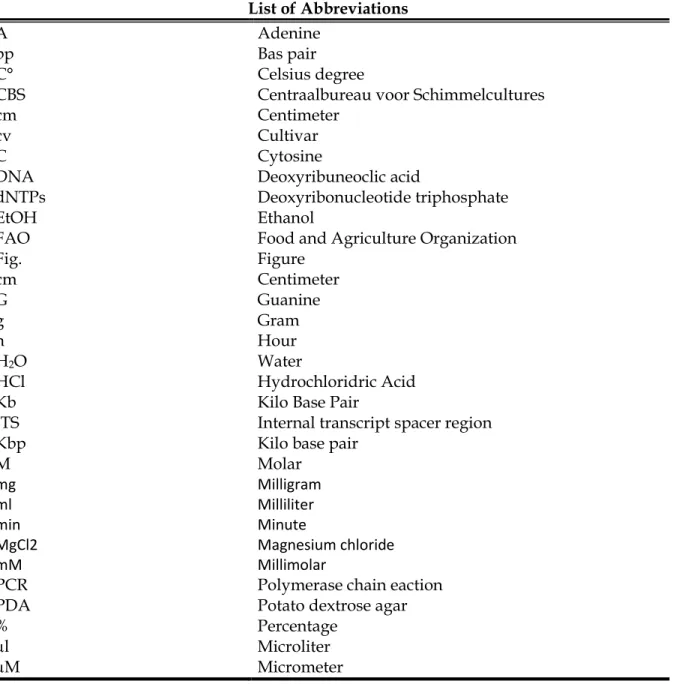
Mango Production: Mango covered an area of 4,946,313 ha with production of 37.12 million tons in the world during the year of 2010 (Table 1.1). India occupies top position among mango growing countries of the world and produces 40.48% of the total world mango production. China and Thailand stand at second and third position among mango producing countries in the world with 4,366 and 2,551 thousand tons respectively. The other major mango producing countries in the world during 2010 were Thailand (2550 thousand tons), Pakistan (1784 thousand tones), Mexico (1633 thousand tones) and Indonesia (1314 thousand tones) respectively (FAOSTAT, 2011). In 2010, Mexico dominated the export trade with shares of 22% followed by Philippines (16%), Pakistan (16%), Brazil (13%) and India (11%). Other major exporters include the Netherlands and Peru (Fig.1.1A). United States imported approximately half of total mango imports (48%), followed by China with shares of (11%) (Fig.1.1B). The Netherlands imports of mangoes are (8%), but most of this is redistributed throughout the European Union. Other major importing redistributors of mangoes are the United Arab Emirates (6%) and Saudi Arabia (4%), with most of these imports being redistributed within the Middle East (FAOSTAT, 2011).
Mango production in Italy: There are no official records available for mango production in Italy since the crop is relatively new.
4
Table 1.1 Major producing countries of mango in the world during 2010 (FAOSTAT,
2011).
Country Area (ha) Production (Tons) Productivity (Tons/Ha) % Share in world Total production
India 2312.3 15026.7 6.5 40.48 China 465.337 4351.29 9.35 11.72 Thailand 311.048 2550.6 8.2 6.87 Pakistan 173.7 1845.5 10.62 4.97 Mexico 174.97 1632.65 9.33 4.4 Indonesia 131.674 1287.29 9.78 3.47 Brazil 75.111 1188.91 15.83 3.2 Bangladesh 170.8 1047.85 6.13 2.82 Philippines 189.437 825.68 4.36 2.22 Nigeria 114.9 790.2 6.88 2.13 Other Countries 827.04 6578.07 7.95 17.72 World 4946.314 37124.74 7.51
Figure 1.1 The major countries share in global mango exports % (A) and global imports % (B).
CHAPTER 1. General Introduction and Literature Review
5
Mango Nutritional Value: Ripe mango is delicious fruits with pleasant flavor, the fruits are highly nutritious that contain carbohydrates, proteins, fats, minerals, and vitamins (Litz, 2009; Anonymous, 2010; Chen et al., 2010; Paull and Duarte, 2010).
Mango Varieties: There are over a thousand mango varieties grown around the world. India having the greatest number of varieties almost over 500 named cultivars (Bally, 2006). Florida (USA) has developed a large number of cultivars, using mainly Indian cultivars, which have shown wide geographic adaptability. Most mango varieties planted in the Americas, the Canary Islands and many African countries were developed in Florida The Florida varieties are also dominant in international trade (Paull and Duarte, 2010). The selection of mango varieties is usually based on keeping quality of fruits and tree growing characteristics. Preferences for mango varieties often differ among countries, regions, traditions, and cuisines of markets in which they are consumed. Among the most important mango varieties are:
Kensington Pride (Australia): It is grown widely in the tropical and sub-tropical regions. Kensington Pride was the first cultivar introduced into Sicily (Southern Italy), because it is resistant to low temperatures particularly well represented in Catania and also along the north-eastern coast (Anonymous, 2010). Its fruits are produced early (late August and September) and sells well. The only problem of this cultivar is fruit color that looks a little pale, with little blush, so when it arrives in the markets, the cultivar Glenn, which has the same period of maturation, the Kensington appears to have less success (Litz, 2009).
Glenn (Florida, USA): The best for the time of maturation (August) for both color and flavor that has far surpassed all other varieties (Litz, 2009).
Kent (Florida, USA): The fruit color is greenish yellow with red blush. Its weight is around at 600-700 grams. The flesh is of excellent flavor and has a pleasant and a rich aroma (Litz, 2009).
6
Tommy Atkins (Florida, USA): Is the most important commercial cultivar in the western hemisphere. It is highly resistant to anthracnose disease, handling and shipping stress (Campbell, 1992).
Zebda (Egypt): Is the most important cultivar in Egypt. The tree is vigorous and regularly productive. It is highly tolerant for anthracnose and resistant to malformation (Litz, 2009).
Mango industry in Italy: In the eighties, mango cultivation was introduced in Italy with limited extensions mainly in Sicily (Southern Italy). Sicily is the largest island in the Mediterranean Sea. It has a typical Mediterranean climate with mild, wet winters and hot, dry summers. Mango industry started as a result of studies conducted by Prof. F. Calabrese and his team of researchers through a project funded by the tropical fruit MIPAAF (Anonymous, 2010). The objectives of these studies were i) trying to cultivate mango under Sicilian mild climate, especially that mango had already been successfully cultivated in countries with comparable climate like Spain, France and Israel, and ii) to provide an alternative crop in areas of Sicily that well suited to tropical and subtropical crops. Therefore, small mango pitches began to arise in different parts of Sicily in order to ascertain what would be the area most suitable for mango cultivation and with which varieties. These researches concluded that the cultivar Kensington Pride could easily be grown even in areas where temperatures down or are close to zero, while for other cultivars was chosen the Tyrrhenian coast that goes from Palermo to Messina. In the near future, it is expected that commercial and backyard plantings of mango trees will increase, being a profitable crop in Sicily (Anonymous, 2010).
Mango Production boundaries: Mangos grow and produce in many tropical and subtropical climates, although fruit production is limited by wet weather during the flowering and fruiting period (Prakash, 2004; Bally, 2006; Paull and Duarte, 2010). Unreliable yields and fruit quality from season to season are also limiting characteristics of many mango varieties. Generally, the low productivity is due to the wide range of climatic conditions, environment situation and the diversity of the associated disease and disorder problems (Ploetz, 2004; Prakash, 2004).
CHAPTER 1. General Introduction and Literature Review
7
Mango Physiological Disorders: Physiological disorders are any aberrations that have not been caused by infecting organisms. True physiological disorders cannot be transmitted from plant to plant, mechanically or by insect bites (Litz, 2009). They are results of imbalances in metabolism induced by some factors in the pre-harvest or postharvest environment that leads to cell collapse. Pre-harvest factors include growing location, orchard conditions and tree nutrition while post-harvest storage conditions such as temperature, oxygen and carbon dioxide levels, packaging and surface coating treatments are contributing factors to the occurrence of the disorders (Campbell, 1992; Prakash, 2004; Litz, 2009; Paull and Duarte, 2010). Internal necrosis of mango fruit is an emerging physiological disorder due to boron deficiency which may also lead to fruit cracking (Chin
et al., 2010; Saran and Kumar, 2011).
Mango Pests: Of the 260 species of insects and mites that have been recorded as pests of mango, 87 are fruit feeders, 127 are foliage feeders, 36 feed on the inflorescence, 33 inhabit buds, and 25 feed on branches and the trunk (Peña et al., 1998). The major key pests (fruit flies, seed weevils, tree borers and mango hoppers) require annual control measures. Secondary pests generally occur at sub-economic levels, but can become serious pests as a result of changes in cultural practices and cultivar or because of indiscriminate use of pesticides against a key pest (Peña et al., 1998; Litz, 2009; Paull and Duarte, 2010).
Mango Diseases: Over 140 pathogens are known to cause damage to mango plants (Prakash, 2004). Mango diseases are caused mainly by some true fungi, fungus-like Oomycetes, nematodes and bacteria. The first step in overcoming the threats from diseases is to accurately identify the problems followed by adequate management and judicious use of fungicides and bactericides (Bally, 2006; Litz, 2009).
8
Literature Review
2.
An Overview of Mango Diseases
Mango as any fruit trees can be affected by different factors, which can influence the healthy status of trees either biotic or abiotic factors. Mango is affected by a number of diseases at all stages of its growth from the seedling and grafted plants in the nursery to the fruits in storage or transit (Ploetz, 2004). Most of mango parts specifically, seeds, root, trunk, branch, twig, leaf, petiole, flower and fruit are attacked by a number of pathogens including fungi and bacteria. They may cause several kinds of rot, die back, wilt, anthracnose, scab, necrosis, blotch, spots, mildew, fruit rot, etc. Diseases in the field result in crop loss, while post-harvest diseases are directly linked with the losses in export and domestic market. In several regions, they are the most important constraint to fruit production as they cause heavy losses in mango yield (Ploetz, 1994a;2004; Bally, 2006; Litz, 2009).
1.1 Fungal Soilborne Diseases of Mango
Soilborne diseases are caused by fungi or pathogenic microbes that persist in soil without plant hosts. These pathogens can induce different kind of symptoms such as damping off, root rot, stem rot and wilt. Plant diseases caused by soilborne pathogens are considered major problems in agricultural production throughout the world. These diseases can reduce yield and quality of many crops. The main fungal and oomycete genera reported as containing species that are pathogenic toward mango, include the fungal genera Fusarium,
Rhizoctonia and Verticillium, and the oomycete genera Phytophthora and Pythium (Prakash
and Singh, 1980; Tsao et al., 1994; Prakash, 2004; Ploetz and Freeman, 2009). Although soilborne diseases of mango are relatively less important than foliar diseases, they can cause significant damage to seedlings, nursery stock and mature trees (Litz, 2009).
CHAPTER 1. General Introduction and Literature Review
9
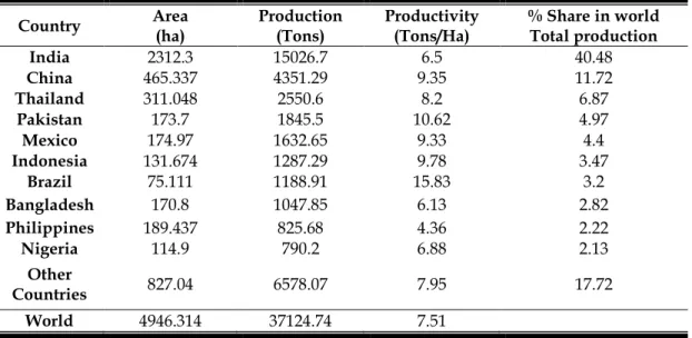
a. Pythium Damping Off and Root rot Diseases
Damping-off and root rot diseases occur worldwide in tropical, temperate climates, and in every greenhouse. The disease affects seeds, seedlings, and plant root. The greatest damage arises during germination either before or after emergence. Losses vary considerably with soil moisture, temperature, and other factors (Spies et al., 2011). In many cases, seedlings in seedbed are entirely destroyed by damping-off or they die soon after they are transplanted. Generally, poor germination of seeds or poor emergence of seedlings is the result of damping-off infections in the pre-emergence stage (Spies et al., 2011). Older plants are rarely killed when infected with the damping-off pathogen, but they develop root and stem lesions and root rots. Their growth may be slowed significantly, and their yields may be reduced severely. In mango damping off of seedlings was reported as a serious problem in nurseries in Indonesia, Pakistan and India (Prakash, 2004).
Symptoms: Lim and Khoo (1985) indicated that overcrowding, excessive moisture and the use of polybags favoured this disease. Symptoms include wilting of foliage, which initially becomes pale green, but later develops necrotic patches. Roots develop a wet, blackened necrosis that begins in fine roots and progresses to larger roots and the root collar. Death of seedlings often occurs (Prakash, 2004; Litz, 2009).
The Causal Organism: The genus Pythium contains several species that are facultative pathogens. These species causing severe diseases in many crops and acting either individually or in complexes with other organisms (André LéVesque and De Cock, 2004). In Malaysia, Pythium vexans de Bary, has been reported as the causal pathogen of mango root rot and wilt of seedlings (Lim and Khoo, 1985) and induced seedling losses of up to 30% in nurseries (Lim and Khoo, 1985; Litz, 2009).
Life Cycle: Pythium species occur in surface waters and soils. They live on dead plant and as saprophytes or as parasites of fibrous roots of plants. The pathogen needs free water for its zoospores to swim and infect the host. When a wet soil is infested heavily with Pythium, any seeds or young seedlings in such a soil may be attacked by the pathogen (Agrios, 2005). Pythium mycelium or spore germ tubes come in contact with mango seeds and seedling tissues then enter by direct penetration (Fig 1.2). The mycelium
10
grows between and through the plant cells. Pectinolytic enzymes secreted by the fungus dissolve the pectins and the protoplasts of invaded cells, and, in some cases, cellulolytic enzymes cause complete collapse and breakdown of the cell walls. As a result, the invaded tissues cannot support the seedling, which falls over and dies. The disease and losses caused by Pythium infections are more severe when the soil is kept wet for long periods
Disease management: Cultural practices are helpful in reducing the amount of infection. Such practices include providing good soil drainage and good air circulation among plants, planting when temperatures are favourable for fast plant growth, avoiding application of excessive amounts of nitrate forms of nitrogen fertilizers. Seed chemical treatment and spraying of seedlings are important when the soil is infested heavily with
Pythium or when the soil stays wet for prolonged periods during the early stages of plant
growth (Agrios, 2005).
CHAPTER 1. General Introduction and Literature Review
11
b. Rhizoctonia Damping Off and Root Rot Disease
Rhizoctonia diseases occur throughout the world. They cause losses on almost all vegetables and fruit trees. The most common symptoms on utmost plants are damping-off of seedlings and root rot. It occurs primarily in cold, wet soils. Very young seedlings may be killed before or soon after they emerge from the soil. Damping off of mango seedlings is a serious problem in nurseries in Indonesia and India (Prakash and Singh, 1980).
Symptoms: The disease is characterised by sudden dropping of leaves after the emergence of seedlings from the soil. During prolonged rainy and humid weather, infection occurs below the ground level with circular to irregular water-soaked patches. These patches enlarge and ultimately girdle the entire base of the stem. On account of rotting, the diseased tissues become soft, dark brown or black and the entire seedling collapses and dies (Litz, 2009). Fungal mycelia and sclerotia are densely present on the severely infected parts. Rotting may spread both above and below the stem down up to roots and roots are disintegrated (Prakash and Singh, 1980; Prakash, 2004).
The Causal Organism: Rhizoctonia is soil inhabitant basidiomycetes. The fungus was recognized as sterile fungi for many years, because they were thought to produce only sclerotia and to be incapable of producing sexual or asexual spores (Sneh et al., 1991). It is known now that at least some species of Rhizoctonia, can produce basidiospores as their sexual spores only under special conditions in the laboratory or are extremely rare in nature. Therefore, fungus basiospores have little value in identifying the fungus (Sneh et
al., 1991). The genus Rhizoctonia is comprised of a diverse group of fungi that can broadly
be divided into multinucleate and binucleate groups based on the number of nuclei per hyphal cell (Sneh et al., 1991). Isolates are classified further into anastomosis groups (AGs), based on their hyphal compatibility with known tester isolates (Tewoldemedhin et al., 2011).
Disease Cycle: The pathogen overwinters usually as mycelium or sclerotia in the soil and in or on infected perennial plants. The fungus spreads with rain, irrigation, and with infected propagative materials. After the seedlings have emerged, the fungus attacks their stem and makes it water soaked, soft, and incapable of supporting the seedling,
12 which then falls over and dies. In older seedlings, invasion of the fungus is limited to the outer cortical tissues, which develop reddish-brown lesions. The lesions may increase in length and width until they finally girdle the stem, and the plant may die (Agrios, 2005).
Disease Management: Conditions that delay seed germination and slow seedling growth, such as cool, moist, poorly drained soils, favor seedling diseases. Because damping-off is most severe when crops are grown in conditions not favorable to rapid seed germination and seedling emergence, avoid planting into cool, wet, and poorly drained soil. Fields should be prepared so that water does not stand. Seed treatments with appropriate fungicides may provide some protection from seedling diseases (Naqvi, 2004).
c. Fusarium Black Root Rot Disease
Black root rot is reported to be an uncommon problem on young mango trees (Lim and Khoo, 1985). Several species of fungi have been recovered from affected plants, mainly the genera Fusarium such as Fusarium solani, F. oxysporum and in addition to
Lasiodiplodia. theobromae, but these were thought to be secondary colonizers of roots (Lim
and Khoo, 1985).
Symptoms: Canopies of affected plants wilt suddenly and subsequently defoliate. Roots show a water-soaked, darkened decay with association with an unpleasant odour. root discoloration may cover the tap root and the stem below the soil line (Lim and Khoo, 1985; Prakash, 2004).
The Causal Organism:Fusarium genus contains over 70 species (Leslie et al., 2006). Many species inhabit soil ecosystems where they are rhizosphere or endophytic colonizers. The interaction of Fusarium species with plants can range from highly pathogenic to beneficial plant growth stimulation (Tewoldemedhin et al., 2011). F. solani
generally produces only asexual spores, although under certain conditions it produces its perithecial stage, Netria haematococca. The asexual spores are microconidia, macroconidia and thick-walled chlamydospores. F. oxysporum is the most widely dispersed of the
CHAPTER 1. General Introduction and Literature Review
13
Disease Cycle: The pathogen can live on dead plant tissue and can overwinter as mycelium or spores in infected or dead tissues (Leslie et al., 2006). The fungus is already present in many soils as spores, which are spread easily by air, equipment, water, and contact. Fusarium root rot disease becomes more severe when plants are stressed by low temperature, by irregular drought or excessive soil water (Prakash, 2004).
Disease Management: Treatment of propagative stock with appropriate fungicides or application of fungicide sprays on the plants has helped reduce Fusarium rots (Naqvi, 2004).
d. Phytophthora Diseases of Mango
Phytophthora crown and root rots are common and destructive diseases of fruit trees throughout the world (Erwin and Ribeiro, 1996). Phytophthora species cause diseases of mango in several areas. It caused wilt, crown rot, root rot and the death of nursery trees in Arizona USA (Matheron and Matejka, 1988), the Philippines and Thailand (Tsao et al., 1994).
Symptoms: Diseased trees are most likely to be found in heavy, wet soils or sections of the orchard where water collects or is slow to drain. Above-ground symptoms include poor growth with sparse off-color foliage and develop of gumming and visible bark lesions develop above ground on these plants, whereas root and crown rots are evident at or below the ground level (Prakash, 2004; Litz, 2009; Ploetz and Freeman, 2009). Mortality of trees is not observed, but substantial stem cracking and bleeding does occur.
The Causal Organism: The Oomycete pathogens of Phytophthora are the most destructive plant pathogens known. There are over 82 species in the genus many with a wide host range. Many Phytophthora species have been associated with mango root rot in many countries. Phytophthora palmivora was isolated from infected mango trees showing crown rot, root rot and wilt symptoms in the Philippines (Tsao et al., 1994). P. palmivora has been reported also as the causal agent of mango root rot and the death of nursery plants in Arizona USA and Thailand (Matheron and Matejka, 1988). P. parasitica has been reported in India to cause leaf blight disease on mango (Prakash and Srivastava, 1987; Prakash, 2004). Recently, P. citricola was reported in Spain, the fungus was isolated from
14 mango trees that were wilted, chlorotic and had sparse canopies and cracked bark (Zea-Bonilla et al., 2007)
Disease Cycle: Phytophthora species can survive in cold winters or hot, dry summers as oospores, chlamydospores, or mycelium in infected roots, stems and in soil. In the spring, the oospores and chlamydospores germinate by means of zoospores, whereas the mycelium grows further and produces zoosporangia that release zoospores. The zoospores swim around in the soil water and infect roots of susceptible hosts with which they come in contact (Fig. 1.3). More mycelium and zoospores are produced during wet, cool weather and spread the disease to more roots (Erwin and Ribeiro, 1996).
Disease Management: All planting stock should be free of infection. In greenhouses, the soil mixture, seedbeds and planting pots should be sterilized with steam before planting. Sanitary measures must be performed to avoid introduction of pathogen in the nurseries (Agrios, 2005). Overcrowding should be avoided and frequency of irrigation must be adjusted. Seed treatments and transplant dips with suitable systemic fungicides. Some protection of trees can be obtained by injections of selected fungicides into their trunks. Also, application of a solution of some fungicides in the soil around trees seems to inhibit the growth and activity of Phytophthora (Naqvi, 2004; and Prakash, 2004).
CHAPTER 1. General Introduction and Literature Review
15
e. Verticillium Wilt Disease of Mango
Verticillium species are soil-borne pathogens responsible for Verticillium wilt diseases in
temperate and subtropical regions. They affect over 200 hosts, including many economically important fruit tree crops (Klosterman et al., 2009). Verticillium wilt of mango was first reported in Florida USA (Marlatt et al., 1970).
Symptoms: The problem is usually observed in young trees planted in soil previously cropped with vegetables that are also susceptible to Verticillium disease. Disease symptoms comprise wilting, chlorosis, stunting, necrosis and vein clearing. In many of these trees the symptoms expanded, leading to decline and eventual death. Killed leaves usually remain attached to the tree and cross sections of affected branches revealed brown vascular discoloration (Baeza-Montanez et al., 2010).
The Causal Organism:: Verticillium wilt of mango was first reported in Florida USA (Marlatt et al., 1970). The disease was originally attributed to Verticillium albo-atrum but this was before Verticillium dahliae was recognized as a distinct species. Recently, Baeza-Montanez et al. (2010) reported Verticillium wilt caused by V. dahliae in Southern Spain.
Disease Cycle: The typical disease-cycle of Verticillium wilt begins with the germination of resting structures (microsclerotia) in soil, and penetration of the root epidermis of host plants to reach the cortex (Fradin and Thomma, 2006). After crossing the endodermis, the hyphae invade the vascular tissues where conidia can be formed (Fig. 1.4). The conidia are drawn up into the plant with the water stream in the xylem, and trapped in pit cavities, where they germinate. The expression of Verticillium wilt symptoms is usually associated with the colonization of adjacent vascular and cortical tissue (Klosterman et al., 2009). The fungus produces microsclerotia, which are returned to the soil with the decomposition of the plant, and the disease cycle starts again (Fradin and Thomma, 2006; Sanei et al., 2008; Klosterman et al., 2009).
16
Disease Management: Control of Verticillium wilt depends on planting disease-free materials in disease-disease-free soil and using resistant rootstocks. Avoid planting where solanaceous crops (i.e. potato, tomato and eggplant) have been grown repeatedly is recommended. New mango orchards should not be planted on such sites (Ploetz and Freeman, 2009). Sharma et al. (1993) found that soil drenching with Carbendazim (0.1%) or Captafol (0.25%) has been found effective in controlling the seedling wilt.
CHAPTER 1. General Introduction and Literature Review
17
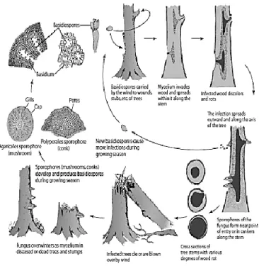
f. Armillaria Root Rot Disease of Mango
The disease is also known as shoestring root rot, mushroom root rot, or oak root fungus disease. Armillaria root rot occurs throughout temperate and tropical regions. It affects fruit trees, vines, shrubs, and forest trees (Aguín-Casal et al., 2004; Matsushita and Suzuki, 2005). The threat of Armillaria root rot is of serious concern in Europe, Australia, America and other developed nations of the world (Popoola, 2004).
Symptoms: The pathogen grows underneath the bark and destroys the structural root system of plant. Chlorosis and abscission of leaves are the common symptoms when the sufficient amount of large roots is destroyed by the fungus. Diagnostic characteristics of Armillaria root rot appear at decayed areas in the bark. White mycelial mats, their margins often veined and shaped like fans, form between the bark and wood (Popoola, 2004). The mycelium may extend for a few feet upward in the phloem and cambium of the trunk and may cause white rot decay. Another characteristic sign of the disease is the formation of black rhizomorphs. These are consisting of a compact outer layer of black mycelium and a core of white or colorless mycelium (Prakash, 2004).
The Causal Organism: The most common Armillaria species that can cause root rot on fruit trees are A. tabescens and A. mellea. The two species are generally very similar but there are a few features that distinguish them. The basidiocarps of A. tabescens lack an annulus or ring just below the cap and can be a darker shade of brown than those of A.
mellea. In contrast, an annulus is present in the basidiocarps of A. mellea (Prakash, 2004). Disease Cycle: Armillaria mellea is a polyphagus pathogen very aggressive on old plants, sometime on young plants. The fungus overwinters as mycelium or rhizomorphs in diseased trees or in decaying roots (Fig.1.5). The main dispersal means are the mycelium and rhizomorphs and not the basidiospores produced by the fungus. Rhizomorphs can grow in the soil 0.5 - 3 m per year, spreading the disease as an oil spot. The fungus can survive for many years in dead or living roots. The pathogen requires cool, moist soil conditions for spreading and disease development. In autumn-winter and a few days after rains, Armillaria often forms clusters of mushrooms at the base of infected trees (Prakash, 2004).
18
Disease Management: Good sanitary measures are the best practice to reduce the disease and to manage Armillaria root. Provide a good growing environment and proper cultural practices. Good drainage is important, as Armillaria fungus is very susceptible to drying (Prakash, 2004). The rhizosphere soil and affected root system should be removed carefully by deep diggings. Removal of stumps and as much as possible root pieces with rhizomorphs from the soil helps in controlling the further spread of the disease (Agrios, 2005).
CHAPTER 1. General Introduction and Literature Review
19
g. Wood Rots And Decays of Mango
Wood rots and decays diseases are considered to be minor diseases on mango.
Symptoms: Depending on the tree part attacked, wood rots may be called root rots, root and butt rots, or stem rots. The wood decay is usually associated with wounds or cankers, whereas in wood pieces the decay is usually at or near the surface of wood that has high moisture content.
The causal organism: Different species belong to basidiomycetes fungi can induce wood rot and decay such as Rigidoporus lignosus and Ganoderma lucidum. Some species are saprophytic that grow on dead or declining heartwood of trees, while several are pathogens that cause decay of roots and stems.
Disease Cycle: Fungi that cause tree or wood decays grow inside the wood cells and utilize the cell wall components (Fig.1.6). Some of them attack softwoods and break down and utilize primarily the cell wall polysaccharides like cellulose and hemicellulose giving the rotten wood symptoms.
20
Disease Management: Reduce the chance of introducing the fungi into healthy orchards. Avoiding or preventing wounds on the trees. Keep the trees in good vigor through adequate irrigation and proper fertilization. Treatment the large cuts or wounds with a wound dressing or tree paint.
1.2 Fungal Foliar Diseases of Mango
Foliar diseases are any disease above ground and localized in the foliage. They are the most important and visible problems on mango. Since many of the pathogens that caused foliar diseases also affect panicles (Litz, 2009; Ploetz and Freeman, 2009).
a. Grey Leaf Spot Disease
Grey leaf spot or grey blight of mango disease caused by Pestalotiopsis mangiferae is prevalent in many mango grown areas.
Symptoms: Initial symptoms are small, yellow-to-brown spots on leaves. Later, the irregularly shaped spots, ranging from a few millimetres to a few centimetres in diameter, turned white to grey and coalesced to form larger grey patches. Lesions had slightly raised dark margins. On mature lesions, numerous black acervuli, may become visible to the naked eye in the central region and more on upper surface of the leaf (Prakash, 2004; Ko et
al., 2007).
The Causal Organism: Pestalotiopsis mangiferae is the main species causes grey leaf spot and stem end rot of mango fruit. Two other species of Pestalotiopsis that have been reported on mango are P. mangifolia and P. versicolor (Litz, 2009).
Disease Cycle: The fungus produces abundant conidia in acervuli that develop in grey leaf spot lesions and necrotic areas on fruit (Lim and Khoo, 1985). Pestalotiopsis does not kill the plant wholly but reduces the photosynthesis activity. The fungus is a weak parasite that infects the injured tissues through wounds. Moist conditions are favorable by the fungus and help in increasing infection.
Disease Management: No specific control measures are required. Control methods used to control other foliar disease are usually sufficient and effective to control grey leaf spot disease (Prakash, 2004).
CHAPTER 1. General Introduction and Literature Review
21 b. Mango Anthracnose
Anthracnose is the most important disease on mango. It is affecting plants by killing inflorescences, causing spots on leaves, and, especially, by causing dark brown to black decay spots on fruit when it nears the ripening stage (Agrios, 2005)). Mango anthracnose occurs throughout the tropics and humid production where mangos are grown and is less important than other diseases in dry production area (Litz, 2009; Ploetz and Freeman, 2009). Mango anthracnose is particularly severe and may destroy the total crop as a postharvest disease (Prakash, 2004).
Symptoms: The disease appears as blossom blight, as leaf blight, and, when moisture conditions are favorable, as tree dieback. Blossom blight kills individual flowers or it affects parts of or the complete inflorescence. Infected leaves develop irregular shaped black necrotic spots that often merge and form large necrotic areas. Young twigs may also be invaded and killed, resulting in dieback of twigs. Under wet or very humid conditions, fruit become infected in the field but remain symptomless until the beginning of ripening, which takes place after harvest. Symptoms on fruit are brownish-black lesions on the surface. The lesions form larger dark lesions that cover large areas of the fruit spreading downward from the stem end toward the distal end of the fruit (Campbell, 1992; Litz, 2009).
The Causal Organism: Colletotrichum gloeosporioides is a major fungal pathogen of anthracnose on mango and avocado fruits. It causes anthracnose and stem-end rot in these crops but has also been identified as the causal pathogen of pepper spot of avocado and tear stain of mango. C. gloeosporioides var. minor and C. acutatum have been also reported (Giblin et al., 2010).
Disease Cycle: Mango anthracnose fungi produce abundant conidia on infected leaves, inflorescences, and on mummified aborted fruit. Conidia are spread by splashing rain and cause new infections on leaves, blossoms, and fruits. In the infected fruit, in the field, the fungus remains quiescent until the fruit is harvested and ripening begins. The fungus then becomes activated and the lesions begin to develop and to enlarge. In storage, however, the fungus does not move from one fruit to the next. Conidia of Colletotrichum spp. produced on hosts, such as avocado, papaya, banana, and citrus, can also infect and