Scuola Normale Superiore
Pisa
Selection of an antibody library identifies a pathway to induce
immunity by targeting CD36 on steady state CD8
α
+dendritic cells
Thesis submitted for the Degree of Doctor Philosophiae
(Perfezionamento in Genetica Molecolare e Biotecnologie)
Academic Year 2006-2007
Candidate: Elisa Tagliani
Supervisor: Dr. Oscar Burrone
CONTENTS
1 INTRODUCTION ... 1
1.1 Dendritic cells and the control of immunity... 2
1.1.1 The “Langerhans cells Paradigm” ... 2
1.2 Dendritic cells general features ... 4
1.2.1 Mechanisms of Antigen uptake and pathogen recognition... 4
1.2.2 Mechanisms of Antigen presentation and cross-presentation... 10
1.2.3 Induction of Dendritic cells activation and signal transduction... 14
1.3 T cells stimulation by Dendritic cells... 19
1.3.1 T cells immunity ... 19
1.3.2 Dendritic cell maturation and T cell priming... 19
1.3.3 Dendritic cell “licensing” by CD4+ T cells... 21
1.3.4 Dendritic cells control of T-cell polarization... 23
1.4 Dendritic cells subsets and T cells priming ... 26
1.4.1 Dendritic cells subsets ... 26
1.4.2 Dendritic cells subset and antigen-presenting functions... 28
1.5 Dendritic cells based cancer vaccines ... 32
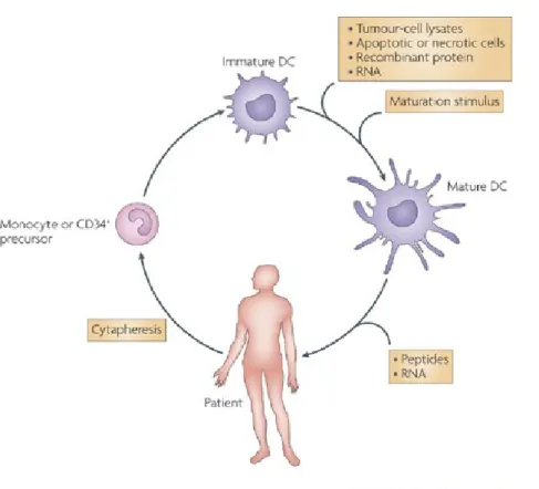
1.5.1 Ex vivo generation of immunocompetent DCs ... 32
1.5.2 Loading dendritic cells with tumor antigens... 34
1.5.3 Clinical trials with DC-based vaccines... 36
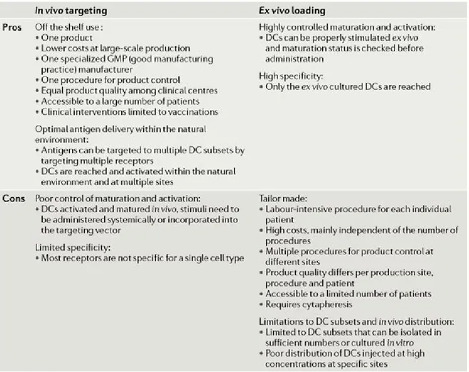
1.5.4 Future perspectives: in vivo dendritic cell targeting ... 37
2 MATERIALS AND METHODS ... 40
3 RESULTS ... 49
3.1 Screening of a scFv phage library on mouse Dendritic Cells... 50
3.1.1 The Phage display technology ... 50
3.1.2 Setting up of the screening procedure... 51
3.1.3 Selection of internalizing phages by panning on BM-DCs ... 55
3.1.4 Selection of internalizing phages by panning on splenic DCs... 59
3.2 Receptor identification ... 61
3.2.1 Binding of scFv-2E5 on DCs subsets ... 62
3.2.2 Binding of scFv-2E5 on hematopoietic cells... 62
3.2.3 Antigen identification ... 64
3.3 Antigen presentation efficiency of the antibody/receptor pairing... 66
3.3.1 Production of scFv-OVA257-264 epitope fusion protein... 66
3.3.2 In vitro antigen cross-presentation assay using scFv-OVA257-264 proteins... 68
3.3.3 In vivo antigen cross-presentation assay using scFv-OVA257-264 proteins ... 70
3.3.4 Production of scFv-OVA fusion proteins ... 71
ABSTRACT
Due to their prominent role in the orchestration of a broad range of immune responses, dendritic cells (DCs) have emerged in the past decade as central target for cancer immunotherapy. Recent advance in the knowledge of DCs functions and subset specialization led to the design of novel immunotherapeutic approaches based on the possibility to target DCs directly in vivo, thus avoiding their ex vivo manipulation.
A promising strategy is to use antibodies to target antigens to cell-surface molecules expressed by DCs, in order to increase T-cell mediated immune responses. The studies performed so far, have revealed that the efficacy of in vivo DC vaccination depend on several factors including the specific DC subset targeted, their maturation status and the nature and biological properties of the receptor targeted. Therefore, the identification of the most appropriate ligand/receptor pairing it is a requisite to improve the modality of delivery tumor Ags to DCs.
To this aim, we screened a library of Ab fragments on mouse DCs to isolate new potential antibodies capable of targeting DCs in vivo and able to induce T-cell mediated immune responses against specific antigens. In this study, we provide the proof of principle that the phage display technology can be successfully used to isolate internalizing antibodies on mouse DCs. We further develop such technology by engineering the selected molecules to create antigen fusion proteins to use in vaccination protocols.
In particular, we focus on a high affinity Ab against CD36, a multiligand scavenger receptor primarily expressed by the CD8α+ subset of conventional DCs. We characterize the antigen
presenting properties of this receptor which help to delineate a novel function of CD36 in adaptive immunity. We show that targeting CD36 on DCs results in the delivery of exogenous Ags to both the MHC class-I and MHC class-II processing pathways. In addition, immunization with the recombinant anti CD36-Ag fusion Ab induces the robust activation of naïve CD4+ and CD8+ Ag specific T lymphocytes and the differentiation of primed CD8+ T cells into long term effector CTLs. Finally, we demonstrate that in vivo targeting of CD8α+ DCs with anti-CD36 Ab elicits humoral and
cell mediated protection from the growth of an Ag specific tumor. Collectively, these results identify CD36 as an appropriate receptor to better elucidate the properties of the lymphoid organ resident CD8+ DCs and indicate it as a novel potential target for cancer immunotherapy.
1.1 Dendritic cells and the control of immunity
Optimal defense against invading pathogens requires specialized classes of non-specific and antigen-specific immune responses, generally referred as the innate and the adaptive components of immunity. Innate immunity or non-specific host defenses that exist prior to exposure to an antigen, specifically detects key features of an infectious process consisting in a limited set of conserved molecular patterns (PAMPs) that are unique to the microbial world and invariant among entire classes of pathogens (1). PAMPs recognition by immune system has two major effects. First, it triggers effector cells of inflammation (macrophages and neutrophils) that limit the infection at the site of pathogen entry. Second, it activates antigen presenting cells (APC) to trigger adaptive immunity, which increases antigenic specificity and generates immunological memory.
Central to this process are dendritic cells (DCs), a heterogeneous family of leukocytes that integrate innate information and convey it to lymphocytes. Upon direct recognition of PAMPs through specific pattern recognition receptors (PRRs) or indirect sensing of infection through inflammatory cytokine or other endogenous “danger” signals, DCs enter an integrated developmental program that triggers their differentiation into immunogenic APCs capable of priming and sustaining the expansion of naïve T cells. Among the “professional” APCs, DCs have a preeminent role in the initiation and modulation of the immune response. Their unique distribution and capacity to accumulate within the T cells area of lymphoid organs optimize antigen capture and clonal selection of rare CD4+ and CD8+ T cells. Their inherent greater efficiency for antigen presentation allows small numbers of DCs and low levels of antigen to induce strong T cell responses (2). In addition, DCs have the unique capacity to translate the environmental signals into specific classes of adaptive immune responses by polarizing T cells development. Finally, DCs are not only implicated in the induction of immunity, but also play an essential role in the generation of both central and peripheral T cells tolerance by inducing deletion, anergy or regulation of T lymphocytes.
1.1.1 The “Langerhans cells Paradigm”
Dendritic cells are described as following a life cycle that was modeled according to observations of studies carried out in the 1970s and 1980s by Steinman and colleagues on DCs found in the epidermis (Langerhans cells) and on DCs purified from the spleen (3). This model, generally referred as “Langerhans cells paradigm”, proposes that DCs can exist in two functional
states: immature and mature. In the periphery, immature DCs efficiently take-up self and non self antigens but are quite inefficient in presenting it to T cells. Only upon encounter of pathogens and mediators of inflammations, DCs enter an integrated developmental program called maturation. During this process, they downregulate their endocytic capacity, activate the antigen processing machinery that generates complex of MHC molecules with peptides derived from the internalized antigens and increase the expression of T cell co-stimulatory molecules. In parallel, modifications in the pattern of chemokine receptors and adhesion molecules and changes in the cytoskeleton organization cause the mobilization from the periphery to secondary lymphoid organs where the antigen are efficiently presented to T cells thus initiating adaptive immune responses.
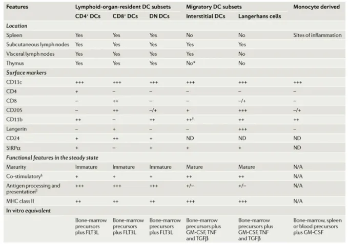
Important findings of the last few years have revealed a far greater complexity of the DCs system of APCs with the consequent need to add few modifications to this original DCs life cycle paradigm. The analysis of the DCs found in the thymus, spleen and lymph nodes has revealed a considerable heterogeneity and has led to the definition of several DCs subtypes, each with a particular location and specialized function in the immune system (4). It is now clear that only a fraction of the DCs found in lymph nodes are derived from cells previously resident in the peripheral tissues. In a steady state condition (i.e in absence of infections or inflammatory stimuli), half of the DCs found in lymph nodes and most of the DCs in the spleen seem to be lymphoid tissue resident cells, with an immature phenotype and great efficiency in antigen uptake and processing (5, 6). Therefore, these lymphoid-organ-resident DCs do not conform to the Langerhans cells paradigm, since they develop from bone marrow precursors within the lymphoid organs and without previously trafficking through peripheral tissues (7).
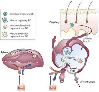
Figure I. Different life cycles of migratory and resident dendritic cells. Migratory DCs
have an immature phenotype in peripheral tissue where they survey the environment and endocytose extracellular material. They migrate through the afferent lymph to the local lymph nodes where they acquire a mature phenotype. Resident DCs, present in lymph nodes and spleen, spend their entire lifespan in
1.2 Dendritic cells general features
1.2.1 Mechanisms of Antigen uptake and pathogen recognition
Dendritic cells have evolved several features that greatly enhance their capacity as APCs. Among these, the property of immature DCs to efficiently take up particles and microbes by phagocytosis and the ability to form large pinocytic vesicles to sample extracellular fluids and solutes in a process referred to as macropinocytosis (8). Moreover, DCs express a great variety of receptors involved in antigen recognition and uptake by mechanisms of receptor mediated endocytosis. Endocytosis is a regulated process in DCs, being highly active in immature DCs and downmodulated upon cells maturation (9). This assures the efficient sampling of the environment in the periphery and concomitantly limits the range of antigens that the cells will be able to present after living the peripheral tissues. The downregulation of internalization is achieved by decreasing the expression of the cell receptors involved in antigen uptake and by downmodulating both macropinocytic and phagocytic activities.
Phagocytosis
The uptake of large particulate antigens (>0,5µm) by phagocytosis is the prevalent form of antigen uptake in vivo for both pathogen-derived and endogenous antigens (8). It is an active and highly regulated process initiated by the engagement of specific cell surface receptors, generally involved also in clathrin mediated endocytosis, that trigger a signaling cascade mediated by the Rho-family GTPases (Rho, Rac and Cdc42). The final outcome is the extensive reorganization of the actin cytoskeleton with the formation of cell-surface extensions that zipper up around the pathogen to engulf it (10). The nature of the particle to be ingested and the receptors that recognizes it determine the mode of protrusive activity and the signaling pathways that are activated. In the case of FcγR mediated phagocytosis, the cells extends pseudopods that engulf the particle and subsequently fuse to form a phagosome. This process requires the activation of Cdc42 for the pseudopods extension and Rac in pseudopods fusion and phagosome closure. In contrast, phagocytosis mediated by complement receptor is controlled by Rho, do not induce pseudopod formation but requires the coordinated action of chemokines and integrin ligation to form the phagocytic cup (11).
Phagocytosis is essential in host defense against infections: immature DCs were reported to phagocytose almost any kind of bacteria, both GRAM+ and GRAM- and mycobacteria, yeast cells
and parasites (12). Phagocysis is also crucial for clearing apoptotic cells, both at sites of inflammation and tissue damage and during development. This process is mediated by the recognition of specific molecules that are normally absent on live cells such as calreticulin and phosphatidylserine, amino sugars and lysophospholipids and involves various types of scavenger receptors, complement receptors and integrins on DCs. The nature of the cargo determines the set of receptors involved in its recognition and uptake, the signaling cascade that is activated upon receptor binding and ultimately regulates the efficiency of processing/presentation and the immunogenicity of the internalized material. For instance, phagocytosis of microbial cells and of apoptotic bodies rely on the same phagocytic machinery, but the cellular responses that accompany these forms of phagocytosis are different, being respectively inflammatory with the production of tumor necrosis factors, IL-1and IL-6 in case of pathogens and anti-inflammatory in case of uptake of apoptotic material (13).
Macropinocytosis
Macropinocytosis contributes to bulk fluid-phase uptake via the formation of membrane protrusions that collapse onto and fuse with the plasma membrane generating large endocytic vesicles (micropinosomes) that allow the sampling of large volumes of extracellular milieu (10). The signaling cascades that induce macropinocytosis involve the Rho-family of GTPases and especially Cdc42, that trigger the actin-dependent formation of the membrane ruffles (11). In immature DCs macropinocytosis is constitutive and like phagocytosis, is downmodulated upon cells maturation. The impact of antigen uptake via macropinocytosis is vivo is not clear since pathogens release few soluble antigens, with most proteins integrated in membranes, cell walls or cytoplasmic compartments that would need to be taken up as particles. Conversely, it has a role in the capture of soluble antigens that become available upon intravenous, intraperitoneal and intradermal injections (8). The degree of internalization of fluid-phase antigens is proportional to their concentration in the medium. The efficiency of endocytosis is increased by non-specific binding of solutes to the cell membrane (adsorptive pinocytosis) and even more by the capture of soluble antigens through
Receptor mediated endocytosis
Uptake of extracellular material through receptor mediated endocytosis greatly increases the efficiency of antigen capture thus allowing DCs to present antigens found at picomolar and nanomolar concentrations (2). This process is initiated by signal cascades triggered by specific endocytic receptors present in specialized regions of the plasma membrane. The nature of the receptors engaged determines the mechanism of internalization that can be mediated by clathrin-coated vesicles, by caveolae or can be both clathrin- and caveolae- independent.
DCs express a wide range of endocytic receptors that belongs to different families. Receptors for the Fc portion of immunoglobulin (FcRs) and complement receptors (CRs) are involved in the uptake of particles that are opsonized by immunoglobulins or complement factors. A second class of endocytic receptors comprises scavenger receptors (SRs) and C-type lectin family receptors (CLRs) that directly recognize specific structures on both self-antigens and pathogens and are generally referred to as innate immunity receptors. A large number of these endocytic molecules are selectively expressed by subpopulations of DCs thus contributing to the functional diversity among different DCs.
Fcγ and complement receptors
Mouse immature DCs express different types of FcRs such as FcγRI, FcγRIIb, FcγRIII and FcγRIV and the complement receptors CR3 and CR4. Both FcRs and CRs may be functionally considered as antigen receptors due to their ability to bind and to mediate the uptake of immune complexes and complement-opsonized particles. The FcγRs differs in terms of antibody isotype specificity and binding affinity with FcγRI binding with the highest affinity exclusively monomeric IgG2a and FcγRIV binding with intermediate affinity IgG2a and IgG2b but not IgG1, whereas both FcγRIIb and FcγRIII binds immune complexes of all IgG isotypes (14). Antigens bound to soluble Ig can follow different fates depending on the cell type that captures them. Among APCs, DCs are unique for their capacity to efficiently deliver antigens internalized via FcγRs to the antigen processing machinery which results in the formation of both MHC class I and MHC class II-peptide complexes (15).
CR3 binds a promiscuous range of ligands, both of host and microbial origin. It was shown to be involved in the uptake of complement opsonized apoptotic cells and also in the direct recognition of pathogens such as M. tuberculosis and yeast-derived zymosan (16).
C-type lectin receptors
Immature DCs differentially express a broad variety of C-type lectin receptors (CLRs) that are involved in the recognition of a wide range of carbohydrate structures in antigens and act as endocytic or phagocytic receptors depending on the size of the antigen. The most characterized examples of DCs CLRs include the mannose receptor, DEC205, DC-SIGN, Dectin-1 and -2 and Langerin. They are generally classified as type I or type II receptors depending on the orientation of their N terminus, which is extracellular or cytoplasmic respectively, and on the number of carbohydrate recognition domains (CRDs) present in their extracellular portion. The cytoplasmic domains of the CLRs are diverse and contain several conserved motifs that are important for antigen uptake and determine the intracellular route followed by the antigen upon receptor-mediated internalization. For instance, most CLRs (DEC205, DC-SIGN, BDCA-2 and Dectin-1) contain a tri-acidic cluster that target internalized antigens to lysosomes and MHC class II+ late endosomes, whereas the MR constitutively delivers antigens to early endosomes and rapidly cycle back to the cell surface (17).
Several CLRs expressed by immature DCs are capable of binding pathogens through the recognition of specific carbohydrate structures present on the cell wall component. Some CLRs have overlapping carbohydrate specificity and recognize a wide variety of carbohydrate structures such as mannose- and fucose-containing carbohydrates, yet others have a unique recognition profile dictated by a specific branching of the mannose structures (17). However, little is known about the antigen specificity of CLRs expressed by DCs and their functions are not yet documented. Only in few cases specific pathogens have been associated to CLRs. For example, the mannose receptor is able to bind yeast, human immunodeficiency virus and Micobacterium tuberculosis, whereas DC-SIGN binds a large variety of pathogens, including viruses (HIV, HCV, CMV, Ebola and Dengue virus), Candida albicans, bacteria (Micobacterium tuberculosis, Helicobacter pylori and Klebsiella pneumonae) and parasites (Leishmania pifanoi and Schistosoma mansoni) (18). Dectin-1 recognizes conserved molecular pattern (β1,3-glucans) in fungal pathogens (P.carinii, C.albicans and A.fumigatus) and triggers a signaling pathway that induces the activation of adaptive immunity (19). To date this is the only documented case of host protection mediated by a CLR. In fact, although DC-SIGN and MR
glycoproteins, such as lysosomal hydrolases (21) and lutotropin (22) confirming an important role for this CLR in the clearance of self antigens.
Scavenger receptors
A third class of endocytic receptors expressed by DCs is the family of scavenger receptors (SRs). SRs encompass a broad range of cell-surface glycoproteins that differ greatly in their structure but are, by definition, all capable to mediate the uptake of selected polyanionic ligands, including modified low-density lipoproteins (LDL). The main feature of SRs is their ability to recognize multiple ligands, both microbial and host derived, and to carry out a multiplicity of cellular functions (ranging from anti-microbial immunity, antigen presentation and cell adhesion to atherosclerosis and Alzheimer’s disease). However, while SR expression and function has been extensively investigated for macrophages, the role of these molecules on DCs is only beginning to be unraveled. Three SRs belonging to different classes, defined on the basis of their molecular structure, are expressed by DCs: Scavenger Receptor A (SR-A) (23), CD36 (24-26) and Lox-1 (27).
SR-A is a trimeric type II glycoprotein containing in the extracellular portion an α helical coiled coil, a collagenous and a C-terminal cysteine-rich domain. Its cytoplasmic tail instead, contains a potential protein kinase C interaction site, but not an internalization motif. SR-A was shown to bind several components of both GRAM- and GRAM+ bacteria (lipid A and lipoteichoic acid respectively). Indeed, its role in microbial clearance has been confirmed in two studies performed on SR-A deficient mice, where the absence of the receptor resulted in increased bacterial loads of Listeria monocytogenes and Staphylococcus aureus (28). Recently, Amiel et al showed that SR-A-/- BMDCs are impaired in their ability to phagocytose and accumulate E.coli bacteria and that the internalization requires lipid raft structures (29). Similarly to other SRs, SR-A recognizes also endogenous ligands and modified host molecules expressed on aged/apoptotic cells, with a possible role in tissue homeostasis (30). Finally, Berwin et al identified SR-A as a predominant endocytic receptor for the ER chaperones gp96 and calreticulin and demonstrate that SR-A can direct gp96-OVA derived peptide complex into an MHC class I presentation pathway (31). However, SR-A is not required for antitumor immunity generated by HSP-based vaccines, rather its lack restored the immunogenicity of poorly immunogenic tumors and enhanced the tumor specific immune response, suggesting a role of SR-A in immune suppression and in the maintenance of host immune homeostasis (32).
LOX-1 is a 48-50 kDa type II membrane glycoprotein with a C type lectin-like domain whose antibacterial potential was revealed by binding studies performed with both GRAM+ and GRAM- bacteria (S. aureus and E.coli). LOX-1 was also identified as a receptor for Klebsiella
pneumoniae OmpA, a major component of the outer membrane of Enterobacteriaceae. Specifically, it was shown to act in a sequential and coordinate way with TLR-2 to mediate OmpA internalization and DCs activation (33). Delneste et al first reported LOX-1 as a receptor for Hsp70 on human DCs and showed its involvement in Hsp70 mediated antigen cross-presentation in vivo thus suggesting a possible role for LOX-1 in the recognition of cellular damage (27).
CD36 belongs to the class B SR family. It is a double-spanning N-linked glycosylated protein of 53 kDa thought to be associated with lipid rafts. The predicted structure orients most of the protein extracellularly, except for two short (9-13 amino acids) cytoplasmic tails (34). The C-terminal cytoplasmic domain was shown to have a role in the localization of CD36 in the plasma membrane and in the uptake of long-chain fatty acid (35) and oxidized LDL (OxLDL) (36). Binding of OxLDL to CD36 leads to endocytosis through a lipid raft pathway that is both clathrin and caveolin independent (37).
As most of SRs, CD36 is a multiligand receptor shown to play a role in diverse cellular processes including foam cell formation, fatty acid transport, suppression of angiogenesis, cell-matrix interaction (34). CD36 is one of the first DCs receptors shown to be involved in the uptake of apoptotic cells (24), probably through interactions with oxidized phosphatidylserine (38) and with thrombosponsdin-1 (39) present on the membrane of apoptotic cells.
CD36 has also been implicated in the innate immune response to GRAM+ bacteria such S. aureus . Hoebe et al first demonstrated the selective and nonredundant role of CD36 as sensor for microbial diacylglycerides such as macrophage-activating lipopetide 2 and lipoteichoic acid (LTA) (40). Upon binding to bacterial LTA, CD36 forms a cluster with the heterodimer TLR2/TLR6 within lipid raft, followed by the internalization of the complex and its targeting to the Golgi apparatus (41). The C-terminal cytoplasmic residues of CD36 were shown to be essential to trigger phagocytic engulfment and specifically, the tyrosine 463 was implicated in signaling to the actin cytoskeleton (42). These data collectively indicated that CD36 acts as a facilitator or co-receptor for diacylglyceride recognition through the TLR2/6 complex.
1.2.2 Mechanisms of Antigen presentation and cross-presentation
After their entry into DCs, antigens are degraded into small immunogenic epitopes that associate with the major histocompatibility complex molecules (MHC) and are transported to plasma membrane where they eventually trigger the activation of naïve T lymphocytes.
Specifically, the activation of CD8+ and CD4+ T lymphocytes requires recognition by the T cell receptor (TCR) of fragments of antigens (peptides) associated with MHC class I and MHC class II molecules, respectively. The antigen processing pathways that lead to the formation of peptide-MHC complexes, rely on ancient proteolytic mechanisms (proteasome, lysosomes) that in DCs were geared towards optimal T cells stimulation (8).
Initial studies on antigen presentation defined a strict compartmentalization of MHC class I and II biogenensis, with MHC class II molecules encountering exogenous antigen in the endocytic pathway and MHC class I loading with endogenous antigens in the endoplasmic reticulum (ER). Although still valid, recent evidence have proved that both boundaries can be crossed and so MHC-II molecules can present intracellular antigens not coming from extracellular space and MHC-I can present peptides derived from exogenous antigens, phenomenon generally referred to as cross-presentation.
MHC class I–restricted antigen presentation
DCs process and present on MHC class I molecules proteins coming from various sources including cytosolic proteins (either endogenous or viral), alternative translation products and defective ribosomal products (DRiPs), protein retrotranslocated to the cytosol from the ER (i.e. secretory and membrane proteins) and internalized proteins transferred to the cytosol and imported into the ER (8). Independently of the route of entry, most of the peptides to be loaded on MHC-I molecules are generated by the proteasome and further trimmed by cytosolic or ER resident peptidases. The proteasome has a critical role in initiating the processing and in most cases, releases precursor peptides that have the correct C terminus for MHC-I anchoring and an N-terminal extension that requires further degradation. The efficiency and rate of epitope generation by the proteasome are increased in response to an inflammatory environment (i.e. IFN-γ) by expression of facultative subunits (referred to as immunosubunits). The proteasome has also been implicated in splicing of peptides to create epitopes from sequences that are not contiguous in the original protein (43, 44). The N-terminal trimming occurs in both cytosol and ER by the tripeptidyl peptidase II (TPPII) and ER-associated aminopeptidase (ERAAP) respectively (45). Once the peptide products
are shuttled into the ER via the transporter associate with antigen processing (TAP) and customized by ERAAP, the final MHC-I peptide complex (pMHC-I) is formed and eventually displayed on the cell surface (46).
Cross-presentation of exogenous antigens
Dendritic cells have evolved a specialized machinery that allows the entry of exogenous antigens into the MHC class I pathway of antigen presentation (47). This unique capacity, generally referred to as “cross presentation,” is a major mechanism for the immune surveillance of tissues, allowing the immune system to monitor parenchymal cells for the presence of foreign antigens. Several types of antigens have been reported to be cross-presented: soluble proteins, immune complexes, pathogens and cellular antigens, independently of the mechanism of internalization (macropinocytosis, phagocytosis or receptor mediated endocytosis) (48). However, the mechanisms by which APCs transfer internalized antigens to the MHC class I loading pathway are not well understood (49). In most of the cases, proteasomal digestion and transport into ER by TAP seem to be required, implicating a mechanism to allow the release of the antigen from the endocytic structures to the cytosol. However, the cellular and molecular bases of endosomal egress into the cytosol remain unknown (49). Reports have suggested that a potential mechanism for cross-presentation relies on the provision of MHC-I loading components to the phagosome by membranes derived from the ER, resulting in ER-phagosome complexes self-sufficient for cross-presentation (50, 51). Although the relative contribution of the ER versus the plasma membrane as source of membrane for phagocytosis is still a matter of debate (49), recent data by the Cresswell group have shown that soluble antigens can gain access to the lumen of the ER after internalization (52) and that the ER protein retrotranslocation machinery (ERAD) is involved during cross-presentation (53). Among APCs, DCs have the highest cross-presentation efficiency. One of the mechanisms that accounts for this specialization is the limited degradation of internalized antigens in the endocytic compartments, which may increase the opportunity for the internalized antigens to reach the cytosol (54). Indeed, experimental reduction of lysosomal proteolysis by choloquine was shown to increase
The uniqueness of DCs with respect to antigen handling reflects the essential role that these cells play in priming in vivo CTL responses to exogenous antigens. In an elegant study, Jung and colleagues demonstrated that the in vivo depletion of DCs abrogates priming of CD8+ T cells by exogenous cell-associated antigens and results in the failure to mount a CTL response to infection with the intracellular bacterium Listeria monocytogenes and the malaria parasite Plasmodium yoelii (57). In addition to intracellular bacterial infection, cross-presentation have an essential role in responses to those viruses, e.g. cytopatic strains or ones with immune evasion molecules, that impair the ability of directly infected DCs to function, or to tissue specific viruses that do no infect DCs (58-61). Finally, cross-presentation is also implicated in the maintenance of both central and peripheral tolerance to self antigens eventually leading to the deletion or anergy of self reacting CTLs (62).
MHC class II–restricted antigen presentation
Antigens loaded on the MHC-II are typically exogenous proteins that are internalized by DCs through different mechanisms of endocytosis or endogenous proteins that reside in the secretory system. Moreover, analysis of the peptides eluted from the MHC-II molecules has revealed that also antigens topologically isolated from the endosomal system (i.e. cytosolic and nuclear proteins) can gain access to MHC-II class antigen presentation pathway (63). Recent studies have implicated autophagy as likely source of peptide derived from various cytosolic and nuclear antigens (64). Schmid and colleagues have recently shown that macroautophagy is a constitutively active pathway in all MHC-II positive cells and that autophagosome continuously fuse with multivesicular MHC-II loading compartments. They also show that targeting antigens to autophagosomes leads to enhanced MHC-II presentation and CD4+ T cells stimulation (63).
De novo synthesized MHC-II αβ dimers assemble in the ER with the chaperon invariant chain (Ii), which inserts its class II associated peptide (CLIP) portion within the peptide binding site thus stabilizing and protecting the MHC-II dimer from other polypeptides. The MHC-II αβIi complexes are transported to early endosomes and further on to late endosomes and lysosomes. Along this pathway, the Ii is progressively processed by various proteases (AEP, cathepsins B and S) and finally displaced to allow the binding of the antigenic peptide, in a reaction favored by the acidic pH and by the chaperone protein HLA-DM (8). MHC-II molecules can bind polypeptide precursors that are trimmed by various proteases along the endocytic route, so that the final MHC-II peptide complex eventually reaches the plasma membrane.
Recent findings by Blander et al have demonstrated how the nature of the cargo internalized by phagocytosis and thus the type of receptors engaged during this process are able to influence the efficiency of presentation on MHC class II molecules. They show how the generation of peptide-MHC class II complexes is controlled by Toll-like receptors (TLRs) by regulating the progressive proteolysis of the Ii to CLIP in a strictly phagosome-autonomous manner. This mechanism allows DCs to distinguish between different forms of antigens as self or non-self at the subcellular level and ensures that only antigens that concomitantly engage TLRs are preferentially delivered to the pool of MHC-II molecules transported to the plasma membrane (13).
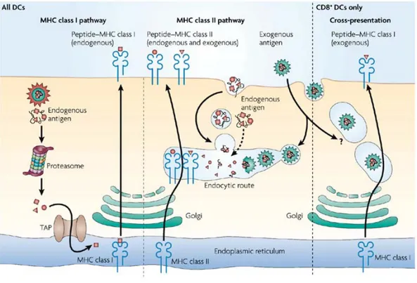
Figure II. The antigen-presentation pathway in dendritic cells. MHC class I molecules present
peptides that are primarily derived from endogenously synthesized proteins of either self of pathogen origin. These proteins are degraded into peptides by the proteasome and transported through TAP molecules into the endoplasmic reticulum for loading on MHC class I molecules. MHC class II molecules acquire peptide cargo that is generated by proteolytic degradation in endosomal compartments. The precursor proteins of these peptides include exogenous material that is endocytosed from the extracellular environment, and also endogenous components, such as plasma membrane proteins and cytosolic proteins that access the endosomes by autophagy.
1.2.3 Induction of Dendritic cells activation and signal transduction
Only upon direct recognition of pathogens associated pattern (PAMPs) or sensing of inflammatory signals, DCs are activated to enter the program of maturation that endows the cells with the ability to stimulate naïve T lymphocytes. DCs response to microbial/inflammatory signals is mediated by different classes of surface and intracellular receptors whose engagement triggers signal cascades that lead to the production of inflammatory cytokines, to the up-regulation of co-stimulatory molecules and to the altered expression of chemokine receptors profile. Mature DCs have an increased antigen-presenting capacity and migrate to the draining lymph nodes where they interact with and stimulate antigen specific T cell. DCs maturation is therefore a critical link between innate and adaptive immunity (65).
Toll like receptor family
One of the best characterized classes of pattern recognition receptors (PRRs) that directly contribute to the inflammatory response to pathogens is the Toll-like receptor family (TLRs). Mammalian TLRs are a family of at least 12 transmembrane proteins that collectively recognize lipids, carbohydrate, peptide and nucleic-acid structures broadly expressed by different groups of micro-organisms. Some TLRs (TLRs 1,2,4,5 and 6) are expressed at the cell surface and seem to specialize in the recognition of mainly bacterial products, whereas others (TLRs 3,7,8, and 9) are expressed on the membrane of endocytic vesicles or other intracellular organelles and are specialized in detection of viral nucleic acids (66). TLRs differ from each other in ligand specificities, expression patterns and presumably in the target genes that they can induce.
TLR4 plays an essential role in the recognition of LPS, a major component of GRAM- bacteria, whereas TLR2 is critical to the recognition of bacteria peptidoglycan and lipoproteins, but it is also able to recognize components from fungi (zymosan) and parasites (glycoinositolphospholipids from Trypanosoma Cruzi). TLR2 promiscuity in ligand recognition derives from its ability to cooperate with other TLRs (TLR1 and TLR6) by formation of different heterodimers which confer discrimination among different microbial components (67). Further complexity is added by recent data showing the involvement of non-TLRs in the presentation of some PAMPs to TLR2. For instance, the scavenger rector CD36 acts as facilitator to mediate the interaction of microbial diacylglicerydes to the heterodimer TLR2/6 (40). Moreover, the potential interaction of TLR2 with a member of CLRs is suggested by data from Wang et al. showing the critical role of TLR2 in the initiation of the adaptive immune response to the polysaccharide A (PSA) from Bacteroides fragilis
(68). Other TLRs are involved in the recognition of microbial proteins such as flagellin by TLR5 or apicomplexan profilins by TLR11 (69).
TLRs have also an essential role in the antiviral immune responses due to their ability to induce type I IFNs, a family of cytokines specialized to coordinate immunity to viruses and other intracellular infections (70). For instance, TLR9 and TLR7 which represent a structurally related subfamily, are involved in the recognition of unmethylated 2’-deoxyribo (cytidine-phosphate-guanosine) (CpG) DNA motifs present in bacteria and viral DNA, or ribonucleic acid homologs (imiquimod and resiquimod) and synthetic ssRNA rich in guanosine/uridine respectively. A further member, TLR3, responds to dsRNA and to its synthetic analog poly-inosinic acid-cytidylic acid (poly(I)●poly(C))
(71).
Upon recognition of their ligands, TLRs transduce signals through two pathways involving distinct adaptor proteins containing Toll/IL-1R (TIR) domains. One of these adaptors, MyD88, is utilized by all of the known TLRs except TLR3. MyD88 is recruited to the receptor-ligand complex in an interaction that is mediated its C-terminal TIR domain. Through its N-terminal domain the adaptor engages the kinase IRAK which is activated by phosphorylation a then associates with TRAF6, leading to the activation of two distinct signalling pathways: JNK and NF-kB (72).
TLR3 and TLR4 are able to activate NF-kB in a MyD88 independent way that involves the adaptor molecule TRIF and other signalling molecules such as RIP1 and TRAF6. Activation of TLR3 and 4, through the adaptor TRIF, induces also the production of type I IFN. TRIF associates with the kinases TBK1 and IKKi which mediate the phosphorylation and the nuclear translocation of IFN-regulatory factor 3 (IRF3) (73). Also TLR7, 8 and 9 elicit type I IFN induction, especially IFN-α, through MyD88, in a pathway that involve the kinase IRAK and the IFN-regulatory factor 7 (IRF7) (73)
Collectively, activation of these signalling cascades induce the expression of a variety of host defence genes that include inflammatory cytokines (IL-12, TNFα and IL-6) and type I interferon cytokines, the up-regulation of co-stimulatory molecules (CD40, CD80 and CD86) and the altered expression of chemokines receptors (CCR2, CCR 5 and CCR7) (65).
MHC class II endocytosis, thus extending the half-life of assembled peptide-MHC complexes at the plasma membrane (76).
Fc receptor family
Some classes of PRRs involved in antigen recognition and uptake are also able to activate a downstream signalling cascade that leads to DCs maturation. Among these, the well characterized class of Fcγ receptors. The potent immunoregulatory functions of immune complexes (ICs) rely on two different classes of DCs FcγRs: the activatory FcγRIII and FcγRIV and the inhibitory receptor FcγRIIb that transduce their signals via immunoreceptor tyrosine-based activation (ITAM) and inhibitory motifs (ITIM), respectively (14).
FcγRIIb is a single-chain receptor that carries an ITIM motif in its cytoplasmic domain. Its expression on DCs controls ICs mediated DCs maturation, as DCs derived from mice deficient for FcγRIIb have an enhanced potential to generate Ag-specific T cells responses (77). FcγRIIb may function as a regulator of DCs activation by setting the threshold that prevents spontaneous maturation of DCs and thus promoting steady-state tolerance (14).
The activating FcγRs transmit their signals through a common accessory γ chain that carries the ITAM motif and that is essential for their cell surface expression. DCs maturation is triggered by FcγRs engagement in a signalling pathway that leads to tyrosine phosphorylation of the ITAM motif by members of the Src-kinase family and subsequent recruitment of members of the Syk-kinase family (78).
The paired expression of activating and inhibitory FcγRs on the same DCs is the key for the generation of balanced immune responses, where the final outcome will depend on the expression levels of activating and inhibitory receptors, on their relative affinity for IgGs and on the isotype of the antibodies produced by the humoral response.
C-type lectin receptor family
Among C-type lectin receptors, Dectin-1 is the first example of PRR that can mediate its own intracellular signals thus coupling PAMP recognition to the induction of adaptive immunity. Dectin-1 contains an ITAM-like motif in its cytoplasmic tail which resembles sequences found in the activating FcγRs and similarly to them, upon receptor engagement, it is phosphorylated by Src-kinases and subsequently it recruits Syk-Src-kinases (79). The tyrosine kinase Syk activates downstream signalling components including the transcription factor NF-kB in a pathway that also involves the
CARMA1-related adaptor protein CARD9. Dectin-1 signalling in DCs results in maturation and production of a distinct combination of cytokines, including IL-2, IL-10, TNF and IL-23 which strongly biased T helper cell differentiation to a T helper 17 fate (19).
Intracellular pattern recognition receptors
In addition to membrane associated PRRs, DCs expresses cytosolic receptors involved in the recognition of bacteria and viruses that gain access into the cytoplasm and are able to induce the production of cytokines and cell activation. These cytosolic receptors are grouped in two main family: the NLRs (nucleotide-binding oligomerization domain (NOD)-like receptor family) which comprises at least 23 members that are either NOD receptors or NALPs; and the RLRs (RIG-I like receptors), a family of receptors that have an RNA-helicase domain joined to two caspase-recruitment domains (CARDs) which include RIG-I (the retinoic-acid inducible gene I) and MDA5 (the melanoma-differentiation-associated gene 5) (80). NLRs are involved in the recognition of bacterial peptidoglycans from both GRAM+ and GRAM⎯ bacteria and drive the activation of MAP kinases and NF-kB in a signalling pathway similar to that triggered by TLRs. In addition to microbial products, some NALP proteins can also sense the presence of host danger signals such as low concentrations of intracellular potassium, monosodium urate and calcium pyrophosphate crystals and drive the formation of a complex called “inflammasome”. This complex has an important role in the activation of pro-inflammatory caspase such as caspase-1 which in turn leads to the processing and the release of the active form of the inflammatory cytokine IL-1β. RLRs are cytoplasmic sensor of virally derived dsRNA and similarly to anti-viral TLRs trigger the activation of NF-kB and transcription factors of the IRF family (IRF3 and IRF7), which cooperate in the induction of antiviral IFN type I (81).
Microorganisms are thus able to simultaneously trigger a complex set of PRRs, both within and outside the TLRs family. It is becoming clear that the combined activation of these different receptors can result in complementary, synergistic or agonistic effects especially in terms of cytokine
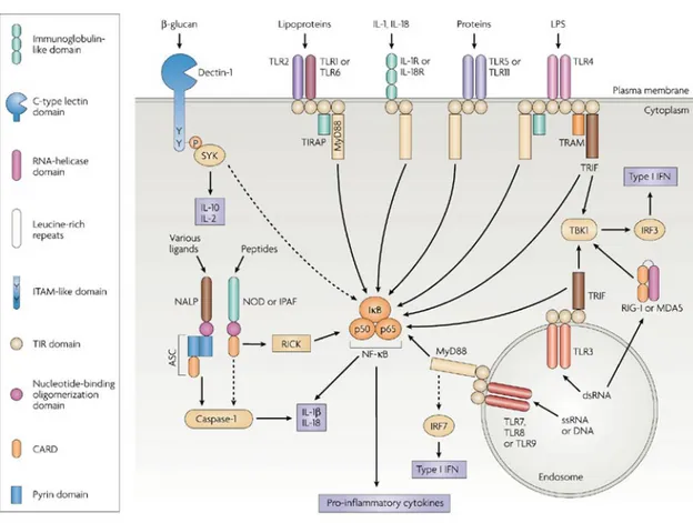
Figure III. Schematic representation of the main signaling pathways of the PRR families. Only
the adaptor molecules and the main signaling pathways that differentiate the different classes of PRRs are shown. In reality, the pathways that are activated by the different receptors are multiple. TLRs (with the exception of TLR3), IL-1R and IL-18R induce NF-kB dependent cytokine production through a pathway involving the adaptor MyD88. However, TLRs signaling may also involve MAP kinases and phosphatidylinositol 3-kinase. TLR3 and TLR4 uses a MyD88-independent signaling pathway that involves the adaptor molecule TRIF and induces type I IFNs production. Dectin-1 (a β-glucan) receptor is shown as an example of various cell-surface PRRs, that like FcRs, signal via their ITAM-like motif to activate the kinase SYK. The NLRs family of intracellular PRRs signal through complex pathways that result in the activation of caspase-1 in case of NALP, or in the induction of NF-kB in case of NOD proteins. The RLRs (RIG-I and MDA5) activate NF-kB, but also induce type I IFNs production via IRF3 and IRF7.
1.3 T cells stimulation by Dendritic cells
1.3.1 T cells immunity
Several unique properties contribute to the prominent role of DCs in the initiation and modulation of adaptive immune responses: the great efficiency in antigen uptake and processing, the unique capacity of sensing and integrating different environmental signals and the ability to migrate and to localize in T cells rich areas of secondary lymphoid organs. Is in this definite area of lymph nodes that antigen specific immune responses initiate by engagement of the T cell receptor (TCR) with the cognate peptide-MHC complex displayed by the DCs. TCR triggering determine the antigen-specificity of the response and represents the fist of the three signals delivered by DCs that dictates the fate of naïve T cells. The second signal is referred to as T cell co-stimulation and is mediated by the engagement of both positive (CD28) and negative (CTLA-4 and PD-1) co-stimulating receptors on T cells by DCs. The fine balance between these positive and negative signals controls the initiation of protective immunity. If the DC is not fully activated, due to the lack of inflammatory stimuli, negative signals prevail leading to T cell inactivation by anergy or deletion and the induction of a state of tolerance (83). Finally, T cells sense and integrate signals, mediated by different cytokines and chemokines (referred to as signals 3) that are produced by DCs and whose nature strictly depends on the conditions under which DCs are primed. The set of secreted cytokines determine the extent of T cells proliferation, the survival and the ability to develop effector functions and specifically the differentiation into effector cytotoxic T cells or the acquisition of a TH1, TH2,
TH17 of T regulatory phenotype in case of CD8+ and CD4+ T lymphocytes respectively.
1.3.2 Dendritic cell maturation and T cell priming
Several studies initiated almost a decade ago, have provided the direct evidence that DCs play a crucial role not only in the induction of adaptive immunity, but also in the maintenance of
ultimately led to an immune tolerant state (84). Further studies by Steinman group implicated DCs as the APCs responsible for tolerance induction. They showed that in a steady state condition (i.e. absence of inflammation), direct targeting of DCs in situ through DEC205 induced the efficient presentation of the peptide HEL (fused to anti-DEC antibody) on MHC class II molecules and the extensive proliferation of adoptively transferred HEL-specific CD4+ T cells. However, the activated T cell were eventually deleted unless an additional DCs activation signal, such as CD40 ligation, was provided (85). A second study by the same group extended these findings on Ag specific CD8+ T cells. Targeting DCs via an anti-DEC-205 Ab coupled to OVA induced the efficient cross-presentation of the Ag and the extensive proliferation of adoptively transferred OT-I cells. However, by 10-12 days, the stimulated T cells underwent a deletional mechanism, being entirely absent from lymph nodes, spleen and blood and were unresponsive upon rechallange with OVA Ag, thus proving the induction of peripheral tolerance. Similarly to the previous study, the co-administration of an agonistic anti-CD40 antibody as an adjuvant, activated DCs in situ and produced a strong CTLs response and memory (86).
The finding that cross-presentation of different forms of Ags, either soluble proteins (85, 86) or dying cells (87) by steady state DCs is an essential mechanism to guarantee tolerance to self, was further extended to the classical pathway of MHC class I presentation by works performed in van den Broek laboratory. Probst et al. used a transgenic mouse model (DIETER) that allows the controlled expression and presentation of lymphocytic choriomeningitis virus (LCMV)-derived CTL epitopes exclusively by DCs. They observed that activated DCs (by systemic administration of anti-CD40 Ab), efficiently primed naïve endogenous CTL to proliferate and to differentiate into protective effectors, whereas resting DCs induced Ag specific tolerance, that could not be broken by a subsequent infection with LCMV (88).
Collectively, all these data demonstrate that DCs maturation state is the critical switch that provides signals for effector T cell development and memory, diverting T cells from anergy or deletion to protective immunity (89).
Several properties of mature DCs could account for their capacity to efficiently prime T lymphocytes and to induce their differentiation into long-lived functional memory. Among these, the increased expression of co-stimulatory, MHC and adhesion molecules which are crucial in controlling the stability and the duration of the DC-T cell contact (90) so that the duration of the TCR signaling may influence the functional outcome of T cells activation (91). In addition, mature DCs secrete immune enhancing cytokines like IL-12 and type I/type II IFNs that favor the differentiation of T lymphocytes into effector cells and T cells survival. On the contrary, immature DCs induce peripheral T cell tolerance through combined mechanisms of antigen specific T cells deletion and functional inactivation or by generation of regulatory T cells. However, the in vivo relative contribution of these mechanisms remains still elusive and may largely depend on the specific experimental setting and model analyzed. For instance, delivery of antigens to steady state DCs via DEC205 leads to T cells tolerance by both deletional mechanisms (85, 86) and induction of CD4+/CD25+ regulatory T cells (92). Alternatively, presentation of cellular antigens expressed by resting DCs results in the depletion of antigen specific T cells upon engagement of the T cells co-inhibitory molecules PD-1 and CTLA-4 (83). Finally, a population of CD103+ DCs found in the
mesenteric LNs is able to promote the conversion of naïve T cells into Foxp3+ T reg cells upon oral
administration of the antigen, in a mechanism dependent on TGFβ and retinoic acid (93).
1.3.3 Dendritic cell “licensing” by CD4
+T cells
Dendritic cells play a prominent role in the generation of CTL responses not merely by presenting antigens, but in supplying critical co-stimulatory and differentiation-inducing mediators that guide the development of lymphocyte responses. Experimental evidences from the late of 1990s led to a model of DCs licensing, in which antigen-specific CD4+ T cells interacted with DCs via CD40/CD40L interactions and conferred upon DCs the ability to prime CTL responses (94, 95). Whether such CD4+ T cells help is essential for the induction of primary CTL responses or in the generation of functional memory CD8+ T cells it is still an open issue and a matter of intense study. The generation of a primary CD8+ T cells response has been shown to be independent from the
this context, the recognition of PAMPs by TLRs and other PRRs may lead to a DCs activation status that allows to bypass the need for CD4+ T cells help (95).
A second critical issue regards the requirement of CD4+ T cell help in generating functional CD8+ T cell memory. In this setting, many studies have clearly demonstrated that even if the primary CTL response to pathogens like Listeria monocytogenes and lymphocytic choriomeningitis virus fully develops in a TH independent manner, the resulting memory CD8+ T cell population is poorly
functional and unable to undergo a second round of clonal expansion following re-encounter with the antigen (96-98). The general accepted view is that CD4+ T cell help makes the initial CD8+ response bigger and programs the differentiation of responding CD8+ T cells into long lived, protective memory. However, exactly what CD4+ T cell provide to maintain CD8+ T cell memory remains a mystery (95). Janssen and colleagues showed that the un-helped CD8+ T cells undergo death, which is mediated by TNF-related apoptosis-inducing ligand (TRAIL) upon antigen restimulation (101). The regulation of Trail expression by CTLs may thus be one of the mechanisms by which CD4+ T cells influence the generation of CD8+ T cell memory.
A critical point is whether different stimulatory signals can bypass the requirement for CD4+
T cell help in the induction of fully efficient memory CTLs. In a recent study, Hervas-Stubbs et al. evaluated whether signals generated by TLR engagement may be sufficient to cross-prime effector/memory CTLs independently of CD4+ T cells help. This was achieved by immunizing mice with synthetic microsphere covalently linked to the OVA class I peptide together with different TRL ligands. This work showed that the requirement for T-cell help to trigger effector CTLs can be bypassed by TLR3-L (poly(I:C)) and TLR9-L (CpG mixed to a lipid formulation), but not with TLR2/4/7-L, although they induced DCs maturation and IL-12 secretion. The production of type I IFNs was necessary to induce a full CTL response but not sufficient alone, since it required the concert action of other proinflammatory cytokines. Finally, the “helpless” CTLs induced by TRL3-L differentiated into functional memory CTLs able to proliferate upon secondary immunization, thus proving that signals derived from TLR triggering can bypass the requirement of CD4+ T cells help (102). Further studies will be required to assess whether the engagement of other activating receptors on DCs would be sufficient to trigger signaling cascades resulting in generation of functional memory.
1.3.4 Dendritic cells control of T-cell polarization
Dendritic cells have a central role in the orchestration of various forms of immunity and tolerance. They can modify and adapt the T-cell response to the type of invading pathogen by developing into functional DC phenotypes that initiate either TH1, TH2, TH17 or T regulatory
responses. It is becoming increasingly clear that the way in which DCs bias the development of TH
cell subsets is related to the engagement of specific receptors that are involved in DC maturation. The integration of the signals triggered by multiple PRRs upon antigen encounter and the factors produced in the surrounding tissues by infected cells, lead to the polarized maturation of DCs and the subsequent production of specific molecules, primarily cytokines, that promote TH1, TH2, TH17
or regulatory T cell development.
In general, TH development is characterized by the presence of reiterative feedback mechanisms that
propagate the early lineage decision once initiated (103), i.e. the cytokines produced by a given helper T cell subset often serve as potent inducers of the differentiation of that subset, as well as negative regulators of the other subsets. Cytokines regulate TH differentiation by repressing or
inducing the transcription of genes encoding for key transcription factors which in turn, activate specific cytokines receptors genes and thus define the growth factor preference of each mature helper T cell lineage (104). If the identification of the key transcription factors that regulate the development of specific TH subsets has shed light on the signaling pathways that sustain TH cells
differentiation once initiated, on the other side, the identity of the DCs PRRs whose engagement drive the polarized maturation of DCs is far from being complete.
TH1 cells polarization evolved to enhance clearance of a broad spectrum of intracellular
bacteria, viruses and some parasites. Engagement TLR3, 4, 7 and 9 on DCs by these pathogens results in the production of both types of IFNs and IL-12, which play a crucial role in inducing TH1
cells differentiation and in the cell proliferation and survival of committed TH1 cells respectively.
TH1 differentiation is coupled to the sequential action of IFNγ and IL-12 through a signalling
pathway that involves STAT1 and the transcription factor T-bet (103), which represent the master regulator of TH1 cells (105).
induces the transcription of Il4, Il5 and Il13 genes while suppressing the factors critical to TH1
pathway.
TH17 cells are a third class of effector CD4+ T lymphocytes that has been implicated in
numerous autoimmune and inflammatory conditions including arthritis, multiple sclerosis, psoriasis and inflammatory bowel diseases and are characterized by production of a distinct profile of effector cytokines, including IL-17 and IL-6 (104). They probably have evolved to enhance the host clearance of a range of pathogens distinct from those targeted by TH1 and TH2 (103). Indeed, IL-17
has been linked to resistance to infection by extracellular bacteria such as Klebsiella pneumoniae as well as by fungi such as Candida albicans (106). Although the PRRs able to induce a DCs phenotype that initiate a TH17 response are largely unknown, a recent work by Reis e Sousa group
showed how the dectin-1-Syk-CARD9 signaling pathway promotes DCs maturation and the secretion of proinflammatory cytokines such as IL-6, IL-2, TNF and IL-23 that strongly bias TH
differentiation to TH17 fate. Indeed, large amount of IL-17 were produced by CD4+ spleen cells from
mice infected with C. albicans thus linking the induction of TH17 responses to fungal infection. The
authors also showed that TH17 differentiation is dependent on the presence of T regulatory cells and
requires TGFβ (19). These data are consistent with reports suggesting that TGFβ and IL-6 act in the first step of TH17 development, by activating the key transcription factor RORγt, and that IL-23 acts
secondarily probably to expand the committed TH17 effectors or maintain their function (104).
Finally, exposure of DCs to cytokines like IL-10 and TGFβ can induce the differentiation of CD4+ T cells into a distinct regulatory subsets characterized by the ability to suppress adaptive T cell responses and prevent autoimmunity. Regulatory T cells that develop from naïve CD4+ T cell precursor in the periphery are called adaptive Treg and differ from the naturally occurring regulatory T cells that develop intrathymically. At least two types of adaptive Treg have been described. Treg1 cells develop under the control of IL-10-conditioned DCs, do not express the transcription factor Foxp3 and are marked by the production of high amount of IL-10. Treg2 instead, are induced from naive precursors under the influence of TGFβ, express Foxp3+ and have suppressive activities
indistinguishable from naturally occurring Treg (104). Indeed, proinflammatory cytokines such as IL-6 and IL-1 that are released by TLR-matured DCs, are able to subvert Treg suppression, but at the same time they also reverse the Treg anergic state thus permitting their proliferation while retaining the suppressive functions once proinflammatory cytokines production ceases (103). Moreover, many pathogens such as Mycobacteria species which bind to DC-SIGN, Schistosoma mansoni and Yersinia species to TLR2 or Plasmodium falciparum infected erythrocytes to CD36 and CD52, have been described to prime DCs to support the development of regulatory T cells as a mechanism of immune evasion (107).
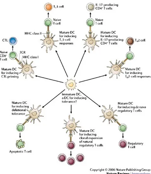
Figure IV. Dendritic cells effector function. Immature DCs can give rise to
multiple types of “effector” DC that instruct distinct T-cell fates, including immunity, tolerance and immune deviation. Maturation refers to the changes that accompany immature DC transition to a given effector state in response to environmental signals of exogenous (microbial) or endogenous (cytokines, hormones, dying cells) origin. The quality of these signals determines the choice of effector DC, although ontogeny can also have a role, predisposing certain DC subtypes towards particular effector states. Some maturation signals can promote the generation of tolerogenic DCs. This does not negate the possibility that some immature DCs have an intrinsic tolerogenic function.
1.4 Dendritic cells subsets and T cells priming
1.4.1 Dendritic cells subsets
The phenotypic and functional analyses of the DCs found in the thymus, spleen and lymph nodes have revealed a considerable heterogeneity among the DCs population, such that different DCs subsets have emerged and described as following a life cycle that differed from the original “Langerhans cell paradigm” (3). Although all DCs are capable of antigen uptake, processing and presentation to naïve T cells, the DCs subtypes differ in hematopoietic origin, location, migratory pathways, immunological functions and dependence on infection or inflammatory stimuli for their generation (7).
A first criteria to classify DCs is to distinguish among conventional DCs (cDCs) that have a dendritic form and exhibit DCs functions in steady state, from precursors of DCs (pre-DCs), that require further development to acquire DCs phenotype and full DCs function. Although the development of some pre-DCs can occur in steady state, in many cases the further development of pre-DCs requires a microbial or inflammatory stimulus (7). An example of pre-DCs are the plasmacytoid DCs that circulate through the blood and lymphoid tissues and only acquire the typical DCs morphology upon activation. Also monocytes can serve as source of pre-DCs and differentiate into functional DCs upon infection or inflammatory conditions (108, 109).
Among conventional DCs, two main categories can be distinguished based on the path they follow to access the lymphoid organs: migratory DCs and lymphoid-tissue resident DCs. Migratory DCs comprise epidermal Langerhans cells and interstitial or dermal DCs. The two subsets can be readily distinguished by the selective expression of the C-type lectin langerin on the surface of epidermal LCs and by LC-specific intracellular organelles known as Birbeck granules (110). Migratory DCs follow a life cycle described by the Langerhans cell paradigm serving as antigen sampling sentinels in the peripheral tissues and trafficking to the lymph nodes in response to danger signals. Migratory DCs display a mature phenotype in LNs also in steady state conditions, indicating that their migration and maturation proceed constitutively and independently of pathogens and are probably triggered by inflammatory compounds that are constitutively released by peripheral tissues (3).
Lymphoid-tissue resident DCs or blood-derived DCs constitute half of the lymph node DCs (the second half being migratory DCs) and all the DCs found in the spleen and thymus (6). They can be further subdivided into three subsets depending on the expression of CD4 and CD8 markers: CD4+CD8-, CD4-CD8+ and CD4-CD8- cells. Resident DCs do not conform to the Langerhans cells
paradigm, since they develop from bone marrow precursors directly in secondary lymphoid organs without previously trafficking through peripheral tissues. In contrast to migratory DCs, blood-derived DCs maintain an immature phenotype in steady state, but can be induced to mature by inoculation of inflammatory stimuli. Thus the function of the immature lymphoid organ DCs may be to promote tolerance in steady state while maintaining their capacity to respond to infections reaching those organs (6).
1.4.2 Dendritic cells subset and antigen-presenting functions
Since the initial characterization of different DCs population less than a decade ago, a great effort has been spent trying to elucidate the role of various DC subsets in the initiation of adaptive immune responses or in the maintenance of a tolerogenic state under normal circumstances. A scenario where the coordinate action of the different DCs subtypes, each with a specific function, operates to mount effective responses against specific pathogens or to prevent such responses in the absence of danger can be envisaged.
The possibility to isolate highly pure populations of DCs from different lymphoid organs taking advantage of specific lineage markers and the generation of transgenic mice that allows the tracking (111) or the conditional ablation of specific DCs types in vivo (57) have allowed to investigate the relative efficiency of DCs in priming T cell responses and their differential contribution to in vivo immune responses.
Although all DCs types express high levels of MHC class I and MHC class II molecules, especially when mature, and are able to generate peptide-MHC complexes, an increasing amount of data is indicating a different ability of the various DCs subset to incorporate peptides, derived from a given antigen, into their presentation pathway and thus a different capacity to activate CD4+ or CD8+ T lymphocytes.
Plasmacytoid DCs (pDCs) are a principal subset of DCs in both human and mouse with a unique ability to sense and respond to viral infections mainly by secreting large amounts of type I IFNs. Differently from conventional DCs, pDCs constitutively express the IFN regulatory factor 7 (IRF-7) that allows the rapid secretion of vast amount of IFN-α in a signaling pathway initiated by TLR7 and TLR9 engagement. Due to their poor phagocytic activity and thus inefficiency to present antigens (112), pDCs were generally considered to have a role in boosting the ability of conventional DCs to mature and stimulate T cells in viral infections, rather than playing a direct role in antigen presentation. However, recent findings by Sapoznikov et al indicate that pDCs are indeed able to initiate productive CD4+ T cell responses in lymph nodes upon antigen challenge. Taking advantage
of mice conditionally depleted of CD11chi cDCs but retaining a normal pool of pDCs, the authors
demonstrate how the ability of this specific subset to trigger antigen specific CD4+ T cell responses is organ dependent, being limited to lymph nodes and absent in spleen. The unique priming of CD4+, but not CD8+, T cell responses by pDCs could result from exclusive presentation of exogenous antigen on MHC class II, but not MHC class I molecules and would predict that pathogens that specifically activate pDCs will elicit exclusively humoral but not citotoxic immunity (113).
Migratory DCs present in the epidermis (Langerhans cells) and in the dermis (interstitial DCs) are characterized by the expression of many PRRs and endocytic receptors and are able to efficiently mediate the uptake of macromolecules and a broad range of microorganisms (114). Residing in most peripheral tissues at site of interface with the environment, migratory DCs constitutively take up antigens and traffic to lymph nodes where they interact with both CD4+ and CD8+ T lymphocytes. Indeed, cutaneous DCs express upon activation high levels of MHC class I and class II molecules and are able to efficiently prime both T helper and cytotoxic T lymphocytes. Conversely, their ability to cross-present soluble or cell-associated antigens is less defined. In a mouse model of autoimmunity, where the OVA class I peptide is expressed under the control of the human keratin 14 promoter (K14-OVAp mice), Langerhans cells have been demonstrated to cross-present this highly expressed antigen in the skin draining LNs and to induce the expansion of antigen specific cytotoxic T cells endowed with effector functions (115). A recent study by Romani group showed that Langerhans cells in epidermal skin explants were able to cross-present soluble and cell-associated antigens in a TAP-dependent manner. Interestingly, the in vitro comparison of the relative efficiency of Langerhans cells and CD8α+ resident DCs to present an exogenous antigen on MHC
class II or cross-present it on MHC class I molecule was different, with CD8α+ spleen cells being
more potent in inducing CD8+ T cells and Langerhans cells better in stimulating CD4+ T cells (116). Whether migratory DCs are the main DCs subset involved in the presentation of peripheral-tissue antigens or antigens derived from pathogens that infect the skin, lungs or gut, to both CD4+ and CD8+ T cells is still a matter of controversy. Although migratory DCs have an essential role in carrying peripheral antigens to the draining lymph nodes, recent studies using various models of viral infection have highlighted the dominance of the non-migrating, lymph node-resident CD8α+
DCs subset in priming CTL responses thus implicating the transfer of peripheral antigens from migratory to resident DCs upon entry in the T cell areas of the lymph nodes (59, 61, 117).
Villadangos et al propose a model where the relative contribution of the two subsets to the presentation of peripheral antigens depends on the pathogen capacity to infect DCs or impair their antigen presenting functions and on the number of antigen-carrying migratory DCs that reach the lymph node (3).
phenotype and the secretion of large amount of IL-12, support the hypothesis that cDCs indeed play a major role in immunosurveillance. On the other hand, if peripheral tolerance is mediated by immature DCs, the large number of blood derived DCs contained in the steady state in the lymphoid organs would provide a strong tolerogenic environment for naïve T cells (118). Due to their crucial role in the control of immunity, the definition of the in vivo relevance of the various subsets of lymphoid organ resident DCs in terms of antigen presenting functions has been subject of intense study.
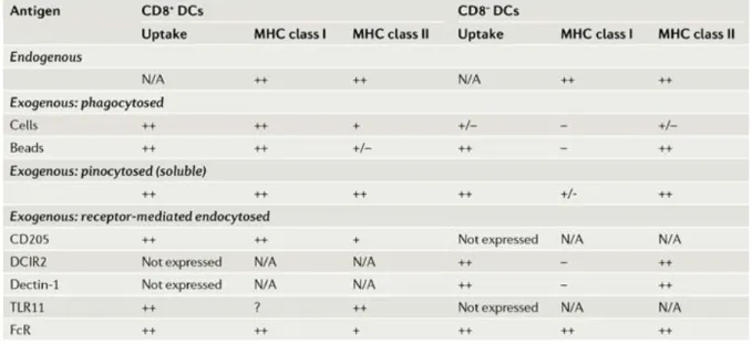
Both in vitro and in vivo analysis of the capacity of blood-derived DCs subsets to present exogenous antigens on MHC class II or cross-present them on MHC class I have revealed a functional dichotomy between the CD8α+ and CD8α- (comprising both CD4+ and double negative DCs)
subsets of resident DCs. Specifically, it emerges that the CD8α+ DCs are by far the most efficient at
cross-presenting cellular (119, 120), soluble (121, 122) or latex bead-associated antigens (122). Indeed, the ability of CD8α+ DCs to cross-present cellular associated antigens is attributable to their
selective capacity to endocytose dying cells in culture and in vivo (120, 123).
Furhtermore, CD8α+ DCs have also been identified as the major subset involved in the MHC class I
presentation of antigens from viruses (HSV1, influenza and vaccinia virus) and cytosolic bacterium L. monocytogenes (61, 124). By contrast, CD8α- DCs seem to be more efficient than CD8α+ DCs at presenting exogenous antigens on MHC class II molecules, especially in the case of phagocytosed antigens (i.e antigens associated to latex beads) (122) and soluble antigens, although less evident (120-122).
The CD8α+ and CD8α- subsets of DCs differentially express multiple putative antigen receptors
belonging to the C-type lectin family. The DEC-205 and mannose receptors are primarily expressed by the CD8α+ DCs (125, 126), whereas dectin-1, DCIR2, FIRE and CIRE are specific for the CD8α
-DCs (127-129). Targeting of soluble antigens coupled to antibodies against these various receptors has shed light on the in vivo role of the two DCs subtypes and allowed to characterize the type of immune response (humoral vs cytotoxic) preferentially triggered by these DCs subsets. Collectively, these studies highlight the greatest efficiency of CD8α+ DCs to activate CTLs and the preferential
induction of CD4+T cells and humoral responses by the CD8α- DCs (127-129).
As suggested by Dudziak et al, a possible explanation of this dichotomy is the differential expression of various components of the MHC class I and MHC class II machinery by the CD8α+ and CD8α
-subsets respectively (127). Alternatively, the MHC class I and class II pathways are fully operational in both subsets, but in addition, the CD8α+ DCs possess a specialized machinery for
cross-presentation which is largely absent in the CD8α- subtype (3). A recent work by Burgdorf et al