Clinical ultrasonography in loggerhead sea turtles
(Caretta caretta): imaging of pathological features
M. De Majo
1, F. Macri
1, M. Masucci
1, G. Coci
2, M.G. Pennisi
11Department of Veterinary Sciences, University of Messina, Messina, Italy 2Veterinary Practitioner, Tremestieri Etneo, Catania, Italy
ABSTRACT: Ultrasound scans were used to image pathological features in sea turtles. Scans were carried out in 19 loggerhead sea turtles, weighing from 2 to 21 kg, during the course of clinical examinations using 3.5 and 7.5 MHz sector transducers. The examination was performed after placing turtles in dorsal recumbency. Turtles were manually restrained and, in order to find the most suitable acoustic windows, were held down either by their heads (left and right cervicobrachial windows), front flippers (left and right axillary windows) or back flippers (left and right prefemoral and postfemoral windows). The right and left cervicobrachial windows allowed visualisation of the liver and gallbladder; the stomach was localised through the left prefemoral acoustic window; the intestinal loops were observed through the left and right prefemoral acoustic windows; the bladder was identified through both prefemoral acoustic windows. The pathological findings were as follows: idiopathic ileus, an intestinal linear foreign body and smooth muscle hypertrophy, presence of calculi and sediment in the gallbladder. These results highlight the importance of ultrasound examination along with clinical examination in sea turtles for the evalu-ation of coelomic pathologies.
Keywords: ultrasound; loggerhead sea turtle; Caretta caretta
The loggerhead sea turtle (Caretta caretta) is a common inhabitant of the Mediterranean Sea. This species nests particularly on the Sicilian and Calabrian coasts. As an endangered species, its conservation involves also the protection of nesting sites, where juveniles and adults are often captured by fisheries (De Florio et al. 2005).
Sea turtles show poor clinical signs and, due to the presence of carapace and plastron, physical examination requires complementary diagnostic procedures to provide useful information for clini-cal diagnosis (Penninck et al. 1991; Lieberman and Rosskopf 1984).
Radiological examination is the best method in order to detect the presence of a swallowed fish-hook, but it has limited value to identify the internal organs of sea turtles, due to the shell and the lack of visceral fat (Silverman 1989; Penninck et al. 1991; Rubel et al. 1991; Deshaw et al. 1996; Silverman and Janssen 1996; Stetter 2000). Laparoscopic exami-nations can be performed, even if there could be problems in using this technique (Blanvillain et al.
2008). In our experience such risks are inherent to post-surgical recovery in wild aquatic animals (i.e. infections or dehiscence of the wound).
In aquatic animal medicine, ultrasound is a very helpful and easy-to-use tool for investigation and diagnosis, particularly in the study of the coelomic organs in chelonians (Penninck et al. 1991; Martorell et al. 2004; Valente et al. 2007; Margheri et al. 2011; Kot et al. 2012; Macri et al. 2013a; Macri et al.2013b; Macri et al. 2015). Regarding chelonian reproduc-tion, ultrasound examinations have proven to be an efficient non-invasive method for monitoring ovar-ian development and activity in sea turtles and re-productive activity in adult males (Rostal et al. 1990; Valente et al. 2007; Manire et al. 2008).
Valente et al. (2007, 2008) described the normal sonographic appearance of coelomic organs in the loggerhead sea turtle and identified ultrasound-accessible blood vessels in Caretta caretta and their Doppler waveform patterns. Since the information regarding pathological patterns are still unsatisfac-tory or too dissimilar from the ones now used for
diagnostic purposes (Valente et al. 2007; Valente et al. 2008), in the present study we hypothesised that ultrasonographic imaging would help practitioners in the diagnosis of the various pathologies affecting sea turtles. Therefore, the aim of this study was to perform ultrasound examination on sea turtles (Caretta caretta) rescued near the Sicilian coast.
Ultrasound techniques, as well as different patho-logical and normal patterns, are reported and dis-cussed.
MATERIAL AND METHODS
Ultrasound examinations were performed on 19 loggerhead sea turtles (Caretta caretta) of dif-ferent sizes and weights belonging to the Rescue Centres of the Sicilian Nature Fund of Catania (Fondo Siciliano per la Natura, Catania, Italy) and the World Wildlife Fund for Nature of Messina (Italy). The scans were carried out using an ultrasound machine (Sigma IRIS 440, Kontron Instruments, Italy), with ultrasound multifrequency probes (3.5–7.5 MHz).
The following acoustic windows were used: left and right cervicobrachial, left and right axillary, left and right prefemoral and postfemoral. The examination was done after placing the animals in dorsal recum-bency; the turtles were manually restrained accord-ing to Valente et al. (2007). In brief, turtles were held down by their heads and their front or back flippers, depending on the most suitable acoustic window. Their eyes and body surface were covered by disposable wet wipes.
On the basis on clinic examination, ultrasound was performed in nine subjects with pathological signs (Group A).Ten additional animals (Group B) underwent ultrasound examination to exclude asymptomatic lesions. The percentage of visualisa-tion of anatomic structures from different acoustic windows was noted.
RESULTS
The turtles had a straight-line carapace length between 32 and 63 cm and a weight ranging from
Table 1. Clinical, radiological and ultrasound findings
Clinical and/or X-ray exam Ultrasound
1 skin lesions at head and nape regions nothing to report
2 fishing line leaking from cloaca absence of peristalsis, intestinal stasis, coelomic fluid
3 ocular changes nothing to report
4 hook in oesophagus; discoloured and soften areas
of the carapax echogenic dots were visible floating within the bladder 5 fishing line leaking from cloaca absence of peristalsis, coelomic fluid, hyperechoic
bladder content
6 hook in oesophagus dilated loops, coelomic fluid
7 dehydration, softened plastron, partial absence
of rhamphotheca nothing to report
8 dehydration, softened plastron, fishing line leaking
from cloaca the ultrasound scan was not performed through the inguinal window due to intestinal gas
9 nothing to report nothing to report
10 nothing to report intestinal idiopathic muscular hypertrophy
11 hook in oesophagus surgically removed 5 days previously absence of peristalsis, liquid in the bowel, coelomic fluid 12 hook in oesophagus surgically removed 5 days previously normal bowel peristalsis, echogenic gallbladder content,
echogenic dots were visible floating within the bladder
13 nothing to report nothing to report
14 hook in oesophagus nothing to report
15 fishing line leaking from cloaca adynamic ileus, coelomic fluid, fishing line
16 nothing to report nothing to report
17 nothing to report nothing to report
18 nothing to report nothing to report
2 to 21 kg; according to Pont and Alegre (2000), the subjects examined were juvenile and sub-adult ones (Pont and Alegre 2000).
Clinical examination
One turtle presented cutaneous lesions in the head region; another presented ocular lesions; three turtles showed the presence of fishing-line protruding from the oral cavity and, as later con-firmed by an X-ray examination, a hook lodged in the oesophagus. Two animals underwent surgery to remove the hook five days before the ultrasound scan. Four turtles had a fishing line attached to the cloaca. One animal showed signs of dehydra-tion, softening of the carapace and a fracture of the rhamphotheca. Seven subjects showed no clinical symptoms. All the above data, along with ultra-sound reports are summarised in Table 1.
Ultrasound examination of Group A
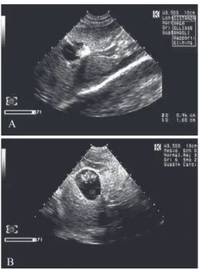
Five subjects showed findings of idiopathic ileus (atonic intestinal loops with accumulation of fluid or dilated by the accumulation of food material); the bowel diameter appeared larger than the con-trol group (Figure 1 A and B). The bowel diameters
of turtles with and without signs of ileus were com-pared using Student’s t- test at P < 0.05.
In six subjects with regular peristalsis, the di-ameter of the loops ranged from 1.30 to 2.25 cm (mean 1.67 cm), while in the subjects with findings indicative of mechanical ileus, the bowel diameter ranged from 1.70 to 3.75 cm (mean 2.79 cm).
The two-tailed P value was 0.02, indicating sig-nificance.
Five subjects showed coelomic fluid between the intestinal loops (Figure 2) or in the bladder area (Figure 3).
In the gallbladder of two subjects, one of which was anorexic and the other which had undergone surgery five days previously, an echogenic/hyper-echoic content, which caused distal acoustic shad-owing, was found.
Figure 1. Intestine –
A and B; adynamic
ileus and dilated loops
Figure 2. Intestine; normal intestinal pattern, free of coelomic fluid
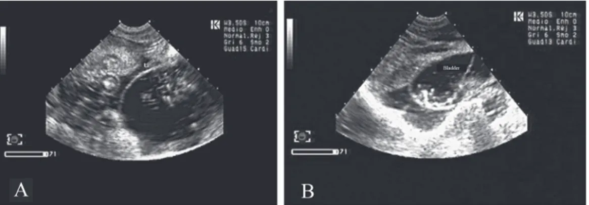
Figure 3. Bladder – A and B; presence of free-floating hypere-choic material within bladder
One case revealed a hyperechoic structure due to the presence of gallstones (Figure 4 A and B).
One subject, with a fishing-line protruding from the cloaca and an ultrasound image of ileus, showed an intestinal loop inside with fluid content and a hyperechoic line connected to the linear foreign body (Figure 5); in these cases, the increased bowel transversal dimension was significant, recalling a similar case described in a cat by Tidwell and Penninck (1992).
Ultrasound examination of Group B
One subject without any anamnestic indication of pathology in the gastrointestinal tract, showed
a thicker intestinal loop at the level of muscularis propria (Figure 6).
In the other nine subjects findings were consid-ered normal according to literature data, as de-scribed below.
Liver and gallbladder. The liver was located
laterally and slightly dorsally to the heart, with the gallbladder housed on the ventral surface of its right lobe. The right and left cervicobrachial windows allowed visualisation of the organ in a percentage of the subjects varying from 52% to 63%, respectively; through the right and left prefemoral windows, visualisation was obtained in 52% to 73% of the subjects. Partial images of the hepatic pa-renchyma with transverse and oblique scan planes were also obtained through the axillary windows.
Figure 4. Gallbladder; A – gallstones; B – biliary sludge
Figure 5. Intestine; linear foreign bodies
Figure 6. Intestine; hypertrophy of the muscle wall of the intestine
Figure 8. Colon; presence of gas in the colon with hyper-echoic content with acoustic shadowing
Stomach. The stomach, curved and situated
to the left of the coelomic cavity, was displayed through the left prefemoral acoustic window only in five subjects (26%). It was identified by the pres-ence of its fluid content. The prespres-ence of echogenic material was found in one subject (Figure 7).
Intestinal loops were observed through the left
and right prefemoral acoustic windows, respective-ly, in 73% and 78% of the subjects examined, as a region that originates from the pyloric sphincter and then curves upward from left to right, becom-ing an entangled organ located medially in the coe-lomic cavity up to the cloaca. Aspects of normal mucosa (Figure 2), the presence of gas in the colon with hyperechoic content and acoustic shadowing (Figure 8), as well as echogenic material without acoustic shadowing (Figure 9A) were observed. In some cases, the presence of fluid in the loops enabled excellent visualisation of the layers of the walls (Figure 9B).
Bladder. This was easily identifiable through the
prefemoral acoustic windows in the turtles of both groups, just above the large intestine and under the pelvis. The presence of hyperechoic material was re-vealed in two subjects due to calculosis (Figure 10). The presence of hyperechoic material with and
with-out comet-tail artefacts was found inside the bladder of another subject (Figure 3 A and B).
DISCUSSION
Loggerhead sea turtles are often recovered along the Ionian Sea, the southern Adriatic and the Sicilian coasts, generally because of ingestion of fishing hooks and other foreign bodies (Tomas et al. 2002; Casale et al. 2004; Oros et al. 2004; De Florio et al. 2005).
The poor radiopacity of foreign bodies (nylon fishing-lines, plastic bags, etc.), along with the frequent absence of clinical signs, can delay the diagnosis of a gastrointestinal obstruction and in-testinal intussusception (De Vico et al. 2003; Di Bello et al. 2006). In our study, the ultrasonogra-phy allowed the diagnosis of ileus likely caused by ingestion of fishing-lines and/or fishing hooks in subjects with only indicative signs of gastroenteric pathology, sometimes accompanied by the presence of coelomic fluid. In one case it was also possible to recognise the presence of the fishing line. This find-ing, unlike what reported by Penninck et al. (1991) in Xerobates agassizii, the presence of coelomic fluid is not a frequent occurrence in C. caretta, and its clinical significance is unknown (Penninck et al. 1991). The thickening of the intestinal muscu-lar layer reported in some mammals, such as cats and horses, has been related to chronic stenosis or chronic inflammatory enteropathies (Diana et al. 2003; Penninck 2008). In our case it was not possi-ble to detect the cause of intestinal wall thickening. In the literature these ultrasound intestinal patterns have never been described in sea turtles. For this rea-son we consider it interesting to report our evaluations. The presence of gallstones in the anorexic subject is likely related to malnutrition. Echogenic dots
Figure 10. Bladder; prefemoral windows; calculus with acoustic shadowing
were visible floating within the bladder and have been linked to the presence of urate crystals, para-sites or faecal material (Wyneken 2001).
The evidence of comet-tail artefacts was indica-tive of gas, which probably entered accidentally via the cloaca.
In this study, we chose to place the examined turtles in dorsal recumbency as suggested by Pease et al. (2010).
In the present study superior visualisation of coe-lomic organs, such as stomach intestinal loops and the urinary bladder, was obtained using the same acoustic windows reported by Valente et al. (2007), with the exception of the liver that was more visible through the prefemoral acoustic window.
The inability to perform histopathological or cy-topathological examinations in order to obtain a diagnosis of some suspect ultrasound patterns may be a limitation of this study, although we still think that it is instructive to report the observed patterns as they have not been described before in the litera-ture. Our study represents a novel contribution to the evaluation of coelomic disorders in sea turtles. Some subjects with pathological findings were sent to rehabilitation centres or were set free into the sea, so we did not have the possibility to perform necroscopies on subjects after death.
Acknowledgement
The authors thank Fondo Siciliano per la Natura of Catania, Italy and the World Wildlife Fund (WWF) of Messina, Italy for their collaboration.
REfERENCES
Blanvillain G, Pease A, Rostal D, Owens D, Segars A (2008): Comparison of ultrasound and laparoscopy to evaluate the reproductive activity in adult male loggerhead (Caretta caretta) sea turtles. In: 28th Annual International Symposium on Sea Turtle Biology and Conservation, January 20–27, Loreto Baja, Mexico.
Casale P, Laurent L, De Metrio G (2004): Incidental capture of marine turtles by the Italian trawl fishery in the north Adriatic Sea. Biological Conservation 119, 287–295. De Florio M, Aprea A, Corriero A, Santamaria N, De
Met-rio G (2005): Incidental captures of sea turtles by sword-fish and albacore longlines in the Ionian sea. Fisheries Science 71, 1008–1016.
Deshaw B, Schoenfeld A, Cook RA, Haramati N (1996): Imaging of reptiles: a comparison study of various radio-graphic techniques. Journal of Zoo and Wildlife Medicine 27, 364–370.
De Vico G, Marino F, Sfacteria A, Macri F, Lorizio R, Peretti B, Restucci B (2003): Retrograde intestinal intussuscep-tion in a wild sea turtle Caretta caretta (in Italian). Summa 8, 9–51.
Diana A, Pietra M, Guglielmini C, Boari A, Bettini G, Cipone M (2003): Ultrasonographic and pathologic fea-tures of intestinal smooth muscle hypertrophy in four cats. Veterinary Radiology Ultrasound 44, 566–569. Di Bello A, Valastro C, Staffieri F, Crovace A (2006):
Con-trast radiography of the gastrointestinal tract in sea tur-tles. Veterinary Radiology Ultrasound 47, 351–354. Kot BCW, Ying MTC, Brook FM, Kinoshita RE, Cheng SCH
(2012): Ultrasonographic assessment of the thyroid gland and adjacent anatomic structures in Indo-Pacific bottle-nose dolphins (Tursiops aduncus). American Journal of Veterinary Research 73, 1696–1706.
Lieberman SS, Rosskopf WS (1984): Blood panel analyses of captive desert tortoises (Gopherus agassizi). Avian/ Exotic Practice 1, 15–29.
Macri F, Liotta L, Bonfiglio R, De Stefano C, Ruscica D, Aiudi G (2013a): Ultrasound measurement of reproduc-tive organs in juvenile European sea bass Dicentrarchus labrax. Journal of Fish Biology 83, 1439–1443.
Macri F, Di Pietro S, Bonfiglio R, De Stefano C, Giorgianni P, Bottari T (2013b): Anatomical evaluation of the organs in the red swamp crayfish, Procambarus clarkii, by diag-nostic ultrasound examination. Journal of Crustacean Biology 33, 586–589.
Macri F, Passantino A; Di Pietro S, Ruscica D, Sfacteria A, De Stefano C, Bottari T (2015): Effects of formalin pres-ervation on eye lens of bogue, Boops boops (Linnaeus, 1758): comparison of direct (post-fixation) vs ultrasound (true) measurements. Journal of Applied Ichthyology 31, 393–394
Manire CA, Byrd L, Therrien CL, Martin K (2008): Mating-induced ovulation in loggerhead sea turtles Caretta caretta. Zoo Biology 27, 213–225.
Margheri L, Ponte G, Mazzolai B, Laschi C, Fiorito G (2011): Non-invasive study of Octopus vulgaris arm morphology using ultrasound. Journal of Experimental Biology 214, 3727–3731.
Martorell J, Espada Y, Ruiz de Gopegui R (2004): Normal echoanatomy of the red-eared slider terrapin (Trachemys scripta elegans). Veterinary Record 155, 417–420. Oros J, Calabuig P, Deniz S (2004): Digestive pathology of
sea turtles stranded in the Canary Island between 1993 and 2001. Veterinary Record 7, 169–174.
Pease A, Blanvillain G, Rostal D, Owens D, Segars A (2010): Ultrasound imaging of the inguinal region of adult male loggerhead sea turtles (Caretta caretta). Journal of Zoo and Wildlife Medicine 41, 69–76.
Penninck DG (2008): Gastrointestinal tract. In: Penninck DG (ed.): Atlas of Small Animal Ultrasonography. 1st ed. Blackwell Publishing, Iowa. 281–318.
Penninck DG, Stewart JS, Paul-Murphy J, Pion P (1991): Ultrasonography of the California desert tortoise (Xero-bates agassizi): anatomy and application. Veterinary Ra-diology Ultrasound 32, 112–116.
Pont SG, Alegre FN (2000): Work of the foundation for the conservation and recovery of marine. Life Marine Turtle Newsletter 87, 5–7.
Rostal DC, Robeck TR, Owens DW, Kraemer DC (1990): Ultrasound imaging of ovaries and eggs in Kemp’s Ridley sea turtles (Lepidochelys kempi) Journal of Zoo and Wild-life Medicine 21, 27–35.
Rubel GA, Kuoni W, Frye FL (1991): Radiology and imaging. In: Frye FL Malabar FL (eds.): Biomedical and Surgical Aspects of Captive Reptile Husbandry. Krieger Publishing Company, Malabar, FL. 85–208.
Silverman S (1989): Advances in avian and reptilian imaging. In: Bonagura JD (ed.): Kirk’s Current Therapy X: Small Animal Practice. WB Saunders, Philadelphia. 786–789.
Silverman S, Janssen DL (1996): Diagnostic imaging. In: Mader RM (ed.): Reptile Medicine and Surgery. WB Saun-ders, Philadelphia. 67–78.
Stetter MD (2000): Diagnostic imaging of reptiles. In: Bo-nagura JD (ed.): Kirk’s Current Therapy XIII: Small Ani-mal Practice. WB Saunders, Philadelphia. 1163–1168. Tidwell AS, Penninck DG (1992): Ultrasonography of
gas-trointestinal foreign bodies. Veterinary Radiology Ultra-sound 33, 51–62.
Tomas J, Guitart R, Mateo R, Ragaa JA (2002): Marine de-bris ingestion in loggerhead sea turtles Caretta caretta from the Western Mediterranean. Marine Pollution Bul-letin 44, 211–216.
Valente AL, Parga ML, Espada Y, Lavin S, Alegre F, Marco I, Cuenca R (2007): Ultrasonographic imaging of logger-head sea turtles (Caretta caretta). Veterinary Record 161, 226–232.
Valente AL, Parga ML, Espada Y, Lavin S, Alegre F, Marco I, Cuenca R (2008): Evaluation of Doppler ultrasonogra-phy for the measurement of blood flow in young logger-head sea turtles (Caretta caretta). The Veterinary Journal 176, 385–392.
Wyneken J (2001): The Anatomy of Sea Turtle. NOAA Tech-nical Memorandum, Miami US Department of Commerce 1–172.
Received: 2014–04–15 Accepted after corrections: 2016–02–05
Corresponding Author:
Francesco Macri, University of Messina, Department of Veterinary Sciences, Viale Annunziata, 98168, Messina, Italy E-mail: [email protected]