ESC Guidelines for the diagnosis and treatment
of acute and chronic heart failure 2012
The Task Force for the Diagnosis and Treatment of Acute and
Chronic Heart Failure 2012 of the European Society of Cardiology.
Developed in collaboration with the Heart Failure Association (HFA)
of the ESC
Authors/Task Force Members: John J.V. McMurray (Chairperson) (UK)
*
,
Stamatis Adamopoulos (Greece), Stefan D. Anker (Germany), Angelo Auricchio
(Switzerland), Michael Bo¨hm (Germany), Kenneth Dickstein (Norway),
Volkmar Falk (Switzerland), Gerasimos Filippatos (Greece), Caˆndida Fonseca
(Portugal), Miguel Angel Gomez-Sanchez (Spain), Tiny Jaarsma (Sweden),
Lars Køber (Denmark), Gregory Y.H. Lip (UK), Aldo Pietro Maggioni (Italy),
Alexander Parkhomenko (Ukraine), Burkert M. Pieske (Austria), Bogdan A. Popescu
(Romania), Per K. Rønnevik (Norway), Frans H. Rutten (The Netherlands),
Juerg Schwitter (Switzerland), Petar Seferovic (Serbia), Janina Stepinska (Poland),
Pedro T. Trindade (Switzerland), Adriaan A. Voors (The Netherlands), Faiez Zannad
(France), Andreas Zeiher (Germany).
ESC Committee for Practice Guidelines (CPG): Jeroen J. Bax (CPG Chairperson) (The Netherlands), Helmut Baumgartner (Germany), Claudio Ceconi (Italy), Veronica Dean (France), Christi Deaton (UK),
Robert Fagard (Belgium), Christian Funck-Brentano (France), David Hasdai (Israel), Arno Hoes (The Netherlands), Paulus Kirchhof (Germany/UK), Juhani Knuuti (Finland), Philippe Kolh (Belgium), Theresa McDonagh (UK), Cyril Moulin (France), Bogdan A. Popescu (Romania), Zˇ eljko Reiner (Croatia), Udo Sechtem (Germany), Per Anton Sirnes (Norway), Michal Tendera (Poland), Adam Torbicki (Poland), Alec Vahanian (France), Stephan Windecker (Switzerland).
Document Reviewers: Theresa McDonagh (CPG Co-Review Coordinator) (UK), Udo Sechtem (CPG Co-Review Coordinator) (Germany), Luis Almenar Bonet (Spain), Panayiotis Avraamides (Cyprus), Hisham A. Ben Lamin (Libya), Michele Brignole (Italy), Antonio Coca (Spain), Peter Cowburn (UK), Henry Dargie (UK), Perry Elliott (UK), Frank Arnold Flachskampf (Sweden), Guido Francesco Guida (Italy), Suzanna Hardman (UK), Bernard Iung
*Corresponding author. Chairperson: Professor John J.V. McMurray, University of Glasgow G12 8QQ, UK. Tel:+44 141 330 3479, Fax: +44 141 330 6955, Email:john.mcmurray@ glasgow.ac.uk
Other ESC entities having participated in the development of this document:
Associations: European Association for Cardiovascular Prevention & Rehabilitation (EACPR), European Association of Echocardiography (EAE), European Heart Rhythm Association (EHRA), European Association of Percutaneous Cardiovascular Interventions (EAPCI)
Working Groups: Acute Cardiac Care, Cardiovascular Pharmacology and Drug Therapy, Cardiovascular Surgery, Grown-up Congenital Heart Disease, Hypertension and the Heart, Myocardial and Pericardial Diseases, Pulmonary Circulation and Right Ventricular Function, Thrombosis, Valvular Heart Disease
Councils: Cardiovascular Imaging, Cardiovascular Nursing and Allied Professions, Cardiology Practice, Cardiovascular Primary Care
The content of these European Society of Cardiology (ESC) Guidelines has been published for personal and educational use only. No commercial use is authorized. No part of the ESC Guidelines may be translated or reproduced in any form without written permission from the ESC. Permission can be obtained upon submission of a written request to Oxford University Press, the publisher of the European Heart Journal and the party authorized to handle such permissions on behalf of the ESC.
Disclaimer. The ESC Guidelines represent the views of the ESC and were arrived at after careful consideration of the available evidence at the time they were written. Health professionals are encouraged to take them fully into account when exercising their clinical judgement. The guidelines do not, however, override the individual responsibility of health professionals to make appropriate decisions in the circumstances of the individual patients, in consultation with that patient, and where appropriate and necessary the patient’s guardian or carer. It is also the health professional’s responsibility to verify the rules and regulations applicable to drugs and devices at the time of prescription.
(France), Bela Merkely (Hungary), Christian Mueller (Switzerland), John N. Nanas (Greece),
Olav Wendelboe Nielsen (Denmark), Stein Ørn (Norway), John T. Parissis (Greece), Piotr Ponikowski (Poland).
The disclosure forms of the authors and reviewers are available on the ESC websitewww.escardio.org/guidelines
-Keywords Heart failure † Natriuretic peptides † Ejection fraction † Renin – angiotensin system † Beta-blockers † Digitalis † Transplantation
Table of Contents
Abbreviations and acronyms . . . 805
1. Preamble . . . 807
2. Introduction . . . 808
3. Definition and diagnosis . . . 808
3.1 Definition of heart failure . . . 808
3.2 Terminology related to left ventricular ejection fraction . 808 3.3 Terminology related to the time-course of heart failure 809 3.4 Terminology related to the symptomatic severity of heart failure . . . 809
3.5 Epidemiology, aetiology, pathophysiology, and natural history of heart failure . . . 810
3.6 Diagnosis of heart failure . . . 810
3.6.1 Symptoms and signs . . . 810
3.6.2 General diagnostic tests in patients with suspected heart failure . . . 811
3.6.3 Essential initial investigations: echocardiogram, electrocardiogram, and laboratory tests . . . 811
3.6.4 Natriuretic peptides . . . 811
3.6.5 Chest X-ray . . . 813
3.6.6 Routine laboratory tests . . . 813
3.6.7 Algorithm for the diagnosis of heart failure . . . 815
4. The role of cardiac imaging in the evaluation of patients with suspected or confirmed heart failure . . . 816
4.1 Echocardiography . . . 816
4.1.1 Assessment of left ventricular systolic dysfunction . 816 4.1.2 Assessment of left ventricular diastolic dysfunction . 816 4.2 Transoesophageal echocardiography . . . 816
4.3 Stress echocardiography . . . 818
4.4 Cardiac magnetic resonance . . . 818
4.5 Single-photon emission computed tomography and radionuclide ventriculography . . . 819
4.6 Positron emission tomography imaging . . . 819
4.7 Coronary angiography . . . 819
4.8 Cardiac computed tomography . . . 819
5. Other investigations . . . 819
5.1 Cardiac catheterization and endomyocardial biopsy . . . 819
5.2 Exercise testing . . . 820
5.3 Genetic testing . . . 820
5.4 Ambulatory electrocardiographic monitoring . . . 820
6. Prognosis . . . 820
7. Pharmacological treatment of heart failure with reduced ejection fraction (systolic heart failure) . . . 820
7.1 Objectives in the management of heart failure . . . 820
7.2 Treatments recommended in potentially all patients with systolic heart failure . . . 820
7.2.1 Angiotensin-converting enzyme inhibitors and beta-blockers . . . 820
7.2.2 Mineralocorticoid/aldosterone receptor antagonists . . . 823
7.2.3 Other treatments recommended in selected patients with systolic heart failure . . . 825
7.2.4 Angiotensin receptor blockers . . . 825
7.2.5 Ivabradine . . . 825
7.2.6 Digoxin and other digitalis glycosides . . . 826
7.2.7 Combination of hydralazine and isosorbide dinitrate . . . 826
7.2.8 Omega-3 polyunsaturated fatty acids . . . 826
7.3 Treatments not recommended (unproven benefit) . . . . 827
7.3.1 3-Hydroxy-3-methylglutaryl-coenzyme A reductase inhibitors (‘statins’) . . . 827
7.3.2 Renin inhibitors . . . 827
7.3.3 Oral anticoagulants . . . 827
7.4 Treatments not recommended (believed to cause harm) 827 7.5 Diuretics . . . 828
8. Pharmacological treatment of heart failure with ‘preserved’ ejection fraction (diastolic heart failure) . . . 828
9. Non-surgical device treatment of heart failure with reduced ejection fraction (systolic heart failure) . . . 829
9.1 Implantable cardioverter-defibrillator . . . 829
9.1.1 Secondary prevention of sudden cardiac death . . . . 829
9.1.2 Primary prevention of sudden cardiac death . . . 829
9.2 Cardiac resynchronization therapy . . . 830
9.2.1 Recommendations for cardiac resynchronization therapy where the evidence is certain . . . 831
9.2.2 Recommendations for cardiac resynchronization therapy where the evidence is uncertain . . . 831
10. Arrhythmias, bradycardia, and atrioventricular block in patients with heart failure with reduced ejection fraction and heart failure with preserved ejection fraction . . . 832
10.1 Atrial fibrillation . . . 832
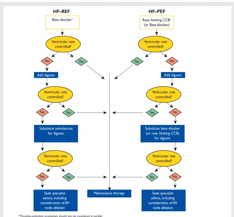
10.1.1 Rate control . . . 832
10.1.2 Rhythm control . . . 833
10.1.3 Thrombo-embolism prophylaxis . . . 834
10.2 Ventricular arrhythmias . . . 834
11. Importance and management of other co-morbidity in heart failure with reduced ejection fraction and heart failure with
preserved ejection fraction . . . 837
11.1 Heart failure and co-morbidities . . . 837
11.2 Anaemia . . . 837
11.3 Angina . . . 837
11.4 Asthma: see chronic obstructive pulmonary disease . . 837
11.5 Cachexia . . . 837
11.6 Cancer . . . 837
11.7 Chronic obstructive pulmonary disease . . . 837
11.8 Depression . . . 838 11.9 Diabetes . . . 838 11.10 Erectile dysfunction . . . 839 11.12 Gout . . . 839 11.13 Hyperlipidaemia . . . 839 11.14 Hypertension . . . 839 11.14 Iron deficiency . . . 840
11.15 Kidney dysfunction and cardiorenal syndrome . . . 840
11.16 Obesity . . . 840
11.17 Prostatic obstruction . . . 840
11.18 Renal dysfunction . . . 840
11.19 Sleep disturbance and sleep-disordered breathing . . . 840
12. Acute heart failure . . . 840
12.1 Initial assessment and monitoring of patients . . . 841
12.2 Treatment of acute heart failure . . . 841
12.2.1 Pharmacological therapy . . . 841
12.2.2 Non-pharmacological/non-device therapy . . . 843
12.3 Invasive monitoring . . . 847
12.3.1 Intra-arterial line . . . 847
12.3.2 Pulmonary artery catheterization . . . 847
12.4 Monitoring after stabilization . . . 847
12.5 Other in-patient assessments . . . 847
12.6 Readiness for discharge . . . 847
12.7 Special patient populations . . . 847
12.7.1 Patients with a concomitant acute coronary syndrome . . . 847
12.7.2 Isolated right ventricular failure . . . 848
12.7.3 Acute heart failure with ‘cardiorenal syndrome’ . . 848
12.7.4 Perioperative acute heart failure . . . 848
12.7.5 Peripartum cardiomyopathy . . . 848
12.7.6 Adult congenital heart disease . . . 848
13. Coronary revascularization and surgery, including valve surgery, ventricular assist devices, and transplantation . . . 848
13.1 Coronary revascularization . . . 848 13.2 Ventricular reconstruction . . . 849 13.3 Valvular surgery . . . 849 13.3.1 Aortic stenosis . . . 849 13.3.2 Aortic regurgitation . . . 849 13.3.3 Mitral regurgitation . . . 849 13.4 Heart transplantation . . . 850
13.5 Mechanical circulatory support . . . 850
13.5.1 End-stage heart failure . . . 851
13.5.2 Acute heart failure . . . 851
14. Holistic management, including exercise training and multidisciplinary management programmes, patient monitoring, and palliative care . . . 852
14.1 Exercise training . . . 852
14.2 Organization of care and multidisciplinary management programmes . . . 853
14.3 Serial natriuretic peptide measurement . . . 854
14.4 Remote monitoring (using an implanted device) . . . 854
14.5 Remote monitoring (no implanted device) . . . 854
14.6 Structured telephone support . . . 854
14.7 Palliative/supportive/end-of-life care . . . 854
15. Gaps in evidence . . . 854
15.1 Diagnosis . . . 854
15.2 Co-morbidity . . . 854
15.3 Non-pharmacological, non-interventional therapy . . . . 855
15.4 Pharmacological therapy . . . 855
15.5 Devices . . . 855
15.6 Acute heart failure . . . 855
15.7 End-of-life care . . . 855
References . . . 855
Abbreviations and acronyms
ACE angiotensin-converting enzyme ACHD adult congenital heart disease AF atrial fibrillation
AF-CHF Atrial Fibrillation and Congestive Heart Failure AHF acute heart failure
AIRE Acute Infarction Ramipril Efficacy ARB angiotensin receptor blocker ARR absolute risk reduction
ATLAS Assessment of Treatment with Lisinopril And Survival
AV atrioventricular AVP arginine vasopressin
BEAUTIFUL MorBidity-mortality EvAlUaTion of the If
inhibi-tor ivabradine in patients with coronary disease and left ventricULar dysfunction
BEST Beta-Blocker Evaluation of Survival Trial BiVAD bi-ventricular assist device
BNP B-type natriuretic peptide b.p.m. beats per minute BTC bridge to candidacy BTD bridge to decision BTR bridge to recovery BTT bridge to transplantation CABG coronary artery bypass graft CAD coronary artery disease
CARE-HF Cardiac Resynchronization in Heart Failure Study CCB calcium-channel blocker
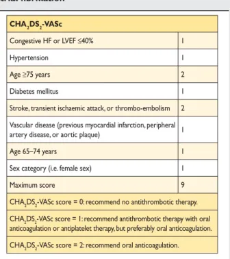
CHA2DS2-VASc Cardiac failure, Hypertension, Age ≥75
(Doubled), Diabetes, Stroke (Doubled)-Vascular disease, Age 65 – 74 and Sex category (Female) CHARM Candesartan in Heart Failure: Assessment of
Re-duction in Mortality and Morbidity CIBIS II Cardiac Insufficiency Bisoprolol Study II CMR cardiac magnetic resonance
COMET Carvedilol or Metoprolol European Trial COMPANION Comparison of Medical Therapy, Pacing, and
De-fibrillation in Heart Failure
CONSENSUS Cooperative North Scandinavian Enalapril Sur-vival Study
COPD chronic obstructive pulmonary disease
COPERNICUS Carvedilol Prospective Randomized Cumulative Survival
CORONA Controlled Rosuvastatin Multinational Trial in Heart Failure
CPAP continuous positive airway pressure CRT cardiac resynchronization therapy
CRT-D cardiac resynchronization therapy-defibrillator CRT-P cardiac resynchronization therapy-pacemaker CT computed tomography
DEFINITE Defibrillators in Non-ischemic Cardiomyopathy Treatment Evaluation
DIG Digitalis Investigation Group DT destination therapy ECG electrocardiogram
ECMO extracorporeal membrane oxygenation EF ejection fraction
eGFR estimated glomerular filtration rate
ELITE II Second Evaluation of Losartan in the Elderly Trial EMPHASIS-HF Eplerenone in Mild Patients Hospitalization and
Survival Study in Heart Failure GFR glomerular filtration rate
GISSI-HF Gruppo Italiano per lo Studio della Sopravvi-venza nell’Infarto miocardico-heart failure H-ISDN hydralazine and isosorbide dinitrate
HAS-BLED Hypertension, Abnormal renal/liver function (1 point each), Stroke, Bleeding history or predis-position, Labile INR, Elderly (.65), Drugs/ alcohol concomitantly (1 point each)
HEAAL Heart failure Endpoint evaluation of Angiotensin II Antagonist Losartan
HF heart failure
HF-ACTION Heart Failure: A Controlled Trial Investigating Outcomes of Exercise Training
HF-PEF heart failure with ‘preserved’ ejection fraction HF-REF heart failure with reduced ejection fraction I-PRESERVE Irbesartan in heart failure with preserved systolic
function
i.v. intravenous
IABP intra-aortic balloon pump
ICD implantable cardioverter-defibrillator LA left atrial
LBBB left bundle branch block LV left ventricular
LVAD left ventricular assist device LVEF left ventricular ejection fraction
MADIT-II Multicenter Automatic Defibrillator Implantation Trial II
MCS mechanical circulatory support MDCT multi-detector computed tomography
MERIT-HF Metoprolol CR/XL Randomised Intervention Trial in Congestive Heart Failure
MRA mineralocorticoid receptor antagonist
MR-proANP mid-regional atrial (or A-type) natriuretic peptide
MUSTIC Multisite Stimulation in Cardiomyopathies NIPPV non-invasive positive pressure ventilation NNT number needed to treat
NSAID non-steroidal anti-inflammatory drug NYHA New York Heart Association
OPTIMAAL Optimal Therapy in Myocardial infarction with the Angiotensin II Antagonist Losartan
PEP-CHF Perindopril for Elderly People with Chronic Heart failure
PET positron emission tomography PUFA polyunsaturated fatty acid
RAFT Resynchronization/Defibrillation for Ambulatory Heart Failure Trial
RALES Randomised Aldactone Evaluation Study RCT randomized controlled trial
RRR relative risk reduction
SAVE Survival and Ventricular Enlargement SCD-HeFT Sudden Cardiac Death in Heart Failure Trial SENIORS Study of Effects of Nebivolol Intervention on
Outcomes and Rehospitalization in Seniors With Heart Failure
SHIFT Systolic Heart failure treatment with the If
inhibi-tor ivabradine Trial
SOLVD Studies of Left Ventricular Dysfunction SPECT single-photon emission computed tomography STICH Surgical Treatment for Ischemic Heart Failure TAPSE tricuspid annular plane systolic excursion TDI tissue Doppler imaging
TOE transoesophageal echocardiography TRACE TRAndolapril Cardiac Evaluation Val-HeFT Valsartan Heart Failure Trial
VALIANT Valsartan In Acute myocardial infarction VO2 maximal oxygen consumption
1. Preamble
Guidelines summarize and evaluate all available evidence at the time of the writing process, on a particular issue with the aim of assisting physicians in selecting the best management strategies for an individual patient, with a given condition, taking into account the impact on outcome, as well as the risk – benefit ratio of particular diagnostic or therapeutic means. Guidelines are no substitutes, but are complements, for textbooks and cover the European Society of Cardiology (ESC) Core Curriculum topics. Guidelines and recommendations should help physicians to make decisions in their daily practice. However, the final decisions con-cerning an individual patient must be made by the responsible physician(s).
A large number of Guidelines have been issued in recent years by the ESC as well as by other societies and organizations. Because of the impact on clinical practice, quality criteria for the develop-ment of guidelines have been established in order to make all deci-sions transparent to the user. The recommendations for formulating and issuing ESC Guidelines can be found on the
ESC website ( http://www.escardio.org/guidelines-surveys/esc-guidelines/about/Pages/rules-writing.aspx). ESC Guidelines repre-sent the official position of the ESC on a given topic and are regu-larly updated.
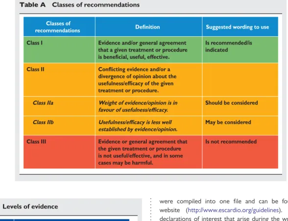
Members of this Task Force were selected by the ESC to rep-resent professionals involved with the medical care of patients with this pathology. Selected experts in the field undertook a comprehensive review of the published evidence for diagnosis, management, and/or prevention of a given condition according to ESC Committee for Practice Guidelines (CPG) policy. A crit-ical evaluation of diagnostic and therapeutic procedures was per-formed including assessment of the risk – benefit ratio. Estimates of expected health outcomes for larger populations were included, where data exist. The level of evidence and the strength of recommendation of particular treatment options were weighed and graded according to pre-defined scales, as outlined in Tables A and B.
The experts of the writing and reviewing panels filled in declara-tions of interest forms of all reladeclara-tionships which might be perceived as real or potential sources of conflicts of interest. These forms
were compiled into one file and can be found on the ESC website (http://www.escardio.org/guidelines). Any changes in declarations of interest that arise during the writing period must be notified to the ESC and updated. The Task Force received its entire financial support from the ESC without any involvement from the healthcare industry.
The ESC CPG supervises and coordinates the preparation of new Guidelines produced by Task Forces, expert groups, or con-sensus panels. The Committee is also responsible for the endorse-ment process of these Guidelines. The ESC Guidelines undergo extensive review by the CPG and external experts. After appropri-ate revisions, it is approved by all the experts involved in the Task Table A Classes of recommendations
Classes of
recommendations Definition Suggested wording to use
Class I Evidence and/or general agreement
that a given treatment or procedure is beneficial, useful, effective.
Is recommended/is indicated
Class II Conflicting evidence and/or a divergence of opinion about the usefulness/efficacy of the given treatment or procedure. Class IIa Weight of evidence/opinion is in
favour of usefulness/efficacy.
Should be considered
Class IIb Usefulness/efficacy is less well established by evidence/opinion.
May be considered
Class III Evidence or general agreement that the given treatment or procedure is not useful/effective, and in some cases may be harmful.
Is not recommended
Table B Levels of evidence
Level of evidence A
Data derived from multiple randomized clinical trials or meta-analyses. Level of
evidence B
Data derived from a single randomized clinical trial or large non-randomized studies.
Level of evidence C
Consensus of opinion of the experts and/ or small studies, retrospective studies, registries.
Force. The finalized document is approved by the CPG for publi-cation in the European Heart Journal.
The task of developing ESC Guidelines covers not only the inte-gration of the most recent research, but also the creation of edu-cational tools and implementation programmes for the recommendations. To implement the guidelines, condensed pocket guidelines versions, summary slides, booklets with essential messages, and an electronic version for digital applications (smart-phones, etc.) are produced. These versions are abridged and, thus, if needed, one should always refer to the full text version which is freely available on the ESC website. The National Societies of the ESC are encouraged to endorse, translate, and implement the ESC Guidelines. Implementation programmes are needed because it has been shown that the outcome of disease may be favourably influ-enced by the thorough application of clinical recommendations.
Surveys and registries are needed to verify that real-life daily practice is in keeping with what is recommended in the guidelines, thus completing the loop between clinical research, writing of guidelines, and implementing them into clinical practice.
The guidelines do not, however, override the individual respon-sibility of health professionals to make appropriate decisions in the circumstances of the individual patients, in consultation with that patient, and, where appropriate and necessary, the patient’s guard-ian or carer. It is also the health professional’s responsibility to verify the rules and regulations applicable to drugs and devices at the time of prescription.
2. Introduction
The aim of this document is to provide practical, evidence-based guidelines for the diagnosis and treatment of heart failure (HF). The principal changes from the 2008 guidelines1relate to:
(i) an expansion of the indication for mineralocorticoid (aldosterone) receptor antagonists (MRAs);
(ii) a new indication for the sinus node inhibitor ivabradine; (iii) an expanded indication for cardiac resynchronization therapy
(CRT);
(iv) new information on the role of coronary revascularization in HF;
(v) recognition of the growing use of ventricular assist devices; and
(vi) the emergence of transcatheter valve interventions.
There are also changes to the structure and format of the guide-lines. Therapeutic recommendations now state the treatment effect supported by the class and level of recommendation in tabular format; in the case of chronic heart failure due to left ventricular (LV) systolic dysfunction, the recommendations focus on mortality and morbidity outcomes. Detailed summaries of the key evidence supporting generally recommended treat-ments have been provided. Practical guidance is provided for the use of the more important disease-modifying drugs and diuretics. When possible, other relevant guidelines, consensus statements, and position papers have been cited to avoid unduly lengthy text. All tables should be read in conjunction with their accompanying text and not read in isolation.
3. Definition and diagnosis
3.1 Definition of heart failure
Heart failure can be defined as an abnormality of cardiac struc-ture or function leading to failure of the heart to deliver oxygen at a rate commensurate with the requirements of the metabolizing tissues, despite normal filling pressures (or only at the expense of increased filling pressures).1 For the pur-poses of these guidelines, HF is defined, clinically, as a syn-drome in which patients have typical symptoms (e.g. breathlessness, ankle swelling, and fatigue) and signs (e.g. ele-vated jugular venous pressure, pulmonary crackles, and dis-placed apex beat) resulting from an abnormality of cardiac structure or function. The diagnosis of HF can be difficult (see Section 3.6). Many of the symptoms of HF are non-discriminating and, therefore, of limited diagnostic value.2–6 Many of the signs of HF result from sodium and water reten-tion and resolve quickly with diuretic therapy, i.e. may be absent in patients receiving such treatment. Demonstration of an underlying cardiac cause is therefore central to the diagno-sis of HF (see Section 3.6). This is usually myocardial disease causing systolic ventricular dysfunction. However, abnormalities of ventricular diastolic function or of the valves, pericardium, endocardium, heart rhythm, and conduction can also cause HF (and more than one abnormality can be present) (see Section 3.5). Identification of the underlying cardiac problem is also crucial for therapeutic reasons, as the precise pathology determines the specific treatment used (e.g. valve surgery for valvular disease, specific pharmacological therapy for LV systol-ic dysfunction, etc.).
3.2 Terminology related to left
ventricular ejection fraction
The main terminology used to describe HF is historical and is based on measurement of LV ejection fraction (EF). Mathematical-ly, EF is the stroke volume (which is the end-diastolic volume minus the end-systolic volume) divided by the end-diastolic volume. In patients with reduced contraction and emptying of the left ven-tricle (i.e. systolic dysfunction), stroke volume is maintained by an increase in end-diastolic volume (because the left ventricle dilates), i.e. the heart ejects a smaller fraction of a larger volume. The more severe the systolic dysfunction, the more the EF is reduced from normal and, generally, the greater the end-diastolic and end-systolic volumes.
The EF is considered important in HF, not only because of its prognostic importance (the lower the EF the poorer the survival) but also because most clinical trials selected patients based upon EF (usually measured using a radionuclide technique or echocardi-ography). The major trials in patients with HF and a reduced EF (HF-REF), or ‘systolic HF’, mainly enrolled patients with an EF ≤35%, and it is only in these patients that effective therapies have been demonstrated to date.
Other, more recent, trials enrolled patients with HF and an EF .40 – 45% and no other causal cardiac abnormality (such as valvular or pericardial disease). Some of these patients did not have an entirely normal EF (generally considered to be .50%)
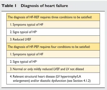
but also did not have a major reduction in systolic function either. Because of this, the term HF with ‘preserved’ EF (HF-PEF) was created to describe these patients. Patients with an EF in the range 35 – 50% therefore represent a ‘grey area’ and most prob-ably have primarily mild systolic dysfunction. The diagnosis of HF-PEF is more difficult than the diagnosis of HF-REF because it is largely one of exclusion, i.e. potential non-cardiac causes of the patient’s symptoms (such as anaemia or chronic lung disease) must first be discounted (Table 1).7,8 Usually these patients do not have a dilated heart and many have an increase in LV wall thickness and increased left atrial (LA) size. Most have evidence of diastolic dysfunction (see Section 4.1.2), which is generally accepted as the likely cause of HF in these patients (hence the term ‘diastolic HF’).7,8
It is important to note that EF values and normal ranges are de-pendent on the imaging technique employed, method of analysis, and operator. Other, more sensitive measures of systolic function may show abnormalities in patients with a preserved or even normal EF (see Section 4.1.1), hence the preference for stating pre-served or reduced EF over prepre-served or reduced ‘systolic function’.9,10
3.3 Terminology related to the
time-course of heart failure
The terms used to describe different types of HF can be confusing. As described above, in these guidelines the term HF is used to de-scribe the symptomatic syndrome, graded according to the New York Heart Association (NYHA) functional classification (see Section 3.4 and Table2), although a patient can be rendered asymptomatic by treatment. In these guidelines, a patient who has never exhibited the typical signs or symptoms of HF is described as having asymptomatic LV systolic dysfunction (or whatever the underlying cardiac abnormality is). Patients who have had HF for
some time are often said to have ‘chronic HF’. A treated patient with symptoms and signs, which have remained generally un-changed for at least a month, is said to be ‘stable’. If chronic stable HF deteriorates, the patient may be described as ‘decom-pensated’ and this may happen suddenly, i.e. ‘acutely’, usually leading to hospital admission, an event of considerable prognostic importance. New (‘de novo’) HF may present acutely, for example as a consequence of acute myocardial infarction or in a subacute (gradual) fashion, for example in a patient who has had asymptom-atic cardiac dysfunction, often for an indeterminate period, and may persist or resolve (patients may become ‘compensated’). Al-though symptoms and signs may resolve in the latter patients, their underlying cardiac dysfunction may not, and they remain at risk of recurrent ‘decompensation’. Occasionally, however, a patient may have HF due to a problem that resolves completely (e.g. acute viral myopericarditis). Some other patients, particularly those with ‘idiopathic’ dilated cardiomyopathy, may also show sub-stantial or even complete recovery of LV systolic function with modern disease-modifying therapy [including an angiotensin-converting enzyme (ACE) inhibitor, beta-blocker, and mineralocor-ticoid receptor antagonist (MRA)]. ‘Congestive HF’ is a term that is sometimes still used, particularly in the USA, and may describe acute or chronic HF with evidence of congestion (i.e. sodium and water retention). Congestion, though not other symptoms of HF (e.g. fatigue), may resolve with diuretic treatment. Many or all of these terms may be accurately applied to the same patient at different times, depending upon their stage of illness.
3.4 Terminology related to the
symptomatic severity of heart failure
The NYHA functional classification (Table 2) has been used to select patients in almost all randomized treatment trials in HF and, therefore, to describe which patients benefit from effective therapies. Patients in NYHA class I have no symptoms attribut-able to heart disease; those in NYHA classes II, III or IV are sometimes said to have mild, moderate or severe symptoms, respectively.
It is important to note, however, that symptom severity corre-lates poorly with ventricular function, and that although there is a clear relationship between severity of symptoms and survival, patients with mild symptoms may still have a relatively high abso-lute risk of hospitalization and death.11–13 Symptoms can also change rapidly; for example, a stable patient with mild symptoms can become suddenly breathless at rest with the onset of an ar-rhythmia, and an acutely unwell patient with pulmonary oedema and NYHA class IV symptoms may improve rapidly with the ad-ministration of a diuretic. Deterioration in symptoms indicates heightened risk of hospitalization and death, and is an indication to seek prompt medical attention and treatment. Obviously, im-provement in symptoms (preferably to the point of the patient be-coming asymptomatic) is one of the two major goals of treatment of HF (the other being to reduce morbidity, including hospital admissions, and mortality).
The Killip classification may be used to describe the severity of the patient’s condition in the acute setting after myocardial infarction.14
Table 1 Diagnosis of heart failure
The diagnosis of HF-REF requires three conditions to be satisfied: 1. Symptoms typical of HF
2. Signs typical of HFa 3. Reduced LVEF
The diagnosis of HF-PEF requires four conditions to be satisfied: 1. Symptoms typical of HF
2. Signs typical of HFa
3. Normal or only mildly reduced LVEF and LV not dilated 4. Relevant structural heart disease (LV hypertrophy/LA enlargement) and/or diastolic dysfunction (see Section 4.1.2)
HF ¼ heart failure; HF-PEF ¼ heart failure with ‘preserved’ ejection fraction; HF-REF ¼ heart failure and a reduced ejection fraction; LA ¼ left atrial; LV ¼ left ventricular; LVEF ¼ left ventricular ejection fraction.
a
Signs may not be present in the early stages of HF (especially in HF-PEF) and in patients treated with diuretics (see Section 3.6).
3.5 Epidemiology, aetiology,
pathophysiology, and natural history of
heart failure
Approximately 1 – 2% of the adult population in developed coun-tries has HF, with the prevalence rising to≥10% among persons 70 years of age or older.15 There are many causes of HF, and these vary in different parts of the world (Appendix A). At least half of patients with HF have a low EF (i.e. HF-REF). HF-REF is the best understood type of HF in terms of pathophysiology and treatment, and is the focus of these guidelines. Coronary artery disease (CAD) is the cause of approximately two-thirds of cases of systolic HF, although hypertension and diabetes are probable contributing factors in many cases. There are many other causes of systolic HF (Appendix A), which include previous viral infection (recognized or unrecognized), alcohol abuse, chemotherapy (e.g. doxorubicin or trastuzumab), and ‘idiopathic’ dilated cardiomyop-athy (although the cause is thought to be unknown, some of these cases may have a genetic basis).16
HF-PEF seems to have a different epidemiological and aetiological profile from HF-REF.17,18 Patients with HF-PEF are older and more often female and obese than those with HF-REF. They are less likely to have coronary heart disease and more likely to have hypertension and atrial fibrillation (AF). Patients with HF-PEF have a better prognosis than those with HF-REF (see below).19
In patients with LV systolic dysfunction, the maladaptive changes occurring in surviving myocytes and extracellular matrix after myo-cardial injury (e.g. myomyo-cardial infarction) lead to pathological ‘re-modelling’ of the ventricle with dilatation and impaired contractility, one measure of which is a reduced EF.11,20 What characterizes untreated systolic dysfunction is progressive worsen-ing of these changes over time, with increasworsen-ing enlargement of the left ventricle and decline in EF, even though the patient may be symptomless initially. Two mechanisms are thought to account for this progression. The first is occurrence of further events leading to additional myocyte death (e.g. recurrent myocardial in-farction). The other is the systemic responses induced by the decline in systolic function, particularly neurohumoral activation.
Two key neurohumoral systems activated in HF are the renin – angiotensin – aldosterone system and sympathetic nervous system. In addition to causing further myocardial injury, these sys-temic responses have detrimental effects on the blood vessels, kidneys, muscles, bone marrow, lungs, and liver, and create a pathophysiological ‘vicious cycle’, accounting for many of the clical features of the HF syndrome, including myocardial electrclical in-stability. Interruption of these two key processes is the basis of much of the effective treatment of HF.11,20
Clinically, the aforementioned changes are associated with the development of symptoms and worsening of these over time, leading to diminished quality of life, declining functional capacity, episodes of frank decompensation leading to hospital admission (which is often recurrent and costly to health services), and prema-ture death, usually due to pump failure or a ventricular arrhythmia. The limited cardiac reserve of such patients is also dependent on atrial contraction, synchronized contraction of the left ventricle, and a normal interaction between the right and left ventricles. Intercurrent events affecting any of these [e.g. the development of AF or conduction abnormalities, such as left bundle branch block (LBBB)] or imposing an additional haemodynamic load on the failing heart (e.g. anaemia) can lead to acute decompensation. Before 1990, the modern era of treatment, 60 – 70% of patients died within 5 years of diagnosis, and admission to hospital with worsening symptoms was frequent and recurrent, leading to an epidemic of hospitalization for HF in many countries.21–23Effective treatment has improved both of these outcomes, with a relative reduction in hospitalization in recent years of 30 – 50% and smaller but significant decreases in mortality.21–23
3.6 Diagnosis of heart failure
3.6.1 Symptoms and signs
The diagnosis of HF can be difficult, especially in the early stages. Although symptoms bring patients to medical attention, many of the symptoms of HF (Table4) are non-specific and do not, there-fore, help discriminate between HF and other problems. Symp-toms that are more specific (i.e. orthopnoea and paroxysmal nocturnal dyspnoea) are less common, especially in patients with milder symptoms, and are, therefore, insensitive.2–6
Many of the signs of HF result from sodium and water retention, and are, therefore, also not specific. Peripheral oedema has other causes as well, and is particularly non-specific. Signs resulting from sodium and water retention (e.g. peripheral oedema) resolve quickly with diuretic therapy (i.e. may be absent in patients receiv-ing such treatment, makreceiv-ing it more difficult to assess patients already treated in this way). More specific signs, such as elevated jugular venous pressure and displacement of the apical impulse, are harder to detect and, therefore, less reproducible (i.e. agree-ment between different doctors examining the same patient may be poor).2–6
Symptoms and signs may be particularly difficult to identify and interpret in obese individuals, in the elderly, and in patients with chronic lung disease.24–26
The patient’s medical history is also important. HF is unusual in an individual with no relevant medical history (e.g. a potential cause of cardiac damage), whereas certain features, particularly previous myocardial infarction, greatly increase the likelihood of HF in a Table 2 New York Heart Association functional
classification based on severity of symptoms and physical activity
Class I
No limitation of physical activity. Ordinary physical activity does not cause undue breathlessness, fatigue, or palpitations.
Class II
Slight limitation of physical activity. Comfortable at rest, but ordinary physical activity results in undue breathlessness, fatigue, or palpitations.
Class III
Marked limitation of physical activity. Comfortable at rest, but less than ordinary physical activity results in undue breathlessness, fatigue, or palpitations.
Class IV
Unable to carry on any physical activity without discomfort. Symptoms at rest can be present. If any physical activity is undertaken, discomfort is increased.
patient with appropriate symptoms and signs.2–5These points high-light the need to obtain objective evidence of a structural or func-tional cardiac abnormality that is thought to account for the patient’s symptoms and signs, to secure the diagnosis of HF (see below).
Once the diagnosis of HF has been made, it is important to establish the cause, particularly specific correctable causes (Appendix A). Symptoms and signs are important in monitoring a patient’s response to treatment and stability over time. Persistence of symptoms despite treatment usually indicates the need for additional therapy, and worsening of symptoms is a serious devel-opment (placing the patient at risk of urgent hospital admission and death) and merits prompt medical attention.
3.6.2 General diagnostic tests in patients with suspected heart failure
In view of the difficulty in grading the evidence for diagnostic tests, all diagnostic recommendations have been given an arbitrary evidence level of C.
3.6.3 Essential initial investigations: echocardiogram, electrocardiogram, and laboratory tests
The echocardiogram and electrocardiogram (ECG) are the most useful tests in patients with suspected HF. The echocardiogram provides immediate information on chamber volumes, ventricular systolic and diastolic function, wall thickness, and valve func-tion.7–10,27–34
This information is crucial in determining appropri-ate treatment (e.g. an ACE inhibitor and beta-blocker for systolic dysfunction or surgery for aortic stenosis). Echocardiography is discussed in detail later (see Section 4). The ECG shows the heart rhythm and electrical conduction, i.e. whether there is sino-atrial disease, atrioventricular (AV) block, or abnormal intraventri-cular conduction (see Table5). These findings are also important for decisions about treatment (e.g. rate control and anticoagulation for AF, pacing for bradycardia, or CRT if the patient has LBBB) (see Section 9.2 on treatment). The ECG may also show evidence of LV hypertrophy or Q waves (indicating loss of viable myocardium), giving a possible clue to the aetiology of HF. HF is very unlikely (likelihood ,2%) in patients presenting acutely and with a com-pletely normal ECG.2,3,35–38In patients with a non-acute presenta-tion, a normal ECG has a somewhat lower negative predictive value (likelihood ,10 – 14%).
The information provided by these two tests will permit an initial working diagnosis and treatment plan in the majority of patients. Routine biochemical and haematological investigations are also important, partly to determine whether renin – angioten-sin – aldosterone blockade can be initiated safely (renal function and potassium) and to exclude anaemia (which can mimic or aggra-vate HF) and because they provide other, useful information (see Section 3.6.6).
Other tests are generally only required if the diagnosis remains unclear (e.g. if echocardiographic images are suboptimal or if an unusual cardiac cause, or a non-cardiac cause, of the patient’s con-dition is suspected) or if further evaluation of the underlying cause of the patient’s cardiac problem is indicated (e.g. perfusion imaging or angiography in suspected CAD or endomyocardial biopsy in certain infiltrating diseases of the myocardium). Special tests are discussed in more detail in Sections 4 and 5.
3.6.4 Natriuretic peptides
Because the signs and symptoms of HF are so non-specific, many patients with suspected HF referred for echocardiography are not found to have an important cardiac abnormality. Where the availability of echocardiography is limited, an alternative approach to diagnosis is to measure the blood concentration of a natriuretic peptide, a family of hormones secreted in increased amounts when the heart is diseased or the load on any chamber is increased (e.g. by AF, pulmonary embolism, and some non-cardiovascular condi-tions, including renal failure).39–42 Natriuretic peptide levels also increase with age, but may be reduced in obese patients.26 A normal natriuretic peptide level in an untreated patient virtually excludes significant cardiac disease, making an echocardiogram un-necessary (investigation for a non-cardiac cause of the patient’s problems is likely to be more productive in such patients).39,42 The use of natriuretic peptides as a ‘rule-out’ test in the diagnosis of HF is discussed in detail elsewhere.39–50Multiple studies have examined the threshold concentration that excludes HF for the Table 4 Symptoms and signs typical of heart failure
Symptoms Signs
Typical More specific
Breathlessness Elevated jugular venous pressure
Orthopnoea Hepatojugular reflux
Paroxysmal nocturnal dyspnoea Third heart sound (gallop rhythm) Reduced exercise tolerance Laterally displaced apical impulse Fatigue, tiredness, increased time
to recover after exercise Cardiac murmur
Ankle swelling
Less typical Less specific
Nocturnal cough Peripheral oedema (ankle, sacral,
scrotal)
Wheezing Pulmonary crepitations
Weight gain (>2 kg/week)
Reduced air entry and dullness to percussion at lung bases (pleural effusion)
Weight loss
(in advanced heart failure) Tachycardia
Bloated feeling Irregular pulse
Loss of appetite Tachypnoea (>16 breaths/min)
Confusion
(especially in the elderly) Hepatomegaly
Depression Ascites
Palpitations Tissue wasting (cachexia)
two most commonly used natriuretic peptides, B-type natriuretic peptide (BNP) and N-terminal pro B-type natriuretic peptide (NT-proBNP).43–50 The exclusion threshold differs for patients presenting with acute onset or worsening of symptoms (e.g. to a
hospital emergency department) and those presenting with a more gradual onset of symptoms.
For patients presenting with acute onset or worsening of symptoms, the optimal exclusion cut-off point is 300 pg/mL Recommendations for the diagnostic investigations in ambulatory patients suspected of having heart failurec
Recommendations Classa Levelb
Investigations to consider in all patients
Transthoracic echocardiography is recommended to evaluate cardiac structure and function, including diastolic function (Section 4.1.2),
and to measure LVEF to make the diagnosis of HF, assist in planning and monitoring of treatment, and to obtain prognostic information. I C
A 12-lead ECG is recommended to determine heart rhythm, heart rate, QRS morphology, and QRS duration, and to detect other relevant abnormalities (Table 5). This information also assists in planning treatment and is of prognostic importance. A completely normal ECG makes systolic HF unlikely.
I C
Measurement of blood chemistry (including sodium, potassium, calcium, urea/blood urea nitrogen, creatinine/estimated glomerular filtration rate, liver enzymes and bilirubin, ferritin/TIBC) and thyroid function is recommended to:
(i) Evaluate patient suitability for diuretic, renin–angiotensin–aldosterone antagonist, and anticoagulant therapy (and monitor treatment)
(ii) Detect reversible/treatable causes of HF (e.g. hypocalcaemia, thyroid dysfunction) and co-morbidities (e.g. iron deficiency)
(iii) Obtain prognostic information.
I C
A complete blood count is recommended to:
(i) Detect anaemia, which may be an alternative cause of the patient’s symptoms and signs and may cause worsening of HF (ii) Obtain prognostic information.
I C
Measurement of natriuretic peptide (BNP, NT-proBNP, or MR-proANP) should be considered to:
(i) Exclude alternative causes of dyspnoea (if the level is below the exclusion cut-point–see Figure 1–HF is very unlikely)
(ii) Obtain prognostic information.
IIa C
A chest radiograph (X-ray) should be considered to detect/exclude certain types of lung disease, e.g. cancer (does not exclude asthma/
COPD). It may also identify pulmonary congestion/oedema and is more useful in patients with suspected HF in the acute setting. IIa C
Investigations to consider in selected patients
CMR imaging is recommended to evaluate cardiac structure and function, to measure LVEF, and to characterize cardiac tissue, especially in subjects with inadequate echocardiographic images or where the echocardiographic findings are inconclusive or incomplete (but taking account of cautions/contraindications to CMR).
I C
Coronary angiography is recommended in patients with angina pectoris, who are considered suitable for coronary revascularization, to
evaluate the coronary anatomy. I C
Myocardial perfusion/ischaemia imaging (echocardiography, CMR, SPECT, or PET) should be considered in patients thought to have CAD, and who are considered suitable for coronary revascularization, to determine whether there is reversible myocardial ischaemia and viable myocardium.
IIa C
Left and right heart catheterization is recommended in patients being evaluated for heart transplantation or mechanical circulatory
support, to evaluate right and left heart function and pulmonary arterial resistance. I C
Exercise testing should be considered: (i) To detect reversible myocardial ischaemia
(ii) As part of the evaluation of patients for heart transplantation and mechanical circulatory support (iii) To aid in the prescription of exercise training
(iv) To obtain prognostic information.
IIa C
BNP ¼ B-type natriuretic peptide; CAD ¼ coronary artery disease; CMR ¼ cardiac magnetic resonance; COPD ¼ chronic obstructive pulmonary disease; ECG ¼ electrocardiogram; HF ¼ heart failure; LV ¼ left ventricular; LVEF ¼ left ventricular ejection fraction; MR-proANP ¼ mid-regional pro atrial natriuretic peptide; NT-proBNP ¼ N-terminal pro B-type natriuretic peptide; PET ¼ positron emission tomography; SPECT ¼ single photon emission computed tomography; TIBC ¼ total iron-binding capacity.
a
Class of recommendation.
b
Level of evidence.
c
This list is not exhaustive and other investigations are discussed in the text. Additional investigations may be indicated in patients with suspected acute HF in the emergency department/ hospital, including troponins and D-dimer measurement and right heart catheterization.
for NT-proBNP and 100 pg/mL for BNP. In one other study, mid-regional atrial (or A-type) natriuretic peptide (MR-proANP), at a cut-off point of 120 pmol/L, was shown to be non-inferior to these thresholds for BNP and NT-proBNP in the acute setting.51
For patients presenting in a non-acute way, the optimum exclu-sion cut-off point is 125 pg/mL for NT-proBNP and 35 pg/mL for BNP. The sensitivity and specificity of BNP and NT-proBNP for the diagnosis of HF are lower in non-acute patients.43–50
3.6.5 Chest X-ray
A chest X-ray is of limited use in the diagnostic work-up of patients with suspected HF. It is probably most useful in identifying an alterna-tive, pulmonary explanation for a patient’s symptoms and signs. It may, however, show pulmonary venous congestion or oedema in a patient with HF. It is important to note that significant LV systolic dysfunction may be present without cardiomegaly on the chest X-ray.
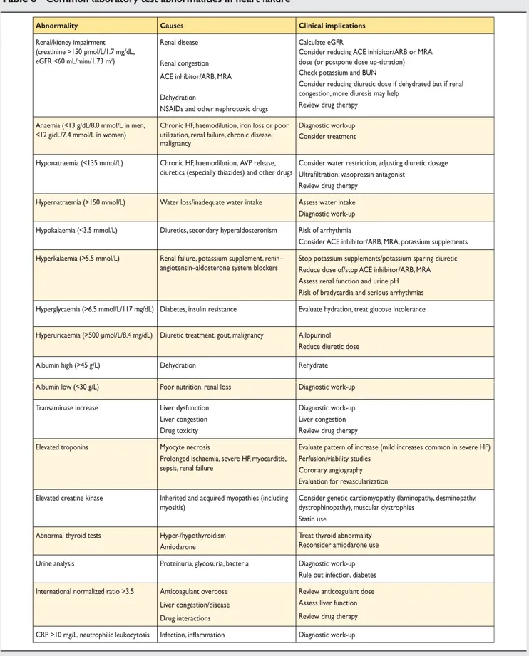
3.6.6 Routine laboratory tests
In addition to standard biochemical [sodium, potassium, creatin-ine/estimated glomerular filtration rate (eGFR)] and haemato-logical tests (haemoglobin, haematocrit, ferritin, leucocytes, and platelets), it is useful to measure thyroid-stimulating hormone (thyrotropin) as thyroid disease can mimic or aggravate HF (Table6). Blood glucose is also worth measuring as undiagnosed diabetes is common in patients with HF. Liver enzymes may also be abnormal in HF (important if considering amiodarone or warfarin).
As well as a pre-treatment check, biochemical monitoring is important after the initiation of renin – angiotensin system block-ers, while the dose is being up-titrated (see Section 7.2) and during longer term follow-up, especially if an intercurrent illness leading to sodium and water loss occurs (e.g. diarrhoea and vomiting) or another drug that affects sodium and water homeo-stasis or renal function is started or the dose altered [e.g. non-steroidal anti-inflammatory drugs (NSAIDs) or diuretics]. Many Table 5 Most common abnormalities on the electrocardiogram in heart failure
Abnormality Causes Clinical implications
Sinus tachycardia Decompensated HF, anaemia, fever, hyperthyroidism Clinical assessment Laboratory investigation Sinus bradycardia Beta-blockade, digoxin, ivabradine, verapamil, diltiazem
Antiarrhythmics Hypothyroidism Sick sinus syndrome
Review drug therapy Laboratory investigation
Atrial tachycardia/flutter/ fibrillation
Hyperthyroidism, infection, mitral valve disease Decompensated HF, infarction
Slow AV conduction, anticoagulation, pharmacological cardioversion, electrical cardioversion, catheter ablation
Ventricular arrhythmias Ischaemia, infarction, cardiomyopathy, myocarditis hypokalaemia, hypomagnesaemia
Digitalis overdose
Laboratory investigation
Exercise test, perfusion/viability studies, coronary angiography, electrophysiology testing, ICD
Myocardial ischaemia/infarction Coronary artery disease Echocardiography, troponins, perfusion/viability studies, coronary angiography, revascularization
Q waves Infarction, hypertrophic cardiomyopathy
LBBB, pre-excitation
Echocardiography, perfusion/viability studies, coronary angiography
LV hypertrophy Hypertension, aortic valve disease, hypertrophic cardiomyopathy
Echocardiography/CMR
AV block Infarction, drug toxicity, myocarditis, sarcoidosis, genetic cardiomyopathy (laminopathy, desminopathy), Lyme disease
Review drug therapy, evaluate for systemic disease; family history/ genetic testing indicated. Pacemaker or ICD may be indicated. Low QRS voltage Obesity, emphysema, pericardial effusion, amyloidosis Echocardiography/CMR, chest X-ray; for amyloidosis consider further imaging (CMR, 99mTc-DPD scan) and endomyocardial biopsy
QRS duration ≥120 ms and LBBB morphology
Electrical and mechanical dyssynchrony Echocardiography
CRT-P, CRT-D
AV ¼ atrioventricular; CMR ¼ cardiac magnetic resonance; CRT-P ¼ cardiac resynchronization therapy pacemaker; CRT-D ¼ cardiac resynchronization therapy defibrillator; ECG ¼ electrocardiogram; HF ¼ heart failure; ICD ¼ implantable cardioverter-defibrillator; LBBB ¼ left bundle branch block; LV ¼ left ventricular. 99mTc-DPD ¼ technetium-99m 3,3-diphosphono-1,2-propanodicarboxylic acid.
Table 6 Common laboratory test abnormalities in heart failure
Abnormality Causes Clinical implications
Renal/kidney impairment (creatinine >150 µmol/L/1.7 mg/dL, eGFR <60 mL/mim/1.73 m2)
Renal disease Renal congestion ACE inhibitor/ARB, MRA Dehydration
NSAIDs and other nephrotoxic drugs
Calculate eGFR
Consider reducing ACE inhibitor/ARB or MRA dose (or postpone dose up-titration) Check potassium and BUN
Consider reducing diuretic dose if dehydrated but if renal congestion, more diuresis may help
Review drug therapy Anaemia (<13 g/dL/8.0 mmol/L in men,
<12 g/dL/7.4 mmol/L in women)
Chronic HF, haemodilution, iron loss or poor utilization, renal failure, chronic disease, malignancy
Diagnostic work-up Consider treatment
Hyponatraemia (<135 mmol/L) Chronic HF, haemodilution, AVP release, diuretics (especially thiazides) and other drugs
Consider water restriction, adjusting diuretic dosage Ultrafiltration, vasopressin antagonist
Review drug therapy Hypernatraemia (>150 mmol/L) Water loss/inadequate water intake Assess water intake Diagnostic work-up Hypokalaemia (<3.5 mmol/L) Diuretics, secondary hyperaldosteronism Risk of arrhythmia
Consider ACE inhibitor/ARB, MRA, potassium supplements Hyperkalaemia (>5.5 mmol/L) Renal failure, potassium supplement, renin–
angiotensin–aldosterone system blockers
Stop potassium supplements/potassium sparing diuretic Reduce dose of/stop ACE inhibitor/ARB, MRA Assess renal function and urine pH Risk of bradycardia and serious arrhythmias Hyperglycaemia (>6.5 mmol/L/117 mg/dL) Diabetes, insulin resistance Evaluate hydration, treat glucose intolerance
Hyperuricaemia (>500 µmol/L/8.4 mg/dL) Diuretic treatment, gout, malignancy Allopurinol Reduce diuretic dose
Albumin high (>45 g/L) Dehydration Rehydrate
Albumin low (<30 g/L) Poor nutrition, renal loss Diagnostic work-up
Transaminase increase Liver dysfunction
Liver congestion Drug toxicity
Diagnostic work-up Liver congestion Review drug therapy
Elevated troponins Myocyte necrosis
Prolonged ischaemia, severe HF, myocarditis, sepsis, renal failure
Evaluate pattern of increase (mild increases common in severe HF) Perfusion/viability studies
Coronary angiography Evaluation for revascularization Elevated creatine kinase Inherited and acquired myopathies (including
myositis)
Consider genetic cardiomyopathy (laminopathy, desminopathy, dystrophinopathy), muscular dystrophies
Statin use
Abnormal thyroid tests Hyper-/hypothyroidism
Amiodarone
Treat thyroid abnormality Reconsider amiodarone use
Urine analysis Proteinuria, glycosuria, bacteria Diagnostic work-up
Rule out infection, diabetes International normalized ratio >3.5 Anticoagulant overdose
Liver congestion/disease Drug interactions
Review anticoagulant dose Assess liver function Review drug therapy CRP >10 mg/L, neutrophilic leukocytosis Infection, inflammation Diagnostic work-up
ACE ¼ angiotensin-converting enzyme; ARB ¼ angiotensin receptor blocker; AVP ¼ arginine vasopressin; BNP ¼ B-type natriuretic peptide; BUN ¼ blood urea nitrogen; CRP ¼ C-reactive protein; eGFR ¼ estimated glomerular filtration rate; HF ¼ heart failure; MRA ¼ mineralocorticoid receptor antagonist; NSAID ¼ non-steroidal anti-inflammatory drug.
routine laboratory tests provide valuable prognostic information (see Section 6).
3.6.7 Algorithm for the diagnosis of heart failure
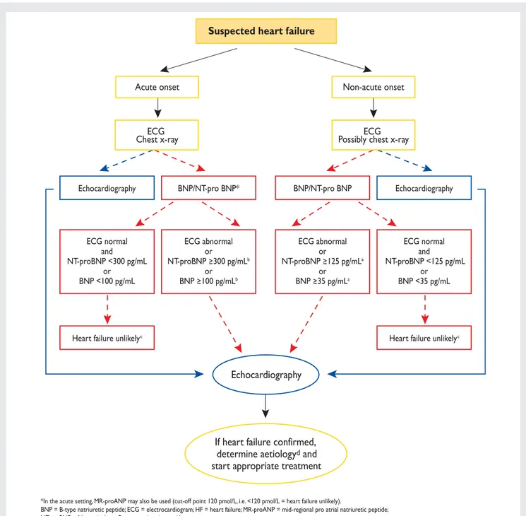
An algorithm for the diagnosis of HF or LV dysfunction is shown in Figure1.
In patients presenting to hospital as an emergency with sus-pected HF and acute onset of symptoms, early echocardiog-raphy is recommended (and immediate echocardiogechocardiog-raphy in shocked or severely haemodynamically compromised patients). If a natriuretic peptide is measured, a high exclusion cut-off point should be used.39–50 In patients presenting non-emergently in primary care, or to a hospital outpatient
Acute onset Non-acute onset
ECG Chest x-ray
Echocardiography BNP/NT-pro BNP* BNP/NT-pro BNP Echocardiography
Heart failure unlikelyc Heart failure unlikelyc
ECG normal and NT-proBNP <300 pg/mL or BNP <100 pg/mL ECG abnormal or NT-proBNP 300 pg/mLb or BNP 100 pg/mLb ECG abnormal or NT-proBNP 125 pg/mLa or BNP 35 pg/mLa ECG normal and NT-proBNP <125 pg/mL or BNP <35 pg/mL ECG Possibly chest x-ray
Suspected heart failure
Echocardiography
If heart failure confirmed,
determine aetiology
dand
start appropriate treatment
*In the acute setting, MR-proANP may also be used (cut-off point 120 pmol/L, i.e. <120 pmol/L = heart failure unlikely).
BNP = B-type natriuretic peptide; ECG = electrocardiogram; HF = heart failure; MR-proANP = mid-regional pro atrial natriuretic peptide; NT-proBNP = N-terminal pro B-type natriuretic peptide.
a Exclusion cut-off points for natriuretic peptides are chosen to minimize the false-negative rate while reducing unnecessary referrals for echocardiography.
b Other causes of elevated natriuretic peptide levels in the acute setting are an acute coronary syndrome, atrial or ventricular arrhythmias, pulmonary embolism, and severe
chronic obstructive pulmonary disease with elevated right heart pressures, renal failure, and sepsis. Other causes of an elevated natriuretic level in the non-acute setting are: old age (>75 years), atrial arrhythmias, left ventricular hypertrophy, chronic obstructive pulmonary disease, and chronic kidney disease.
c Treatment may reduce natriuretic peptide concentration, and natriuretic peptide concentrations may not be markedly elevated in patients with HF-PEF. dSee Section 3.5 and Appendix A.
Figure 1 Diagnostic flowchart for patients with suspected heart failure—showing alternative ‘echocardiography first’ (blue) or ‘natriuretic peptide first’ (red) approaches.
clinic, with slow onset of symptoms (and signs) suggestive of HF, an ECG and natriuretic peptide measurement may be used as a means of identifying patients who most need echocardiography (an echocardiogram is indicated if the natriuretic peptide level is above the exclusion threshold/ECG is abnormal). In these patients, a lower exclusion natriuretic peptide cut-off point should be used to prevent a ‘false-negative’ diagnosis of HF.39–50 Patients with a high pre-test likelihood of HF, such as those with a history of myocardial infarction, may be referred directly for echocardiography.
4. The role of cardiac imaging in
the evaluation of patients with
suspected or confirmed heart
failure
Imaging plays a central role in the diagnosis of HF and in guiding treatment. Of the several imaging modalities available, echocardi-ography is the method of choice in patients with suspected HF for reasons of accuracy, availability (including portability), safety, and cost.27–34 It may be complemented by other modalities, chosen according to their ability to answer specific clinical ques-tions and taking account of contraindicaques-tions to, and risks of, spe-cific tests (see Table 7).9,10,52–60 All imaging examinations, regardless of type, should be performed only by individuals compe-tent and experienced in the specific technique.32
4.1 Echocardiography
Echocardiography is a term used here to refer to all cardiac ultra-sound imaging techniques, including two-dimensional/three-dimensional echocardiography, pulsed and continuous wave Doppler, colour flow Doppler, and tissue Doppler imaging (TDI).8,27–34,61–64 Echocardiography provides information about cardiac anatomy (e.g. volumes, geometry, mass) and function (e.g. LV function and wall motion, valvular function, right ventricu-lar function, pulmonary artery pressure, pericardium).
4.1.1 Assessment of left ventricular systolic dysfunction LVEF is not an index of contractility as it depends on volumes, preload, afterload, heart rate, and valvular function, and is not the same as stroke volume. Stroke volume may be maintained by LV dilation in a patient with HF-REF, whereas it may be reduced in patients with HF-PEF and concentric LV hypertrophy. EF may also be preserved (and stroke volume reduced) in patients with significant mitral regurgitation. Thus EF must be interpreted in its clinical context.
The recommended echocardiographic method for measure-ment of EF is the apical biplane method of discs (the modified Simpson’s rule).8,27–34,61 However, because this method relies on accurate tracing of the endocardial border, use of a contrast agent to better delineate the endocardial border is recom-mended when image quality is suboptimal (i.e. where ,80% of the endocardial border is adequately visualized).61The Teichholz and Quinones methods of calculating EF from linear dimensions may result in inaccuracies, particularly in patients with regional
LV dysfunction; the same is true for another technique for asses-sing LV systolic function—fractional shortening. These and visual assessment of EF (‘eye-balling’) are not recommended.61 Three-dimensional echocardiography of adequate quality further improves the quantification of ventricular volumes and EF calcu-lation.62 The LV wall motion score index may be an acceptable alternative to EF but is not widely used. Other indices of LV sys-tolic function include AV plane syssys-tolic excursion, syssys-tolic tissue Doppler velocities, and measurements of deformation (strain and strain rate). Deformation imaging is more sensitive than EF in detecting minor changes in LV systolic function. However, issues of reproducibility and standardization currently limit the routine clinical use of deformation imaging. Stroke volume and cardiac output can also be calculated by measuring the velocity time integral at the LV outflow tract area.
The most common echocardiographic abnormalities seen in patients with HF and their clinical significance are presented in Table8.
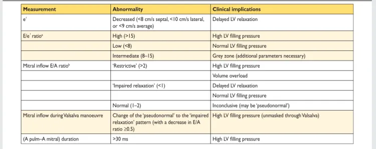
4.1.2 Assessment of left ventricular diastolic dysfunction LV diastolic dysfunction is thought to be the underlying patho-physiological abnormality in patients with HF-PEF, and thus its identification is fundamental to the diagnosis of this type of HF (Table 9).7,8,27–34,63,64 The Doppler echocardiographic diastolic indices commonly measured in patients with HF are shown in Table9. Of note, normal values for functional echocardiographic indices of LV diastolic dysfunction may also depend on age, heart rate, and body size.63,64 Importantly, no single echocardiographic parameter is sufficiently accurate and reproducible to be used in isolation to make a diagnosis of LV diastolic dysfunction. There-fore, a comprehensive echocardiographic examination incorporat-ing all relevant two-dimensional and Doppler data is recommended.8,63,64 This should include the evaluation of both structural (LV hypertrophy, LA dilation) and functional abnormal-ities (Table 1). Tissue Doppler imaging-derived early diastolic myocardial velocities (e’), measured at the mitral annulus, allow the assessment of myocardial relaxation. A normal e’ (.8 cm/s septal, .10 cm/s lateral, or .9 cm/s average, measured using real-time pulsed TDI) is very unusual in a patient with HF. The E/e’ ratio correlates with LV filling pressure.63,64 (Table 9). Thus, echocardiographic evidence of LV diastolic dysfunction may consist of a reduced e’ (e’ average ,9 cm/s) or an increased E/e’ ratio (.15), or a combination of these parameters (Table9). The presence of at least two abnormal measurements and/or AF increases the likelihood of the diagnosis.
4.2 Transoesophageal echocardiography
Transoesophageal echocardiography (TOE) is not needed in routine diagnostic assessment unless the transthoracic ultrasound window is inadequate (e.g. because of obesity, chronic lung disease, ventilated patients) and an alternative modality [e.g. cardiac magnetic resonance (CMR) imaging] is not available or applicable.
TOE is, however, valuable in patients with complex valvular disease (especially mitral disease and prosthetic valves), suspected endocarditis, and in selected patients with congenital heart disease.
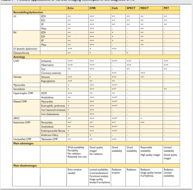
Table 7 Possible applications of various imaging techniques in the diagnosis of HF
Echo CMR Cath SPECT MDCT PET
Remodelling/dysfunction LV: EDV ++ +++ ++ ++ ++ ++ ESV ++ +++ ++ ++ ++ ++ EF ++ +++ ++ ++ ++ ++ Mass ++ +++ - - ++ -RV: EDV ++ +++ + - ++ -ESV ++ +++ + - ++ -EF ++ +++ + - ++ -Mass ++ +++ - - ++ -LV diastolic dysfunction +++ + +++ - - -Dyssynchrony ++ + - + - -Aetiology CAD: Ischaemia +++a +++ +++b +++ - +++ Hibernation +++a +++a - +++ - +++ Scar ++ +++ - ++ - ++ Coronary anatomy - - +++ - +++ -Valvular: Stenosis +++ + +++ - ++c -Regurgitation +++ ++ ++ - - -Myocarditis + +++ +++d - - -Sarcoidosis + +++ ++d - - ++ Hypertrophic CMP: HCM +++ ++ ++ - - -Amyloidosis ++ +++ +++d - - -Dilated CMP: Myocarditis + +++ +++d - - -Eosinophilic syndromes + +++ +++d - - -Iron: haemochromatosis + +++ - - - -Iron: thalassaemia + +++ - - - -ARVC ++ +++ +++d - + -Restrictive CMP: Pericarditis ++e ++ f ++e - ++g -Amyloidosis ++ +++ +++d - - -Endomyocardial fibrosis + +++ +++d - - -Anderson–Fabry + + - - - -Unclassified CMP Takotsubo-CMP ++ ++ +++ - - -Main advantages Wide availability Portability No radiation Relatively low cost
Good quality images No radiation h Good availability Good availability Reasonable availability Limited availability High quality images Good quality
imagesi
Main disadvantages
Echo window needed
Limited availability Radiation Invasive
Radiation Radiation Image quality limited if arrhythmia Radiation Limited availability Contraindications Functional analysis Image quality limited if arrhythmia k
Selection of a test in daily practice should consider availability, local expertise, advantages/disadvantages, and, in the case of several questions to address, which test could best answer several of them.
ARVC ¼ arrhythmogenic right ventricular cardiomyopathy; CAD ¼ coronary artery disease; Cath ¼ cardiac catheterization; CMP ¼ cardiomyopathy; CMR ¼ cardiac magnetic resonance; EDV ¼ end-diastolic volume; EF ¼ ejection fraction; ESV ¼ end-systolic volume; HCM ¼ hypertrophic cardiomyopathy; LV ¼ left ventricular; MDCT ¼ multidetector computed tomography; PET ¼ positron emission tomography; RV ¼ right ventricular; SPECT ¼ single photon emission computed tomography.
a
Stress (dobutamine) imaging.
b
Fractional flow reserve or ‘Doppler’ flow reserve measurements.
c
Including measurements of aortic annulus for transcatheter aortic valve implantation.
d
Endomyocardial biopsy.
e
Haemodynamic evaluation (constriction).
f
Describes disease activity by contrast-enhanced CMR.
g
Calcifications.
h
Good quality irrespective of patient habitus.
i
Excellent attenuation correction.
k