SHORT REPORT
Pancreaticoduodenal Artery Aneurysm Ruptured into
Duodenum, Associated with Celiac Trunk Stenosis.
Case Report and Review of Literature
F. Messina,*G. Azzena, G. Anania, R. Galeotti, D. Pelligrini, G. Cavallesco, D. de Tullio, L. Biaino and S. Occhionorelli
Istituto di Clinica Chirurgica, Dipartimento di Scienze Chirurgiche, Anestesiologiche e Radiologiche, Universita` degli Studi di Ferrara, Italy
Pancreaticoduodenal artery (PDA) aneurysm associated with a celiac artery (CA) occlusion or stenosis is an uncommon event. We report the case of a 63-years old man who presented with acute abdominal pain radiating to the back. During the hospital stay, the patient had an episode of severe hematemesis. He had a gastroscopy and then a surgical exploration. How-ever only with arteriography we found a PDA, which had ruptured into duodenum. The aneurysm was associated with a stenosis of the celiac trunk and was supplied by a dense network of collateral vessels from the SMA. The patient was successfully treated with embolization and was discharged on the 64th postoperative day. Short term and mid term fol-low-up was uneventful. This case shows the difficulty in diagnosing these rare events in time, indicating that angiography is indispensable to establish a diagnosis and enable nonsurgical treatment.
Keywords: Pancreaticoduodenal artery aneurysm; Celiac artery stenosis; Surgery; Embolization.
Introduction
Pancreaticoduodenal artery (PDA) aneurysm associ-ated with celiac artery (CA) occlusion or stenosis is extremely rare, and occurs in 2% of all visceral aneu-rysms.1The number of reported cases has gradually increased due to recent advances in non-invasive di-agnostic imaging modalities such as computed to-mography (CT) and ultrasonography.
Aneurysms involving PDA, often develop sponta-neously, however some authors suggest they are re-lated to specific hemodynamic disorders. An early diagnosis before the occurrence of a fatal rupture is still difficult because of the rare incidence, non-specific symptoms, and deep location in the body of such cases. We report herein the case of a PDA aneu-rysm ruptured into duodenum, associated with a CA
stenosis in a patient successfully treated with emboli-zation. Our case shows the difficulty in diagnosing these rare events in time, and in activating prompt treatment to stop bleeding.
We reviewed the literature of true ruptured and non-ruptured pancreaticoduodenal artery aneurysms associated with a celiac artery lesion.
Case Report
A 63-year old man (weight, 65 kg; height 166 cm), was admitted with acute abdominal pain radiating to the back and anorexia. There was no history of smoking or excessive alcohol consumption and there had been no episodes of pancreatitis, hypertension, portal hypertension, or abdominal injury. No signs of vascu-litis or collagen disease could be found on examina-tion. On admission, he was pale and in pain.
On examination he was apyrexial, his blood pres-sure was 110/70 mmHg and his pulse 84 beats/min. He had a markedly distended abdomen with pain in *Corresponding author. Dr Federico Messina, MD, Istituto di
Clinica Chirurgica, Universita` degli Studi di Ferrara, Arcispedale S. Anna, Corso della Giovecca, 203, 44100 Ferrara, Italy.
E-mail address:[email protected]
EJVES Extra 12, 15e18 (2006)
doi:10.1016/j.ejvsextra.2006.03.002, available online athttp://www.sciencedirect.comon
the right hypochondrium and weak bowel sounds, but no rebound tenderness or guarding. Blood tests revealed a haemoglobin of 10.1 g/dl, a white blood cell count of 11 900/mm,3 and a serum albumin of 2.8 g/dl. The electrolytes, liver function parameters, and serum amylase levels were within the normal ranges. His abdominal pain increased and the pa-tient had nausea and vomiting. A nasogastric tube was inserted and revealed active bleeding. After pa-tient’s hemodynamic stabilization, an operative gas-troscopy revealed massive bleeding coming from the second part of the duodenum, but failed to stop the bleeding.
The patient therefore underwent an emergency lap-arotomy which revealed a duodenum filled with blood and involved in a fibrotic process. After kocher-isation of the duodenum a duodenal ulcer-like lesion, sized 1,5 cm in maximal diameter was detected, ap-parently causing the bleeding. After duodenal sutur-ing and accurate hemostasis, the bleedsutur-ing stopped. However at the end of the operation, there was a re-prise of bleeding. The patient therefore, underwent an emergency operative arteriography.
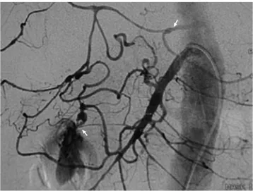
The arteriography revealed a pancreaticoduodenal artery aneurysm 10 mm in diameter ruptured into du-odenum associated with a tight stenosis involving the celiac trunk and a supplied by a particularly dense network of collateral vessels of pancreaticoduodenal arcades and superior mesenteric artery (Fig. 1).
A selective catheterization and embolization of infe-rior-anterior pancreaticoduodenal artery and inferior-posterior pancreaticoduodenal artery was performed. Catheterization and embolization of mesenteric-hepatic anastomotic arcades, permitted a complete
thrombosis of the vessels supplying the aneurysm, while preserving the gastroduodenal artery (Fig. 2).
The postoperative course was complicated by a du-odenal fistula, treated with conservative therapy. The patient was discharged on the 64th postoperative day. Short term and mid term follow-up was uneventful.
Discussion
Aneurysms of the pancreaticoduodenal artery are rare (2% of all visceral artery aneurysms) and nearly 30% of these aneurysms evolve as complication of acute or chronic pancreatitis. There seems to be no gender predilection and the most common age of involve-ment is the sixth decade. The association with celiac artery occlusion was first reported by Sutton and Lawton in 1973.2Since then, 57 cases have been repor-ted.3e15Celiac artery occlusion or stenosis was found in 63% of all cases with PDA aneurysm,8and this was an incidental finding in about 40% of the cases where there was no rupture.
With the increasing efficiency of CT and angiogra-phy, many aneurysms are being discovered before rupture or thrombosis in patients being evaluated for abdominal pain or other disease. Despite these advances, a significant number of vascular aneurysms are not discovered until patients present with rupture. Pancreaticoduodenal artery aneurysms often have symptoms of abdominal pain before rupture.1The dif-ficulty to reach a correct diagnosis explains the high mortality rate with rupture (26% to 50%) and supports the need for early diagnosis and treatment.1,5
Our literature review, wants to point out the atten-tion to true pancreaticoduodenal artery aneurysms
Fig. 1.Arteriography showing the celiac trunk stenosis and the pancreaticoduodenal artery aneurysm ruptured into duodenum.
Fig. 2.Selective catheterization and embolization of the pan-creaticoduodenal and superior mesenteric artery arcades supplying the aneurysm.
16 F. Messina et al.
associated with celiac trunk stenosis, evaluating its correct management.
True aneurysms are often hard to differentiate from false aneurysms. Several findings suggested that our patient had a true aneurysm of the pancreaticoduode-nal artery: the associated celiac trunk lesion, the ab-sence of acute or chronic pancreatitis, and the dense arterial network connecting the superior mesenteric artery to the celiac artery.
False pancreaticoduodenal aneurysms, secondary to local injury, are mainly seen with acute or chronic pancreatitis.
True aneurysms are found in men and women equally and in patients with a wide age range: mean 60 years.16,17 True pancreaticoduodenal artery aneu-rysms are especially rare and various causes have been reported including arteriosclerosis, infection, fibrodysplasia,18and mainly, stenosis or occlusion of the celiac trunk.
A celiac lesion in fact is acknowledged as a major cause of development of an aneurysm of the pancrea-ticoduodenal artery. Its prevalence varies from 63% to 74% in different series.2,8,17 Sutton and Lawton2 proposed that the increased blood flow in the peri-pancreatic arterial network provided collateral supply for revascularization of the celiac trunk, thereby dilat-ing the vascular walls until an aneurysm developed.
Their hypothesis receives support from four re-ported cases that showed that simple revasculariza-tion of the celiac trunk, with surgical decompression through arcuate ligament section, or direct revascular-ization, led to complete aneurysm regression at angio-graphic followup.19e21
Our patient had a relatively small aneurysm in di-ameter. Most pancreaticoduodenal artery aneurysms range from 8 to 30 mm in diameter.8
Our review suggests that size is not a factor for rup-ture; all non-ruptured aneurysms measured more than 10 mm in diameter, whereas most ruptured aneurysms measured less than 10 mm. Most ruptured aneurysms manifest clinically with non-specific abdominal pain,11,17,22e33and in a few cases there is an acute ab-dominal syndrome associated with bleeding into the peritoneal cavity, and ultimately hemorrhagic collapse. These aneurysms usually rupture into the retro-peritoneal space around the pancreas, but also in the peritoneal cavity,28,29 or, exceptionally, into the digestive tract, the duodenum,26 or the Wirsung ca-nal.31 In our patient, abdominal ultrasonography could not identify any relevant finding because of the presence of intestinal gas. Moreover a CT scan was deemed unsafe because of hemodynamic insta-bility. The first diagnostic step to identify the exact site of bleeding was a gastroscopy because the
clinical presentation simulated a peptic ulcer. The surgical operation did not identify the presence of a visceral artery aneurysm, because of its appearance with an ulcer-like duodenal lesion. Only arteriogra-phy, identified the presence of a visceral artery aneu-rysm and showed an associated lesion of the celiac trunk. Our case report emphasizes therefore the role of angiography in localising the source of bleed-ing and its treatment by embolization.
The literature review showed that the current man-agement of pancreaticoduodenal artery aneurysm depends on the presentation of the patient, comorbid-ities and risk factors. It consists of no treatment, sur-gery, embolization therapy, and treatment of the celiac trunk lesion. No treatment is an exceptional op-tion, reported only twice in 39 case for non-ruptured aneurysms: in one case the patient refused therapy; in the second case there was life-threatening risk in a patient with cirrhosis, who died within a few days, of hepatic insufficiency reports.2,17 Surgery was most often used.
The surgical procedure involved resection or sim-ple exclusion of the aneurysm from the circulation. Surgical management through overseeing the aneu-rysm and ligating the feeding arteries, associated with decompression by section of the large left pillar of the arcuate ligament, may be hazardous, because of the high risk of rupture owing the presence of mul-tiple pancreatic vessels that communicate with the an-eurysm.8 Therefore transcatheter embolization is the only alternative treatment in such cases.16,34
Conclusion
Aneurysm of the pancreaticoduodenal artery associ-ated with celiac trunk stenosis is an exceptional event, but requires a prompt management because of its risk of rupture regardless of size. Rupture is often the ini-tial symptom so, intraoperative isolation and control, can be difficult and hazardous. Our patient had no risk factors or symptoms that could justify a visceral artery aneurysm rupture, so he was treated for a gastro-intestinal bleeding.
Otherwise, in this case, the right management would have been not to perform surgery after endos-copy failed to stop the bleeding, but a minimally inva-sive treatment through angiography.
Arteriography can establish a diagnosis, and en-able non-surgical treatment with embolization.
However in cases of a stenosis of the celiac trunk, caused by median arcuate ligament compression, an elective surgical decompression should be considered, to prevent the risk for recurrent aneurysm.
17 Case Report and Review of Literature
References
1 STANLEY JC, WAKEFIELD TW, GRAHAMLM, WHITEHOUSE Jr WM, ZELENOCKGB, LINDENAUERSM. Clinical importance and manage-ment of splanchnic artery aneurysms. J Vasc Surg 1986;3:836e840. 2 SUTTOND, LAWTONG. Coeliac stenosis or occlusion with
aneu-rysm of the collateral supply. Clin Radiol 1973;24:49e53. 3 SUZUKIK, KASHIMURAH, SATOM, HASSANM, YOKOTAH, NAKAHARAA
et al. Pancreaticoduodenal artery aneurysms associated with coe-liac axis stenosis due to compression by median arcuate ligament and coeliac plexus. J Gastroenterol 1998;33:434e438.
4 CHIANGKS, JOHNSONCM, MCKUSICKMA, MAUSTP, STANSONAW. Management of inferior pancreaticoduodenal artery aneurysms: a 4-year, single center experience. Cardiovasc Intervent Radiol 1994;17:217e221.
5 PATYPS, CORDEROJr JA, DARLINGIII RC, CHANGBB, SHAH DM, LEATHER RP. Aneurysms of the pancreaticoduodenal artery. J Vasc Surg 1996;23:710e713.
6 KOBAYASHI S, YAMAGUCHI A, ISOGAI M, HORI A, KANEOKA Y, TAKEUCHIY. Successful transcatheter embolization of a pancreati-coduodenal artery aneurysm in association with coeliac axis oc-clusion: a case report. Hepatogastroenterology 1999;46:2991e2994. 7 NESCHISDG, SAFFORDSD, GOLDENMA. Management of pancrea-ticoduodenal artery aneurysms presenting as catastrophic intra-abdominal bleeding. Surgery 1998;123:8e12.
8 DEPERROTM, BERNEYT, DELEAVALJ, BUHLERL, MENTHAG, MORELP. Management of true aneurysms of the pancreaticoduodenal ar-teries. Ann Surg 1999;229:416e420.
9 IMAMURAM, SHIIYAN, MURASHITAT, SASAKIS, MATSUIY, YASUDAK. Pancreaticoduodenal artery aneurysm with celiac trunk occlu-sion. Report of a case. J Cardiovasc Surg (Torino) 1998;39:565e567. 10 TARAZOVPG, IGNASHOVAM, PAVLOVSKIJAV, NOVIKOVAAS. Pancrea-ticoduodenal artery aneurysm associated with celiac axis steno-sis: combined angiographic and surgical treatment. Dig Dis Sci 2001;46:1232e1235.
11 OGINOH, SATOY, BANNOT, ARAKAWAT, HARAM. Embolization in a patient with ruptured anterior inferior pancreaticoduodenal arterial aneurysm with median arcuate ligament syndrome. Cardiovasc Intervent Radiol 2002;25:318e319.
12 PETERSONBG, RESNICKSA, ESKANDARIMK. Coil embolization of an inferior pancreaticoduodenal artery aneurysm associated with coeliac artery occlusion. Cardiovasc Surg 2003;11:515e519. 13 KABAROUDISA, PAPAZIOGASB, PAPAZIOGAST. Spontaneous
retroper-itoneal hematoma caused by aneurysm of the inferior pancreati-coduodenal artery. Am J Surg 2002;184:174e175.
14 UHERP, NYMANU, IVANCEVK, LINDHM. Aneurysms of the pan-creaticoduodenal artery associated with occlusion of the coeliac artery. Abdom Imaging 1995;20:470e473.
15 DUCASSEE, ROYF, CHEVALIERJ, MASSOUILLED, SMITHM, SPEZIALEF et al. Aneurysm of the pancreaticoduodenal arteries with a coeliac trunk lesion: current management. J Vasc Surg 2004;39(4):906e911. 16 COLLDP, IERARDIR, KERSTEINMD, YOSTS, WILSONA, MATSUMOTOT. Pancreaticoduodenal artery aneurysms: treatment evolution. Ann Vasc Surg 1998;12:286e291.
17 QUANDALLE P, CHAMBON JP, MARACHEP, SAUDEMONTA, MAES B. Pancreaticoduodenal artery aneurysms associated with celiac axis stenosis: report of two cases and review of the literature. Ann Vasc Surg 1990;4:540e545.
18 SERLETHHJ, COGBILTH, GUNDERSENSB. Ruptured pancreaticoduo-denal artery aneurysms and pheochromocytoma in a pregnant patient with neurofibromatosis. Surgery 1998;124:100e102. 19 NAGANON, TAKEUCHIY, GOMIA, NAKATANIH, KOHNOK. A case
report of multiple aneurysms of pancreaticoduodenal region with celiac obstruction. Nippon Geka Gakkai Zasshi 1997;98: 968e971.
20 MORAJD, OBSTD. Celiac axis artery stenosis with aneurysmal calcification of the collateral supply. Aust Radiol 1976;20:252. 21 PROUD G, CHAMBERLAIN J. Aneurysm formation on the small
pancreatic arteries in association with celiac axis compression. Ann R Coll Surg Engl 1978;60:294e297.
22 POONHK. Ruptured mycotic aneurysm of the inferior pancreati-coduodenal artery. J Ark Med Soc 1981;7:184e188.
23 STAAT P, MOHAMMEDI I, RABATEL F, DUPERRET S, VEDRINNE JM, MOTIN J. Selective embolization of ruptured mycotic aneurysm of the duodenopancreatic arcade disclosing infectious endocar-ditis. Arch Mal Coeur Vaiss 1996;89:1431e1435.
24 FOSTERDR. Inferior pancreaticoduodenal artery aneurysm pre-senting in a young woman. Br J Radiol 1985;58:1127e1129. 25 SCHEFLANM, KADIRS, ATHANASOULISCA, HEDBERGSE.
Pancreati-coduodenal artery aneurysm simulating carcinoma of the head of the pancreas. Arch Surg 1977;112:1201e1203.
26 KADIRS, ATHANASOULISCA, YUNEHY, WILKOVH. Aneurysms of the pancreaticoduodenal arteries in association with celiac axis occlusion. Cardiovasc Radiol 1978;1:173e177.
27 VERMYNCKJP, BERTHOUXJP, REMONDA, GAMAINJ, ABETD, PIETRIJ. Aneurysms of the pancreaticoduodenal arteries associated with thrombosis of the celiac artery. Ann Chir 1979;33:430e432. 28 MARIANOEG, GIEGORS. Aneurysm of the pancreaticoduodenal
artery. J Med Soc N J 1981;78:191e193.
29 VERNHET J, CORCOS J. Aneurysms of the pancreaticoduodenal arteries. Chirurgie 1982;108:617e624.
30 THEVENETA, JOYEUXA. Aneurysms of the pancreaticoduodenal arteries (apropos of 2 cases). Chirurgie 1983;109:668e670. 31 GANGAHARDM, CARVETHSW, REESEHE, BUCHMANRJ, BREINERMA.
True aneurysm of the pancreaticoduodenal artery: a case report and review of the literature. J Vasc Surg 1985;2:741e742. 32 AMBROSETTI P, MEYER P, MENTHA G, BATTIKHA J, SCHNEIDER PA,
FAIBUTTI B. Aneurysms of pancreaticoduodenal and hepatic arteries. Chirurgie 1987;11:36e46.
33 SHIBAHARA T, KURUSU J, FUNAYAMA N, TOMITA S, SAITO Y, KOBAYASHIH. Pancreatico-duodenal artery aneurysm associated with complete occlusion of the celiac axis and its rupture. Nippon Shokakibyo Gakkai Zasshi 1993;90:1606e1610.
34 TARAZOV PG, POYSALOV VN, RYZHLOV VK. Transcatheter treat-ment of splenic artery aneurysms (SAA): report of two cases. J Cardiovasc Surg 1991;32:128e131.
Accepted 18 March 2006
18 F. Messina et al.