microRNA-181b is increased in
cystic fibrosis cells and impairs
lipoxin A
4
receptor-dependent
mechanisms of inflammation
resolution and antimicrobial
defense
Anna Maria Pierdomenico
1,3, Sara Patruno
2,3, Marilina Codagnone
2,3, Felice Simiele
2,3,
Veronica Cecilia Mari
2,3, Roberto Plebani
2,3, Antonio Recchiuti
2,3& Mario Romano
2,3The involvement of microRNA (miR) in cystic fibrosis (CF) pathobiology is rapidly emerging. We previously documented that miR-181b controls the expression of the ALX/FPR2 receptor, which is recognized by the endogenous proresolution ligand, lipoxin (LX)A4. Here, we examined whether the
miR-181b-ALX/FPR2 circuit was altered in CF. We examined human airways epithelial cells, normal (16HBE14o-), carrying the ΔF508 mutation (CFBE41o-) or corrected for this mutation (CFBE41o-/ CEP-CFTR wt 6.2 kb), as well as monocyte-derived macrophages (MΦs) from CF patients. CFBE41o- cells exhibited higher miR-181b and reduced ALX/FPR2 levels compared to 16HBE14o- and CFBE41o-/ CEP-CFTR wt 6.2 kb cells. An anti-mir-181b significantly enhanced ALX/FPR2 expression (+ 60%) as well as LXA4-induced increase in transepithelial electric resistance (+ 25%) in CFBE41o- cells. MΦs
from CF patients also displayed increased miR-181b (+ 100%) and lower ALX/FPR2 levels (− 20%) compared to healthy cells. An anti-mir-181b enhanced ALX/FPR2 expression (+ 40%) and normalized receptor-dependent LXA4-induced phagocytosis of fluorescent-labeled zymosan particles as well
as of Pseudomonas aeruginosa by CF-MΦs. These results provide the first evidence that miR-181b is overexpressed in CF cells, impairing some mechanisms of the ALX/FPR2-dependent pathway of inflammation resolution. Thus, targeting miR-181b may represent a strategy to enhance anti-inflammatory and anti-microbial defense mechanisms in CF.
Non resolving lung inflammation is the main cause of disability and death in patients with cystic fibrosis (CF), the most common autosomal recessive genetic disease1. Mutations in the gene encoding the CF transmembrane
con-ductance regulator (CFTR), a regulatory protein of ion transport expressed in a broad variety of cells and tissues, are the cause of CF2,3. Although the respiratory and the intestinal districts are primarily affected, CF is a systemic
disease in which nearly every cell type, including blood cells4–6, and tissue is dysregulated as consequence of
CFTR mutations7. Inflammation in CF begins early in life even before infections8, can be exacerbated by bacterial
colonization promoted by the CFTR-loss-of-function-dependent reduction in the periciliary fluid volume that impairs mucociliary clearance9, and is exaggerated relatively to the bacterial burden8. Given the high incidence of
inflammation-related disabilities and life losses among patients, understanding why inflammation fails to resolve in CF is overtly important for structuring better therapeutic approaches.
1Department of Medicine and Aging Sciences, “G. D’Annunzio” University of Chieti-Pescara, 66013, Chieti, Italy. 2Department of Medical, Oral, and Technological Sciences, “G. D’Annunzio” University of Chieti-Pescara, 66013, Chieti, Italy. 3Center on Aging Science and Translational Medicine (CeSI-MeT) “G. D’Annunzio” University of Chieti-Pescara, 66013, Chieti, Italy. Correspondence and requests for materials should be addressed to M.R. (email: [email protected])
Received: 25 April 2017 Accepted: 6 October 2017 Published: xx xx xxxx
www.nature.com/scientificreports/
The concept that the resolution phase of the inflammatory response is an active process governed by an array of peptide and lipid mediators and their cognate receptors is supported by numerous observations10. Small lipid
mediators, generated in vivo during inflammation resolution, are derived from polyunsaturated fatty acids, i.e arachidonic, eicosapentaenoic, docosahexaenoic acid, and comprehensively termed specialized proresolving lipid mediators. These, include lipoxins (LX), resolvins (Rv), protectins and maresins11, which are generated by the
catalytic activity of lipoxygenases (LO) and exert regulatory and counterregulatory functions on key processes of the inflammatory response to promote resolution and tissue repair11.
LXA4 and RvD1 activate a G protein-coupled receptor termed ALX/FPR212,13. This proresolving receptor is
also recognized by the endogenous anti-inflammatory peptide, Annexin A114. In vivo, myeloid-targeted
over-expression of human ALX/FPR2 in mice reduced PMN infiltration during acute peritonitis and a left-shifted the dose response to its ligands15, whereas ALX/FPR2 KO mice manifested an impaired resolution phenotype16.
Consistent with this, ALX/FPR2 and its proresolution agonists are defective in human diseases characterized by non resolving inflammation, such as asthma, obesity and atherosclerosis17–20. Thus, mechanisms that control
ALX/FPR2 expression may represent relevant targets to potentiate endogenous anti-inflammatory pathways. Evidence indicates that endogenous mechanisms that normally limit the severity and duration of inflam-mation, promoting its timely resolution, are defective in CF patients. For instance, LXA4 levels are significantly
decreased in CF pediatric patients21. Also, AnxA1 is downregulated in nasal cells from CF patients22, and platelets
from CF patients have an impaired LX-biosynthesis capability4. Finally, a recent study indicates that Pseudomonas aeruginosa, one of the main opportunistic pathogens colonizing CF airways, impairs the transcellular
biosynthe-sis of 15-epi-LXA4 by secreting a soluble CFTR inhibitory factor23. Of interest, in preclinical models of CF, the
ALX/FPR2 agonists LXA4, AnxA1, RvD1, and 15-epi-LXA4 proved beneficial in dampening neutrophil
infil-tration, reducing the release of pro-inflammatory mediators by CF cells, and limiting collateral tissue damage, demonstrating their therapeutic potential for treating chronic lung inflammation21,23–25. Hence, uncovering the
molecular mechanisms by which ALX/FPR2 is regulated in CF can offer the basis for a better understanding of CF pathophysiology and the designing of innovative therapeutics that activate endogenous resolution circuits.
We recently uncovered genetic and epigenetic mechanisms that regulate ALX/FPR2 expression26,27,
includ-ing its inhibition by microRNA (miR)-181b26, which also blunts receptor-dependent anti-microbial and
anti-inflammatory cellular mechanisms26. Therefore, we asked whether the ALX/FPR2- LXA
4 circuit and its
reg-ulation by miR-181b was altered in CF.
Herein, we provide evidence that miR-181b is overexpressed in CF cells, impairing the ALX/FPR2-dependent pathway of inflammation resolution. Thus, targeting miR-181b may represent a novel strategy to enhance anti-inflammatory and anti-microbial defense mechanisms in CF.
Results
ALX/FPR2 expression is downregulated in CF airways cells: involvement of transcriptional and
epigenetic events.
We and others reported that LXA4 generation is impaired in CF4,21. Here, we askedwhether the expression of ALX/FPR2, the LXA4 receptor, was also altered in CF cells. To this end, we evaluated
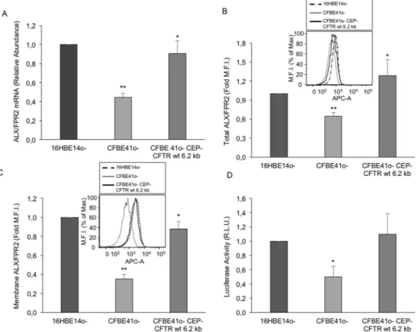
ALX/FPR2 mRNA and protein expression in 16HBE14o- (normal), CFBE41o- (CF) and CFBE41o-/CEP-CFTR wt 6.2 kb (CF corrected) human respiratory epithelial cells. CFBE41o- cells displayed lower ALX/FPR2 mRNA (~ − 60%) and protein, both total and cell membrane (~ − 40–50%), compared to 16HBE14o- cells (Fig. 1A–C).
To investigate on mechanisms underlying these changes, we analyzed ALX/FPR2 transcriptional activity. CFBE41o-cells, transfected with a plasmid expressing the ALX/FPR2 core promoter upstream the luciferase gene28, exhibited significantly lower luciferase activity compared to 16HBE14o- or CFBE41o-/CEP-CFTR wt
6.2 kb cells (Fig. 1D). These results indicate that the ALX/FPR2 transcriptional machinery may be altered in CF cells.
On the other hand, we recently uncovered epigenetic regulatory mechanisms of ALX/FPR2 expression, namely chromatin post-translational modifications27 and mir-181b expression26. Therefore, we examined whether
this miR was involved in ALX/FPR2 expression in CF cells. Figure 2 shows that CFBE41o- cells expressed higher miR-181b levels compared to 16HBE14o- and CFBE41o-/WT cells. We observed a similar increment in bronchial airway primary cells collected from 3 non-CF subjects and 3 CF patients homozygous for the ΔF508 mutation (Fig. 2B).
To analyze the relationship between miR-181b and ALX/FPR2 expression, we downregulated miR-181b using a specific inhibitor (Fig. 2C). miR-181 downregulation enhanced by ~ 60% ALX/FPR2 protein expres-sion in CFBE41o-cells (Fig. 2D), although it did not change ALX/FPR2 mRNA expression (results not shown). To determine whether this increment was associated with enhanced LXA4-induced signaling, we evaluated
trans epithelial electrical resistance (TEER). LXA4 increased TEER by ~ 50% in CFBE41o- cells (Fig. 2E). When
miR-181b expression was downregulated, the effect of LXA4 was enhanced by ~ 25% (Fig. 2E). This effect was
receptor-dependent, since it was abrogated by WRW4, an established ALX/FPR2 antagonist29 (Fig. 2E). We
also evaluated the impact of miR-181b inhibition on the release of selected cytokines (IL-8, IL-6, RANTES) by CFBE41o- cells, exposed or not to LXA4. However, we were unable to detect significant changes in this
experi-mental setting (results not shown).
miR-181b and ALX/FPR2 expression in CF macrophages.
Given the pivotal role of macrophages (MΦs) in inflammation resolution30 and the capability of LXA4 to stimulate ALX/FPR2-dependent pro-resolving
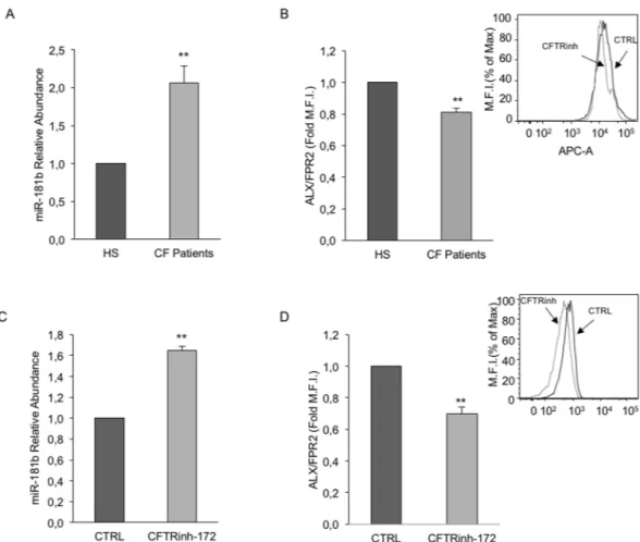
signaling in these cells31, we determined miR-181b and ALX/FPR2 levels in MΦs isolated from CF patients and
age-matched healthy subjects. CF-MΦs displayed higher miR-181b (~ + 100%, p = 0.009) and lower ALX/FPR2 levels (~ − 20%; p = 0.0019) compared to cells from HS (Fig. 3A and B). Notably, in normal MΦs CFTRinh-172, a selective CFTR inhibitor29, for 24 h, enhanced miR-181b (~ + 60%; p = 0.001) and reduced ALX/FPR2 expression
(~ − 30%; p = 0.0019), reminiscent of the CF- MΦs profile (Fig. 3C and D). These results indicate that CFTR con-trols miR-181b expression, which in turn downregulates ALX/FPR2 levels.
Inhibition of miR-181b upregulates ALX/FPR2 expression and LXA
4-induced responses in CF-
MΦs.
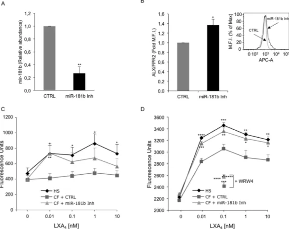
To obtain further evidence of a “cause and effect” relationship between miR-181b and ALX/FPR2, we transfected CF-MΦs with a miR-181b inhibitor. Real-time PCR analysis showed that miR-181b levels were reduced by ~ 70% (p = 0.0013) in transfected cells. In these cells, ALX/FPR2 protein expression increased by ~ 40% (p = 0.0019) (Fig. 4A and B). Next, we determined whether miR-181b-mediated regulation of ALX/FPR2 had an impact on agonist-evoked biological responses. Because macrophage phagocytosis is the hallmark of resolution and it is strongly enhanced by pro-resolving lipid mediators, such as LX and Rv [13, 31], we examined phago-cytosis of fluorescent-labeled zymosan particles as well as of the Pseudomonas aeruginosa strain PAO1 to mimic bacterial clearance from inflamed tissue. We exposed MΦs to increasing concentration of LXA4 (0.001–10 nM)and compared the phagocytic capability of healthy (HS) MΦs, CF-MΦs and CF-MΦs transfected with a miR-181b inhibitor. The zymosan phagocytic activity of unstimulated CF-MΦs was slightly, although not significantly, reduced compared to HS-MΦs (results not shown). When exposed to LXA4, HS-MΦs displayed a significant
incre-ment in zymosan and PAO1 uptake, which was maximal with 1 and 0.1 nM LXA4, respectively (Fig. 4C and D).
In contrast, CF- MΦs showed a smaller increment in phagocytic activity when incubated with LXA4 (Fig. 4C and D).
However, when these cells were transfected with the miR-181b inhibitor, an almost full recovery of LXA4- induced phagocytosis was observed, particularly at lower LXA4 concentrations (Fig. 4C and D).
Receptor dependence was assessed using the WRW4 peptide, which abrogated LXA4-induced phagocytosis of
Figure 1. ALX/FPR2 expression in normal and CF human respiratory epithelial cells. ALX/FPR2 expression
was evaluated in 3 human respiratory epithelial cell lines: normal (16HBE14o-), CF (CFBE41o-) and CF normalized by overexpression of wild type CFTR (CFBE41o- CEP-CFTR wt 62 kb). (A) ALX/FPR2 was evaluated by real-time PCR as reported in material and methods. Results are mean ± SEM from n = 3. *p = 0.03 vs CFBE41o-; **p = 0.002 vs 16HBE14o-. (B) Total ALX/FPR2 protein was assessed by flow cytometry in permeabilized cells. Bars depict mean ± SEM from n = 3. *p < 0.05 vs CFBE41o-; ***p = 0.0008 vs 16HBE14o-. The inset shows a representative histogram. (C) Membrane ALX/FPR2 was evaluated in non permeabilized cells as in (b). Data are mean ± SEM from n = 3. **p = 0.04 vs CFBE41o-; ***p = 0.0006 vs 16HBE14o-. The inset shows a representative histogram. (D) ALX/FPR2 promoter activity was measured in cells transfected with a pGL4 plasmid containing or not the 346 bp sequence of the ALX/FPR2 core promoter upstream the luciferase reporter gene. Luciferase activity was measured 48 h post-transfection. Values were normalized for protein concentration. Results are mean ± SEM from 3 independent experiments carried out in duplicate. *p = 0.01 vs 16HBE14o-.
www.nature.com/scientificreports/
zymosan, both in normal and CF cells (Supplementary Fig. 1). Under these experimental settings, we were unable to detect significant changes in the release of selected cytokines (IL-8, IL-10, IL-1β, RANTES, GM-CSF).
Collectively, these results demonstrate that changes in miR-181b expression impair the ALX/FPR2 proreso-lution signaling in CF cells.
Discussion
Exuberant lung inflammation and infection, leading to respiratory insufficiency and death are trademarks of CF. Recent evidence indicates that in addition to a marked overproduction of pro-inflammatory mediators, an impairment in endogenous anti-inflammatory proresolving mechanisms does occur in CF4,21.
Here, we investigated on the ALX/FPR2 receptor, a GPCR that transduces signals by the endogenous prore-solving mediators LXA4, RvD1 and ANXA112–14 and it is regarded as a main proresolving receptor that intercepts
multiple proresolution pathways. Since changes in ALX/FPR2 levels influence the outcome of an inflammatory response32 we evaluated ALX/FPR2 expression in two cellular models of CF, i.e. airway epithelial cells as a
para-digma of the respiratory pathology and macrophages as key effectors of innate immune responses. In both models ALX/FPR2 levels were reduced compared to the normal counterpart (Figs 1 and 3). Combining this evidence with early data showing reduced LXA4 concentration in BAL from CF patients21; impaired LX biosynthesis, due
to deficient 12-LO activity in CF platelets4; and lower ANXA1 in CF neutrophils25, we can conclude that the
proresolution circuit converging on the ALX/FPR2 receptor is impaired in CF. Together with data showing bene-ficial actions of LXA4 in CF settings33,34 these results encourage further research to test whether the upregulation
of the ALX/FPR2 pathway can be regarded as a suitable strategy to combat inflammation in CF patients. Along these lines, ALX/FPR2 and its proresolution agonists are defective in other human diseases characterized by non resolving inflammation17–20, confirming the relevant immunomodulatory role of this system.
The proresolving actions of LXA4 can be strengthened by the upregulation of ALX/FPR2 expression27.
Therefore, acting on these mechanisms may help to develop innovative anti-inflammatory, proresolving
Figure 2. miR-181b is overexpressed in CF airway cells and controls ALX/FPR2 expression and signaling. (A)
miR-181b levels were evaluated by real-time PCR in normal (16HBE14o-), CF (CFBE41o-) and normalized CF (CFBE41o- CEP-CFTR wt 62 kb) epithelial respiratory cells. RNU6 and SNORD95 were used for normalization. Bars are mean ± SEM from n = 3. *p = 0.04 vs CFBE41o-; **p = 0.02 vs 16HBE14o-. (B) miR-181b levels in primary bronchial epithelial cells from 3 non-CF subjects and 3 CF patients carrying the ΔF508/ΔF508 mutation. Bars are mean ± SEM; **p = 0.0027 (C) CFBE41o- were transfected with a miR-181b inhibitor. miR-181b expression was evaluated by real-time PCR 24 h post transfection. Results are mean ± SEM from 3 separate transfections. ***p = 0.0001. (D) Total ALX/FPR2 protein expression was evaluated by flow cytometry in CFBE41o-cells, transfected with either a negative control or a miR-181b inhibitor. ALX/FPR2 levels were evaluated 48 h post-transfection. Bars depict mean ± SEM from 3 separate transfections. ***p = 0.0005. The inset shows a representative histogram. (E) CFBE41o- were transfected with a negative control or a miR-181b inhibitor. Transepithelial electrical resistance (TEER) was measured 48 h post-transfection using a EVOM Voltohmmeter. Results are expressed as mean ± SEM from 3 separate transfections. **p = 0.0026 vs CTRL.
pharmacology. We previously identified miR-181b as a main epigenetic regulator of ALX/FPR2 expression26.
Here, we tested the hypothesis that the lower ALX/FPR2 expression in CF cells was related to changes in miR-181b level.
We present evidence of miR-181b upregulation in CF respiratory epithelial cells as well as in CF monocyte-derived macrophages (Figs 1 and 3). Although the role of this miR in inflammatory disorders remains to be conclusively defined, evidence indicates that it may exert pro-inflammatory functions, at least in selected settings. For instance, miR-181b regulates TNF-α-induced transcription of pro-inflammatory genes in liver cells35. It also regulates vascular inflammation mediated by NFkB36. More recently, a role of miR-181b in
ath-erosclerosis and aneurysms has been proposed37. Our results with CF cells confirm that miR-181b levels can be
elevated in pathological settings characterized by unresolved inflammation and provide clues to decipher the pathophysiological significance of this miR.
In the specific case of CF, a direct correlation between the genetic defect and miR-181b upregulation appears to occur. We in fact observed that miR-181b expression is under the control of CFTR. This was established in MΦs by experiments with CFTRinh-172, a selective CFTR inhibitor38 (Fig. 3), and in respiratory epithelium
by using cells carrying the ΔF508 mutation and cells where the genetic defect was corrected by the overex-pression of wild type CFTR (Fig. 3). Together, these data support the emerging concept of CFTR as epigenetic modulator39. Along these lines, it has been recently reported that CFTR regulates miR-125b expression with a
bicarbonate-dependent mechanism40. Whether ion fluxes are also involved in CFTR-dependent miR-181b
regu-lation remains to be determined.
On the other hand, our present results indicate that, at least in the CF model, the regulatory mechanisms of ALX/FPR2 expression may be cell specific. In fact, ALX/FPR2 mRNA was reduced in CF airway cells (Fig. 1), but not in CF-MΦs (data not shown), suggesting that transcriptional regulatory events are altered in CF respiratory
Figure 3. miR-181b and ALX/FPR2 expression in normal and CF-MΦs. MΦs and CF- MΦs were obtained
by exposing peripheral blood monocytes from 3 healthy volunteers (HS) and 3 CF patients to GM-CSF for seven days. (A) miR-181b levels were determined by real-time PCR. Bars represent mean ± SEM. **p = 0.009. (B) ALX/FPR2 expression in MΦs and CF-MΦs were evaluated by flow cytometry. Data are mean ± SEM. **p = 0.0019. The inset shows a representative histogram. (C) Normal MΦs were exposed to vehicle (CTRL) or CFTRinh-172 (10 µM) for 30 min. miR-181b levels were determined by real-time PCR. Results are mean ± SEM from n = 3 with duplicates. **p = 0.001. (D) Total ALX/FPR2 levels in normal MΦs exposed to vehicle (CTRL) or CFTRinh-172 (10 µM) levels in were quantitated by flow cytometry. Bars represent mean ± SEM from n = 3. **p = 0.0019. The inset shows a representative histogram.
www.nature.com/scientificreports/
epithelium. We previously documented the impact of epigenetic modifications of the ALX/FPR2 promoter and H3 histones on ALX/FPR2 expression27,28. Although the relevance of these changes remains to be determined
in CF cells, our results clearly demonstrate that miR-181b controls ALX/FPR2 protein expression in both CF respiratory cells and MΦs (Figs 2 and 4). Moreover, in both cell types, by acting on miR-181b it was possible to enhance the functional responses of the pro-resolution agonist LXA4. In CF airway epithelial cells, we observed
a significant increase in TEER (Fig. 2), an indirect readout of the monolayer integrity and of the strength of intercellular junctions, which are altered in CF cells and promote local inflammation41. In MΦs, a miR-181b
inhibitor normalized LXA4-triggered phagocytic activity (Fig. 4). This was observed with both zymosan particles
and Pseudomonas aeruginosa, which chronically colonizes the airways of the majority of CF patients and it is very difficult to eradicate. These results confirm that by blocking miR-181b it is possible to enhance LXA4 bioactions
useful to control bacterial colonization in CF. On the other hand, under the present experimental settings, we were unable to detect significant changes in the release of selected cytokines by airway cells, in the presence or not of miR-181b inhibitor. This is likely to be related to the predominant antagonist action of LXA4 on the release
Figure 4. miR-181b inhibition upregulates ALX/FPR2 expression and agonist-induced phagocytosis in
CF-MΦs. (A) MΦs from healthy subjects were transfected with either a negative control (CTRL) or a miR-181b inhibitor for 24 h miR-181b expression was evaluated by real-time PCR. Bars represent mean ± SEM from 3 independent transfections. **p = 0.0013. (B) MΦs were treated as in (A) and total ALX/FPR2 expression was determined by flow cytometry. WRW4 (10 μM) was added to samples incubated with 0.1 nM LXA4. Bars are
mean ± SEM from 3 independent transfections. *p = 0.019. The inset shows a representative histogram. (C) HS or CF-MΦs (5 × 10⁵/well) transfected either with a negative control or with a miR-181b inhibitor, were
exposed to the indicated concentrations of LXA4. Cells were incubated with FITC-labelled zymosan particles
for 30 min at 37 °C and phagocytosis was assessed by measuring fluorescence with a Synergy H1 microplate reader. Data points are mean ± SEM from separate experiments with cells from 4 healthy donors and 4 CF patients. *p = 0.017 (HS vs CF, 0.01 nM LXA4); *p = 0.032 (HS vs CF, 0.1 nM LXA4); *p = 0.011 (HS vs CF,
1 nM LXA4); *p = 0.045 (HS vs CF, 10 nM LXA4); **p = 0.003 (CF vs CF + miR-181b inhibitor). (D) HS or
CF-MΦs (2 × 10⁵/well) were incubated for 1 h at 37 °C with PAO1-GFP at 1:200 cell:bacteria ratio. Phagocytosis
of PA01-GFP was determined by measuring total fluorescence (Ex 485 nm/Abs 530 nm) using a plate reader (Synergy, BioTek). Data points are mean ± SEM from separate experiments with cells from 3 healthy donors and 3 CF patients. *p = 0.021 (CF vs CF + miR-181b inhibitor, 1 nM LXA4); *p = 0.012 (CF vs CF +
miR-181b inhibitor, 10 nM LXA4); ** p = 0.009 (CF vs CF + miR-181b inhibitor, 0.1 nM LXA4); **p = 0.004 (HS
vs CF, 1 and 10 LXA4); ***p = 0.0003 (CF vs CF + miR-181b inhibitor, 0.01 nM LXA4); ***p = 0.0007 (HS vs
CF, 0.1 nM LXA4); ***p = 0.00062 (CF vs CF + WRW4, 1 nM LXA4); ****p = 0.000006 (HS vs CF, 0.01 nM
LXA4); ****p = 0.000000006 (CF + miR-181b inhibitor vs CF + miR-181b inhibitor + WRW4, 1 nM LXA4);
of inflammatory cytokines, which can be uncovered by pre-exposing cells to pro-inflammatory stimuli. Along these lines, the downstream signaling leading to the enhanced LXA4-induced phagocytic activity, observed when
miR-181 was inhibited, requires further investigation. Whether elements of the autophagy cascade are involved, as recently reported in murine and human macrophages, remains to be determined31. This aspect is
particu-larly relevant in CF, since autophay activators can correct misfolded ∆F508 CFTR and promote clearance of Pseudomonas aeruginosa by ∆F508 CFTR macrophages42–44.
Regardless of these limitations, our present data indicate that the LXA4-ALX/FPR2 signaling may represent a
novel target to stimulate the anti-inflammatory, anti-microbial defense in CF.
In conclusion, here we uncovered the upregulation of miR-181b in CF cells, which contributes to impair the endogenous anti-inflammatory, anti-microbial defense pathway centered on the ALX/FPR2 receptor. Together, these results expand our knowledge of mechanisms of CF inflammation and point to regulatory mechanisms of ALX/FPR2 expression as to potential targets for the development of innovative pharmacology for CF.
Methods
LXA4 (5S,6R,15S-trihydroxy-7E,9E,11Z,13E-eicosatetraenoic acid) was purchased from Calbiochem, (Millipore,
Billerica, MA), stored at −80 °C in ethanol and dissolved in the appropriate aqueous buffer immediately before use. The WRW4 peptide was purchased from Abcam (Cambridge, UK). CFTR inhibitor-172 was from Calbiochem. Growth media, fetal bovine serum (FBS), and supplements were from Gibco (Waltham, MA USA) unless otherwise indicated.
Monocyte isolation and macrophage differentiation.
Monocytes were isolated from peripheral blood (15 ml collected in sodium citrate-containing tubes) of healthy subjects [mean age 34 ± 4.6 (SD), 44% female] and age- and sex-matched CF patients [mean age 27 ± 5.4 (SD), 47% female] referring to the Regional Reference Center for Cystic Fibrosis, Atri (TE), Italy. Patients recruited for this study were free of pulmonary exacerbations at the time of recruitment and had not received i.v. antibiotics, nor steroids and non-steroidal anti-inflammatory drugs in the 2 weeks preceding blood withdrawal.After dextran (6%) sedimentation, mononuclear cells were separated using Histopaque-1077 Ficoll (Sigma, Milan, Italy). Cells (12 × 106) were suspended with serum-free RPMI medium and allowed to adhere to
poly-styrene plates for 1–2 h. Lymphocytes were aspirated and adherent monocytes were analyzed for purity by flow cytometry using an anti-CD-14 antibody (TÜK4 clone, Miltenyi Biotech, Calderara di Reno, Bologna, Italy). MΦ differentiation was obtained by exposing monocytes to RPMI supplemented with 10% FBS, 1% L –glutamine, 1% penicillin/streptomycin, and GM-CSF (10 ng/µl, Prospec, East Brunswick, NJ) for 7 days13. Purity of the isolated
MΦ, as assessed by flow cytometric analysis of CD14 staining, was 100% (M.F.I. ratio = 7.6).
The study was approved by the Ethics Committee of the ASL Teramo, and carried out in accordance with the Declaration of Helsinki, as revised in 2004 and following the guidelines for observational studies published by AIFA (20.03.2008 GU n. 76 of 31.03.2008). Written informed consent was obtained by all human participants.
Bronchial Epithelial Cells.
Normal respiratory epithelial cells (16HBE14o-), CF airway cells homozygous for the ∆F508 mutation (CFBE41o-) and the genetically corrected CF airway cells CFBE41o- CEP-CFTR wt 6.2 kb (CFBE41o-/WT), all kindly provided by Dr. Dieter Gruenert, UCSF, USA, were seeded in fibronectin-coated plates and maintained in MEM medium supplemented with 10% FBS, 1% L-glutamine, 1% penicillin/ streptomycin.Primary human CF airway epithelial cells, isolated from different patients harboring the ∆F508 CFTR gen-otype subjected to lung transplantation, were obtained and provided by Dr. LJV Galietta (Telethon Institute of Genetics and Medicine, Pozzuoli, NA) as a public service of the Italian Cystic Fibrosis Research Foundation. Cells were grown in serum free growth medium (LHC9:RPMI 1640 1:1) supplemented with growth factors45. For
differentiation, epithelia cells were cultured at high density (5 × 10⁵ cells/cm2) in air liquid interface condition
on Transwell filters (0.4 µm pore Ø) for 8–10 days in differentiation medium (Ham’s F12, 2% FBS)46. Epithelium
formation was confirmed by measuring TEER.
Transfection of MΦs and CFBE41o- cells.
Human MΦs were transfected with 10 nM miR-181b inhibitor (single-stranded modified RNA, miScript, Qiagen, Milan, Italy) or non-targeting single-strain RNA (Qiagen) using the INTERFERin reagent (Polyplus Transfection) as reported26. For CFBE41o- transfection, miR-181binhibitor and negative control were diluted with 200 µl of Opti-MEM (Invitrogen, ThermoFisher Scientific, Waltham, MA USA) and combined with 4 µl of INTERFERin for 10 min at room temperature. This solution was immediately added to cells that were incubated at 37 °C.
miR-181b analysis.
miR-181b levels were determined as previously reported26. In brief, we used asilica-based spin column system (Norgen, Thorold, ON, Canada) for extraction of miRNA-enriched fractions. Samples were reverse-transcribed with the miScript II RT kit (Qiagen). Real-time PCR analyses were carried out with 1.5 ng of cDNA using specific primers (miScript Primer Assays) and a SYBR Green master mix (also from Qiagen) with a 7900HT Fast Thermal cycler (Invitrogen). miR-181b relative abundance was quantitated using the 2−ΔΔCt method47. The exogenous cel-miR-39 or endogenous RNU6 and SNORD95 were used to normalize
input cDNA.
ALX/FPR2 expression.
ALX/FPR2 mRNA was evaluated as in Pierdomenico et al.26. Total RNA wasextracted using a silica-based spin column system (Norgen, Thorold, ON, Canada), quantified using a NanoDrop spectrophotometer (Thermo Scientific, Waltham, MA), and reverse-transcribed with the M-MLV Reverse Transcriptase (Sigma). Real-time PCR determinations were carried out using 500 ng of cDNA using the following primers: 5′-GGCCAAGACTTCCGAGAGAG-3′ (forward); 5′-CCGTGTCATTAGTTGGGGCT-3′ (reverse) and
www.nature.com/scientificreports/
a SYBER ROX Real Master Mix (5 Prime, Hilden, Germany) with a 7900HT Fast Thermal cycler (Invitrogen). The GUSB gene was used to normalize cDNA input and ALX/FPR2 relative abundance was determined by the 2−ΔΔCt method47.
ALX/FPR2 protein was assessed by flow cytometry. To this end, cells (1 × 10⁶ /sample) were incubated with
0.5 µg of the Human FPRL1/FPR2 APC-conjugated Antibody (R&D Systems, Minneapolis, MN, USA). Analyses were carried out using a FACS Canto flow cytometer equipped with the Diva software (BD Bioscience). Total ALX/FPR2 was measured in cells permeabilized with Permeabilizing Solution 2 (BD Bioscience). Membrane ALX/FPR2 was assessed using non permeabilized cells.
MΦs phagocytosis.
MΦs were transfected with 10 nM miR-181b inhibitor or non-targeting single-strain RNA vector using INTERFERin (Polyplus Transfection TM) and seeded in 24-well plates (2.5 − 5 × 10⁵ cells/well) 24 h post transfection. The following day, cells were washed twice with Dulbecco’s Phosphate-Buffered Saline (DBPS) and exposed to LXA4 or vehicle (0.01% EtOH) for 15 min at 37 °C. Fluorescein isothiocyanate
(FITC)-labeled serum opsonized zymosan (Zym) A (from Saccharomyces cerevisiae) particles (15 ng/well) were added to cells for 30 min at 37 °C. MΦs were washed twice with DPBS and added of 100 µl of trypan blue (0.03% in DPBS) to quench extracellular fluorescence. Phagocytosis was assessed by measuring fluorescence with a Synergy H1 microplate reader (Biotek, Milan, Italy).
For phagocytosis of Pseudomonas aeruginosa, the PA01 strain, constitutively expressing the green fluorescent protein (GFP), kindly provided by Dr. Gerald Pier (Department of Medicine, Brigham and Women’s Hospital, Harvard Medical School, Boston, MA) upon material transfer agreement, was grown to sub-confluence in 20 ml of tryptic soy broth until reaching an optical density (OD600) = 0.45 ± 0.05, corresponding to ~ 2 × 10⁸
colony forming units/ml. After centrifugation (2,700 rcf, 15 min, 4 °C) and addition of 1 ml of Ca/Mg-free PBS, PA01-GFP (10 µl) was added to monocyte-derived MΦs (2 × 10⁵ cell/well) in 24 well plates at a 1:200 cell:bacteria
ratio. After 1 h at 37 °C in a humidified 5% CO2 incubator, the excess of bacteria was aspirated, plates were washed,
and extracellular PA01-GFP was quenched with trypan blue (0.02% w/vol, 200 µL/well, ~ 1 min). Phagocytosis of PA01-GFP was determined by measuring total fluorescence (Ex 485 nm/Abs 530 nm) using a plate reader (Synergy, BioTek).
Trans epithelial electrical resistance (TEER).
CFBE41o- cells, transfected with 10 nM miR-181b inhib-itor or non-targeting negative control single-strain RNA vector using INTERFERin (Polyplus Transfection TM) were seeded (6 × 10⁵ cells/well) in Transwell permeable supports (6.5 mm insert, 24 well plate) (Corning,NY, USA) 24 h post transfection. The following day, cells were washed twice with DPBS and treated with LXA4,
LXA4 + WRW4 or vehicle (0.01% EtOH) in DPBS. After 4 h, TEER was measured by a EVOM Voltmeter (World
Precision Instruments, Sarasota, FL, USA).
ALX/FPR2 promoter activity.
CFBE41o- and 16HBE14o- cells were seeded in 6-well plates. After 24 h, cells were washed twice with PBS and supplemented with DMEM medium containing 1% FBS. Cells were then transfected with 4 μg of the pGL4 basic plasmid or pGL4 basic + the 346 bp sequence of the ALX/FPR2 core promoter as reported28. Luciferase activity was measured 48 h post-transfection using the luciferase assaykit (Promega) according to the manufacturer’s protocol. Values were normalized for protein concentration as reported28.
Statistical analysis.
Results are reported as mean ± SEM. Statistical significance was evaluated by the Student’s T-test. P values < 0.05 were taken as statistically significant.References
1. Nichols, D. P. & Chmiel, J. F. Inflammation and its genesis in cystic fibrosis. Ped. Pulmonol. 50, S39–56 (2015).
2. Riordan, J. R. et al. Identification of the cystic fibrosis gene: cloning and characterization of complementary DNA. Science 245, 1066–1073 (1989).
3. Anderson, M. P. et al. Demonstration that CFTR is a chloride channel by alteration of its anion selectivity. Science 253, 202–205 (1991).
4. Mattoscio, D. et al. Cystic fibrosis transmembrane conductance regulator (CFTR) expression in human platelets: impact on mediators and mechanisms of the inflammatory response. FASEB J. 24, 3970–3980 (2010).
5. Del Porto, P. et al. Dysfunctional CFTR alters the bactericidal activity of human macrophages against Pseudomonas aeruginosa.
PLoS One 6, e19970 (2011).
6. Painter, R. G. et al. Expression in human neutrophils and the phagolysosomal chlorination defect in cystic fibrosis. Biochemistry 45, 10260–10269 (2006).
7. Castellani, C. & Assael, B. M. Cystic fibrosis: a clinical view. Cell. Mol. Life Sci. 74, 129–140 (2017).
8. Stoltz, D. A., Meyerholz, D. K. & Welsh, M. J. Origins of cystic fibrosis lung disease. N. Engl. J. Med. 372, 1574–1575 (2015). 9. O’Sullivan, B. P. & Freedman, S. D. Cystic fibrosis. Lancet 373, 1891–1904 (2009).
10. Serhan, C. N. et al. Resolution of inflammation: state of the art, definitions and terms. FASEB J. 21, 325–332 (2007). 11. Serhan, C. N. Pro-resolving lipid mediators are leads for resolution physiology. Nature 510, 92–101 (2014).
12. Fiore, S., Maddox, J. F., Perez, H. D. & Serhan, C. N. Identification of a human cDNA encoding a functional high affinity lipoxin A4 receptor. J. Exp. Med. 180, 253–260 (1994).
13. Krishnamoorthy, S. et al. Resolvin D1 binds human phagocytes with evidence for proresolving receptors. Proc. Natl. Acad. Sci. USA 107, 1660–1665 (2010).
14. Perretti, M. et al. Endogenous lipid-and peptide-derived anti-inflammatory pathways generated with glucocorticoid and aspirin treatment activate the lipoxin A4 receptor. Nature Med. 8, 1296–1302 (2002).
15. Devchand, P. R. et al. Human ALX receptor regulates neutrophil recruitment in transgenic mice: roles in inflammation and host defense. FASEB J. 17, 652–659 (2003).
16. Dufton, N. et al. Anti-inflammatory role of the murine formyl-peptide receptor 2: ligand-specific effects on leukocyte responses and experimental inflammation. J. Immunol. 184, 2611–2619 (2010).
18. Planagumà, A. et al. Airway lipoxin A4 generation and lipoxin A4 receptor expression are decreased in severe asthma. Am. J. Respir.
Crit. Care Med. 178, 574–582 (2008).
19. Kosicka, A. et al. Attenuation of plasma annexin A1 in human obesity. FASEB J. 27, 368–378 (2013).
20. Ho, K. J. et al. Aspirin-triggered lipoxin and resolvin E1 modulate vascular smooth muscle phenotype and correlate with peripheral atherosclerosis. Am. J. Pathol. 177, 2116–2123 (2010).
21. Karp, C. L. et al. Defective lipoxin-mediated anti-inflammatory activity in the cystic fibrosis airway. Nat. Immunol. 5, 388–392 (2004).
22. Bensalem, N. et al. Down-regulation of the anti-inflammatory protein annexin A1 in cystic fibrosis knock-out mice and patients.
Mol. Cell. Proteomics 4, 1591–1601 (2005).
23. Flitter, B. A. et al. Pseudomonas aeruginosa sabotages the generation of host proresolving lipid mediators. Proc. Natl. Acad. Sci. USA 114, 136–141 (2017).
24. Codagnone, M. et al. Resolvin D1 Enhances the Resolution of Lung Inflammation Caused by Long-term Pseudomonas aeruginosa Infection. Mucosal Immunol. In press (2017).
25. Dalli, J., Rosignoli, G., Hayhoe, R. P., Edelman, A. & Perretti, M. CFTR inhibition provokes an inflammatory response associated with an imbalance of the annexin A1 pathway. Am. J. Pathol. 177, 176–186 (2010).
26. Pierdomenico, A. M. et al. MicroRNA-181b regulates ALX/FPR2 receptor expression and proresolution signaling in human macrophages. J. Biol. Chem. 290, 3592–3600 (2015).
27. Simiele, F. et al. Epigenetic regulation of the formyl peptide receptor 2 gene. Biochim. Biophys. Acta 1859, 1252–1258 (2016). 28. Simiele, F. et al. Transcriptional regulation of the human FPR2/ALX gene: evidence of a heritable genetic variant that impairs
promoter activity. FASEB J. 26, 1323–1333 (2012).
29. Shin, E. H. et al. Trp-Arg-Trp-Trp-Trp-Trp antagonizes formyl peptide receptor like 2-mediated signaling. Biochem. Biophys. Res.
Commun. 341, 1317–1322 (2006).
30. Dalli, J. & Serhan, C. N. Macrophage Proresolving Mediators-the When and Where. Microbiol. Spectr. 4(3), https://doi.org/10.1128/ microbiolspec.MCHD-0001-2014 (2016).
31. Prieto, P. et al. Activation of autophagy in macrophages by pro-resolving lipid mediators. Autophagy 11, 1729–1744 (2015). 32. Morris, T. et al. Dichotomy in duration and severity of acute inflammatory responses in humans arising from differentially expressed
proresolution pathways. Proc. Natl. Acad. Sci. USA 107, 8842–8847 (2010).
33. Verrière, V. et al. Lipoxin A 4 stimulates calcium-activated chloride currents and increases airway surface liquid height in normal and cystic fibrosis airway epithelia. PLoS One 7, e37746 (2012).
34. Higgins, G. et al. Lipoxin A4 prevents tight junction disruption and delays the colonization of cystic fibrosis bronchial epithelial cells by Pseudomonas aeruginosa. Am. J. Physiol. Lung Cell. Mol. Physiol. 310, L1053–1061 (2016).
35. Zhao, J. et al. (2012) Downregulation of PCAF by miR-181a/b provides feedback regulation to TNF-α-induced transcription of proinflammatory genes in liver epithelial cells. J. Immunol. 188, 1266–274 (2012).
36. Sun, X. et al. MicroRNA-181b regulates NF-κB-mediated vascular inflammation. J. Clin. Invest. 122, 1973–1990 (2012).
37. Di Gregoli, K. et al. MicroRNA-181b Controls Atherosclerosis and Aneurysms Through Regulation of TIMP-3 and Elastin. Circ. Res. 120, 49–65 (2017).
38. Ma, T. et al. Thiazolidinone CFTR inhibitor identified by high-throughput screening blocks cholera toxin-induced intestinal fluid secretion. J. Clin. Invest. 110, 1651–1658 (2002).
39. Chan, H. C., Jiang, X. & Ruan, Y. C. Emerging role of cystic fibrosis transmembrane conductance regulator as an epigenetic regulator: linking environmental cues to microRNAs. Clin. Exp. Pharmacol. Physiol. 41, 615–622 (2014).
40. Lu, Y. C. et al. CFTR mediates bicarbonate-dependent activation of miR-125b in preimplantation embryo development. Cell Res. 22, 1453–1466 (2012).
41. Castellani, S. et al. Emerging relationship between CFTR, actin and tight junction organization in cystic fibrosis airway epithelium.
Histol. Histopathol. 32, 445–459 (2017).
42. Tosco, A. et al. A novel treatment of cystic fibrosis acting on-target: cysteamine plus epigallocatechin gallate for the autophagy-dependent rescue of class II-mutated CFTR. Cell Death Differ. 24, 1305 (2017).
43. Ferrari, E. et al. Cysteamine re-establishes the clearance of Pseudomonas aeruginosa by macrophages bearing the cystic fibrosis-relevant F508del-CFTR mutation. Cell Death Dis. 8, e2544 (2017).
44. Romani, L. et al. Thymosin α1 represents a potential potent single-molecule-based therapy for cystic fibrosis. Nat Med. 23, 590–600 (2017).
45. Zegarra-Moran, O., Sacco, O., Romano, L., Rossi, G. A. & Galietta, L. J. V. Cl− Currents Activated by Extracellular Nucleotides in
Human Bronchial Cells. J. Membr. Biol. 156, 297–305 (1997).
46. Scudieri, P. et al. Association of TMEM16A chloride channel overexpression with airway goblet cell metaplasia. J. Physiol. 590, 6141–6155 (2012).
47. Livak, K. J. & Schmittgen, T. D. Analysis of relative gene expression data using real-time quantitative PCR and the 2−ΔΔCT method. Methods 25, 402–408 (2001).
Acknowledgements
We thank Dr. Paolo Moretti and Maria Di Sabatino (Cystic Fibrosis Regional Center, San Liberatore Hospital of Atri, Teramo, Italy) for patient evaluation and blood sample collection. We also thank Dr. Dieter Gruenert (UCFS, USA) for providing airway epithelial cells and Paola Lanuti, Giuseppina Bologna and Alessia Lamolinara (“G. D’Annunzio” University of Chieti-Pescara) for technical assistance. A special thank to Drs. Loretta Ferrera and Emanuela Caci (Istituto “G. Gaslini”, Genoa, Italy) for their skillful training on primary CF epithelial cell culture and Drs. Domenico Mattoscio and Elisa Isopi (“G. d’Annunzio” University of Chieti-Pescara) for preparing primary CF airway epithelial cell samples. This work was supported in part by the Italian Cystic Fibrosis Foundation (grants FFC#23/2014 to M.R. and FFC #19/2016 to AR) and by the American Cystic Fibrosis Foundation (Grant RECCHI17I0 to AR).
Author Contributions
A.M.P. conducted the experiments, analyzed the results and wrote the paper; S.P. conducted the experiments and analyzed the results; M.C. conducted the experiments and analyzed the results; F.S. conducted the experiments and analyzed the results; R.P. conducted the experiments and analyzed the results; V.C.M. conducted the experiments and analyzed the results; A.R. conceived the experiments, analyzed the results and wrote the paper; M.R. conceived the experiments, analyzed the results and wrote the paper. All authors reviewed the manuscript.
Additional Information
www.nature.com/scientificreports/
Competing Interests: The authors declare that they have no competing interests.
Publisher's note: Springer Nature remains neutral with regard to jurisdictional claims in published maps and
institutional affiliations.
Open Access This article is licensed under a Creative Commons Attribution 4.0 International
License, which permits use, sharing, adaptation, distribution and reproduction in any medium or format, as long as you give appropriate credit to the original author(s) and the source, provide a link to the Cre-ative Commons license, and indicate if changes were made. The images or other third party material in this article are included in the article’s Creative Commons license, unless indicated otherwise in a credit line to the material. If material is not included in the article’s Creative Commons license and your intended use is not per-mitted by statutory regulation or exceeds the perper-mitted use, you will need to obtain permission directly from the copyright holder. To view a copy of this license, visit http://creativecommons.org/licenses/by/4.0/.