includes segmental resection with complete debridement of all non-viable tissue and subsequent reconstruction,3typically with vascu-larized bone flaps.
To the best of our knowledge, we report the first case of a patient who developed metachronous ORN of the mandible requiring re-section and subsequent reconstruction with 2 sequential fibula free flaps.
CLINICAL REPORT
A 55-year-old man who had been treated 2 years previously with chemoradiation for stage IV cancer of the base of tongue was re-ferred for treatment of a necrotic left mandibular body, resistant to local debridement and hyperbaric oxygen therapy. A segmental man-dibulectomy from the left ramus to the left parasymphysis was per-formed, and the defect was reconstructed with a right free fibula osteocutaneous flap. The patient did well from that initial surgery. Two and a half years later, the patient presented with ORN of the right mandibular body, resulting in a pathologic fracture. A second segmental mandibular resection was performed from the right angle to the previously placed contralateral fibula flap. Robust intramed-ullary bleeding was noted from the resected end of the previously placed fibula flap. The defect was reconstructed with a left fibula osteocutaneous free flap fixed to the previously placed vascularized fibula bone. The patient had an uneventful postoperative course. A solid bony union between the 2 fibulas was achieved both clinically and radiologically within 6 months, and the patient was left with satisfactory aesthetic and functional results.
DISCUSSION
Owing to advances in the nonsurgical treatment of head and neck malignancies, patients are more frequently treated with aggressive chemoradiation protocols.4Although they may enjoy longer peri-ods of disease-free survival, they are at a greater risk for compli-cations of their treatment. While the exact pathophysiology of ORN remains to be elucidated, radiation dose and fractionation, poor oral hygiene, tissue trauma during surgery, and tumor location within the mandible are thought to be contributing factors.3Nonsurgical treat-ment modalities for superficial or partial-thickness cases of ORN include hyperbaric oxygen therapy,2,3 oral antibiotics, and dental care.3More aggressive measures should be taken, however, when conservative treatments fail or when patients develop pathologic fractures or fistulae.1,3
While other authors have reported the reconstruction of onco-logic defects using sequential bilateral free fibular flaps anchored in the midline by means of osteosynthesis4,5or of bilateral ORN of mandible treated with a single free fibula flap1or staged, bilateral free fibular flaps anchored to native symphysis,3ours is the first re-ported case, to our knowledge, of metachronous ORN of the man-dible treated by sequential bilateral free fibula osteocutaneous flaps with end-to-end osteosynthesis of the 2 free flaps.
CONCLUSIONS
Even in the presence of prior complex microvascular reconstruc-tions, patients can successfully undergo additional reconstructive procedures to restore their function, appearance, and quality of life.
REFERENCES
1. Jacobson AS, Buchbinder D, Urken ML. Reconstruction of bilateral osteoradionecrosis of the mandible using a single fibular free flap. Laryngoscope 2010;120:273Y275
2. Sandel HD 4th, Davison SP. Microsurgical reconstruction for radiation necrosis: an evolving disease. J Reconstr Microsurg 2007;23:225Y230 3. Kildal M, Wei FC, Chang YM, et al. Reconstruction of bilateral
extensive composite mandibular defects after osteoradionecrosis with two fibular osteoseptocutaneous free flaps. Plast Reconstr Surg 2001;108:963Y967
4. Jones NF, Elahi MM. Osteosynthesis and bony healing between two consecutive free fibular bone grafts. Plast Reconstr Surg
2000;105:166Y170
5. Jones NF, Taub PJ. Sequential second free bone flaps for reconstruction of metachronous mandibular defects. Plast Reconstr Surg
2005;116:939Y945
Mandibular Symphyseal
Fracture Simulated by
a Foreign Body in the Chin
Francesco Arcuri, MD, Matteo Brucoli, MD, Fabrizio Grivetto, MD, Arnaldo Benech, MD, PhD
Introduction: Penetrating foreign bodies occurring after maxillo-facial injuries are a diagnostic challenge for the trauma surgeon. Different materials and various sites of penetration in the maxillo-facial region are described in the literature. We present the peculiar course of a patient with an endoral retained foreign body after a pen-etrating facial injury. The diagnostic pitfall in this type of trauma is highlighted owing to the hyperdensity of the foreign body that, at the computed tomographic (CT) axial scan, simulated a vestibular corti-cal fracture of the mandibular body and deceived both the radiolo-gist and the surgeon.
Clinical Report: We introduce the case of a boy who fell from his bicycle. Computed tomography was performed to detect any bone injuries. The radiologic report stated that a left condylar fracture was presented, associated to a vestibular cortical fracture of the man-dibular body. Anamnestic questions revealed that the boy fell from his bicycle in a dug-up street. Clinical examination revealed 2 ex-traoral open wounds in the subnasal and periorbital areas and an endoral linear wound in the inferior fornix at the mandibular sym-physeal region. Consequently, the left condylar fracture was surgi-cally treated, and the mandibular body was explored by the endoral wound revealing an intact cortex: the road metal was removed from the soft tissue of the chin. The initial diagnostic pitfall was clarified: the radiodense foreign bodies penetrated the endoral wound in the soft tissue of the chin during the fall. They simulated a vestibular cortical fracture of the mandibular body at the CT scan deceiving both the radiologist and the surgeon.
From the Department of Maxillo-Facial Surgery Azienda Ospedaliera Maggiore della Carita` University of Piemonte OrientaleBAmedeo Avogadro,[ Novara, Italy.
Received February 2, 2011
Accepted for publication April 3, 2011.
Address correspondence and reprint request to Francesco Arcuri, MD, SCDU di Chirurgia Maxillo-Facciale, Ospedale Maggiore della Carita`, Corso Mazzini 18, 28100 Novara, Italy; E-mail: [email protected] The authors report no conflicts of interest.
Copyright* 2012 by Mutaz B. Habal, MD ISSN: 1049-2275
DOI: 10.1097/SCS.0b013e318246839a
The Journal of Craniofacial Surgery
&
Volume 23, Number 2, March 2012 Brief Clinical Studies* 2012 Mutaz B. Habal, MD
e91
Discussion: According to the literature, soft tissue foreign bodies can be detected by ultrasonography, plain radiography, CT, and mag-netic resonance imaging. Superficially retained foreign bodies are easily detected with ultrasonography if they are not covered by over-lying bone or gas. If this easily available technique had been applied initially in this case, the correct diagnosis might have been established at the initial admittance. Deeply located foreign bodies are best visu-alized by CT. The foreign body in the case introduced was made by radiopaque substance it presented the same radiodensity as the bone.
Key Words: Foreign body, mandibular fracture, facial trauma
P
enetrating foreign bodies occurring after maxillofacial injuries are a diagnostic challenge for the trauma surgeon; approximately one third of all foreign bodies are initially missed. Different materials and various sites of penetration in the maxillofacial district such as nose, orbit, mouth, and ear are described in the literature.1When a facial fracture occurs, plain radiography alone is rarely indicated because of the poor sensitivity and specificity. It does not allow accurately identification of the fractures; moreover,
radi-ologists and clinicians may misinterpret normal suture lines as non-displaced fractures.2
Conversely, in the management of a facial trauma, radiologic im-aging by computed tomography (CT) is very often indicated to ex-clude other fractures and to plan a proper surgical procedure. A CT scan by coronal, sagittal, and three-dimensional reconstructions per-mits a more precise visualization of the facial structures than stan-dard radiography does.3,4
We present the peculiar course of a patient with an endoral re-tained foreign body after a penetrating facial injury. The diagnostic pitfall in this type of trauma is highlighted owing to the hyperden-sity of the foreign body that, at the CT axial scan, simulated a vesti-bular cortical fracture of the mandivesti-bular body and deceived both the radiologist and the surgeon.
CLINICAL REPORT
We introduce the case of a 16-year-old boy who fell from his bi-cycle during a race; it was admitted at the Novara Major Hospital on July 2010 with the diagnosis of facial trauma. Neurological exami-nation was negative for abnormality. Anamnestic questions revealed that the boy fell in a dug-up street.
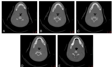
Computed tomography was performed to detect any bone inju-ries. The radiologic report stated that a left condylar fracture was presented (Fig. 1), associated to a vestibular cortical fracture of the mandibular body (Figs. 2AYE).
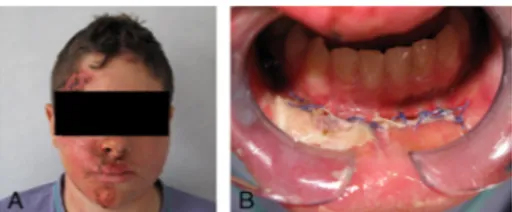
Clinical examination revealed 2 extraoral open wounds in the subnasal and periorbital areas, perioral bruising, posttraumatic mal-occlusion, and an endoral linear wound (1,5 cm) in the inferior for-nix at the mandibular symphyseal region, associated to road metal that penetrated the soft tissue of the chin (Figs. 3A, B).
Informed consent was obtained for open reduction and internal fixation of the fractures. The left condylar fracture was surgical treated and the mandibular body was explored by the endoral wound reveal-ing an intact cortical: the road metal was removed from the soft tissue of the chin.
The initial diagnostic pitfall was clarified: the radiodense foreign bodies penetrated the endoral wound in the soft tissue of the chin during the fall. They simulated a vestibular cortical fracture of the mandibular body at the CT deceiving both the radiologist and the surgeon.
FIGURE 1. Computed tomographic axial scan demonstrating the left condylar fracture.
FIGURE 2. A to E, Computed tomographic scan sequence showing a peculiar hyperdense image resembling a vestibular cortical fracture of the mandibular body.
Brief Clinical Studies The Journal of Craniofacial Surgery
&
Volume 23, Number 2, March 2012e92
* 2012 Mutaz B. Habal, MDThe postoperative course was uneventful, and the radio-graphic control demonstrated a successful osteosynthesis (Fig. 4). The patient was discharged 4 days after the surgical procedure.
DISCUSSION
The correct diagnosis of a retained foreign body is often difficult be-cause it may follow relatively minor trauma, and it may be not clini-cally identified.2In the case presented, the retained foreign body in the chin was initially missed, and its radiologic appearance was misinterpreted. In the appropriate trauma setting, a penetrated and retained for-eign body must always be suspected; especially in cases with lac-eration of the facial soft tissue, a foreign body must be excluded.5,6 According to the literature, metal, glass, and stone are the most common foreign bodies found in the maxillofacial region, and they can be detected by ultrasonography, plain radiography, CT, and mag-netic resonance imaging. Superficially retained foreign bodies are easily detected with ultrasonography if they are not covered by over-lying bone or gas.1,2
If this easily available technique had been applied initially in this case, the correct diagnosis might have been established at the initial admittance. Deeply located foreign bodies are best visualized by CT. The foreign body in the case introduced was made of radiopaque sub-stance, and it presented the same radiodensity as the bone.
Foreign bodies with low radiopacity, which could be detected in air with CT, became less visible in muscle tissue and between bone and muscle. Performing ultrasonography to detect foreign bodies with low radiopacity is relatively better than performing CT.7
Computed tomography is a more useful technique for detecting foreign bodies in air than ultrasound and conventional plain radi-ography. Ultrasonography visualizes superficial foreign bodies with low radiopacity in the tissues of the body more effectively than CT and conventional plain radiography.8
Foreign bodies are usually dirty and carry many microorga-nisms; wound infection is very frequent. Although a foreign body may lead to an aseptic foreign body reaction, antibiotic treatment must be started with good anaerobic coverage. Both types of inflam-mation demand complete surgical removal of the retained foreign body to resolve the symptoms.8
Clinically, the infection can be very severe; Guyennet et al9 re-ported the case of a cephalic tetanus after extraction of a wooden for-eign body with right facial nerve palsy, disorders of swallowing, contralateral III cranial nerve palsy, and trismus. Other studies from the literature report of necrotizing mediastinitis and cervical abscess.10Y12
In summary, the vestibular cortical fracture of the mandibular body was actually simulated by the retained foreign body, which was
penetrated in the submucosal layer of the chin by the vestibular labial wound.
This experience taught us that a high index of suspicion, along with meticulous examination of the wound, is necessary in any patient presenting with a penetrating injury of the face, particularly if the nature of the injury is unknown or atypical; radiologic and clinical examination must be complementary to avoid potential diagnostic pitfalls.
REFERENCES
1. Robinson PD, Rajayogeswaran V, Orr R. Unlikely foreign bodies in unusual facial sites. Br J Oral Maxillofac Surg 1997;35:36Y39 2. Ginsburg MJ, Ellis GL, Flom LL. Detection of soft-tissue foreign
bodies by plain radiography, xerography, computed tomography, and ultrasonography. Ann Emerg Med 1990;19:701Y703
3. Rhea JT, Rao PM, Novelline RA. Helical CT and three dimensional CT of facial and orbital injury. Radiol Clin North Am 1999;37:489Y513 4. Dalley RW. Intraorbital wood foreign bodies on CT. Use of wide bone
window settings to distinguish wood from air. AJR Am J Roentgenol 1995;164:434Y435
5. Dort JC, Robertson D. Nonmetallic foreign bodies of the skull base: a diagnostic challenge. J Otolaryngol 1994;24:69Y72
6. Glatt HJ, Custer PL, Barrett L, et al. Magnetic resonance imaging and computed tomography in a model of wooden foreign bodies in the orbit. Ophthal Plast Reconstr Surg 1990;6:108Y114
7. Specht CS, Varga JH, Jalali MM, et al. Orbitocranial wooden foreign body diagnosed by magnetic resonance imaging. Dry wood can be isodense with air and orbital fat by computed tomography. Surv Ophthalmol 1992;36:341Y344
8. Aras MH, Miloglu O, Barutcugil C, et al. Comparison of the sensitivity for detecting foreign bodies among conventional plain radiography, computed tomography and ultrasonography. Dentomaxillofac Radiol 2010;39:72Y78
9. Guyennet E, Guyomard JL, Barnay E, et al. Cephalic tetanus from penetrating orbital wound. Case Report Med. 2009;2009:548343 10. Mihos P, Potaris K, Gakidis I, et al. Management of descending
necrotizing mediastinitis. J Oral Maxillofac Surg 2004;62:966Y972 11. Makeieff M, Gresillon N, Berthet JP, et al. Management of
descending necrotizing mediastinitis. Laryngoscope 2004;114:772Y775 12. Sancho LM, Minamoto H, Fernandez A, et al. Descending
necrotizing mediastinitis: a retrospective surgical experience. Eur J Cardiothorac Surg 1999;16:200Y205
Intraorbital Epidermoid Cyst: A
5-Year-Old With Exophthalmos
and Strabismus
Bengu Ekinci, MD,* Ender Koktekir, MD,§ Ali Kal, MD,Þ Aylin Karalezli, MD,Þ Hilal Erinanc, MDþ
FIGURE 3. Photographs showing the clinical view (A) and the endoral wound (B).
FIGURE 4. Radiographic control showing the successful anatomic reduction.
From the *Department of Ophthalmology, Selcuk University; and Depart-ments of †Ophthalmology and ‡Pathology, Konya Hospital, Baskent University, and §Department of Ophthalmology, Selcuklu Faculty of Medicine, Selcuk University, Konya, Turkey.
Received February 23, 2011.
Accepted for publication April 3, 2011.
Address correspondence and reprint requests to Bengu Ekinci, MD, Selcuklu Tip Fakultesi, Selcuk Universitesi, Sel0uklu, Konya, Turkey; E-mail: [email protected]
The authors report no conflicts of interest. Copyright* 2012 by Mutaz B. Habal, MD ISSN: 1049-2275
DOI: 10.1097/SCS.0b013e31824683ba
The Journal of Craniofacial Surgery
&
Volume 23, Number 2, March 2012 Brief Clinical Studies* 2012 Mutaz B. Habal, MD