ORIGINAL ARTICLE
Peri-implant bone reactions around immediately loaded
conical implants with different prosthetic suprastructures:
histological and histomorphometrical study on minipigs
Bartolomeo Assenza&Antonio Scarano&
Vittoria Perrotti&Iole Vozza&Alessandro Quaranta&
Manlio Quaranta&Adriano Piattelli&Giovanna Iezzi
Received: 7 August 2008 / Accepted: 18 May 2009 / Published online: 4 June 2009 # Springer-Verlag 2009
Abstract The aim of this study was to evaluate peri-implant bone reactions around immediately loaded conical implants with metal and acrylic resin prosthetic restorations. Five splinted conical implants were inserted in each hemimand-ible of six minipigs at the alveolar crest level. Ten implants were inserted in each minipig. All the implants were immediately loaded. The implants were divided into a group with an acrylic resin prosthetic restoration and into another group with a metal prosthetic restoration. No postoperative complications or deaths of the minipigs occurred. All minipigs were killed after 3 months. No implant was lost. A total of 60 implants were retrieved and processed to obtain thin ground sections. Histology and histomorphometry showed the presence of compact, mature bone around all the implants. Bone was in close contact with the implant surface starting from the first or second implant threads. A high quantity of mineralized bone was present around immediately loaded conical, root form implants. No differ-ences in the peri-implant bone response were found in the groups with different prosthetic reconstructions.
Keywords Animal study . Bone tissue . Conical implants . Immediate loading . Prosthetic superstructures
Introduction
Immediate loading of dental implants is currently one of the most interesting and studied topics in implant dentistry [1]. Although it has been shown to be clinically successful under long-term function [2,3], limited knowledge exists regarding the healing and remodeling processes of the bone around these implants. Animal studies demonstrated osseointegra-tion of immediately loaded definitive [4,5] and provisional [6] implants. Histologic evaluations of immediately loaded implants showed a high degree of osseointegration under long-term function [7, 8]. Using screw implants with a microstructured surface and bone quality-adapted insertion procedures, osseointegration was achieved when implants were initially stable and splinted within the prosthetic suprastructures [9]. The importance of the implant geometry and surface characteristics, in an effort to achieve better bone anchorage, has been demonstrated [10] and various implant systems have been introduced over the past years in order to achieve a faster bone integration [11, 12]. Some authors focused their attention on the conical implant design. Nordin et al. [13] showed that a conical implant had a wider diameter in the cortical passage and that it resulted in bone resorption along the conical surface down to the first thread. Sakoh et al. [14], in an in vitro study, showed that conical implants had a higher primary stability than cylindrical implants. These authors found that the torque and push-out values of the conical implants were significantly higher, while the mean Periotest values of the conical implants were significantly lower. Quaresma et al. [15], in a finite element analysis, showed that a conical implant connected to a solid, internal, conical abutment produced lower stresses on the
B. Assenza
:
A. Scarano:
V. Perrotti:
A. Piattelli (*):
G. IezziDental School, University of Chieti–Pescara,
Via F. Sciucchi 63, 66100 Chieti, Italy e-mail: [email protected]
I. Vozza
:
A. Quaranta:
M. QuarantaDental School“Sapienza”,
alveolar bone and prosthesis and greater stresses on the abutment compared to a cylinder implant connected to a screw-retained, internal hexagonal abutment. The use of miniature pigs in dental research increased significantly due to their similarity in gross anatomy and physiology to humans, as well as for other scientific, economic, and ethical reasons [16]. The roots of the teeth in the minipig are curved distally and the teeth have a higher number of roots than humans (e.g., the molars have four to six roots) [16]. The minipig is a valuable preclinical model that can be used in oral and craniofacial research [17-21] although the rate of bone regeneration in pigs is different from humans (pigs 1.2– 1.5 mm/day; human 1.0–1.5 mm/day) [22]. The restorative materials used for implant-supported prostheses are another important point in implant dentistry due to the fact that they play a relevant role in the long-term success of osseointe-grated implants [23]. However, in vivo studies histologically quantifying the reaction of the peri-implant hard tissues to different restorative superstructure materials are lacking [24]. The three most common groups of materials used for fixed prostheses are porcelain, acrylic, and metal. Impact loads are the lowest with acrylic, increase with metal, are greater with enamel, and further increase with porcelain. Porcelain and acrylic fractures can occur under excessive loads or even with a lesser load of longer duration, angulation, or frequency, and acrylic fractures more easily [25]. Few data are available regarding biomechanical reactions between immediately loaded implants rehabilitated with acrylic and metal suprastructures [26].
The aim of this study was to evaluate peri-implant bone reactions around immediately loaded conical implants with either metal or acrylic resin prosthetic restorations.
Materials and methods
A total of six Göttingen minipigs, 14 to 16 months of age and with an average body weight of 35 kg, were used in this study. The study was approved by the Ethical Committee of the Veterinary Clinic of the University of Madrid, Spain. A total of 60 root form conical implants (RF, Bone System, Milan, Italy) were used. The animals were sedated with an intramuscular injection of ketamine (10 mg/kg), atropine (0.06 mL/kg), and stresnil (0.03 mL/kg). In the areas exposed to surgery, 4 mL of local anesthesia (2% lidocaine with 12.5 µg/mL epinephrine; Xylocain/Adrenalin®, Astra, Wedel, Germany) was injected and three premolars and three molars were extracted in each hemimandible.
The tooth extractions were difficult in every case because the roots were divergent and usually curved distally. It was always necessary to separate the roots before extracting them. After a healing period of 3 months, no migration of the neighboring teeth was observed and five splinted implants
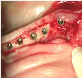
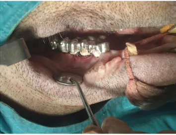
10 mm in length and 4.1 mm in diameter were inserted in each hemimandible at the level of the alveolar crest (Fig.1) under continuous external sterile saline irrigation. All the implants were immediately loaded (the same day of the insertion). The implants were divided into two groups; the implants in the first group, inserted in the left hemimandible, were rehabil-itated with prosthetic restorations in acrylic resin (Fig. 2), while the implants in the second group, inserted in the right hemimandible, were rehabilitated with a prosthetic restora-tion in metal (Fig.3). Both kinds of restorations were put in centric occlusion and checked with articulating paper [27]. The animals were inspected after the first few postoperative days for signs of wound dehiscence or infection and weekly thereafter to assess general health. No postoperative compli-cations or death occurred. All minipigs were killed after 3 months. The animals were euthanized with an overdose of ketamine hydrochloride given intravenously. No implants were lost. A total of 60 implants were retrieved (Figs.4and
5). The implants and surrounding tissues were stored immediately in 10% buffered formalin and processed to obtain thin ground sections. The specimens were processed using the Precise 1 Automated System (Assing, Rome, Italy). The specimens were dehydrated in a graded series of ethanol rinses and embedded in a glycol methacrylate resin (Technovit 7200 VLC, Kulzer, Wehrheim, Germany). After polymerization, the specimens were sectioned, along the longitudinal axis of the implants, with a high-precision diamond disk at about 150 µm and ground down to about 30 µm with a specially designed grinding machine. Three slides were obtained for each implant. These slides were stained with acid fuchsin and toluidine blue and examined with transmitted light under a Leitz Laborlux microscope (Leitz, Wetzlar, Germany).
Histomorphometry was carried out using a light micro-scope (Laborlux S, Leitz, Wetzlar, Germany) connected to a
Fig. 1 Five implants are placed in each hemimandible at the level of the alveolar crest
high-resolution video camera (3CCD, JVC KY-F55B, JVC®, Yokohama, Japan) and interfaced to a monitor and personal computer (Intel Pentium III 1200 MMX, Intel®, Santa Clara, CA, USA). This optical system was associated with a digitizing pad (Matrix Vision GmbH, Oppenweiler, Germany) and a histometry software package with image-capturing capabilities (Image-Pro Plus 4.5, Media Cyber-netics, Immagini & Computer Snc, Milano, Italy).
The histomorphometric measurements involved the mean percentage of bone to implant contact (BIC) in the three best consecutive threads in the cortical region, the bone area in all threads, as well as the bone area in the three best consecutive threads in the cortical region.
Results
The histological results in both groups were similar and will be presented together. Histological analysis of the bone–
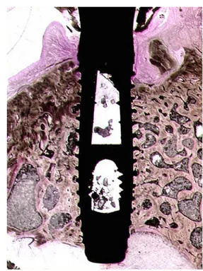
implant interface revealed compact, mature bone found around all implants (Figs. 6 and 7). Many osteons were present. Only a few marrow spaces were present. Bone was in close contact with the implant surface (Fig. 8). Only a few remodeling areas were present. Only a few osteoblasts were present at higher magnification. No inflammatory cell infiltrate was present in the marrow spaces. A slight inflammatory cell infiltrate was observed in the peri-implant soft tissues. No osteoclasts were observed. No gaps or fibrous, connective tissue was found at the bone– implant interface (Fig. 9). No epithelial downgrowth was present. First bone to implant contact was present at the level of the first or second implant thread. The quantitative histomorphometric analysis showed that:
1. bone to implant contact in all available threads around the implant = 69.8±3.2 (acrylic prosthetic restorations) vs 68.1±2.1 (metal prosthetic restorations;p=0.38); 2. bone to implant contact in the three best consecutive
threads in the cortical region = 84 ± 2.7 (acrylic
Fig. 2 Implants rehabilitated with acrylic resin restoration
Fig. 3 Implants rehabilitated with metal restoration
Fig. 4 Radiograph showing, before retrieval, the implants splinted and rehabilitated with acrylic resin restoration
prosthetic restorations) vs 83±3.6 (metal prosthetic restorations;p=0.41);
3. bone area in all threads = 91±0.9 (acrylic prosthetic restorations) vs 89±1.1 (metal prosthetic restorations; p=0.32);
4. bone area in the three best consecutive threads in the cortical region = 93±1.6 (acrylic prosthetic restorations) vs 91±1.8 (metal prosthetic restorations;p=0.39).
Discussion
Primary stability is related to mechanical interlocking, which is one of the most important factors for the development of osseointegration [28]. The implant geom-etry leads to a homogenous strain distribution in loaded peri-implant bone [29]. It has been demonstrated in animal experimental studies that immediate loading of dental implants could be performed without disturbing the early osseointegration process [30]. Several studies emphasized
Fig. 6 Low-power magnification of a histologic section of an implant rehabilitated with an acrylic resin restoration. Bone is present over a large portion of the implant surface. Acid fuchsin–toluidine blue, ×8
Fig. 7 Low-power view of a histologic slide of an implant rehabilitated with a metal restoration. Mature bone lines the implant
perimeter. Acid fuchsin–toluidine blue, ×8
Fig. 8 Compact, mature bone with many osteons are found at the implant interface of an implant supporting an acrylic restoration. Acid
fuchsin–toluidine blue, ×40
Fig. 9 Peri-implant bone found around an implant restored with a
metal prosthesis. No gaps or soft tissues are found at the bone–metal
that the osseointegration process depended on the implant design [10-20]. In the present study, a very high bone–
implant contact percentage was present around immediately loaded conical, root form implants. The implant system used in this study was designed to allow implants to have a direct bone–implant contact over the whole implant surface directly after insertion. The use of a rough, sandblasted, and acid-etched surface was most probably helpful in obtaining a high bone–implant contact percentage in both groups. Clinical, radiographic, and histologic evidence supported the use of metal [24] and acrylic resin suprastructures as fixed prostheses for the restorations of endosseous dental implants [31]. However, when comparing porcelain and acrylic resin occlusal surfaces on osseointegrated implant-supported prostheses opposing natural teeth, no differences related to material could be detected in the load rates [32]. In the present animal study, no differences were found in the bone response in specimens retrieved after 3 months in the two groups (implants with metal superstructures and implants with acrylic resin superstructures). Acrylic resin had a low module of elasticity and could decrease the occlusal impact forces on the bone–implant area if compared to metallic suprastructures. Gracis et al. [33] evaluated the damping effect of five restorative materials rigidly connected to a Brånemark implant and subjected to an impact force. These materials included a gold alloy, a noble metal ceramic alloy, porcelain, a laboratory-processed light-activated microfilled resin, and a heat- and pressure-polymerized polymethyl methacrylate resin. The two resins were found to reduce the impact force by about 50% when compared to porcelain or the alloys. However, this potential protective role has never been fully demonstrated and, on the contrary, a significantly better distribution of bending moments was observed with the metal prostheses in comparison to the acrylic resin prostheses [34]. Sertgöz [35] used a three-dimensional field emission microscopy to study the effect of the superstructure material and occlusal surface material on the stress distribution in an implant-supported fixed prosthesis. The conclusion was that using a superstructure material with a lower modulus did not lead to substantial differences in the stresses in any of the parts of the model (e.g., prosthesis, screws, implants, surround-ing bone), although the lower-modulus material did tend to concentrate stresses in the retaining screws.
The data of the present study were in agreement with other studies that observed no statistically significant differences of the force absorption quotient between the occlusal surfaces of gold, porcelain, and resin [36]. More recently, a study [37] used strain-gauged abutments to measure the force transferred to the implant after the application of a shock. This was measured in vitro and in vivo in five patients, and the different occlusal materials did not lead to different forces generated on the implants.
In the present histological specimens, a marginal bone resorption up to the first or second thread was observed. This could be related to the lack of oral hygiene or to an overloading of the implants. The data to assess the importance of inflammation of the peri-implant tissues and of occlusal overload are, however, still insufficient [38]. In a recent review, it has been stated that, even if in several clinical studies marginal bone loss around implants had been associated with high occlusal stress of the implants, a causative relationship with overload has not been established [39].
Even if there was a difference in resilience between acrylic resin and other materials, this difference was, probably, only measurable in vitro. Therefore, it seemed reasonable that prosthesis materials had no significant effects on the peri-implant bone stress, and the results of the present study showed that a protective role of resin for the bone–implant interface could not be demonstrated.
Additional studies are certainly necessary to evaluate the different types of implant-supported prostheses before final restorative recommendations can be made.
Acknowledgments This work was partially supported by the
Ministry of University, Education and Research (M.I.U.R.), Rome, Italy. The help of Prof. F. San Roman, Madrid, Spain, is gratefully acknowledged.
Conflicts of interest The authors declare that they have no conflicts
of interest.
References
1. Esposito M, Grusovin MG, Willings M, Coulthard P, Worthington HV (2007) The effectiveness of immediate, early, and conventional loading of dental implants: a Cochrane systematic review of randomized controlled clinical trials. Int J Oral Maxillofac Implants
22:893–904
2. Schnitman PA, Wohrle PS, Rubenstein JE, DaSilva JD, Wang NH (1997) Ten-year results for Branemark implants immediately loaded with fixed prostheses at implant placement. Int J Oral
Maxillofac Implants 12:495–503
3. Tarnow DP, Emtiaz S, Classi A (1997) Immediate loading of threaded implants at stage 1 surgery in edentulous arches: ten consecutive case reports with 1- to 5-year data. Int J Oral Maxillofac Implants 12:319–324
4. Piattelli A, Corigliano M, Scarano A, Costigliola G, Paolantonio M (1998) Immediate loading of titanium plasma-sprayed implants: an
histologic analysis in monkeys. J Periodontol 69:321–327
5. Romanos GE, Toh CG, Siar CH, Swaminathan D (2002) Histologic and histomorphometric evaluation of peri-implant bone subjected to immediate loading: an experimental study with Macaca fascicularis. Int J Oral Maxillofac Implants 17:44–51 6. Zubery Y, Bichacho N, Moses O, Tal H (1999) Immediate loading
of modular transitional implants: a histologic and
histomorphomet-ric study in dogs. Int J Periodontics Restorative Dent 19:343–353
7. Ledermann PD, Schenk RK, Buser D (1998) Long-lasting osseointegration of immediately loaded, bar-connected TPS
screws after 12 years of function: a histologic case report of a
95-year-old patient. Int J Periodontics Restorative Dent 18:552–563
8. Proussaefs P, Lozada JL (2002) Evaluation of two vitallium blade-form implants retrieved after 13 to 21 years of function: a clinical report. J Prosthet Dent 87:412–415
9. Neugebauer J, Traini T, Thams U, Piattelli A, Zöller JE (2006) Peri-implant bone organization under immediate loading state. Circularly polarized light analyses: a minipig study. J Periodontol 77:152–160
10. Albrektsson T, Brånemark P-I, Hasson HA, Lindstrom J (1981) Osseointegrated titanium implants. Requirements for ensuring a long-lasting direct bone to implant anchorage in man. Acta Orthop
Scand 52:155–170
11. Büchter A, Kleinheinz J, Wiesmann HP, Seper L, Joos U, Meyer U (2004) Peri-implant bone formation around cylindrical and conical
implant systems. Mund Kiefer Gesichtschir 8:282–288
12. Joos U, Vollmer D, Kleinheinz J (2000) Effect of implant geometry on strain distribution in peri-implant bone. Mund Kiefer
Gesichtschir 4:143–147
13. Nordin T, Jonsson G, Nelvig P, Rasmusson L (1998) The use of a cortical fixture design for fixed partial prostheses. A preliminary report. Clin Oral Implants Res 9:343–347
14. Sakoh J, Wahlmann U, Stender E, Al-Nawas B, Wagner W (2006) Primary stability of a conical implant and a hybrid cylindric screw-type implant in vitro. Int J Oral Maxillofac Implants 21: 560–566
15. Quaresma SE, Cury PR, Sendyk WR, Sendyk C (2008) A finite element analysis of two different dental implants: stress distribu-tion in the prosthesis, abutment, implant, and supporting bone. J
Oral Implantol 34:1–6
16. Wang S, Liu Y, Fang D, Shi S (2007) The miniature pig: a useful large animal model for dental and orofacial research. Oral Dis
13:530–537
17. Nkenke E, Lehner B, Fenner M et al (2005) Immediate versus delayed loading of dental implants in the maxillae of minipigs: follow-up of implant stability and implant failures. Int J Oral
Maxillofac Implants 20:39–47
18. Buchter A, Kleinheinz J, Wiesmann HP et al (2005) Biological and biomechanical evaluation of bone remodelling and implant stability after using an osteotome technique. Clin Oral Implants Res 16:1–8
19. Büchter A, Joos U, Wiesmann HP, Seper L, Meyer U (2006) Biological and biomechanical evaluation of interface reaction at conical screw-type implants. Head Face Med 2(1):5
20. Joos U, Büchter A, Wiesmann HP, Meyer U (2005) Strain driven
fast osseointegration of implants. Head Face Med 1:1–6
21. Wurzler KK, Heisterkamp M, Bohm H, Kubler NR, Sebald W, Reuther JF (2004) Mandibular reconstruction with autologous bone and osseoinductive implant in the Gottingen minipig. Mund
Kiefer Gesichtschir 8:75–82
22. Laiblin C, Jaeschke G (1979) Clinical study of bone and muscle
metabolism under stress conditions in the Goettingen minipig—an
experimental study. Berl Munch Tierarztl Wochenschr 92:124–128
23. Albrektsson T, Johansson C (2001) Osteoinduction,
osteoconduc-tion and osseointegraosteoconduc-tion. Eur Spine J 10:96–101
24. Hürzeler MB, Quiñones CR, Schüpbach P, Vlassis JM, Strub JR, Caffesse RG (1995) Influence of the suprastructure on the peri-implant tissues in beagle dogs. Clin Oral Implants Res 6:139–148 25. Misch CE (2005) Dental implant prosthetics. Elsevier Mosby, St.
Louis, USA, pp 502–503
26. Brunski JB, Puleo DA, Nanci A (2000) Biomaterials and biome-chanics of oral and maxillofacial implants: current status and future
developments. Int J Oral Maxillofac Implants 15(1):15–46
27. Davies SJ, Gray RM, McCord JF (2001) Good occlusal practice in
removable prosthodontics. Br Dent J 191(9):491–494 497–502
28. Meyer U, Vollmer D, Bourauel C, Joos U (2001) Sensitivity analysis of bone geometries around oral implants upon bone loading using finite element method. Comput Methods Biomech
Biomed Eng 3:553–559
29. Ivanoff CJ, Sennerby L, Lekholm U (1996) Influence of initial implant mobility on the integration of titanium implants. An
experimental study in rabbits. Clin Oral Implants Res 7:120–127
30. Meyer U, Vollmer D, Runte C, Bourauel C, Joos U (2001) Bone loading pattern around implants in average and atrophic edentu-lous maxillae: a finite element analysis. J Craniomaxillofac Surg 29:100–105
31. Lindquist LW, Carlsson GE, Glantz PO (1987) Rehabilitation of the edentulous mandible with a tissue integrated fixed prosthesis: a six year longitudinal study. Quintessence Int 18:89–96 32. Hobkirk JA, Psarros KJ (1992) The influence of occlusal surface
material on peak masticatory forces using osseointegrated implant-supported prostheses. Int J Oral Maxillofac Implants
7:345–352
33. Gracis SE, Nicholls JI, Chalupnik JD, Yuodelis RA (1991) Shock-absorbing behavior of five restorative materials used on implants.
Int J Prosthodont 4:282–291
34. Duyck J, Van Oosterwyck H, Vander Sloten J, De Cooman M, Puers R, Naert I (2000) Influence of prosthesis material on the loading of implants that support a fixed partial prosthesis: in vivo
study. Clin Implant Dent Relat Res 2:100–109
35. Sertgöz A (1997) Finite element analysis study of the effect of superstructure material on stress distribution in an implant supported fixed prosthesis. Int J Prosthodont 10:19–27
36. Cibirka RM, Razzoog ME, Lang BR, Stohler CS (1992) Determining the force absorption quotient for restorative materials used in implant occlusal surfaces. J Prosthet Dent 67:361–364 37. Bassit R, Lindström H, Rangert B (2002) In vivo registration of
force development with ceramic and acrylic resin occlusal materials on implant-supported prostheses. Int J Oral Maxillofac
Implants 17:17–23
38. Miyata T, Kobayashi Y, Araki H, Ohto T, Shin K (2002) The influence of controlled occlusal overload on peri-implant tissue. Part 4: a histologic study in monkeys. Int J Oral Maxillofac
Implants 17:384–390
39. Isidor F (2006) Influence of forces on peri-implant bone. Clin Oral