UNIVERSITY OF URBINO CARLO BO
Department of Biomolecular Sciences
Ph.D. Course in Life Sciences, Healthcare and Biotechnologies
Curriculum: Exercise science and health
XXXII cycle
Role of Physical Exercise and Metformin
in in vitro models of tumor progression control
SSD: MED/42
Supervisor:
Ph.D. student:
Prof. Giorgio Brandi
Dr. Giulia Baldelli
Co-Advisor:
Prof. Giuditta Fiorella Schiavano
Organization of the thesis
After the Introduction, the First Chapter will give a complete view of the possible mechanisms involved in exercise-induced control of triple-negative breast cancer, reviewing recent data about exercise modulation of mTOR pathway and the possible effects resulted from different exercise protocols.
In the Second Chapter, the optimization procedure of a three-dimensional growth assay to assess the effects of an intense session of exercise on triple-negative breast cancer clonogenic potential will be described.
The Third Chapter will focus on the central project of the present thesis, in which the evaluation of systemic responses to structured high-intensity endurance cycling sessions of exercise and to a high-intensity interval training period will be considered. The modulation of the proliferative and tumorigenic capacities of triple-negative breast cancer and prostate cancer cells stimulated with the exercise-conditioned human sera will be evaluated. Moreover, the possible relations between the in vitro results and the biological responses measured during the exercise will be assessed.
In the Fourth Chapter, a possible pharmacological approach to prevent cancer initiation will be taken into account, with the analysis of metformin effects in preventing cell tumorigenesis.
Finally, after the Conclusions, the Appendix will focus on a work related to a different research line, in comparison to that analyzed in the central Ph.D. project. In this last part, the possible chemopreventive activity of an in vitro culture derived from an apple will be considered in two colon cancer cell lines and in a pre-neoplastic cell line, in terms of antiproliferative and antitumorigenic activity, cell cycle blockade and molecular mechanisms involved.
CONTENT
INTRODUCTION 1
1.1 Cancer incidence and mortality 2
1.1.1 Worldwide Data 2
1.1.2 Cancer Incidence and Mortality: Italian Data 3
1.2 Breast cancer 4
1.2.1 Epidemiology 4
1.2.2 Breast cancer classification 4
1.3 Prostate cancer 6
1.3.1 Epidemiology 6
1.4 Physical Exercise and cancer prevention 6
1.5 Metformin and cancer 9
AIMS OF THE THESIS 10
CHAPTER 1 12
New Insights into the Role of Exercise in Inhibiting mTOR Signaling
in Triple-Negative Breast Cancer 13
Abstract 14
1. Introduction 14
2. mTOR Signaling 15
2.1 mTOR Pathway and mTOR Activation in BC 15
2.2 MicroRNAs and mTOR Signaling in BC 18
2.3 Autophagy and mTOR Signaling. 19
3. Evidence of mTOR Modulation by Exercise in TNBC 20
3.1 mTOR and Exercise 20
3.1.1 Aerobic Exercise and Muscular Effects 21
3.1.2 Resistance Exercise and Muscular Effects 22 3.1.3 Systemic and Microenvironment Effects of Exercise 22
3.2 Experimental Evidence of mTOR Inhibition 27
4. Energy Intake in TNBC and mTOR Modulation 30
5. Exercise Prescription in BC Survivors 33
6. Benefits of Exercise Pre- and Post-diagnoses 37
7. Conclusions 38
References 40
CHAPTER 2 53
A dataset on the effect of exercise-conditioned human sera
in three-dimensional breast cancer cell culture 54
Abstract 55
Specifications Table 55
Data 57
Experimental Design, Materials, and Methods 61 References 63 CHAPTER 3 64
Effects of human sera conditioned by high-intensity endurance cycling sessions and a high-intensity interval training period on cancer cell proliferation and tumorigenesis 65
Abstract 66
Introduction 66
Materials and Methods 68
Results 76
Discussion 84
References 89
CHAPTER 4 92
Metformin prevents cell tumorigenesis through autophagy-related cell death 93
Abstract 94 Introduction 94
Results 96
Discussion 105
Materials and Methods 109
References 113
CONCLUSIONS 117
Conclusions and Future Perspectives for Intervention Programs 120
REFERENCES 121
References for the Introduction 122
References for the Conclusions 126
APPENDIX 127
Chemopreventive Potential of Apple Pulp Callus Against Colorectal Cancer Cell Proliferation and Tumorigenesis 128
Abstract 129
Introduction 129
Materials and Methods 131
Results 133
Discussion 139
Original Papers
The present thesis is based on the following original research articles
I. Agostini D*, Natalucci D*, Baldelli G*, De Santi M, Donati Zeppa S, Vallorani L,
Annibalini G, Lucertini F, Federici A, Izzo R, Stocchi V and Barbieri E. New Insights into the Role of Exercise in Inhibiting mTOR Signaling in
Triple-Negative Breast Cancer. Oxidative Medicine and Cellular Longevity, 2018 Sep 30; 2018:5896786. doi: 10.1155/2018/5896786.
*Authors contributed equally to this work.
II. De Santi M, Baldelli G, Lucertini F, Natalucci V, Brandi G and Barbieri E. A dataset on the effect of exercise-conditioned human sera in three-dimensional breast cancer cell culture. Data in Brief. Published Online 2019 Oct 21; 27:104704. doi: 10.1016/j.dib.2019.104704. eCollection 2019 Dec.
III. Baldelli G. et al. Effects of human sera conditioned by high-intensity endurance cycling sessions and a high-intensity interval training period on cancer cell proliferation and tumorigenesis
In preparation
IV. De Santi M, Baldelli G, Diotallevi A, Galluzzi L, Schiavano GF, Brandi G. Metformin prevents cell tumorigenesis through autophagy-related cell death. Scientific Reports. 2019 Jan 11; 9(1):66. doi: 10.1038/s41598-018-37247-6.
V. Appendix. Baldelli G, De Santi M, Fraternale D, Brandi G, Fanelli M, Schiavano GF. Chemopreventive Potential of Apple Pulp Callus Against Colorectal Cancer Cell Proliferation and Tumorigenesis. Journal of Medicinal Food. 2019 Jun;22(6):614-622. doi: 10.1089/jmf.2018.0188.
1
2 1.1 Cancer incidence and mortality
1.1.1 Worldwide Data
Noncommunicable diseases (NCDs) are responsible for most of the worldwide deaths and cancer is one of the most commonly diagnosed pathologies and one of the most common leading death diseases [1].
The estimation of total cancer cases worldwide and of the cancer incidence and mortality rates in 2018 have been described by the GLOBOCAN project [2], which showed the occurrence of 18.1 million new cancer cases and 9.6 million cancer deaths in 2018 worldwide; the 20% risk of getting cancer before 75 years old and a 10% risk of dying from it have been detected.
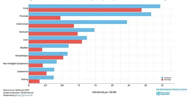
In men, prostate cancer was the most commonly diagnosed cancer type in 12 regions of the world, followed by lung cancer. The lung cancer was also the most frequent cause of death from cancer, followed by prostate and liver cancers (Figure 1).
Figure 1: Estimated age-standardized incidence and mortality rate (Word) in 2018, in males, all ages. Slightly modified from Ferlay et al. [3].
In women, breast cancer was the most frequently diagnosed cancer and the most frequent cause of death from cancer, followed by lung cancer (Figure 2).
3 Figure 2: Estimated age-standardized incidence and mortality rate (Word) in 2018, in females, all ages. Slightly modified from Ferlay et al. [3].
About 25% of the new cancer cases (4,229,662 cases) and 20% of the cancer-related deaths (1,943,478 deaths) occurred in the European regions, even if they account for only 9% of the global population.
1.1.2 Cancer Incidence and Mortality: Italian Data
According to AIOM ("Associazione Italiana di Oncologia Medica", Italian Association of Medical Oncology), about 371,000 new cancer cases are diagnosed each year in Italy; about 53% of cancer cases are diagnosed in men (196,000 cases) and 47% in women (175,000 cases) [4].
The most frequent tumor sites assessed in Italy considering the entire population are breast (14%), colon-rectum (13%), lung (11%), prostate (10%) and bladder (8%). Excluding skin cancers, the most frequently diagnosed cancers in men are prostate (19%), lung (15%) and colorectal (14%), and in women breast (30%), colorectal (12%) and lung (12%) cancers.
In Italy 175,741 cancer-related deaths were estimated in 2018 (98,513 in men and 77,228 in women) [3]. Based on these data, on average, one out of three 3 men and one out of 6 women are likely to die from cancer every year, in Italy.
Last available data about cancer survival rate indicated that women have a 5-year survival rate of 63% and men of 54% and that the survival rate resulted better in young cancer patients than in older ones (in men: 79% in population between 15 and
4 44 years old and 44% in population older than 75; in women: 86% and 42%, in population between 15 and 44 years old and in those older than 75, respectively) [4]. AIOM estimated 3.5 million people as cancer-diagnosed patients in Italy in 2019, which corresponds to 6% of the entire Italian population.
Being the cancer pathology the second cause of deaths worldwide, and being the cancer incidence increasing every year, the study on prevention strategies, on the determination of possible subtypes and targeted therapies is essential, nowadays.
1.2 Breast cancer 1.2.1 Epidemiology
Breast cancer (BC) is the most common invasive tumor diagnosed worldwide in women; epidemiological data about cancer showed that 2.1 million women have been newly diagnosed BC worldwide, in 2018, and that about 627,000 women died for BC [5]. The BC incidence is increasing by about 3% every year [6]. The survival rate varies worldwide, according to the prevention strategies applied and the exposition to risk factors: the highest survival percentage has been showed in high-income countries, in which BC is often diagnosed in the early stages, thanks to the availability of prevention strategies and screening programs. On the other hand, the delayed diagnosis of BCs and the absence of effective treatments lead to a higher mortality rate in low- and middle-income countries, even if the BC incidence is lower than in high-income ones [7].
1.2.2 Breast cancer classification
According to the diagnosed subtype, the prognosis, therapies and survival rates for BCs are different. The tumor size and tumor grade at the diagnosis, the lymphovascular invasion of tumor cells and the analysis of the status of axillary lymph nodes are some of the factors which determine the prognosis of BC. Furthermore, the analysis of gene expression profile and the presence or absence of hormonal receptors in the tumor cell surface are other analyses to be considered to understand BC biology [8]. Different molecular subtypes of BC have been identified in the last 15 years, which have been associated with different treatment responses and outcomes. Basing on receptors expressed by cancer cells, BCs have been classified as Estrogen Receptor, Progesterone Receptor and Human Epidermal Receptor 2 (HER2) positive or negative BCs or Triple-Negative BCs.
5 The estrogen receptor (ER) is a nuclear hormone receptor that acts as a transcription regulator factor and which is one of the prognostic biomarkers for BC. The isoform alpha of ER (ERα) is the measured one during the immunohistochemical examination of tumor and patients whose tumor expresses ER (ER-positive tumor, ER+) are about 80% of the entire BC patients population; ER+ BC patients usually have a better prognosis than ER-negative ones, having a great response to endocrine therapies which targets ER (such as aromatase inhibitors or tamoxifen) [9]; moreover, this type of BC has a lower recurrence rate and cancer-related mortality risk than other BC subtypes [10]. Another receptor analyzed during the classification of BC is the progesterone receptor (PR). PR-positive (PR+) BC patients account for about 55-65% of BCs; in most cases, PR and ER expressions correlate and in PR+ BCs the outcome is usually well responsive to endocrine therapy and often leads to a greater outcome than ER- and PR-negative BC [9].
The human epidermal receptor 2 (HER2) has been found in 13-15% of BCs and it is linked to the HER2 pathway activation, which induces tumor cell proliferation and survival and metastatic process [11]. In HER2-positive (HER2+) BCs, the standard therapy is composed of chemotherapy associated with anti-HER2 blockade strategies. Importantly, HER2+ BCs show a high rate of radioresistance; for this reason, radiotherapy is not usually prescribed in BC patients which expresses high levels of HER2. HER2+ BC is associated with the highest death rate, followed by the triple-negative BC subtype [11].
The last BC subtype includes 15% of BC cases and is triple-negative breast cancer (TNBC). Epidemiological studies showed that TNBC patients usually have a higher waist to hip ratio, early menarche, short periods of breastfeeding, higher body mass index than other BC subtypes patients and that it is the most commonly diagnosed in pre-menopausal women [12]. TNBCs do not express ER, PR, and HER2 and, for this reason, hormonal treatment and anti-HER2 therapies are not effective for TNBC subtype. After the biopsy and immunohistochemical identification, TNBCs are treated with surgery, radiotherapy, and chemotherapy, alone or in combinations [13]. The evaluation of new therapies for TNBC is an evolving field and new approaches are emerging, such as phosphatidylinositol-3-kinase/Akt/mammalian target of rapamycin (PI3K-Akt-mTOR) signaling inhibitors [14].
TNBC is very aggressive and it shows a poor prognosis, high risk of lung and brain metastases and a high rate of recurrence; the highest recurrence peak for TNBC
6 occurs in the first three-five years after the diagnosis, with local or distant recurrence, and decreases to minimal levels after 8 years from the diagnosis [15].
Concluding, the immunohistochemical classification of receptors expressed by BC cells is essential and it is often performed before the surgery, enabling a rapid therapeutic plan establishment.
1.3 Prostate cancer 1.3.1 Epidemiology
Prostate cancer is the second most frequently diagnosed cancer type in men worldwide. In 2018, 1,276,106 new cases have been diagnosed and 358,989 prostate cancer-caused deaths have been counted; prostate cancer cases represent 7.1% of all cancer cases in men, and the number of prostate cancer-related deaths represent 3.8% of all deaths caused by cancer in men worldwide [5].
The incidence and the mortality rates correlate with age, which is a non-modifiable risk factor for prostate cancer, and the average age for the diagnosis is 66 years. Prostate cancer incidence rates are highly variable worldwide, and it is mainly due to genetic predisposition and preventive screening strategies [16].
Even if the incidence of prostate cancer is high in men worldwide, the 5-year survival rates are around 98% in the USA and 83% in Europe [17]; this is mainly because prostate cancer is often diagnosed at the beginning, in an early stage and effective therapeutic approaches are available.
About 20% of prostate cancer cases are diagnosed in patients which report a family history, not only because of the genetic predisposition acquired but also because of the same exposure to environmental risk factors and unhealthy lifestyles, such as a diet rich in fat and red meat [18] and physical inactivity [19].
1.4 Physical Exercise and cancer prevention
Epidemiological studies analyzed the correlation between physical exercise and the risk of different types of cancer and suggested a beneficial effect of physical exercise in primary prevention, decreasing the risk of different cancer types, like breast, colon and prostate cancer [20-23].
As reported by a review published in 2015, about 90% of cancer cases are also correlated to environmental risk factors and lifestyle-related risk factors, of which physical inactivity is part [24].
7 The World Health Organization (WHO) affirmed that more active individuals have a lower risk of death related to high blood pressure, stroke, metabolic syndrome, colon and breast cancer and other non-communicable diseases (NCDs); starting from this state, WHO strongly suggests at least 2.5 hours per week of moderate-intensity physical activity (PA), 1.25 hours per week of vigorous-intensity PA, or a combination, to reduce risks of NCDs [25].
In last decades, growing evidence showed an inverse association between PA level and cancer risk, especially considering breast cancer; moreover, a linear association between high PA intensity and reduced BC risk has been found, with a better beneficial effect of PA in case of vigorous PA than moderate one [26-29].
Furthermore, several articles highlighted the beneficial effects of post-diagnosis physical exercise in decreasing cancer recurrence risk and cancer-related mortality, considering different cancer types. Analyzing studies about breast, colorectal and prostate cancer patients, different works reviewed a 35-37% reduction in risk of cancer-caused mortality, comparing the patients most active versus the less active ones [30, 31].
The investigation about the possible correlation between different PA level (expressed in MET-hours/week) and reduced BC-related mortality risk in cancer patients have shown that high level of PA (higher than 10 MET-hours/week) is associated with 25% reduction in BC-related mortality risk, in comparison to the patients who do not perform that level of PA [32]. Moreover, Kenfield and colleagues showed that men more physically active have higher survival rates than men less active; prostate cancer patients who practice more than 3 hours per week of vigorous PA had a 61% lower risk of prostate cancer-caused death in comparison to patients who performed less PA [33].
Different systemic responses have been hypothesized for PA induced reduction in the risk of cancer and in cancer recurrence and mortality, including the effects of PA in reducing adiposity, sex hormones level increasing, inflammation, myokines production, and immune cell increased mobilization [21, 34].
A growing body of studies explored the correlation between PA and cancer progression, both in vivo or in vitro models, considering different types of cancers, like breast, prostate and lung cancer [35-40]; in vivo studies agreed on the reduction of tumor volume in exercised mice, in comparison to control group, with a reported
8 60% reduction in both tumor incidence and growth in five different tumor models [35]. Moreover, to better investigate the effects of PA on tumor growth inhibition, several groups evaluated the effects of exercise-conditioned sera on cancer cell proliferation, in vitro; also in that case, all the studies showed a great beneficial effects of sera obtained post PA, in decreasing the proliferative capacity of tumor cell, in comparison to sera taken at rest, resulting in about 10-15% of proliferative capacity decreasing [37-39].
Studies about the possible molecular mechanisms involved in the highlighted beneficial effects of PA on cancer prevention have been performed by several groups, resulting in the suggestion of different pathways that could be modulated by exercise.
One of the essential mechanisms for organ formation, and which is induced at high levels in tumor formation, is the Hippo signaling pathway, through which several pro-proliferation genes are transcribed [41]. In this pathway, if the protein YAP is de-phosphorylated, it translocates into the nucleus and acts as a transcriptional coactivator; on the other hand, if it is phosphorylated, it remains in the cytosol and it is degraded [42]. Recent evidence showed that exercise and exercise-conditioned human sera led to phosphorylation of YAP in BC cells, in an epinephrine-dependent manner, resulting in tumor formation blockade and cancer cell viability inhibition [43].
Another mechanism considered by several preclinical studies is the Akt/mTOR pathway, which has been found as deregulated in tumors, during PA sessions [44]. It is a signaling pathway involved in protein synthesis and endurance PA has been shown to deactivate it, in cancer murine models, being able to reduce cellular protein amount and cell proliferation.
The releasing of myokines by muscles during PA is another mechanism reviewed by Hojman and colleagues [45], involved in PA-induced decreased viability of cancer cells, in vitro. Currently, 600 potential myokines have been estimated and the best characterized one is the Interleukin-6 (IL-6) [46]. Preclinical studies demonstrated that some myokines inhibit BC cell viability, in vitro, and the tumorigenic process of colon cancer cells, in mice models [47]; furthermore, Pedersen et al. [35] showed that epinephrine and IL-6 are essential in BC responses to PA, because of the epinephrine-dependent mobilization of Natural Killer (NK) cells and the IL-6-dependent redistribution in tumor tissues. High levels of NK cells and cytotoxic T
9 cells in tumors have been associated with better prognosis [48], suggesting that PA could lead to a control of the tumor growth also inducing this specific mechanism.
1.5 Metformin and cancer
Metformin is a biguanide with a hypoglycemic effect, used as first-line treatment of Type 2 Diabetes Mellitus (T2DM), being approved by the Food and Drug Administration in 1957 [49]. Several studies showed a lower risk of cancer incidence [50-53], and cancer-related mortality [54, 55] in T2DM patients who were treated with metformin, showing a positive correlation with the dosage of metformin administered and leading to a great interest about the possible anticancer effects of metformin.
Even if the data about the possible antitumorigenic potential of metformin are a lot, the epidemiological literature relates only to diabetic individuals and the effects of metformin in non-diabetic subjects must be considered and demonstrated.
Moreover, the in vitro and in vivo experiments reported did not fully elucidate the possible mechanisms through which metformin could exert those beneficial effects on cancer control.
Some possible cellular and molecular mechanisms have been hypothesized, which comprise the induction of cell cycle arrest and apoptosis and the inhibition of the unfolded protein response, which is considered a mechanism able to protect cancer cells from stress conditions, as hypoxia or glucose deprivation [56]. Another possible mechanism by which metformin exerts its antitumor effects is the IGF1 receptor pathway, by which metformin could reduce insulin and IGF-1 levels in the blood and inactivate the downstream pathway, normally involved in cell proliferation [57]. Moreover, metformin is e well-known autophagic process inducer and autophagy has been suggested as a possible mechanism that could have a role in cancer prevention, eliminating damaged proteins and organelles and avoiding the accumulation of mutations and genetic instability [58]. All these mechanisms could explain the hypothesized chemopreventive properties of metformin, but further investigations in non-diabetic patients are needed.
10
11 The aim of this thesis was to provide new evidence about the beneficial effects of exercise on cancer prevention and progression, with particular attention on triple-negative breast cancer and prostate cancer.
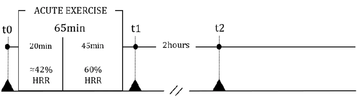
After reviewing the effects of exercise in mTOR pathway inhibition in Triple-Negative Breast Cancer (TNBC), the first goal of this work was to optimize the three-dimensional growth technique (soft agar assay) to assess the effects of an intense bout of physical exercise in modulating the clonogenic capacity of TNBC cells MDA-MB-231. The soft agar assay mimics the formation of the tumor in an anchorage-independent manner and is usually performed to assess the tumorigenic process of cancer cells in vitro, but it has never been used before to assess the exercise-conditioned human sera effects on cancer cell colonies growth.
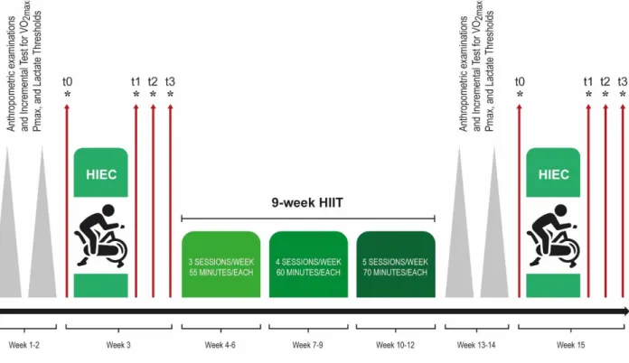
After the optimization of the technique with a first pilot study, a second project has started, in collaboration with the Unit of Exercise and Health Sciences of the Department of Biomolecular Sciences of the University of Urbino “Carlo Bo”, Italy, in which the primary outcome was to evaluate the systemic responses to structured high-intensity endurance cycling (HIEC) tests in decreasing cancer cell proliferative ability and tumorigenic capacity, considering TNBC cells and prostate cancer cells. Moreover, the results obtained by exercise-conditioned human sera taken after the HIEC tests were compared to those obtained from a structured high-intensity interval training (HIIT) period, to understand if the beneficial effects of physical exercise can be due to a HIEC test or to HIIT period, too.
Particular attention will be given to the possible mechanisms involved in the physical exercise beneficial effects in cancer prevention and control, and to the possible relation between the results obtained in vitro and the systemic factors usually measured during physical exercise to understand the amount of exercise performed and the individual response to it.
In the last part of the present study, a possible pharmacological approach to prevent cancer will be taken into account, with the analysis of metformin in preventing cell tumorigenesis.
Starting from the idea that autophagy could be one of the mechanisms involved in the physical exercise positive effects on cancer prevention and that metformin is an autophagic process inducer, a possible synergism between physical exercise and metformin would be considered in TNBC and prostate cancer prevention and modulation.
12
13
Review Article
New Insights into the Role of Exercise in
Inhibiting mTOR Signaling in Triple-Negative
Breast Cancer
Deborah Agostini1, Valentina Natalucci1, Giulia Baldelli1, Mauro De Santi1, Sabrina Donati Zeppa1, Luciana Vallorani1, Giosuè Annibalini1, Francesco Lucertini1, Ario Federici1, Riccardo Izzo1, Vilberto Stocchi1, and Elena Barbieri1,2
1. Department of Biomolecular Sciences, University of Urbino Carlo Bo, Urbino, Italy
2. Interuniversity Institute of Myology (IIM), University of Urbino Carlo Bo, 61029 Urbino, PU, Italy.
Correspondence should be addressed to Elena Barbieri; [email protected] Received 13 April 2018; Revised 3 August 2018; Accepted 12 August 2018; Published 30 September 2018
Academic Editor: Flávio Reis
Copyright © 2018 Deborah Agostini et al. This is an open access article distributed under the Creative Commons Attribution License, which permits unrestricted use, distribution, and reproduction in any medium, provided the original work is properly cited.
Published in Oxidative Medicine and Cellular Longevity. Volume 2018, Article ID 5896786; doi: 10.1155/2018/5896786.
14 Abstract
Triple-negative breast cancer (TNBC) does not express estrogen receptor, progesterone receptor, and human epidermal growth factor receptor 2 and is characterized by its aggressive nature, lack of targets for targeted therapies, and an early peak of recurrence. Due to these specific characteristics, chemotherapy does not usually yield substantial improvements and new target therapies and alternative strategies are needed. The beneficial responses of TNBC survivors to regular exercise, including a reduction in the rate of tumor growth, are becoming increasingly apparent. Physiological adaptations to exercise occur in skeletal muscle but have an impact on the entire body through systemic control of energy homeostasis and metabolism, which in turn influence the TNBC tumor microenvironment. Gaining insights into the causal mechanisms of the therapeutic cancer control properties of regular exercise is important to improve the prescription and implementation of exercise and training in TNBC survivors. Here, we provide new evidence of the effects of exercise on TNBC prevention, control, and outcomes, based on the inhibition of the phosphatidylinositol-3-kinase (PI3K)/protein kinase B (PKB also known as Akt)/mammalian target of rapamycin (mTOR) (PI3K-Akt-mTOR) signaling. These findings have wide-ranging clinical implications for cancer treatment, including recurrence and case management.
1. Introduction
Breast cancer (BC) is one of the most common carcinomas and one of the main causes of cancer-related death worldwide [1]. Among the various subtypes, triple-negative BC (TNBC) accounts for approximately 20% of BC cases. The absence of estrogen and progesterone receptors and human epidermal receptor 2 (HER2) in malignant cells reduces treatment options and increases the risk of recurrence and death, especially in the first 3–5 years of follow-up after surgery [2]. Thus, TNBC exhibits a more aggressive clinical course than non-TNBC. Most TNBC cases are diagnosed in women under the age of 60, and in 20% of diagnosed cases, there is a mutation of the germinal BC (BRCA) gene [3–7]. In patients with metastatic TNBC, there are currently no available targeted therapies and chemotherapy is the only possible treatment option. In addition to the biological-molecular aspects associated with prognosis and BC development, a growing body of evidence highlights the impact of lifestyle on disease-related outcomes. Unhealthy lifestyles with low levels
15 of physical activity (PA) result in overweight and obesity, which appear to have a negative impact on BC [8], increasing the risk of recurrence and death in all subtypes, including TNBC [9]. Conversely, proper diet, weight loss, and increased PA lead to more favorable outcomes in the short and long term [10, 11]. The mechanisms underlying the effects of exercise on breast carcinogenesis are not clear, but experimental evidence suggests that PA induces phosphatidylinositol-3-kinase (PI3K)/protein kinase B (PKB also known as Akt)/mammalian target of rapamycin (mTOR) (PI3K-Akt-mTOR) signaling inhibition and slows TNBC tumor cell growth [12–14]. Physiological adaptations to exercise occur primarily in skeletal muscle, but the effects of exercise and training also impact other tissues through systemic control of energy homeostasis and metabolism, thus influencing the TNBC tumor microenvironment and mTOR inhibition [15]. Given the scope of this review, we summarise recent discoveries related to the underlying biology of exercise-induced modulation of the mTOR pathway in TNBC, examining the benefits induced by different exercise and training protocols. We also consider how exercise affects the level of microRNAs (miRNAs) linked to the mTOR pathway involved in TNBC initiation and progression [16, 17], and how nutrients can influence mTOR signaling. Finally, we discuss how exercise induces beneficial adaptations and why it should be prescribed as a coadjuvant “medicine,” which has the potential to improve TNBC outcomes.
2. mTOR Signaling
2.1. mTOR Pathway and mTOR Activation in BC. mTOR is a serine-threonine kinase that interacts with several proteins to form two distinct complexes, mTORC1 and mTORC2, which show different sensitivities to rapamycin [18]. mTORC1 is acutely sensitive to rapamycin and responds to growth factors, stress, amino acids, and energy, promoting protein translation and synthesis, cell growth, mass, division, and survival. mTORC1 comprises mTOR, the regulatory associated protein of mTOR (Raptor), the G-protein βsubunit-like protein (GβL), also known as mLST8, DEP domain-containing mTOR-interacting protein (Deptor), proline-rich Akt substrate of 40 kDa (PRAS40), and Tti1/ Tel2 complex. mTORC2 is insensitive to acute rapamycin treatment and contains mTOR, the rapamycin-insensitive companion of mTOR (Rictor), the mammalian stress-activated map kinase-interacting protein 1 (mSIN1), GβL, Deptor, protein observed with Rictor-1/2 (Protor
16 1/2), and Tti1/Tel2. Raptor and PRAS40 are unique to mTORC1, while Rictor, mSIN1, and Protor 1/2 are unique to mTORC2 [18]. The various components of mTORC1, which is the most widely studied complex, have several regulatory effects: Raptor, Tti1, and Tel2 are positive regulators, whereas PRAS40 and Deptor are negative regulators [19]. Several factors regulating mTORC1 activation converge in the tubular sclerosis complex (TSC), consisting of hamartin (TSC1), tuberin (TSC2), and TBC1 domain family member 7 (TBC1D7) [20]; the complex works via the Ras homolog enriched in brain (Rheb) GTPase, negatively regulating mTORC1 [21]. An upstream regulator of TSC is the PI3K/Akt pathway activated by growth factors such as insulin-like growth factor 1 (IGF-1) and insulin. PI3K phosphorylates phosphatidylinositol (3,4)-bis-phosphate (PIP2) lipid to phosphatidylinositol (3,4,5)-trisphosphate (PIP3), which recruits phosphoinositide-dependent kinase-1 (PDK1) and Akt. Akt phosphorylates TSC2 and PRAS40 inactivating them and inducing, in turn, mTORC1 activation [22]. TSC2 can also be phosphorylated and inactivated by the activated Ras/extracellular signal-regulated kinase (ERK)/mitogen-activated protein kinase (MAPK) signaling pathway [19]. Another critical regulator of mTORC1 is the adenosine monophosphate-activated protein kinase (AMPK), which is activated when the cellular energy level is low. AMP linking to AMPK allows its phosphorylation (while ATP availability prevents it) triggering repression of energy-consuming processes, also inhibiting mTOR, and enhancing energy-producing processes. AMPK phosphorylates TSC2 in different sites than Akt, activating rather than inactivating TSC2, and phosphorylates Raptor, thus achieving mTORC1 repression [23]. mTORC1 activation requires sufficient amino acid levels, though it is not clear how these levels are sensed. Amino acid regulation requires the formation of a Rag GTPase complex, which binds Raptor, in order to translocate mTORC1 to the lysosome allowing its association with Rheb, and thus its activation [24]. The activation of mTORC1 leads to several downstream effects, including protein synthesis promotion. Raptor binds to the eukaryotic translation initiation factor 4E- (eIF4E-) binding protein 1 (4E-BP1) and the ribosomal protein S6 kinase beta-1 (S6K1), recruiting them to the mTORC1 complex and allowing their phosphorylation [25, 26]. Hyperphosphorylation of 4E-BP1 by mTOR prevents the association of 4E-BP1 and eIF4E, allowing eIF4E to bind eIF4G to begin translation. Phosphorylation of S6Ks, including several S6K1 isoforms and S6K2, by mTOR promotes their activation and thus the phosphorylation of their targets involved in
17 mRNA translation. S6K1 is also involved in negative feedback on mTORC1 and mTORC2 [27]. The mTORC1 complex and AMPK also regulate the autophagic process, a cellular mechanism through which cells eliminate damaged components associated with a wide range of diseases, including cancer. After glucose deprivation, AMPK associates with, and directly phosphorylates, the serine/threonine Unc-51-like autophagy activating kinase (ULK1), an upstream component of the autophagy mechanism. By contrast, when nutrients are plentiful, mTORC1 phosphorylates ULK1, preventing its association with and activation by AMPK, inhibiting autophagy [28]. Aberrant activation of the PI3K/Akt/mTOR pathway is often found in human cancers and promotes cell proliferation [29]. Activation has been shown in the lung, head, and neck and breast, gynecologic, colorectal, and prostate cancers and glioblastoma multiforme [30] and also in B-lineage acute lymphoblastic leukemia [31]. PI3Ks are pivotal molecules in this pathway and possess eight isoforms grouped into class I, class II, and class III. Class I PI3Ks (PI3Kα, β, γ, and δ), stimulated by Tyr kinases, G protein-coupled receptors, and Ras, are currently the focus of research in drug development. Mutation of the PIK3CA gene, which encodes the catalytic subunit α (p110α), one of the class I PI3K isoforms, is found in several cancers [32]. The signaling and biological roles of class II and III PI3Ks are not clear, and they have not been implicated in oncogenesis [32]. In TNBC, the activation of the PI3K/Akt/mTOR pathway is induced by an overexpression of upstream regulators (i.e., growth hormone receptors), mutations of the PIK3CA gene, and by decreased activity of the phosphatase and tensin homolog (PTEN) and of the proline-rich inositol polyphosphatase, which are downregulators of PI3K [33–35]. By contrast, activation of downstream effectors of PI3K (e.g., Akt and mTOR) and activation of downstream effectors of parallel pathways (MAPK and Ras) are rare events in TNBC [36]. Furthermore, other oncogenic pathways (i.e., FGFR, cMET, and RAF) regulated by P53 inactivation converge to activate the PI3K pathway [37]. Due to the frequent activation of the PI3K/Akt/mTOR pathway in human cancers, more than 50 inhibiting drugs are in development, and several clinical trials are ongoing [38]. The first established therapeutic anticancer agents targeting this pathway are everolimus and temsirolimus, which abrogate mTOR signaling, and have been approved by the U.S. Food and Drug Administration. Based on the results obtained with everolimus in pancreatic neuroendocrine tumors [39], and temsirolimus for advanced renal cell cancer [40], these agents are now approved for
18 the treatment of these diseases. Therapies targeting other pathway members have been described. Monotherapy using pan-class I PI3K, which inhibits all class I PI3K isoforms, has effects at dose-limiting toxicity, leading to prolonged disease stabilization in some patients with advanced solid tumors (especially the lung) during phase I clinical trials [41]. Isoform-specific PI3K inhibitors have also been tested and have shown antitumor activity in tumors such as p110δ- (isoform δ-) driven hematologic malignancies [42] or PIK3CA-mutant HR-positive BC [43]. Akt inhibitors and mTORC1/2 inhibitors aimed to suppress not only mTORC1, but also the feedback activation of Akt by mTORC2 [44], are currently being investigated in clinical studies [45]. The use of PI3K/Akt/mTOR pathway inhibitors is often associated with MAPK inhibitors, growth factor receptor inhibitors, and endocrine therapy. Furthermore, they might sensitize tumors to chemotherapy synergistically inducing apoptosis, as shown in sarcomas [46]. These promising strategies are now under investigation for the treatment of several tumors, including nonsmall cell lung cancer [47], colorectal cancer [48], nonmedullary thyroid carcinoma [49], and B-lineage acute lymphoblastic leukemia [31]. Although these strategies have been shown to be effective, there is great variability in the duration and quality of their benefits and the long-term side effects for patients. Thus, the identification of protein and/or genetic biomarkers to recognize subjects that will benefit the most from these therapeutic strategies is essential [50]. In TNBC, the development of PI3K/Akt/mTOR-targeted therapies, taking into account the inhibitors of this pathway alone or in combination with other strategies, will provide new tools to control disease progression and improve outcomes [51]. In a recent phase 2 clinical trial, the efficacy of ipatasertib (an Akt inhibitor) in association with paclitaxel (an antineoplastic agent used in TNBC treatment) was shown [52].
2.2. MicroRNAs and mTOR Signaling in BC. Several studies highlight the role of circulating microRNAs (miRNAs), in different tumors, including BC and the TNBC subtype [16]. In particular, recent evidence has shown that miR10a is downregulated in triple-negative BC cells [53]. Furthermore, overexpression of miR-10a decreases the proliferation and migration of TNBC cell lines via PI3K/Akt/mTOR signaling and through the mitochondrial apoptotic pathway [53]. Recently, Phua et al. [54] demonstrated that 184 is also downregulated in TNBC patients and that miR-184 overexpression in TNBC cells leads to a reduced expression of mTOR. The
19 decreased cancer cell proliferation, due to mTOR reduction, has been confirmed in
vivo: mice injected with mir-184-transfected MDA-MB-231 cells showed a delayed
primary tumor formation and reduced metastatic burden. Emerging evidence points to epigenetic silencing by hypermethylation as a possible mechanism through which these tumor suppressor/growth inhibitor miRNAs are downregulated in TNBC [55]. In metastatic breast tumors, miR-184 has been found to be hypermethylated compared to the methylation status of miR-184 in normal breast tissue, suggesting a selective pressure in silencing this miRNA during the metastatic process [54]. The upregulation of miR-21 was detected in TNBC tissues and in MDA-MB-468 cells by Fang et al. [56]. Inhibition of this miRNA resulted in decreased proliferation, viability, and invasiveness of TNBC cells and enhanced apoptosis. Experiments to identify miR-21 targets have shown that PTEN is downregulated, suggesting an activation of mTOR and the oncogenic properties of miR-21 in TNBC, with increased proliferation and invasion by TNBC cells. Another miRNA that has been found to be upregulated in TNBC tissues in comparison to non-TNBC or adjacent tissues is miR-146a. Indeed, it has been reported to be significantly related to tumor size and histological stage: patients with elevated miR-146a expression have lower survival rates and worse prognoses than low-expression individuals [57]. In addition, miR-146a has been shown to bind the 3′-UTR region of BRCA1, inhibiting its expression; the BRCA1 protein is absent or present at very low levels in about one third of sporadic BCs [58]. Evidence suggests that the downregulation of BRCA1 expression leads to Akt/mTOR oncogenic pathway activation [59]. Hence, strategies that could modify the deregulated status of these miRNAs in TNBC could have a pivotal role in inhibiting the Akt/mTOR pathway and could affect TNBC initiation and progression. It is not yet known how these miRNAs might be modulated by exercise and whether they can be associated positively or negatively with TNBC progression, for which there are no reliable prognostic factors.
2.3. Autophagy and mTOR Signaling. Autophagy is the cellular mechanism responsible for the degradation of cytoplasmic components. It is through this mechanism that cells maintain cellular homeostasis by eliminating damaged proteins and organelles and by providing substrates for energy generation and biosynthesis under stress conditions. The mTOR complex is a major negative regulator of autophagy. It suppresses autophagy in response to nutrients, growth factors, and
20 hormone availability, promoting protein synthesis, cell division, and metabolism. The mTOR signaling pathway is frequently activated in tumor cells, resulting in the activation of its growth-promoting functions and the inhibition of autophagy [60]. In cancer, the cytoprotective role of autophagy could prevent tumorigenic transformation by inhibiting chronic tissue damage. By contrast, once cancer occurs, cancerous cells could utilize autophagy to enhance fitness and survive in the hostile tumor microenvironment, providing energy via substrate degradation. Autophagy could, therefore, be tumor suppressive (for example, via elimination of damaged cellular components), as well as tumor-promoting in established cancers [61]. In addition, autophagy has recently been shown to play a role in necroptosis, and, together with apoptosis, autophagy also regulates other death pathways, including immunogenic cell death, entosis, and pyroptosis [62]. It has been demonstrated that suppression of autophagy in epidermal growth factor receptor- (EGFR-) driven nonsmall cell lung adenocarcinoma xenografts promotes cell proliferation, tumor growth, and dedifferentiation, as well as resistance to EGFR tyrosine kinase inhibitor therapy [63]. Moreover, autophagy suppresses early oncogenesis in lung adenocarcinoma through effects on regulatory T cells [64], and autophagy genes are often required for the cytotoxic effects of chemotherapy [65]. In view of the complex- and context-dependent role of autophagy in cancer progression and response to therapy, it could be hypothesized that the inhibition of the mTOR pathway and the consequent induction of autophagy may be useful in certain cancers through autophagy-dependent antitumor immunity, autophagy-dependent cytotoxic effects, or other tumor suppressor effects [66]. In addition to its effects on skeletal muscle, exercise has also been found to induce autophagy in the liver, pancreas, adipose tissue, and cerebral cortex in transgenic mouse models [67, 68]. Whether exercise-induced stress activates autophagy in healthy cells (or cells primed for malignant transformation), or cancer cells themselves, and whether such effects inhibit or potentiate tumorigenesis, is not known and needs further investigation [15].
3. Evidence of mTOR Modulation by Exercise in TNBC
3.1. mTOR and Exercise. PA reduces mortality for all diseases, including tumors [69], reducing the incidence of primary development and ameliorating the prognosis [15]. Hence, it should be prescribed like a medication indicating the correct typology, dose, and timing, i.e., the type, intensity, duration, and frequency of exercise as
21 described in Exercise Prescription in BC Survivors. Physiological adaptations to exercise occur not only in skeletal muscle but also systemically in other metabolically active tissues involved in the exercise response (such as the bone, heart, adipose, endothelium tissue, and brain) profoundly altering the systemic milieu, in turn influencing the tumor microenvironment and cancer hallmarks [15]. In order to understand the effect of PA on mTOR and BC, muscular, systemic, and microenvironment effects should be considered.
3.1.1. Aerobic Exercise and Muscular Effects. In skeletal muscle, aerobic exercise activates several adaptive pathways, including protein kinases, transcription, and coregulatory factors that, by gene expression modification, increase mitochondrial biogenesis and stimulate metabolic reprogramming [70]. Exercise induces a depletion of nutrients, energetic substrates, and nicotinamide adenine dinucleotide (NAD)H that elevate the ratios of AMP:ATP and NAD+ : NADH, directly activating AMPK and other metabolic sensors, including NAD-dependent protein deacetylase sirtuin 1 (SIRT1) and kinases, such as ERK1/ 2, p38 MAPK, and Jun N-terminal kinase (JNK) [71]. These energy sensors trigger the transcriptional regulator peroxisome proliferator-activated receptor-γ coactivator 1α (PGC1α), which regulates the expression of mitochondrial biogenesis, increases the expression of mitochondrial transcription factor A (TFAM), which, once transferred to the mitochondria, controls transcription of mitochondrial DNA [71]. Moreover, aerobic exercise, through PGC1α phosphorylation, influences other transcription factors, including peroxisome proliferator-activated receptor-γ (PPARγ), an important regulator of fatty acid oxidation and estrogen-related receptor-α (ERRα) and ERRγ, which directly regulate mitochondrial energy metabolism by oxidative phosphorylation, fatty acid oxidation, and the tricarboxylic acid (TCA) cycle [72, 73]. In this regard, the reactive oxygen species (ROS) and reactive nitrogen species produced by exercise also directly or indirectly regulate contraction-induced mitochondrial biogenesis [74] and skeletal muscle metabolic reprogramming via AMPK and PGC-1α [75]. AMPKmediated cell survival requires inhibition of mTOR. Therefore, AMPK and mTOR play antagonistic roles in cells and inhibition of mTOR is essential for AMPK-mediated metabolic homeostasis [76].
22 3.1.2. Resistance Exercise and Muscular Effects. In skeletal muscle, resistance exercise causes an increase in muscle size and strength via mTOR activation. In canonical growth factor signaling, mTOR is activated by PI3K/Akt, through IGF-1 and insulin signaling, but a considerable body of evidence suggests that mTORC1 is also likely activated by a growth factor-independent movement of proteins to and from the lysosome, via resistance exercise-induced phosphorylation of TSC2 [77]. Cellular trafficking of mTOR and its association with positive regulators that occur in human skeletal muscle leading to protein synthesis after resistance exercise, in fed condition, were recently confirmed by Song and colleagues [78].
3.1.3. Systemic and Microenvironment Effects of Exercise. Exercise stimulates the release of molecular signals such as muscle derived regulatory RNAs, metabolites, and myokines with autocrine, paracrine effect on energetic substrate oxidation, hypertrophy, angiogenesis, inflammation, and regulation of the extracellular matrix. To better evaluate the systemic response to PA, a distinction must be drawn between long term (training) and acute exercise. Training induces a reduction of the basal concentration of circulatory sex hormones and lowers adiposity, both recognized risk factors [79], while acute exercise causes a sharp increase in circulating hormones, cytokines, and immune cells [80–82]. Both the systemic adaptations to training and the strong response to acute exercise support plausible mechanisms that inhibit carcinogenesis by suppressing the activation of mTOR signaling network. Hence, exercise may improve BC outcomes [14] (Figure 1). Moreover, both long-term training and a single bout of exercise control energy availability and induce a hormetic response that accounts for the physiological cellular stress adaptation [83, 84].
23
Figure 1: In this figure, we consider potential mechanisms regulated by physical activity and
caloric restriction in inhibiting the mTOR pathway. Both refer to energy availability inhibiting carcinogenesis by suppressing the activation of the mTOR signaling network in this subtype of mammary carcinoma. The mTOR inhibition is mediated through the effects of vigorous PA or long-term exercise on systemic responses such as concentrations of the circulating growth factors and hormones (i.e., IGF-1 and insulin) that regulate the mTOR network. The network is controlled through the PI3K/Akt signaling pathway, the glycemia and glutamine levels, inducing apoptosis and reversing malignancy-associated metabolic programming. Moreover, the control of energy availability by both exercise and CR induces a mitohormetic response that accounts for a physiological cellular stress adaptation through AMPK activation inducing mTOR inhibition. In this context, exercise should be considered in terms of its four components: frequency, intensity, time, and type; however, dose-dependent effects of each component on cancer protection via mTOR inhibition have not yet been clarified. Most data indicate that vigorous PA, either long-term or in adulthood, may reduce a woman’s risk of mammarian cancer, especially TNBC relapse. The inhibition of the mTOR complex and its cell growth-promoting functions leads to a reduction of cell proliferation, control of cancer progression, and consequent autophagy induction probably involved in tumorigenesis prevention. Thus, we hypothesized that exercise-induced inhibition of the mTOR pathway may be useful in the control of cancer progression, including TNBC. PA: physical activity; CR: caloric restriction; CHOs: carbohydrates; mTOR: mammalian target of rapamycin; IGF-1: insulin-like growth factor 1; IGF-1R: insulin-like growth factor receptor 1; IR: insulin receptor; IGFBPs: insulin-like growth factor binding proteins; PI3K: phosphatidylinositol-3-kinase; AMPK: adenosine monophosphate-activated protein kinase; TNBC: triple-negative breast cancer. FITT-VP principle, which reflects the frequency (F), intensity (I), time (T), and type (T) of exercise, and its volume (V) and progression (P) over time, in an individualized exercise training program.
24 Hormesis is a process whereby exposure to a low dose of potential stress favours adaptive changes in the cell that enables it to better tolerate subsequent stress [85, 86]. This type of stress is often related to reactive oxygen species (ROS) originating from the mitochondrial respiratory chain [87]. The accumulation of transient low doses of ROS through exercise influences signaling from the mitochondrial compartment to the cell [88]. Remarkably, this coordinated response to mild mitochondrial stress appears to induce mitochondrial metabolism, increase stress resistance, stimulate various long-lasting cytoprotective pathways, and favour the establishment of an oxidant-resistant phenotype, hence preventing oxidative damage and chronic diseases. Accordingly, low levels of ROS elicit positive effects on physiological cellular and systemic responses and ultimately increase lifespan [83, 88–93]. The hormetic nature of the exercise, which produces low levels of ROS, emerges as a key feature for cancer control. Indeed, in the tumor microenvironment, the activation of exercise-induced hormesis of the AMPK-p38- PGC1-α axis supports oxidative metabolism maintaining the cellular ATP pool and conserving cellular energy and viability during the metabolic stress condition: AMPK regulates metabolism and energy homeostasis [94, 95]. Exercise-induced mitochondrial biogenesis improves mitochondrial function in addition to the upregulation of antioxidant defenses that function as back regulators of intracellular ROS levels and leads to improved redox homeostasis [96, 97] as well as significantly improved insulin sensitivity. By contrast, high levels of ROS cause functional oxidative damage to proteins, lipids, nucleic acids, and cell components, induce a significant increase in intracellular Ca2+, and promote signaling cascades for apoptosis or autophagy via NFκB or forkhead box subgroup O (FoxO) pathways. High ROS levels are therefore reputed to act as etiological, or at least exacerbating factors in chronic/aging-related diseases. The typical hormetic response modulated by exercise involves kinases, deacetylases, and transcription factors; many of which have also been shown to be involved in the carcinogenic process [86]. The most studied are sirtuins (SIRT), which are histone deacetylases, and the FoxO family of transcription factors. The pathways in which NF-kappaB and the Nrf-2/ARE are components are also involved in hormetic responses and implicated in carcinogenesis and are modulated by exercise [86]. FoxO transcription factors play a critical role in cell cycle control and cellular stress responses. FoxOs are known to be regulated by the insulin signaling pathway; however, recently, the research group of Burnet
25 demonstrated that AMPK phosphorylates 6 specific residues on FoxO and opposes the phosphorylation of other FoxO sites by Akt [98]. Phosphorylation of FoxO by AMPK affects the conformation of the protein in such a way that sirtuin-mediated deacetylation is also modified [99]. The dependence of sirtuins on nicotinamide adenine dinucleotide (NAD(+)) links their activity to cellular metabolic status. Emerging evidence indicates that the deacetylation of FoxO by SIRT1 favours the expression of cell survival/stress resistance and the downregulation of proapoptotic genes [85, 100, 101]. Sirtuins, therefore protect against cancer development as they regulate the cellular stress responses and ensure that damaged DNA is not propagated and that mutations do not accumulate [99]. However, how FoxO activation is influenced by exercise remains unclear. In addition, cytokines such as those that we and others have found to be regulated by exercise and training [14, 102–104] have been reported to have direct and indirect effects on cellular stress responses modulated by acetylation/deacetylation reactions, and these effects can be further modified by cortical steroids, which exercise dramatically induces [105]. Similarly, various chemical mimetics of PA and caloric restriction (CR) such as AICAR, PPARδ agonist, resveratrol, and metformin can trigger a beneficial response by activation of key regulators of stress tolerance at the level of transcription, post-transcriptional modifications, and regulation of energy metabolism [92, 106]. Cross talk between major CR hormesis-induced pathways, especially AMPK/PPAR and antioxidant systems, IGF-1, and homeostatic energy balance, reveals the correlation between CR and exercise mimetics [107]. Likewise, depending on the exercise, the level/persistence could induce an adaptive response that might turn the same process from “physiologic” into “pathologic,” as in the case of inflammation. Careful titration of ROS levels within specific tumor microenvironments may lie at the crossroads between the prevention, protection, and/or initiation and progression of the disease, in particular, as regards the induction of mitochondrial functionality, cellular homeostasis, and more generally, cellular metabolic health. Considering the type of exercise, both aerobic and resistance training increase glucose uptake in skeletal muscle via insulin-independent mechanisms, with a subsequent decrease in circulating levels of insulin, IGF-1, and glucose [108]. In a model of mammary carcinogenesis, PA caused a delay in carcinogenesis with concomitant activation of AMPK and a reduction in Akt and mTOR activation and reduction in insulin and IGF-1 in circulation [12]. Reduction of insulin levels is an important aspect given
26 that hyperinsulinemia and insulin resistance are commonly observed in obesity with adipokine alterations, conditions associated with increased risk of BC and poor prognosis [8]. Insulin resistance is a condition in which the target tissues of insulin such as skeletal muscle, adipose tissue, and liver show a reduction in their response to physiological concentrations of the insulin hormone. As a consequence, the pancreatic β-cells produce more of the hormone to compensate for the defective response of target tissues, thus leading to hyperinsulinemia. BC cells express high levels of the insulin receptor (IR), and increased circulating insulin is associated with BC recurrence and death [109]. In contrast, PA has a fundamental role in reducing muscle insulin resistance and normalizing circulating insulin levels. Regular exercise in both healthy and oncological conditions ameliorates glycemic control including glycated hemoglobin (HbA1c) and insulin sensitivity in a “dose”-dependent manner according to duration and intensity [110, 111]. Skeletal muscle in virtue of its mass and high rate of insulin and exercise-stimulated glucose transport represents the most important tissue in glucose uptake. Exercise per sè increases the trafficking of glucose transporter 4 (GLUT4) to the plasma membrane through insulin-independent mechanisms [112]. Under normal physiological conditions, in skeletal muscle, insulin actions are mediated by the IR-catalyzed phosphorylation of the IR substrates 1 and 2 (IRS1 and IRS2). The tyrosine-phosphorylated IRS proteins then interact with and activate PI3K, a critical player in insulin signaling, particularly with regard to glucose homeostasis. Activation of PI3K generates PIP3 that induces membrane translocation of the serine/threonine kinase Akt. PIP3 activation of PDK1 and the Rictor/mTOR complex 2 leads to phosphorylation and subsequent activation of Akt [113]. Akt phosphorylates TBC1D4 (also known as Akt substrate of 160 kDa, AS160) and TBC1D1 promoting the translocation of GLUT4 vesicles from intracellular compartments to the membrane for glucose uptake [114]. Although recent findings help to better understand the effect of exercise on glycemic control, the specific exercise-induced signaling mechanisms leading to the acute and long-term adaptations favouring enhanced glycemic control are less clear [112, 115]. Endurance and, to a lesser extent, resistance exercise represents significant metabolic stress, activating AMPK and thus inhibiting mTOR also in nonmuscular tissue such as liver, fat, and tumor tissues. In order to better evaluate the impact of exercise on mTOR in the BC microenvironment, not only AMPK but also other circulating factors, should be considered. IGF-1, as well as insulin, activates the MAPK pathway
27 and the PI3K pathway, which are both involved in cancer development and progression. The importance of IGF-1 axis in the development and progression of BC has been clearly shown [116]. The overexpression of IGF-1R in BC has been reported and related to poorer survival rates [117]. The IGF signaling system is composed of IGF-1 and IGF-2, insulin-like growth factor binding proteins (IGFBPs), a family of binding proteins regulating IGF half-lives and available in circulation and extracellular fluids, IGF receptors, and insulin receptors. Furthermore, we recently evaluated the complexity of the 1 gene [118] and the biological activity of IGF-1 isoforms in BC cell lines [IGF-1IGF-19] showing that the IGF-IGF-1 isoforms induced cell proliferation via IGF1R phosphorylation. Some studies have reported conflicting results regarding the regulation of IGF-1. Such studies report an increase, no difference or a decrease in circulating IGF-1 levels associated with PA [120–123]. These results are not surprising because the IGF-1 levels are influenced by several clinical factors such as gender, age, body mass index (BMI), sex steroid concentrations, nutrition, stress, level of PA, and intervening illness. Thus, exercise prescription should take into consideration most of these variables. Another process through which exercise might regulate tumor metabolism is the autophagic machinery [15], as described in Autophagy and mTOR Signaling. It is clear that exercise can ameliorate the BC microenvironment and can be very important in reducing BC risk and tumor burden when canonical radiochemotherapy or chemical mTOR inhibitors are not working, as in TNBC. Exercise workouts for these subjects will be explained in Exercise Prescription in BC Survivors. Ex vivo experimental data, using TNBC cell lines stimulated with sera collected before and after a single aerobic exercise bout (pre- or post-exercise serum/a), are described in Experimental Evidence of mTOR Inhibition.
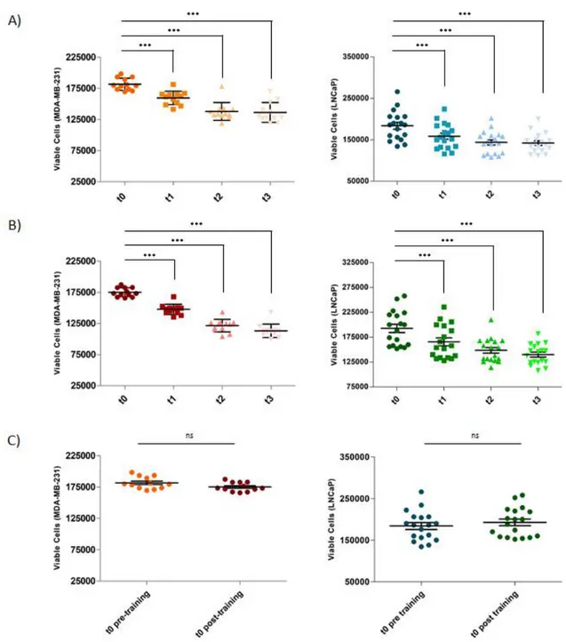
3.2. Experimental Evidence of mTOR Inhibition. As regards the mechanisms involved in the exercise-induced reduction of TNBC risk and tumorigenesis, few data are available. Ex vivo experiments, working with TNBC cells stimulated with sera collected before and after a single aerobic exercise bout (pre- or post-exercise serum/a), are a good starting point to understand how exercise could affect the progression and recrudescence of TNBC. The research group of Dethlefsen has demonstrated that incubation of MCF-7 estrogen responsive BC cells and MDA-MB-231 TNBC cells treated with post-exercise serum, from both healthy volunteers [124]
28 and operated cancer patients [14, 124], resulted in a reduction of BC cell viability in comparison with BC cells incubated with pre-exercise sera. In particular, it has been demonstrated that MCF-7 and MDA-MB-231 stimulation with sera leads to a viability reduction of 11% in MCF-7 cells and 9% in MDA-MB-231 cells in the case of supplementation with post-exercise serum from operated cancer patients receiving adjuvant chemotherapy compared to pre-exercise serum [124]. Furthermore, the viability of both BC cell lines supplemented with sera from healthy women was also significantly reduced by the exercise-conditioned sera, resulting in a 10% and 19% reduction in MCF-7 viability and a 14% and 13% reduction in MDA-MB-231 viability by 1 h and 2 h post-exercise sera, respectively. The reduced viability of MDA-MB-231 supplemented with 5% of healthy women 2-hour post-exercise serum has also been confirmed by a pilot study that we performed working with culture medium with a physiological concentration of glucose (80mg/dl), resulting in a statistically significant reduction in cell proliferation of about 10% compared to cells supplemented with pre-exercise human serum [103]. Promising data on the tumorigenic potential of cancer cells in mice are also available. As reported by Dethlefsen et al. in 2017 [124], different outcomes in incidence and growth of tumors were detected inoculating NMRI-Foxn1nu mice with MCF-7 or MDAMB-231 BC cells preincubated for 48 hours with pre or post-exercise sera from healthy volunteers. In particular, only 45% of the mice inoculated with MCF-7 supplemented with post-exercise human serum formed tumors compared with 90% of mice inoculated with MCF-7 preincubated with at rest sera, and the volume of tumors was reduced by 76%. Moreover, tumor incidence in mice inoculated with MDAMB-231 cells preincubated with post-exercise sera tended to be lower than it was in mice inoculated with MDA-MB-231 cells preincubated with rest sera, but no difference in tumor volume was observed between the two groups. These results show that exercise-stimulated changes suppress BC cell proliferation and reduce the tumorigenic potential of BC cells, also in the case of TNBC cells. Another important aspect to be considered is the fact that PA has been reported to lead to an increased level of the catecholamines epinephrine (EPI) and norepinephrine (NE) [82]; this result has also been confirmed in BC survivors two hours after a single exercise session [124]. Moreover, by blocking the β-adrenergic signaling pathway in BC cells, the effects of post-exercise sera in BC cell viability is completely blunted, indicating the crucial role of catecholamines in inhibiting BC cells viability and