Case Letters
Lung India • Volume 36 • Issue 3 • May-June 2019 261
Conflicts of interest
There are no conflicts of interest.
Roopali Khanna, Neeraj Jain1, Praveen K Goel, Sunil Kumar1 Departments of Cardiology and 1Radio Diagnosis, SGPGIMS,
Lucknow, Uttar Pradesh, India E‑mail: [email protected]
REFERENCES
1. Kinoshita F, Saida Y, Okajima Y, Honda S, Sato T, Hayashibe A, et al.
Thoracolithiasis: 11 cases with a calcified intrapleural loose body. J Thorac Imaging 2010;25:64‑7.
2. Kim Y, Shim SS, Chun EM, Won TH, Park S. A pleural loose body mimicking a pleural tumor: A case report. Korean J Radiol 2015;16:1163‑5.
3. Bhayana R, Chen YA, Deva DP. Bilateral mobile thoracolithiasis. J Radiol Case Rep 2014;8:16‑20.
4. Strzelczyk J, Holloway BJ, Pernicano PG, Kelly AM. Rolling stones in the pleural space: Thoracoliths on CT, and a review of the literature. Clin Radiol 2009;64:100‑4.
5. Suwatanapongched T, Nitiwarangkul C. Thin‑section CT findings of thoracolithiasis. Jpn J Radiol 2017;35:350‑7.
6. Peungjesada S, Gupta P, Mottershaw AM. Thoracolithiasis: A case report. Clin Imaging 2012;36:228‑30.
7. Tanaka D, Niwatsukino H, Fujiyoshi F, Nakajo M. Thoracolithiasis – A
mobile calcified nodule in the intrathoracic space: Radiographic, CT, and MRI findings. Radiat Med 2002;20:131‑3.
8. Oestmann JW, Bridenbaugh S, Eggeling I. Gallstone migration into the pleural space. Am J Gastroenterol 2000;95:836‑7.
9. Brazinsky SA, Colt HG. Thoracoscopic diagnosis of pleurolithiasis after laparoscopic cholecystectomy. Chest 1993;104:1273‑4.
How to cite this article: Khanna R, Jain N, Goel PK, Kumar S.
Mouse in the pleural cavity. Lung India 2019;36:259-61. © 2019 Indian Chest Society | Published by Wolters Kluwer - Medknow
This is an open access journal, and articles are distributed under the terms of the Creative Commons Attribution-NonCommercial-ShareAlike 4.0 License, which allows others to remix, tweak, and build upon the work non-commercially, as long as appropriate credit is given and the new creations are licensed under the identical terms.
Access this article online
Quick Response Code:
Website:
www.lungindia.com
DOI:
10.4103/lungindia.lungindia_445_18
Acute parenchymal lung involvement in a 68‑year‑old
woman with systemic lupus erythematosus, anemia, and
thrombocytopenia
Sir,
Pulmonary manifestations are present in up to 50% of patients with systemic lupus erythematosus (SLE) and can involve pleura, parenchyma, airways, pulmonary vessels, and respiratory muscles.[1]
Acute lupus pneumonitis (ALP) and diffuse alveolar hemorrhage (DAH) are acute parenchymal lung diseases which may occur in SLE.[1]
We hereby present a case of a female participant with SLE associated to acute parenchymal lung involvement and hematological disorders.
A nonsmoker, 68-year-old woman, referred to our hospital with a 5-month history of fatigue and dyspnea that significantly worsened in the last week. She denied fever, cough, sputum, and hemoptysis.
She reported arterial hypertension and documented allergy to nonsteroidal anti-inflammatory drugs and several antibiotics. She denied any occupational exposure.
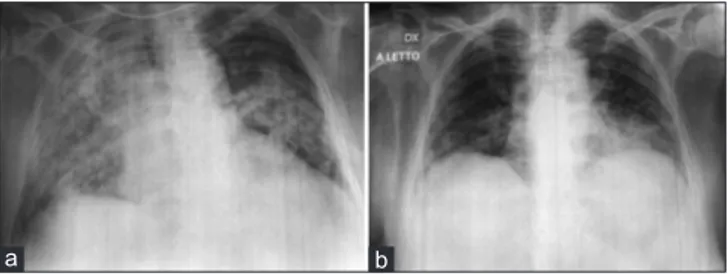
On physical examination, the patient showed hair loss. She had tachypnea (respiratory rate 25 breaths/min) despite breathing oxygen FiO2 60% by the venturi mask. On auscultation, a widespread vesicular murmur and Velcro-like in expiratory crackles were heard. Blood gas analysis revealed a severe hypoxemic respiratory failure (PaO2:65 mmHg; pCO2:45 mmHg; P/F ratio: 108). A two-projection chest radiograph was performed revealing the right lung and left central pulmonary consolidation [Figure 1].
Laboratory findings were the following: white blood cells 7160/µL (86.7% neutrophils, 7.8% lymphocytes, and 4.8% monocytes), hemoglobin 7.3 g/dL, platelets 15000 per µl, and troponin I 0.00 ng/mL; N-terminal pro-B-type natriuretic peptide 132 pg/mL; increased serum inflammatory markers (C-reactive protein 53.8 and erythrocyte sedimentation rate 26 mm/h); pro-calcitonin 0.05 ng/mL. Moreover, there was an alteration of hemolysis indexes (erythropoietin 45.2 mU/mL, total reticulocytes 5.7%, lactate dehydrogenase 275 U/L; total bilirubin 1.50 mg/dL, and indirect bilirubin 0.99 mg/dL).
Case Letters
262 Lung India • Volume 36 • Issue 3 • May-June 2019
forced vital capacity (FVC) 85% of predicted values, FEV1/FVC ration 84.01;), and diffusing capacity of the lungs for carbon monoxide was 39% of predicted value. At discharge, a therapy with prednisone 25 mg/die and azathioprine 100 mg/die was prescribed.
During the follow-up visit, patient’s clinical conditions and laboratory tests were found to be stable.
In line with the rheumatologist’s recommendations, the corticosteroid and immunosuppressive treatment were continued.
On high-resolution CT scan, repeated after 40 days from the discharge, significant reduction of the ground glass areas and the persistence of nonspecific interstitial pneumonia were found [Figure 3].
ALP is reported to affect 1%–12% of SLE patients.[2] It
can be the first expression of the disease in about 50% of participants.
According to the hematological evaluation, the Coombs test was performed, and corticosteroid therapy (40 mg/die of intravenous methyl-prednisolone) was immediately started.
Direct and indirect Coombs tests were positive; therefore, blood transfusion was not performed.
Empirical antibiotic therapy was not prescribed because of the patient’s documented multi-drug allergy.
Microbiological investigations on blood, urinary, and respiratory samples were carried out, and complete autoimmune tests were performed.
During the admission, patient clinical condition deteriorated; a worsening of thrombocytopenia (platelets 4000) and respiratory failure were revealed. Blood gas analysis in high-flow nasal cannula oxygen therapy (airflow 50 L/min, FiO2 90%) showed P/F ratio of 69.
Therefore, methylprednisolone therapy was increased to 80 mg/die.
Immunophenotyping of peripheral blood lymphocytes and bone marrow aspirate did not show abnormalities. The microbiological examinations and sputum cytology were negative.
The rheumatological workup showed the following positive findings: antinuclear antibody (ANA) 1/640 and anti-double strand DNA (anti-dsDNA) 1145 UI/mL. Furthermore, platelet-specific antibodies were positive. Low serum C3 level was found.
In light of nonscarring alopecia, hemolytic anemia, thrombocytopenia, ANA and anti-dsDNA antibody level above laboratory reference range, a diagnosis of SLE was made. The patient was treated by increasing steroid doses (until methylprednisolone up to 1 mg/kg/die intravenous) and intravenous immunoglobulin for 5 days with the improvement of general conditions.
Chest computed tomography (CT) showed “increased density areas mainly located in the left upper lobe, lingular, middle lobe, and anterior segments of both lower lobes associated with interstitial subpleural thickening [Figure 2].” Blood chemistry values (hemoglobin 9. 6 g/dL and platelets 302,000/µL) and blood gas analysis improved (at room air: pH 7. 48; pCO2 of 39 mmHg; pO2 of 72 mmHg; P/F ratio of 342).
Finally, respiratory function tests were performed. Global spirometry parameters were in the normal range (forced expiratory volume in 1 s (FEV1) 86% of predicted values;
Figure 1: Two-projections chest X-ray performed the day of the admission
showing the right lung (a) and left central pulmonary consolidation (b)
a b
Figure 2: Chest computed tomography scan performed during the
admission, revealing increased density areas mainly located in the left upper lobe, lingular, middle lobe (a), and anterior segments of both lower lobes (b) associated with interstitial subpleural thickening
a b
Figure 3: High-resolution chest computed tomography scan performed
40 days after discharge showed significant reduction of the ground glass areas (a) and the persistence of opacities (b)
a b
Case Letters
Lung India • Volume 36 • Issue 3 • May-June 2019 263 In our patient, indirect hyperbilirubinemia plus positivity
of the Coombs test suggested anemia as a complication of SLE (autoimmune hemolytic anemia; AIHA).
AIHA affects 5%–10% of SLE patients and up to 40% of SLE patients show thrombocytopenia.[3,4]
The concomitant or consecutive manifestation of AIHA and immune thrombocytopenia, which is called Evans’ syndrome (ES), is a rare complication,[5] accounting for
0.47% in SLE.[6,7]
The ES could rise alone or in association with other diseases such as autoimmune conditions.[2] The multisystem
involvement of SLE occurs in 3%–15% of ES patients during the follow-up.
Acute pulmonary involvement in SLE is characterized by a poor prognosis. Indeed, both ALP and DAH could be potentially fatal also because of opportunistic infections, cardiac and renal failure, diaphragmatic dysfunction, and drug toxicity that may occur.[2]
To conclude, in our patient, SLE started with the most life-threatening respiratory and hematological complications. The diagnosis was difficult because of the coexistence of ALP and ES, which is a rare event that quickly worsened the patient’s clinical conditions. Declaration of patient consent
The authors certify that they have obtained all appropriate patient consent forms. In the form the patient(s) has/have given his/her/their consent for his/ her/their images and other clinical information to be reported in the journal. The patients understand that their names and initials will not be published and due efforts will be made to conceal their identity, but anonymity cannot be guaranteed.
Financial support and sponsorship Nil.
Conflicts of interest
There are no conflicts of interest.
Michela Dimitri, Vito Liotino, Silvano Dragonieri, Agnese Caringella, Luciana Labate, Pierluigi Carratu, Onofrio Resta Department of Respiratory Diseases, University of Bari, Bari, Italy
E‑mail: [email protected]
REFERENCES
1. Carmier D, Marchand‑Adam S, Diot P, Diot E. Respiratory involvement in systemic lupus erythematosus. Rev Mal Respir 2008;25:1289‑303. 2. Kamen DL, Strange C. Pulmonary manifestations of systemic lupus
erythematosus. Clin Chest Med 2010;31:479‑88.
3. Durán S, Apte M, Alarcón GS, Marion MC, Edberg JC, Kimberly RP,
et al. Features associated with, and the impact of, hemolytic anemia in
patients with systemic lupus erythematosus: LX, results from a multiethnic cohort. Arthritis Rheum 2008;59:1332‑40.
4. Sultan SM, Begum S, Isenberg DA. Prevalence, patterns of disease and outcome in patients with systemic lupus erythematosus who develop severe haematological problems. Rheumatology (Oxford) 2003;42:230‑4. 5. Costallat GL, Appenzeller S, Costallat LT. Evans syndrome and systemic lupus erythematosus: Clinical presentation and outcome. Joint Bone Spine 2012;79:362‑4.
6. Zhang L, Wu X, Wang L, Li J, Chen H, Zhao Y, et al. Clinical features of
systemic lupus erythematosus patients complicated with Evans syndrome: A case‑control, single center study. Medicine (Baltimore) 2016;95:e3279. 7. Michel M, Chanet V, Dechartres A, Morin AS, Piette JC, Cirasino L, et al.
The spectrum of Evans syndrome in adults: New insight into the disease based on the analysis of 68 cases. Blood 2009;114:3167‑72.
How to cite this article: Dimitri M, Liotino V, Dragonieri S,
Caringella A, Labate L, Carratu P, et al. Acute parenchymal lung involvement in a 68-year-old woman with systemic lupus erythematosus, anemia, and thrombocytopenia. Lung India 2019;36:261-3.
© 2019 Indian Chest Society | Published by Wolters Kluwer - Medknow
This is an open access journal, and articles are distributed under the terms of the Creative Commons Attribution-NonCommercial-ShareAlike 4.0 License, which allows others to remix, tweak, and build upon the work non-commercially, as long as appropriate credit is given and the new creations are licensed under the identical terms.
Access this article online
Quick Response Code:
Website:
www.lungindia.com
DOI:
10.4103/lungindia.lungindia_400_18
Rupture of occlusion balloon during transbronchial lung
cryobiopsy
Sir,
Transbronchial lung cryobiopsy (TBLC) is increasingly being utilized for the diagnosis of diffuse parenchymal lung diseases.[1] The procedure can be performed under
rigid bronchoscopy as well as flexible bronchoscopy. It is
preferable to have airway protection during the procedure as significant bleeding may occur unpredictably during the procedure. Furthermore, use of an occlusion balloon (such as Fogarty balloon catheter/Arndt endobronchial blocker) is desirable during the procedure to prevent bleeding complications following biopsy. At our center, [Downloaded free from http://www.lungindia.com on Thursday, May 2, 2019, IP: 212.189.235.67]
View publication stats View publication stats