di Pisa
Characterization of FATZ-3 protein and its interaction with
PDZ containing proteins
Thesis submitted for the Degree of Doctor Philosophiae (Perfezionamento in Genetica Molecolare e Biotechnologie)
Academic Year 2007-2008
Candidate: Mohamed Soliman Ismail
gÉ Åç uxÄÉäxw Ytà{xÜ tÇw `Éà{xÜ
gÉ Åç uxÄÉäxw Ytà{xÜ tÇw `Éà{xÜ
gÉ Åç uxÄÉäxw Ytà{xÜ tÇw `Éà{xÜ
gÉ Åç uxÄÉäxw Ytà{xÜ tÇw `Éà{xÜ
ACKNOWLEDGMENTS
1SUMMARY
2ABBREVIATIONS
4Chapter 1: INTRODUCTION
7 1 Muscle 7 1.2 The sarcomere 121.2.1 The structure of the sarcomere 12
1.2.2 Actin 15
1.3 The Z-disk 16
1.3.1 The structure of the Z-disk 16
1.3.2 Z-disk proteins 18
1.3.2.1 Titin 19
1.3.2.2 MLP 21
1.3.2.3 MURFs 21
1.3.2.4 Calpain 3 22
1.3.2.5 Nebulin and Nebulette 22
1.4 Alpha-actinin 23
1.5 NF-AT3 25
1.6 Filamins 25
1.7 Telethonin 27
1.8 The MARP family 29
1.9.1 Myotilin 32
1.9.2 Palladin 33
1.9.3 Myopalladin 34
1.10 The FATZ family 35
1.10.1 FATZ-1 (calsarcin-2, myozenin-1) 37
1.10.2 FATZ-2 (calsarcin-1, myozenin-2) 39
1.10.3 FATZ-3 (myozenin-3/calsarcin-3) 41
1.11 The Enigma family 42
1.11.1 Enigma 44
1.11.2 Enigma homologue protein (ENH) 44
1.11.3 ALP (Actinin associated LIM protein) 45
1.11.4 RIL (Reversion induced protein) 46
1.11.5 CLP-36 (hCLIM1, Elfin) 47
1.11.6 ZASP/Cypher/Oracle 49
1.12 PDZ domains 51
1.12.1 Structure and binding characteristics of PDZ domains 52 1.12.2 Why do PDZ domains bind specifically to the C-terminal 53
of proteins?
1.12.3 Classification of PDZ domains 56
1.12.4 The effect of phosphorylation on C-terminus binding 58 1.12.5 The multiplicity and function of PDZ domains 59
Chapter 2: MATERIAL AND METHODS
642.1 AlphaScreen 64
2.1.1 AlphaScreen Experiments 65
2.2 Antibodies 67
2.2.1 Primary Antibodies 67
2.5 Cell culture 71
2.5.1 C2C12 cells 71
2.5.2 COS-7 cell line 71
2.5.3 Sf9 cell line 71
2.6 Gene amplification and cloning 72
2.6.1 Amplification and cloning of full length FATZ-3 72 2.6.2 Amplification and cloning of regions of FATZ-3 73
2.6.3 Myotilin 75
2.6.4 Amplification and cloning of FATZ-1, -2, -3 and 75 myotilin lacking last 15 bp
2.6.6 PDZ-ZASP pPROEXHTa 76
2.6.7 PDZ-ZM-ALP pGEX-6P 76
2.7 In vitro binding 76
2.8 In vitro transcription and translation 77
2.9 PDZ Array 77
2.10 Peptides 78
2.11 Protein Production 79
2.11.1 Production and purification of native 6His-tagged proteins 79 2.11.2 Production and purification of GST recombinant proteins 80
2.12. Structural Biology Protocols 83
2.12.1 Production and purification of the C-terminal FATZ-3 83 (81-251aa) for protein
12.2.2 Large scale production and purification of C-terminal 85 FATZ-3 protein
2.12.3 Dynamic light scattering (DLS) 87
2.12.4 Circular dichroism (CD) 89
2.12.5 Stura Footprint screens 89
2.12.6 Protein crystallization 90
Chapter 3 Results
953.1 Amplification, cloning and expression of FATZ-3 95 3.2 The FATZ family, myotilin, palladin and myopalladin share 97
high similarity at their extreme C-termini (last 5 amino acids)
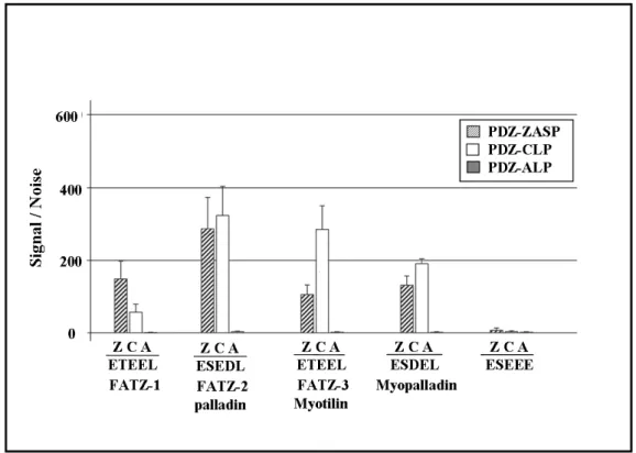
3.3 Preparation of native protein for use in AlphaScreen Experiments 101 3.4 The C-terminal (last 5 amino acids) of the FATZ family and 103
myotilin bind the PDZ domains of ZASP, ALP and CLP-36 proteins
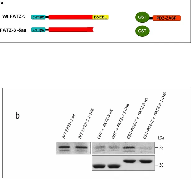
3.5 The PDZ domain of ZASP binds the IVTT full-length FATZ-3 106 but not truncated FATZ-3 (minus C-terminal 5 aa’s)
3.6 Peptides of the C-terminal amino acids of the FATZ family, 108 myotilin, palladin and myopalladin bind to the PDZ domains of
ZASP, ALP and CLP proteins
3.7 Phosphorylation affects the binding activity of the C-terminal amino 110 acid peptides to the PDZ domains of ZASP, CLP-36 and ALP
3.8 The C-terminal FATZ-1 (CD2) competes with the binding 114 between the phosphorylated and non phosphorylated peptides of
FATZ-3/myotilin and the ZASP-1 protein
3.9 The ACTN2 protein competes with the binding between the 116 phosphorylated and non-phosphorylated peptides of FATZ-3/myotilin
and the ZASP-1 protein
3.10 PDZ array experiments 119
3.10.1 FATZ-1 peptides bind to the PDZ domain of hCLIMI 119 (CLP-36) and RIL
3.10.2 FATZ-2/palladin peptides bind to the PDZ domain of 120 hCLIMI (CLP-36) and RIL
3.10.3 FATZ-3/myotilin peptides bind to the PDZ domain of CLP 121 and RIL
3.10.6 The C-terminal FATZ-3 protein behaves as the 124 non-phosphorylated FATZ-3/myotilin peptide
3.11 FATZ-3 interacts with Ankrd2 127
3.11.1 The N-terminal FATZ-3 binds Ankrd2 128
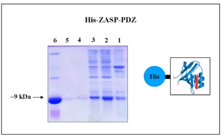
3.12 Expression and purification of FATZ-3 protein 130 3.12.1 Expression and purification of His tagged FATZ-3 131
full-length protein in Bacteria
3.12.2 Protein expression and purification of recombinant 132 GST-FATZ-3 full length protein in Bacteria
3.12.3 Protein expression and production of full-length His tagged 133 FATZ-3 protein using the Baculovirus expression system
3.12.3.1 Dose efficiency experiment for the P2 virus 134 expressing His-FATZ-3 full-length protein
3.12.3.2 Time course experiment for the P2 virus expressing 135 full-length His-FATZ-3 protein
3.12.3.3 Purification of native FATZ-3 protein expressed 136 by baculovirus in Sf9 cells
3.13 Expression and purification of C-terminal FATZ-3 protein in bacteria 138 3.13.1 Comparison of the expression and purification of GST and 138
His tagged C-terminal FATZ-3
3.13.2 Optimization of the purification of C-terminal FATZ-3 protein 140 3.14 Medium scale production and purification of C-terminal FATZ-3 protein 143 3.15 Large scale production and purification of C-terminal FATZ-3 protein 145 3.16 Circular Dichroism (CD) shows high percentage of random coil for the 147
C-terminal FATZ-3 (81-251aa) protein
3.17 Dynamic light scattering (DLS) shows a low percentage of protein 148 polydispersity
3.18 Stura footprint screen experiment 149
Chapter 5 CONCLUSIONS
166This thesis is the harvest of four years full of resisting and insisting to overcome many hard circumstances, I managed to learn many things working in the Muscle Molecular Biology (MMB) laboratory at ICGEB under the supervision of Dr. Georgine Faulkner. I would first like to thank Dr. Georgine Faulkner for her help to me on the professional level by guiding me through my PhD project and teaching me many useful things from her wide experience and helping me to become independent student. I would also like to thank her for her great help on my personal level and supporting me through my hard time (Georgine I thank you very much for all the help you gave to me in the last four years and until this very moment).
This work has been done in collaboration with three different groups and I would like to take the chance to thank all of them, Dr. Olli Carpen, University of Helsinki, Finland, Professor Kristina Carugo, University of Vienna, Austria and especially Professor Giorgio Valle, University of Padua, Italy and the members of his lab for their help in particular Dr. Ivano Zara and Chiara Gardin. Special thanks as well to my colleague and friend Laura Muzzolini for spending with me a lot of time and effort to help me in the protein purification work.
I would also like to thank my colleagues in the MMB lab Anna Belgrano, Snezana Miocić, Valentina Martinelli and my previous colleagues Elisa and Helena.
In my four years at ICGEB I managed to meet great people and friends and I would like to thank them all for giving me the chance to know them and for their great help and support.
I would like to gift this thesis to my great family brothers and sisters and especially to my beloved mother that she will always be in my mind and heart, and to my great father Dr. Soliman Ismail; he is to me not only a father but a brother and a very special friend (father, I thank you very much for your great support and help, you were and still are my guiding light in this life, I wish you a great long happy life).
Last not the least, I would like to thank my tutor Professor Francisco Baralle, for his kind support and precious advice and for ICGEB for giving me the chance to do my PhD in the Muscle Molecular Biology Group.
SUMMARY
The FATZ/calsarcin/myozenin family of proteins has three members FATZ-1 2/myozenin-1), FATZ-2 1/myozenin-2) and FATZ-3 (calsarcin-3/myozenin-3) all localized in the Z-disk and with have high homology to each other at their N- and C-terminals. The FATZ family has been shown to interact with γ-filamin, α-actinin, telethonin, ZASP (Cypher/Oracle) and calcineurin. Together with Prof. O. Carpen we noted that all three members of the FATZ family share high homology with myotilin, palladin and myopalladin at their extreme C-terminal. In fact the final 5 amino acids of FATZ-3 and myotilin are identical (ESEEL), as are those of FATZ-2 and palladin (ESEDL). Computer analysis of the sequences of these proteins indicated that the last four amino acids were considered a probable class III PDZ binding motif (X[DE]X[IVL]). Searches of protein sequence databases revealed that the C-terminal E[ST][DE][DE]L motif is present almost exclusively in the FATZ family of proteins, myotilin, palladin and myopalladin and is evolutionary conserved in these proteins from zebrafish to humans indicating its importance for their biological function.
A main object of this study was to determine if the proteins with this new type of Class III PDZ binding motif at their C-terminal could indeed bind PDZ domains. Since both the FATZ family and myotilin bind ZASP-1 which is a PDZ domain protein we wanted to discover if these interactions occurred via the PDZ domain of ZASP. To do this we collaborated with Prof. G Valle whose group use the AlphaScreen technique to study protein-protein interactions. Biotinylated phosphorylated and non-phosphorylated peptides were designed based on the final C-terminal 5 amino acids of the FATZ
instead of L as its last amino acid. Native protein was also produced for use in AlphaScreen experiments, both to full length proteins and proteins lacking the terminal 5 amino acids. Since ZASP belongs to the Enigma family of proteins the PDZ domains of other members such as ALP and CLP-36 were also checked for their interactions. The results presented here show that the terminal amino acids were responsible and crucial for the interaction with the PDZ domain and that phosphorylation modulated this interaction. These results were further confirmed by in vitro binding experiments such as GST pull-down and PDZ Array. Another Enigma family member RIL was found to bind to this new motif based on PDZ array experiments. The results presented in this thesis demonstrate that proteins of the FATZ family, myotilin, palladin and myopalladin interact with the PDZ domains of Enigma family members via their final C-terminal amino acids. Therefore these final 5 amino acids may represent a new type of class III PDZ binding motif specific for the PDZ domain of Enigma proteins.
I found another binding partner for FATZ-3, namely the I-band protein Ankrd2 whose binding I mapped to the N-terminal of FATZ-3.
The second object of this thesis was to study the tertiary structure FATZ-3 by using protein crystallography. I faced a lot of difficulties expressing and producing native full-length FATZ-3 protein first in bacteria and then using the Baculovirus expression system. It was challenging to obtain enough native purified protein for crystallization studies. I was able to do so using the C-terminal region of FATZ-3 protein (171 amino acids) and undertook crystallization studies at the laboratory of Prof. Kristina Carugo. I tried about 300 crystallization conditions and started to obtain crystals after 1 month. However none of the crystals gave a good diffraction pattern when checked by X-ray.
ABBREVIATIONS
A-band
Anisotropic in polarised lightABD
Actin-binding domainADP
Adenine di-phosphateALP
Actinin-associated LIM proteinAlphaScreen
Amplified Luminescent Proximity Homogeneous AssayAnkrd2
Ankyrin repeat domain 2 proteinAP
Alkaline phosphataseAR LGMD
Autosomal recessive Limb-Girdle Muscular DystrophyATP
Adenine tri-phosphateBCIP
5-bromo-4-chloro-3-indolyl phosphateBSA
Bovine serum albuminCalsarcin
denotes calcineurin associated sarcomeric proteinCD
Circular dichroismCH
calponin homologyCHO
Chinese hamster overyCnA
Calcineurin subunit ACLP
C-terminal LIM domain proteinCOS-7
monkey, African green, kidneyCo-IP
Co-immunoprecipitationDARP
Diabetes-related Ankyrin Repeat ProteinDCM
Dilated cardiomyopathyDHPLC
Performance liquid chromatographyDlg
Discs large proteinDMD
Duchenne Muscular DystrophyEBP50
Ezrin-radixin-moesin binding phosphoprotein-50ECL
Enhanced chemiluminescenceENH
Enigma homologue proteinFATZ
γ-filamin, Alpha-actinin and Telethonin binding proteinFL
Full lenghtGFP
Green fluorescent proteinGST
Glutathione S-transferaseHA
HemagglutininHCM
Hypertrophic cardiomyopathyhCLIM1
carboxyl terminal LIM domain proteinHMERF
Hereditary myopathy with early respiration failureHRP
Horseradish peroxidiseIg
ImmunoglobulinIKEPP
Intestinal and kidney enriched PDZ proteinIPTG
Isopropyl-β-D-thiogalactosideIVTT
In vitro transcribed and translatedkDa
Kilo DaltonLB
Luria BrothLGMD
Limb girdle muscular dystrophyLIMK
LIM domain kinaseLIM domains
Designated LIM for the first three proteins in which it was discovered in, Lin11, Isl-1 & Mec-3MARP
Muscle ankyrin repeat proteinM-band
Mittel, meaning middle in GermanMDM2
Mouse double minute 2MHC
Myosin heavy chainMLP
Muscle LIM proteinMRF
Myogenic regulatory factorMURFs
Muscle Specific RING-finger proteinsNBT
Nitro blue tetrazoliumNFAT
Nuclear factor of activated T-cellsNi-NTA
Nitrilo-tri-acetic acidOD600
Optical density at 600 nmORF
Open reading framePAGE
PolyAcrylamide gel electrophoresisPBS
Phosphate buffered salinePDVF
PolyvinyldifluoridePDZ domains
The name comes from the first three proteins that has been discovered in, PSD-95 , Dlg and ZO-1PEG
Poly ethylene glycol
PKC
Protein kinase CPML
Promyelocytic leukemia proteinPSD-95
Post-synaptic density 95 kDaRIL
Reversion induced proteinSDS
Sodium dodecyl-sulphateSf9
Spodoptera frugiperdaSLRs
spectrin-like repeatsTB
Terrific brothTEV
Tobacco Etch VirusTID
Telethonin interacting domainUV
Ultraviolat lightZASP
Z-band alternatively spliced PDZ domain proteinChapter 1 INTRODUCTION
1 Muscle
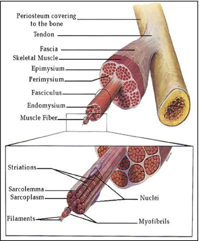
The function of muscle in the body is to produce force and cause motion, either locomotion or movement within internal organs. Most muscle contraction occurs without conscious thought and is necessary for survival. Our hearts contract without us thinking about it, our intestines contract while digesting food and we breathe all without conscious thought. Although movements such as walking, running, writing or even turning our head require conscious thought they are so automatic that usually we are unaware that they result from signals sent to the brain to tell our body parts to move. Muscle is the contractile tissue of the body and is derived from the mesodermal layer of embryonic germ cells. Muscle fibres are bound together by perimysium into bundles which group together to form a muscle which is enclosed in a sheath of epimysium (Fig 1). Muscle spindles are distributed throughout the muscles and provide sensory feedback information to the central nervous system. Individual muscle fibres are surrounded by endomysium. The muscle myofibre is composed of several nuclei and myofibrils. Myobrils are made up of sarcomeres, the basic contractile unit of muscle that are composed of fibres of actin and myosin.
Muscle can be classified into three types: 1) Skeletal muscle (striated, voluntary muscle) is anchored by tendons to bone and enables movement of the skeleton. These muscles are highly specialised for rapid force production. An average adult male has 40-50% of skeletal muscle whereas an average adult female has 30-40%. 2) Cardiac muscle
(striated, involuntary muscle) is an intermittently contracting muscle that maintains the blood supply to the organs of the body. 3) Smooth muscle (non-striated, involuntary muscle) is found within the walls of organs and structures such as the oesophagus, stomach, intestines, bronchi, uterus, urethra, bladder, and blood vessels, and unlike skeletal muscle, smooth muscle is not under conscious control. Smooth muscles are involuntary muscles. Skeletal muscles can be divided into two main types; Type I, slow oxidative, slow twitch, or "red" muscle is dense with capillaries and is rich in mitochondria and myoglobin, giving the muscle tissue its characteristic red colyes our. It can carry oxygen and sustain aerobic activity. Type II which is less dense in mitochondria and myoglobin it is the fastest muscle type in humans. It can contract more quickly and with a greater amount of force than oxidative muscle, but can sustain only short, anaerobic bursts of activity before muscle contraction becomes painful. Cardiac and skeletal muscles are "striated" in that they contain sarcomeres and are packed into highly-regular arrangements of bundles; smooth muscle has neither. While skeletal muscles are arranged in regular, parallel bundles, cardiac muscle connects at branching, irregular angles. Striated muscle contracts and relaxes in short, intense bursts, whereas smooth muscle sustains longer or even near-permanent contractions. Although the three (skeletal, cardiac and smooth) types of muscle have significant differences all three use the movement of actin against myosin to create contraction. In skeletal muscle, contraction is stimulated by electrical impulses transmitted by nerves, the motor nerves and motoneurons in particular. Cardiac and smooth muscle contractions are stimulated by internal pacemaker cells which regularly contract, and propagate contractions to other muscle cells with which they are in contact. All skeletal
muscle and many smooth muscle contractions are facilitated by the neurotransmitter acetylcholine.
Muscular activity accounts for much of the body's energy consumption. All muscle cells produce adenosine triphosphate (ATP) molecules which are used to power the movement of the myosin heads. Muscles contain ATP in the form of creatine phosphate which is generated from ATP and can regenerate ATP when needed by the action of creatine kinase. Muscles also store glucose in the form of glycogen. Glycogen can be rapidly converted to glucose when energy is required for sustained, powerful contractions. Within the voluntary skeletal muscles, the glucose molecule is metabolized in a process called glycolysis, the oxidation of glucose. Glucose is oxidized to either lactate or pyruvate. Under aerobic conditions, the dominant product in most tissues is pyruvate and the pathway is known as aerobic glycolysis. Aerobic glycolysis of glucose to pyruvate, requires two equivalents of ATP to activate the process, with the subsequent production of four equivalents of ATP and two equivalents of NADH. One mole of glucose is oxidised to two moles of pyruvate accompanied by the net production of two moles each of ATP and NADH. Muscle cells also contain globules of fat, which are used for energy during aerobic exercise. The aerobic energy systems take longer to produce the ATP and reach peak efficiency, and require many more biochemical steps, but produce significantly more ATP than anaerobic glycolysis
When oxygen is depleted, as for instance during prolonged vigorous exercise, the dominant glycolytic product in many tissues is lactate and the process is known as anaerobic glycolysis. Under anaerobic conditions and in erythrocytes under aerobic conditions, pyruvate is converted to lactate by the enzyme lactate dehydrogenase (LDH), and the lactate is transported out of the cell into the circulation. The conversion
of pyruvate to lactate, under anaerobic conditions, provides the cell with a mechanism for the oxidation of NADH (produced during the G3PDH reaction) to NAD+; which occurs during the LDH catalyzed reaction. This reduction is required since NAD+ is a necessary substrate for G3PDH, without which glycolysis will cease. Normally, during aerobic glycolysis the electrons of cytoplasmic NADH are transferred to mitochondrial carriers of the oxidative phosphorylation pathway generating a continuous pool of cytoplasmic NAD+. Aerobic glycolysis generates substantially more ATP per mole of glucose oxidized than does anaerobic glycolysis. The utility of anaerobic glycolysis, to a muscle cell when it needs large amounts of energy, stems from the fact that the rate of ATP production from glycolysis is approximately 100X faster than from oxidative phosphorylation. During exertion muscle cells do not need to energize anabolic reaction pathways. The requirement is to generate the maximum amount of ATP, for muscle contraction, in the shortest time frame. This is why muscle cells derive almost all of the ATP consumed during exertion from anaerobic glycolysis.
Cardiac muscle can readily consume any of the three macronutrients (protein, glucose and fat) without a 'warm up' period and always extracts the maximum ATP yield from any molecule involved. The heart and liver will also consume the lactic acid produced and excreted by skeletal muscles during exercise.
Figure 1. The relationship between muscle fibres and the connective tissues of the tendon, epimysium, perimysium and endomysium. Enlargement shows an expanded view of a single muscle fibre containing several nuclei, myofibrils and filaments. (the Figure is taken from Fox, S.I. Human Physiology, 4th Ed.)
2 The sarcomere
2.1 The structure of the sarcomere
All striated muscles are built of subunits known as sarcomeres. These sarcomeres are approximately 2-3 µm long and 1-2 µm in diameter and link end to end to form thin strands known as myofibrils (Craig and Padron, 2004). The striation pattern in muscle results from alternating ordered arrays of myofilaments, i.e. myosin and actin filaments (Cooke, 1995). Figure 2 is a schematic diagram of the sarcomere depicting the following features; thick and thin filaments, A band, M-band, I –band and Z-disk. Thick filaments are composed of mainly of myosin whereas thin filaments are composed of actin filaments associated with tropomyosin and troponin. The bands in the sarcomere were originally named according to their features under a polarized light microscope. The central A-band (Anisotropic in polarised light) is the region where thick and thin filaments interdigitate and is composed of a hexagonal array of aligned, bipolar myosin filaments and adjacent filaments that are cross-linked at their centres by proteins of the M-band (Mittel, meaning middle in German). Electron microscopy images of the A-band showed that there are six thin filaments surrounding one thick filament in a hexagonally arrangement and myosin heads extend to interact with actin forming crossbridges. The region of the A-band without any actinomyosin overlap is called the H-zone (Heller, meaning bright in German). The regions containing only actin filaments are termed I-bands (Isotropic in polarised light) and this surrounds the Z-disk. Actin overlaps partly with myosin and extends to the Z-disk (Zwischen, meaning between in German) at both ends of the sarcomere therefore the Z-disk is the border between each sarcomere. The actin filaments are polar structures, and Z-disc proteins
serve to provide a mechanical link between actin arrays of opposite polarity from adjacent sarcomeres (Huxley and Niedergerke, 1954; Huxley and Hanson, 1954).
The thick and thin filaments represent the main contractile units of the sarcomere. The myosin heads slides along the actin (thin filaments) through a binding interaction which is stimulated by ATP, ADP and inorganic phosphate (Pi). The contraction starts with myosin binding to actin, and the binding of ATP to myosin reduces the affinity of this interaction resulting in the separation of the myosin head from the actin protein, then the myosin head hydrolyses ATP and producing ADP; this reaction generates the power needed to move the myosin head along the actin filament. The replacement of ADP with ATP increases the affinity of binding of the myosin head towards actin. This cycle of ATP hydrolyses and addition of ADP represents the main force generation that is required for filament movement which results in the contraction of the sarcomere which turns the whole muscle to contract.
F ig u re 2 : A s ch em at ic r ep re se nt at io n of t he s ar co m er e. T he t hi ck f il am en ts (b lu e) , th in f il am en ts ( re d) , I-ba nd , Z -d is k (y el lo w ), A -b an d an d M -l in es a re in di ca te d (A u, 2 00 4) . F ig u re 2 : A s ch em at ic r ep re se nt at io n of t he s ar co m er e. T he t hi ck f il am en ts (b lu e) , th in f il am en ts ( re d) , I-ba nd , Z -d is k (y el lo w ), A -b an d an d M -l in es a re in di ca te d (A u, 2 00 4) .
1.2.1 Actin
Thin filaments are mainly composed of Actin. This protein is responsible for diverse cellular functions such as motility, cytokinesis and contraction. Actin filaments form two twisted α-helices that associate with the regulatory proteins tropomyosin and troponin. CapZ stabilizes and prevents depolymerization of actin filaments by binding their plus (+) ends. Moreover it attaches actin filaments to the Z-disk and also binds α-actinin-2 (Casella et al., 1987; Papa et al., 1999). Early in myofibril assembly, thin filament components associate with nascent Z-lines to form the first identifiable structures called I-Z-I complexes (Holtzer et al., 1997). Several Actin isoforms exist, each encoded by separate genes whose expression patterns are regulated developmentally and in a tissue specific manner. Their sequences and molecular structures are very similar. Two muscle-specific isoforms, cardiac and skeletal actins, are co-expressed at varying levels depending on the species, muscle fibre and developmental stage. Vascular and visceral actins are expressed in smooth muscle and also in striated muscle fibres transiently during development. Two non-muscle actins, the cytoplasmic isoforms, are co-expressed with other actin isoforms in many tissues. Mutations in actin can lead to different diseases such as actin myopathy and nemaline myopathy characterized by myofibrillar structural abnormalities and muscle weakness (Ilkovski et al., 2001; Nowak et al., 1999) and also familial dilated cardiomyopathy (DCM) and heart failure, characterized by impaired force transmission and hypertrophic cardiomyopathy (HCM) (Olson et al., 2000; Olson et al., 1998). Thus single amino acid substitutions in actin can result in distinct clinical manifestations, depending on the particular functional domain affected.
1.3 The Z-disk
1.3.1 The structure of the Z-disk
At the beginning of myofibrillogenesis α-actinin rich Z bodies are formed that are the precursors of the Z-disk (Rhee et al., 1994; Sanger et al., 2000). These Z-bodies are just below the plasma membrane and are connected with stress fibre-like structure made up of actin and non-muscle myosin resembling adhesion junctions found in non-muscle cells. The Z-body matures to a striated structure of three filament components; thin filament made up of actin and actin-binding proteins and thick filament consisting of oligomerized myosin (muscle) and titin (Clark et al., 2002; Granzier and Labeit, 2004) as well as intermediate filaments mainly composed of desmin that surround the myofilaments at the Z disk in cardiomyocytes (Clark et al., 2002) and link it to desmosomes, focal adhesion junctions, and the nucleus.
Sarcomeres are separated from one another by the Z-disk, the borderline between sarcomeres. The Z-disk keeps the structure of the sarcomere in register by tethering actin and transmits tension during muscle contraction. Filaments from neighbouring sarcomeres overlap at the Z-disk and are cross-linked by tight interaction with α-actinin-2 which is a major component of the Z-disk (Hoshijima, α-actinin-2006). Alpha-actinin-α-actinin-2 is assembled in the Z-disk as homodimers and aligned in an antiparallel fashion, cross-linking actin and organizing its polarized orientation (Frank et al., 2006) (fig 3). The width of the Z-disk varies depending on the type of muscle. The Z-disc in skeletal muscle fast-twitch fibres has a width of about 30-50 nm (Luther, 1991; Luther, 2000), while in cardiac muscle and skeletal slow-twitch fibres its width is about 100-140 nm (Luther et al., 2002; Yamaguchi et al., 1985).
F ig u re 3 A s ch em at ic d ia gr am d ep ic tin g th e st ru ct ur al c om po ne nt s of a s ec tio n of th e sa rc om er e. A ll st ru ct ur es a re d is pl ay ed u si ng a ri bb on r ep re se nt at io n an d ar e la be ll ed w ith th ei r PD B a cc es si on c od es a nd s ub do m ai ns or c ha in n am e. M on om er ic st ru ct ur es a re co lo ur ed fr om b lu e to r ed , f ro m t he N -to C -t er m in i; a si ng le c ol ou r pe r ch ai n ha s be en u se d fo r m ul tim er ic st ru ct ur es , w ith b lu e an d re d sp he re s in di ca tin g N -an d C -t er m in i ( ex ce pt m yo si n-S1 a nd th e tr op on in co m pl ex ) (A u, 2 00 4) . F ig u re 3 A s ch em at ic d ia gr am d ep ic tin g th e st ru ct ur al c om po ne nt s of a s ec tio n of th e sa rc om er e. A ll st ru ct ur es a re d is pl ay ed u si ng a ri bb on r ep re se nt at io n an d ar e la be ll ed w ith th ei r PD B a cc es si on c od es a nd s ub do m ai ns or c ha in n am e. M on om er ic st ru ct ur es a re co lo ur ed fr om b lu e to r ed , f ro m t he N -to C -t er m in i; a si ng le c ol ou r pe r ch ai n ha s be en u se d fo r m ul tim er ic st ru ct ur es , w ith b lu e an d re d sp he re s in di ca tin g N -an d C -t er m in i ( ex ce pt m yo si n-S1 a nd th e tr op on in co m pl ex ) (A u, 2 00 4) .
1.3.2 Z-disk proteins
It is now clear that the Z disk is not just a simple mechanical structure but a centre for interactions and signalling in the sarcomere. In recent years the number of proteins localised in the Z-disk has increased dramatically but not as much as their interactions among themselves as well as other proteins not normally localised in the Z-disc such as cytoskeletal proteins, nuclear proteins and kinases to mention just a few.
MLP (CSPR3) is a LIM protein downregulated in the adult skeletal muscle up regulated (Arber et al., 1994). MLP is highly expressed in developing and adult heart. Its importance is demonstrated by the fact that the MLP-null mouse develops severe cardiac dysfunction which resembles human dilated cardiomyopathy (Arber et al., 1997). Muscle PDZ-LIM proteins are thought to act as adapters in transmitting mechanical stress signals from the Z-disk to the nucleus will be dealt with in more detail later in the Introduction. Some PDZ-LIM proteins are also present in heart for example ZASP, ALP, enigma and CLP-36. The Z-disk contains many other proteins such as the FATZ (Calsarcin/Myozenin) family, ZASP (Cypher/Oracle), myotilin, myopalladin, myopodin, enigma homology protein, filamin-C, telethonin, desmin, obscurin and titin to mention just a few. As mentioned before most of these proteins interact with each other as well as signalling proteins; for example the FATZ family interacts with and negatively controls calcineurin which it localises to the Z-disk (Frey and Olson, 2002; Frey et al., 2000). Mutations in several Z-disk proteins has been shown to cause cardiomyopathies and or muscular dystrophies, for instance mutations in ZASP (Vatta et al., 2003), FATZ-2 (Osio et al., 2007) and myotilin (Hauser et al., 2002) have been found in patients suffering from cardiomyopathy. The network of interactions of the Z-disk proteins strengthens the Z-Z-disk and allows it to withstand the force caused by the
contraction of the sarcomere. As noted before this places the Z-disk at the centre of the sarcomeric structure (Hoshijima, 2006). Some Z-disk proteins are discussed below and in other sections however I am aware that this is by no means a fully detailed or comprehensive review of all Z-disk proteins.
1.3.2.1 Titin
Titin spans the entire sarcomere and can be divided in three principal regions: Z-disk Titin, I-line Titin and M-line Titin. The Z-disk Titin represents the N-terminal part of the molecule and contains several Ig-like repeats that bind α-actinin (Ohtsuka et al., 1997) and telethonin (Gregorio et al., 1998). Titin, together with telethonin and MLP constitutes a stretch sensor complex in cardiomyocytes (Knoll et al., 2002). The difference in number of alternatively spliced Ig-like repeats in titin isoforms varies between different muscle types and has been suggested to affect the number of cross-links between α-actinin-2 and actin and hence to cause variations in Z-disk thickness (Gautel and Goulding, 1996). Moreover Z-disk titin interacts with Obscurin and small ankyrin 1 (sAnk1) (Kontrogianni-Konstantopoulos and Bloch, 2003), suggesting the existence of a complex implicated in the organization of the sarcoplasmic reticulum around the myofibril (Bang et al., 2001a). In addition to the structural and elastic properties of titin there is evidence that it is involved in signalling pathways. In fact I-line Titin interacts and forms a complex with Calpain protease 94 (Sorimachi et al., 1995) and all the members of the MARP family (CARP, Ankrd2 and DARP) (Miller et al., 2003). Remarkably, all I-band ligands of Titin are also found in the nucleus where they can participate in transcriptional and cell cycle regulation. It seems likely that this dual localization (I-band and nucleus) is not just a coincidence but reflects a dual
function for these proteins: being part of a Titin-based stretch sensing complex in the I-band and regulating transcription in the nucleus. Furthermore, such dual localization may also provide a communication pathway between the I-band and nucleus that links stretch sensing to gene expression (Granzier and Labeit, 2004).
The C-terminal region of titin is located in the M-line. This region contains a Serine/Threonine kinase activity that phosphorylates telethonin in developing muscle (Mayans et al., 1998). However, the upstream elements controlling the Titin kinase activation, its range of cellular substrates and its role in mature muscle are largely unknown. The elastic properties of the Titin molecule and the mechanical deformation of the M-band during stretch and contraction suggest that the signalling properties of the Titin kinase might be modulated by mechanically induced conformational changes (Lange et al., 2005). Also M-line Titin binds to the RING finger protein MURF-1 (Centner et al., 2001) and nbr-1, which interacts with the MURF-2 binding protein p62 (Lange et al., 2005). Nbr-1 acts as a cytoskeleton-associated kinase scaffolding protein that assembles large sarcomeric “signalosomes” through interactions with multiple elements, linking the Titin kinase to p62 and MURF-1 (Lange et al., 2005).
Titin is involved in muscle disease; in fact mutations in the Titin gene, that affect the binding sites for Telethonin and α-actinin, have been found in patients with dilated cardiomyopathy and are thought to be correlated with the disease (Itoh-Satoh et al., 2002). Mutations in the Titin gene that lead to autosomal dominant tibial muscular dystrophy were found to cause LGMD type 2J (Vainzof and Zatz, 2003) and a point mutation in the kinase domain that disrupts the binding with nbr-1 resulted in hereditary myopathy with early respiration failure (HMERF) (Lange et al., 2005).
1.3.2.2 MLP
The Muscle Lim Protein is localized at the periphery of the Z-disk (Arber et al., 1997) and at the intercalated disc (Ehler et al., 2001). The ultrastructural analysis of MLP -/- murine cardiomyocytes revealed a misalignment of the Z-disk (Arber et al., 1997) and morphological defects at costameres and intercalated discs (Ehler et al., 2001) suggesting that MLP plays a primary role in maintaining the stability of these structures. The genetic ablation of MLP in the mouse results in DCM (Arber et al., 1997) and a point mutation in the human MLP gene has also been associated with this pathological condition. This single nucleotide mutation resulted in a severe charge change at position 4 in the MLP protein (W4R) that lies within the N-terminal telethonin interacting domain (TID) of MLP, thus disrupting the binding between the two proteins and causing telethonin miss-localization and Z-line disruption. Since MLP binds telethonin, it was suggested that, together with titin, they could constitute a stretch sensor (Knoll et al., 2002). MLP is known to interact with MyoD (Kong et al., 1997) and has also been implicated in the communication with the nucleus especially in response to hypertrophic signals (Ecarnot-Laubriet et al., 2000).
1.3.2.3 MURFs
The MURFs (Muscle Specific RING-finger proteins) are a family of RING/B-box proteins expressed in striated muscle. Three MURF genes encode three highly similar proteins that can form homo- and heterodimers: MURF-1, MURF-2 and MURF-3 (Centner et al., 2001). MURF-1 acts as a ubiquitin ligase, thereby controlling proteasome-dependent degradation of muscle proteins. Among its targets there are M-line Titin, Nebulin, Myotilin and Telethonin. MURF-1 is part of the structural scaffold
of the M-line lattice but has also been implicated in the regulation of gene expression and myocardiocytic contractility (Witt et al., 2005). MURF-2 co-localizes both with MURF-1 in the M-line and with MURF-3 in microtubules (McElhinny et al., 2004). MURF-3 is a microtubule associated protein differentially expressed during myogenesis and up-regulated during differentiation, probably to stabilize microtubules (Spencer et al., 2000). This is the isoform of MURF located in the Z-line of skeletal muscle where it is though to act as a link between the sarcomeric and the microtubules compartments since it can bind MURF-1, MURF-2 and Z-disk and M-line proteins such as Titin (Spencer et al., 2000).
1.3.2.4 Calpain 3
Muscle-specific Calpain3/p94 is a Ca2+ dependent cysteine protease present both in the M-line and the I-line, where it binds to Titin (Sorimachi et al., 1995). Calpain3/94 is thought to have a regulatory function in the modulation of transcription factors and to be involved in the disassembly of sarcomeric proteins (Vainzof and Zatz, 2003). The importance of its function is suggested by the observation that mutations in the Calpain3/94 gene, with consequent loss of function, lead to LGMD type 2A (Richard et al., 1995).
1.3.2.5 Nebulin and Nebulette
Nebulin is an actin-binding protein which is localized in the I-band of skeletal muscle. It is a large protein (600-900 kDa) that together with Titin forms the third filament system of the myofibril. The C-terminal of the protein is located in the Z-disc, while the rest extends until the end of the thin filaments and its length is proportional to that of the
thin filament. Its binding with actin monomers is sensitive to both calcium and calmodulin (Wright et al., 1993). Nebulin also binds α-actinin (Nave et al., 1990). Mutations in nebulin can cause nemaline myopathy, an autosomal recessive disease. It has been suggested that nebulin acts as a template for the formation of actin filaments (Kruger et al., 1991). Nebulin is found only in skeletal muscle not in heart where a smaller protein called nebulette (109 kDa) that is highly homologous to the C-terminal of nebulin (Millevoi et al., 1998) is found. The C-terminal homology between nebulin and nebulette is thought to be necessary to conserve their binding to the Z-disk (Moncman and Wang, 1995).
1.4 Alpha-actinin
Alpha-Actinin was first discovered in 1964 (Ebashi and Ebashi, 1964). The α-actinin family of proteins plays a very important role in muscle contraction and in the organization of the cytoskeleton. It is a highly conserved family of actin binding proteins, which belong to the spectrin superfamily which includes spectrins, dystrophin and utrophin. Alpha-actinin is an antiparallel homodimer whose structure consists of three major domains; an N-terminal actin-binding domain (ABD) which is a highly conserved region with two calponin homology (CH1 and CH2) domains. The CH1 is responsible for binding actin, while the CH2 increases the binding affinity (Gimona et al., 2002). The central rod domain of α-actinin has tandem spectrin-like repeats (SLRs) which are less conserved than the ABD domain. The number of SLRs in α-actinin has changed during evolution, in vertebrates there are 4 SLRs. Some members of the spectrin super family can contain up to 30 SLRs. In α-actinin the SLRs are involved in
the formation of anti-parallel homodimers and they also bind other interacting proteins (Djinovic-Carugo et al., 2002; MacArthur and North, 2004; Thomas et al., 1997). The C-terminal region which consists of two EF hand (EFh) domains is important for the functional diversities of the α-actinin isoforms.
In humans there are four isoforms of α-actinin, 1, 2, 3, and 4: which are encoded by ACTN1, ACTN2, ACTN3, and ACTN4, respectively. These isoforms share an 80% identity (Beggs et al., 1992; Honda et al., 1998; Millake et al., 1989) and can be characterized according to their biochemical properties, expression patterns and subcellular localisation into two groups; the non-muscle cytoskeletal (calcium sensitive) isoforms alpha-actinin-1 and 4 (Tang et al., 2001) and the muscle sarcomeric (calcium insensitive) isoforms α-actinin-2 and -3 (Blanchard et al., 1989; MacArthur and North, 2004). Alpha-actinin-1 is concentrated at the ends of stress fibres in focal contacts and alpha-actinin-4 is localized in stress fibres and can also be translocated to the nucleus (Honda et al., 1998). The muscle isoforms lack five amino acids at the EFh domain which eliminates their ability to bind calcium. The EFh domain is also responsible for the interaction with titin (Ohtsuka et al., 1997) and ZASP (Faulkner et al., 1999). Alpha-actinin-2 and -3 can be found mainly in the Z-disc, and to a lesser extent in sarcolemma. However α-actinin-3 is absent in cardiac muscles (Beggs et al., 1992; Hance et al., 1999; Lazarides and Granger, 1978) and is not found in 16% of the world population, suggesting that other isoforms can compensate for its absence from the Z-disc of skeletal muscles, especially since there is no phenotypic changes seen when α-actinin-3 is absent (North and Beggs, 1996).
Several studies have shown the importance of α-actinin in muscle function since mice lacking α-actinin die due to degeneration of myofibrils and disruption of the Z-disc
(Fyrberg et al., 1998; Fyrberg et al., 1990; Roulier et al., 1992). It has also been found that over expression of a C-terminal deletion of α-actinin causes Z-disc hypertrophy (Lin et al., 1998; Schultheiss et al., 1992) and a mutation in α-actinin-2 (Q9P) has been found to be associated with dilated cardiomyopathy, this mutation disrupts the binding of α-actinin-2 with MLP (Mohapatra et al., 2003).
1.5 NF-AT3
NF-AT3 is a transcription factor localized both in the Z-disk and in the nucleus. In normal conditions NF-AT3 is tethered to the Z-disk by calcineurin. NF-AT3 resides in the Z-disk because of its interaction with calcineurin. When a signal activates calcineurin, it de-phosphorylates NF-AT3 provoking its translocation into the nucleus. Both NF-AT3 and calcineurin are found in hypertrophy pathways (Olson, 2000).
1.6 Filamins
The Filamins are a family of cytoskeletal proteins which organize filamentous actin in networks and stress fibres. Filamins were first discovered as a family of non-muscle actin-binding proteins (Popowicz et al., 2006; Stossel et al., 2001; van der Flier and Sonnenberg, 2001). The name filamin refers to is filamentous colocalization with actin stress fibres (Wang et al., 1975). There are three main human isoforms of filamin A, B and C encoded respectively by the genes FLNA , FLNB and FLNC (Gorlin et al., 1990). The three isoforms share from 70-80% identity with each other and the exon-intron structure of all the isoforms is highly conserved (Chakarova et al., 2000; Xie et al.,
1998). These isoforms are expressed in most human tissues with some difference in the expression pattern: filamin A is expressed in heart, lung, blood vessels and haematopoietic cells; filamin B is expressed in kidney and pancreas; filamin C is restricted to striated muscle and found predominantly in the Z-disc and in the intercalated disks (Maestrini et al., 1993; Takafuta et al., 1998).
The structure of filamin consists of 24 Ig-like domains (or repeats), each domain is composed of 96 amino acids (Davies et al., 1978). The N-terminal of filamin contains two calponin homology (CH) domains containing 274 amino acids that form the ABD domain (Banuelos et al., 1998). The filamin protein forms a homodimer and electron micrographs showed this homodimer as an extended Y-shaped. The filamin dimer has a molecular weight of 280 kD (Hartwig and Stossel, 1981; Tyler et al., 1980). Himmel and colleagues have shown that the filamin Ig-like domain is sufficient for its dimerisation (Himmel et al., 2003) and serves as a platform for other molecules that participate in the regulation of filamin (Stossel et al., 2001).
Apart from actin cross-linking in cytoskeleton (Hartwig and Stossel, 1981), filamin has many other functions such as linking extra-cellular proteins and the actin cytoskeleton with transmembrane receptors (Meyer et al., 1997). It also plays an important role in signal transduction by serving as a platform for a variety of signalling molecules. A mutation in the filamin C gene (FLNC) can cause myofibrillar myopathy (MFM), this mutation has been mapped to the Ig-like domain 24 (Vorgerd et al., 2005).
1.7 Telethonin
Telethonin (Valle et al., 1997), also known as T-cap (Gregorio et al., 1998) is a small sarcomeric protein with a molecular weight of 19kD. The human telethonin shares around 90% homology with the mouse telethonin (Mason et al., 1999). The N-terminal of telethonin binds to the N-terminal of titin at the Z1 and Z2 Ig-like domains and both these domains and telethonin are localized in the Z-disk (Gregorio et al., 1998; Mues et al., 1998). The binding of telethonin to the N-terminal of titin is very important for Z-disk stability since it has been shown in primary cultures of cardiomyocytes that overexpression of the titin N-terminal resulted in severe myofibril disruption. This observation as well as others suggests that telethonin plays a key role in positioning and anchoring the N-terminal of titin to the Z-disk (Gregorio et al., 1998; Zou et al., 2003). Telethonin is phosphorylated by titin kinase, located in the C-terminal of titin and activated by calcium/calmodulin binding during myocyte differentiation. Since the titin kinase domain is situated in the M-line, whereas telethonin is in the Z-disk, it has been proposed that during myofibrillogenesis the cytoskeleton undergoes reorganization and the titin C-terminal becomes transiently located in close proximity to telethonin, thus allowing its phosphorylation (Mayans et al., 1998).
Crystal structure studies have shown that telethonin and titin form a sandwich complex through the interaction of both N-terminals, forming a dimeric assembly between titin and telethonin however the C-terminus of telethonin would also appear to be involved in the assembly of the titin/telethonin complexes (Pinotsis et al., 2006). Studies on the mechanical strength of the titin Z1Z2-telethonin complex have shown that it is able to withstand the force generated passive muscle stretch due to beta strand crosslinking as titin Z1Z2 in the absence of telethonin had reduced resistance to mechanical stress (Lee
et al., 2006). This data strengthens the hypothesis that telethonin is a key component of the N-terminal titin anchor.
Telethonin was the first sarcomeric protein found to cause an autosomal recessive Limb-Girdle Muscular Dystrophy (AR LGMD). Two different mutations in the telethonin gene were identified in patients with LGMD 2G, both mutations giving rise to premature stop codons; the first mutation was a C157T transition in exon 2 and the second mutation was a deletion of two guanine nucleotides within four guanines at the junction of exon 1 and intron 1 (Moreira et al., 2000). The resulting truncated telethonin protein was not detected suggesting that it was rapidly degraded which may cause instability in the sarcomere structure (Moreira et al., 2000). Interestingly, the C-terminal truncation eliminates the domain of telethonin that is phosphorylated by titin kinase. Mutations in telethonin have been found in other muscle disorders for instance in patients suffering from hypertrophic cardiomyopathy (HCM) (two mutations T137I and R153H) and in a patient suffering from dilated cardiomyopathy (E132Q) (Hayashi et al., 2004). In one case, an individual suffering from severe HCM with a heart septal wall thickness of 46mm had a mutation in telethonin (R70W). This individual had no other known mutations in genes associated with HCM, this mutation was in the binding site of telethonin with MLP (Bos et al., 2006). It has been suggested that the titin/MLP/telethonin complex together with α-actinin may play an important role as a stretch sensors controlling cardiac hypertrophy (Hayashi et al., 2004). It is worth noting that telethonin also binds to Ankrd2 (Kojic et al., 2004) that has been suggested as a stretch sensor in skeletal muscle and that the MARP family, of which Ankrd2 is a member, binds to the N2A region of titin and may form a complex with titin, calpain3 and myopalladin (Miller et al., 2003). In cardiomyocytes telethonin binds the potassium
channel (IKS) β-subunit minK suggesting that a T-tubule-myofibril linking system may
contribute to a stretch-dependent regulation of K+ flux (Furukawa et al., 2001). Telethonin can also bind protein kinase D (Haworth et al., 2004), MURF-1, a RING finger protein found in striated muscle (Witt et al., 2005) and the growth factor myostatin. It has been reported that over expression of telethonin inhibits the secretion of myostatin which would indirectly lead to an increase in the number of myoblasts since myostatin is a negative regulator of myoblast proliferation (Nicholas et al., 2002). Recently it was reported that MDM2 binds telethonin and that high levels of MDM2 can change the sub-cellular localization of telethonin, both proteins co-localised in the nucleus. It would appear that MDM2 can down regulate the protein level of telethonin through a ubiquitin-independent proteasomal pathway (Tian et al., 2006). Pertinent to this thesis telethonin is also able to bind members of the FATZ family of proteins (Faulkner et al., 2000; Kojic et al., 2004).
1.8 The MARP family
Since Ankrd1 (C-193/CARP/MARP) and Ankrd2 are similar both in sequence and in behaviour, it is possible that they play similar roles in heart and in skeletal muscle, respectively. Ankrd1 (CARP), Ankrd2 (ARPP) and DARP (Diabetes-related Ankyrin Repeat Protein, have been grouped together into the MARP family of Muscle Ankyrin Repeat Proteins (Miller et al., 2003). The up-regulation in expression of each MARP is induced by different stress stimuli: injury and hypertrophy in heart for Ankrd1 (Kuo et al., 1999); stretch and denervation in skeletal muscle for Ankrd2 (Kemp et al., 2000) and recovery following starvation for DARP (REF) indicating that these proteins could
be involved in muscle stress response pathways. The MARPs bind to the N2A region of titin and they are part of a Titin-N2A based complex together with Calpain3 and Myopalladin. This complex could be a stress sensor sending signals to the nucleus thereby regulating gene expression (Miller et al., 2003).
1.8.1 Ankrd1 (also known as CARP)
The Ankrd1 protein can shuttle between the sarcomere (I-band) and the nucleus. It is a nuclear co-factor downstream of the homeobox gene Nkx2.5 pathway and forms a physical complex with the ubiquitous transcription factor YB-1, thus acting as a negative regulator of HF-1 dependent pathways for ventricular muscle gene expression (Jeyaseelan et al., 1997; Zou et al., 1997) Ankrd1 expression is predominantly in cardiac muscle, with low levels in skeletal muscle. Its expression can be strongly induced in the regenerating myofibres of Congenital and Duchenne Muscular Dystrophy patients (Nakada et al., 2003). It can also be detected in the sarcomeric I-Z-I areas of regenerating skeletal muscle (Nakada et al., 2003) where it interacts with the I-band protein Myopalladin suggesting it may have a role in the maintenance of sarcomeric integrity (Bang et al., 2001b).
1.8.2 Ankrd2 (ANKyrin Repeat Domain 2, also known as ARPP)
Ankrd2 was first discovered as a protein that was up regulated during muscle stretch (Kemp et al., 2000). It is localized in the I-band and near the Z-line (Tsukamoto et al., 2002). The Ankrd2 gene is located in the same region of human chromosome 10 as that of Ankrd1 (position 10q23.1) and its 9 exons encode a protein with a predicted molecular weight of 37 kD; exons 5, 6, 7, and 8 encoding the ankyrin repeats
(Moriyama et al., 2001; Pallavicini et al., 2001). There is a 43 % homology between Ankrd1 and Ankrd2 (Pallavicini et al., 2001). The human Ankrd2 protein shares a 89% similarity at the amino acid level with the mouse Ankrd2 and it is expressed in skeletal muscle and at a lower level in heart, kidney and prostate (Moriyama et al., 2001; Pallavicini et al., 2001). Ankrd2 expression in foetal heart is lower than that in adult heart and it has been suggested by Moriyama and colleagues (Moriyama et al., 2001) that Ankrd2 could be involved regulation of development in heart. In addition to ankyrin repeats domains, Ankrd2 has a nuclear localization signal (KKKRK) and a PEST-like sequence (Kemp et al., 2000; Moriyama et al., 2001; Pallavicini et al., 2001). It also contains several predicted phosphorylation sites for the following kinases: casein kinase I (CKI), casein kinase II (CKII), protein kinase C (PKC), extra-cellular signal regulated kinase (ERK), cAMP-dependent protein kinase, calmodulin-dependent protein kinase II and cGMP-dependent protein kinase (Moriyama et al., 2001; Pallavicini et al., 2001). In myotubes Ankrd2 is localized both in the nuclei in PML nuclear bodies and diffusely in the cytoplasm (Pallavicini et al., 2001). Ankrd2 has been shown to interact with other Z-disk proteins such as telethonin and non-muscle proteins such as p53 and transcription factors such as PML, and YB-1 (Kojic et al., 2004). These interactions support the idea that Ankrd2 could act a transcription co-factor and it has also been hypothesized that Ankrd2 could be a stretch sensor in skeletal muscle shuttling between the nucleus and the cytoplasm (Kojic et al., 2004).
1.9 Myotilin, Palladin and myopalladin family
1.9.1 Myotilin
The myotilin gene maps at the chromosome locus 5q31 and mutations in this gene are responsible for a dominantly inherited limb-girdle muscular dystrophy (LGMD1A). Myotilin is a 57kD myofibrillar protein that is highly expressed in skeletal muscle and only weakly in heart. It has a unique N-terminal rich in serine residues, a 23 amino acid hydrophobic region (57-79 aa), two Ig-like domains (252-341 aa ; 351-441 aa) that share a high sequence homology with the Ig domains 7 and 8 of human titin and a short C-terminal tail (Salmikangas et al., 1999). Myotilin binds F-actin and efficiently cross-links actin filaments into bundles as well as being able to decrease the rate of F-actin depolymerization which suggests that it could be involved in thin filament stabilization. It is able to crosslink actin since it can dimerize via Ig-like domains. Myotilin is localized in the Z-disk where it binds directly to α-actinin-2 and filamin C, the first 214 residues of myotilin bind to the spectrin-like repeats 3 and 4 of α-actinin-2 and the myotilin C-terminal region containing the Ig-like domains binds to the Ig-like domains 19-21 of filamin C. (van der Ven et al., 2000). Over expression of the C-terminal myotilin had an effect on the assembly of myofibrils in differentiating C2C12 myotubes whereas over expression of full-length myotilin induced formation of thick actin cables in non-muscle cells devoid of endogenous myotilin suggesting that myotilin may have a role in stabilizing the assembled actin bundles (Salmikangas et al., 2003). Not only filamin C but also filamin-A, filamin-B and filamin-Bvar-1 variant bind to myotilin
(Gontier et al., 2005). Myotilin has also been shown to bind to FATZ-2 (calsarcin-1 /myozenin-2) and to the C-terminal region of FATZ-1 (calsarcin-2/myozenin-1) (Gontier et al., 2005).
Patients suffering from Limb girdle muscular dystrophy type 1 A (LGMD1A) have mutations in myotilin. These mutations were a C450T missense mutation which converted residue 57 from threonine to isoleucine but did not disrupt the binding with α-actinin (Hauser et al., 2000). The other two mutations were S55F and T57I, both located in the N-terminal of myotilin but outside the filamin C binding site (Hauser et al., 2002). Mutations in myotilin as well as ZASP, filamin C, desmin and αβ-crystallin can cause myofibrillar myopathy (Griggs et al., 2007; Selcen and Engel, 2004).
1.9.2 Palladin
Palladin was simultaneously discovered by two different laboratories, the mouse palladin was discovered by (Parast and Otey, 2000), while the human palladin was discovered by (Mykkanen et al., 2001). The human palladin shares a 91% homology with the mouse palladin. Palladin is expressed in muscle and non-muscle fibres. In muscles palladin is highly expressed in smooth and poorly expressed in skeletal, while in non-muscle fibres it is highly expressed in prostate, testis, ovary, small intestine, and colon (Mykkanen et al., 2001). There are three isoforms of palladin and their expression depends on cell type; the first isoform is a 90-92 kD protein which is the most abundant isoform of palladin and widely expressed in many cells and tissues, another isoform is around 140 kD which is less abundant than the previous isoform and is mostly expressed in embryonic mice while in adult mice it is mostly detected in smooth muscles (Mykkanen et al., 2001; Parast and Otey, 2000), the third isoform is a 200 kD isoform first found in developing heart and then in embryonic and neonatal bone (Otey et al., 2005). Palladin contains three IgC2 domains, the first IgC2 domain towards the N-terminal shares high homology with the first IgC2 domain, Z-disk-associated IgC2
domains of titin, and with the C-terminal IgC2 domains of myosin light chain kinase, while the middle and the C-terminal IgC2 domains of palladin share high homology with the myotilin N-terminal IgC2 domain; palladin and myotilin share a high homology at their N-terminal and C-terminal. Alpha-actinin colocalizes with palladin in stress fibres, focal adhesions, cell-cell junctions and the embryonic Z-disk. Palladin plays an important role in the organization of actin cytoskeleton and focal adhesions in cultured trophoblasts and fibroblast cells (Parast and Otey, 2000). The expression level of palladin is highly reduced in a number of tissues including heart, skeletal muscle, liver, and kidney, which indicates that palladin could be involved in establishing the cytoskeletal organization of cells during differentiation being replaced by other proteins when the cells are fully differentiated (Parast and Otey, 2000). Recently palladin has been found mutated members of a family suffering from pancreatic cancer, the mutation caused a change of an amino acid proline (hydrophobic) to serine (hydrophilic) (P239S) in a highly conserved region of palladin, the mutation was not found in healthy members of the family. It has been suggested that this mutated palladin in pancreatic cancer may cause cytoskeletal changes in the pancreatic cancer cells and may be responsible for strong invasive and migratory abilities of these tumours (Ronty et al., 2007).
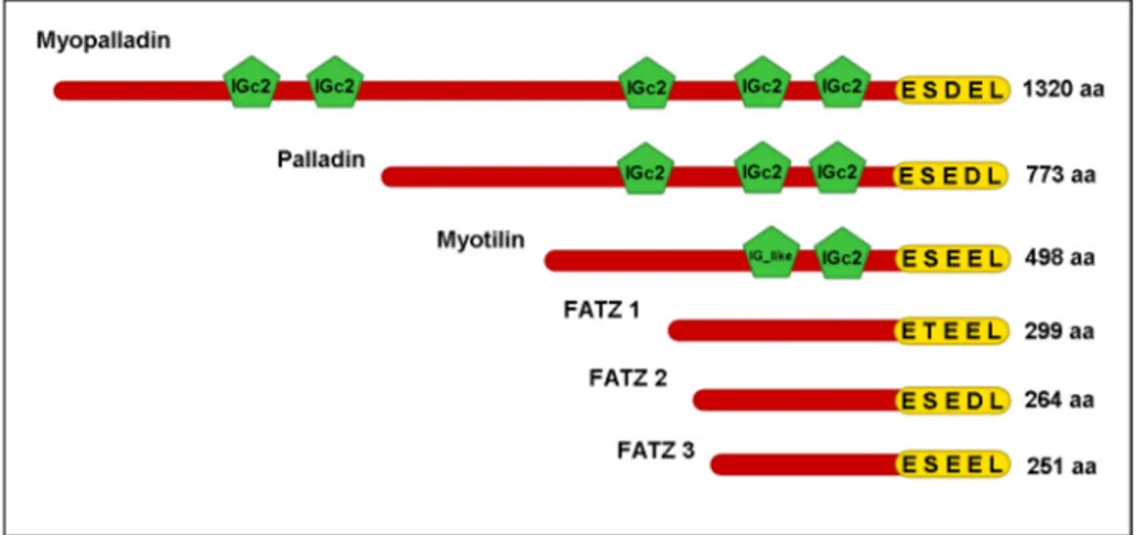
1.9.3 Myopalladin
Myopalladin is a 145 kD protein that interacts with nebulin, nebulette and α-actinin in the Z-disk and with CARP in the I-band. These interactions are important for the integrity of the sarcomere since over expression of myopalladin leads to the disruption of the sarcomeric structure suggesting that it could link the regulatory mechanisms of
the Z-disk to muscle gene expression through its interaction with Ankrd1 (Bang et al., 2001b). Myopalladin is expressed in skeletal and cardiac muscle and it is localized predominantly in the Z-disk and to narrow segments of the I-band on either side of the Z-disk. Myopalladin contains five Ig domains, the last three Ig domains towards the C-terminal are most related to the Ig domains of palladin, whereas the last two Ig domains towards the C-terminal of myopalladin shares high homology with the Ig domains of myotilin. Apart from the homology at the Ig domain level that myopalladin shares with palladin and myotilin, myopalladin shares 63% homology through its C-terminal with the C-terminal of palladin which includes the last three Ig domains (Bang et al., 2001b). Myotilin, myopalladin and palladin form a family which shares high amino acid homology and also high homology at the level of the Ig domains. All three proteins function as scaffolds that regulate actin organization (Otey et al., 2005).
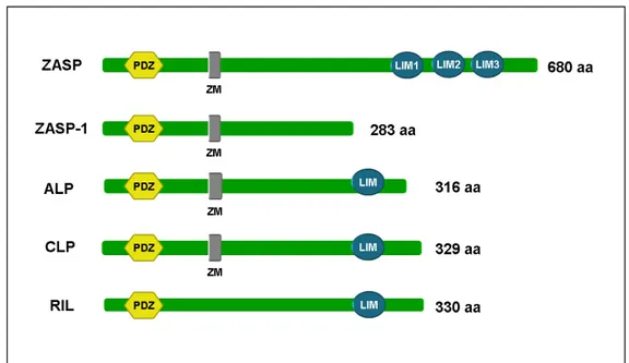
1.10 The FATZ family
The FATZ (also known as calsarcin and myozenin) family was independently discovered by three different groups (Faulkner et al., 2000; Frey et al., 2000; Takada et al., 2001). The family consists of three proteins FATZ-1 (myozenin-1 or calsarcin-2), FATZ-2 (myozenin-2 or calsarcin-1) and FATZ-3 (myozenin-3 or calsarcin-3) (Frey and Olson, 2002) that are localised in the Z-disk (Faulkner et al., 2000; Takada et al., 2001). Then name FATZ denotes filamin C, alpha-actinin and telethonin binding protein of the Z-disk (Faulkner et al., 2000), while the name calsarcin denotes calcineurin associated sarcomeric protein (Frey et al., 2000). The FATZ family are
sarcomeric proteins localized in the Z-disk of striated muscle (Faulkner et al., 2000; Frey et al., 2000).
FATZ-1 (myozenin-1 or calsarcin-2) and FATZ-3 (myozenin-3 or calsarcin-3) are highly expressed in differentiated skeletal muscle in fast-twitch fibres, whereas FATZ-2 (myozenin-2 or calsarcin-1) is expressed in adult cardiac muscle in slow-twitch fibres (Frey and Olson, 2002; Frey et al., 2000). The three proteins share a high homology at the N- and C-termini and less homology in the intervening region, which may indicate that these conserved regions have functional properties and could be protein binding domains (Frey and Olson, 2002; Frey et al., 2000). The family members tend to interact with the same proteins, for example filamin A, B and C, α-actinin-2 and -3, telethonin (Faulkner et al., 2000; Takada et al., 2001) as well as calcineurin (Frey et al., 2000), myotilin (Gontier et al., 2005) and ZASP. FATZ-1, FATZ-2 and FATZ-3 bind strongly to nine different splice variants of ZASP, some of these splice variants had only the N-terminal PDZ domain and lacked the three C-N-terminal LIM domains, suggesting that the LIM domains were not involved in the interaction between ZASP and FATZ (Frey and Olson, 2002; Zhou et al., 2001).
The FATZ family members also interact with calcineurin (calcium/ calmodulin-dependent serine, threonine phosphatase). The catalytic activity of calcineurin is not crucial for the interaction with all three FATZ proteins, since it has been shown that these proteins bind to a mutated subunit of CnA lacking enzymatic activity (Frey and Olson, 2002; Frey et al., 2000). Immunostaining experiments showed that the interaction between calcineurin and FATZ occurs in the Z-disk (Frey et al., 2000). Calcineurin can be detected in the nucleus as well as the cytoplasm suggesting that FATZ may have a role in interacting with and localizing calcineurin to the Z-disk (Frey
et al., 2000). There are other examples of intracellular proteins that bind and localize signalling proteins to specific sites, for example; a family of anchoring proteins called AKAPs (A-kinase anchoring proteins) has been shown to tether protein kinase A (PKA) to centrosomes (Edwards and Scott, 2000). There is evidence that over expression of the FATZ proteins can inhibit calcineurin in vitro, which allows speculation that the FATZ family can serve as inhibitors or activators of calcineurin (Frey et al., 2000).
1.10.1 FATZ-1 (calsarcin-2, myozenin-1)
Human FATZ-1 was mapped to chromosome 10q22.1 (Faulkner et al., 2000). The FATZ-1 gene has six exons that encode for a 32 kD sarcomeric protein that is localized in the Z-disk. (Faulkner et al., 2000; Takada et al., 2001). This protein is expressed in skeletal muscle and to a lesser extent in heart; its expression is upregulated during differentiation (Frey et al., 2000). The human and mouse FATZ-1 proteins share a 90% identity (Faulkner et al., 2000).
Secondary structure predictions show that FATZ-1 has two alpha helical domains at the N-terminal (A1; 1-72 aa ) and at the C-terminal (A2; 87-244 aa) regions, with a glycine rich region (in the middle between residues 95-175) and a short C-terminal tail of approximately 55 amino acids (Takada et al., 2001). These three regions, the two alpha helical and the glycine-rich, have also been named as CD1 (1-71 aa), CD2 (171-299 aa) and GRD (75-171 aa) respectively (Faulkner et al., 2000). As mentioned previously, FATZ-1 binds to several muscle proteins and some of these binding sites have been mapped; both α-actinin and filamin C bind to the full length FATZ-1 protein more particularly its to its C-terminal region (Faulkner et al., 2000). The FATZ-1 binding site on α-actinin-2 was mapped to the region that spans the SR3 and SR4 spectrin-like
![Table 1 Results of the protein database scan for the motif E[ST][DE][DE]L](https://thumb-eu.123doks.com/thumbv2/123dokorg/4786159.48642/107.892.148.834.764.1126/table-results-protein-database-scan-motif-e-st.webp)